Trauma to the developing brain constitutes a poor- ly explored field. Some recent studies attempting to model and study pediatric head trauma, the leading cause of death and disability in the pediatric popu- lation, revealed interesting aspects and potential targets for future research. Trauma triggers both excitotoxic and apoptotic neurodegeneration in the developing rat brain. Excitotoxic neurodegeneration develops and sub- sides rapidly (within hours) whereas apoptotic cell death occurs in a delayed fashion over several days following the initial traumatic insult. Apoptotic neu- rodegeneration contributes in an age-dependent fashion to neuronal injury following head trauma, with the immature brain being exceedingly sensi- tive. In the most vulnerable ages the apoptosis con- tribution to the extent of traumatic brain damage far outweighs that of the excitotoxic component. Molecular and biochemical studies indicate that both extrinsic and intrinsic mechanisms are involved in pathogenesis of apoptotic cell death fol- lowing trauma. Interestingly, in infant rats a pan- caspase inhibitor ameliorated apoptotic neurode- generation with a therapeutic time window of up to 8 h after trauma. These results help explain unfavorable outcomes of very young pediatric head trauma patients and imply that regimen which target slow active forms of cell death may comprise a successful neuropro- tective approach. Key words: Apoptosis; Excitotoxicity; Neuroprotection INTRODUCTION Traumatic brain injury (TBI) constitutes a major cause of morbidity and mortality in the industrialized world (Goldstein, 1990; Sosin et al., 1995; Thurman et al., 1999). According to the National Center for Injury Prevention and Control, an estimated 1.5 million Americans sustain TBI each year. As a result, 50,000 people die, 230,000 are hospitalized and survive and an estimated 80,000-90,000 people experience the onset of long-term disability every year (Thurman et al., 1999). A large proportion of TBI patients are never hospitalized but may suffer varying degrees of cogni- tive impairment, behavioral and personality changes, irritability, post-traumatic vertigo, sleep disturbances, attentional deficits and headaches. Although children under six years of age sustain TBI more frequently than any other age group (Adelson and Kochanek, 1998; Diamond, 1996), there has been lim- ited research focusing on traumatic injury to the devel- oping brain. The assumption that pathophysiology of TBI is identical in the adult and developing central nervous system is incorrect. Clinical studies suggest that age decidedly influences both morbidity and mor- tality after head injury in children, with those under 4 years of age showing the worst outcomes (Mahoney et al., 1983; Koskiniemi et al., 1995; Adelson and Kochanek, 1998). A study by Koskiniemi and col- leagues (1995) demonstrated that in a cohort of chil- dren suffering severe head injury prior to the age of 4 years none was able to work independently outside a structured environment years later. Children older than 4 years at the time of injury had a significantly better outcome. Differences in the mechanisms by which TBI F.P. Graham Publishing Co. Neurotoxicity Research, 2003, VOL. 5(7). pp. 475-490 Neuropathological and Biochemical Features of Traumatic Injury in the Developing Brain PETRA BITTIGAU a , MARCO SIFRINGER a , URSULA FELDERHOFF-MUESER b , HENRIK H. HANSEN a,c and CHRYSANTHY IKONOMIDOU a, * a Departments of Pediatric Neurology and b Neonatology, Charité Children's Hospital, Humboldt University, Augustenburger Platz 1, 13353 Berlin, Germany; c Department of Pharmacology, The Danish University of Pharmaceutical Sciences, DK-2100, Copenhagen, Denmark. [email protected] (Received 19 August 2003; Revised 26 September 2003; In final form 26 September 2003) *Corresponding author. Tel: +49-30-450566103; Fax: +49-30-450566920; E-Mail: [email protected] ISSN 1029 8428 print/ ISSN 1476-3524 online. © 2003 FP Graham Publishing Co., www.fpgrahamco.com

Welcome message from author
This document is posted to help you gain knowledge. Please leave a comment to let me know what you think about it! Share it to your friends and learn new things together.
Transcript

Trauma to the developing brain constitutes a poor-ly explored field. Some recent studies attempting tomodel and study pediatric head trauma, the leadingcause of death and disability in the pediatric popu-lation, revealed interesting aspects and potentialtargets for future research.
Trauma triggers both excitotoxic and apoptoticneurodegeneration in the developing rat brain.Excitotoxic neurodegeneration develops and sub-sides rapidly (within hours) whereas apoptotic celldeath occurs in a delayed fashion over several daysfollowing the initial traumatic insult. Apoptotic neu-rodegeneration contributes in an age-dependentfashion to neuronal injury following head trauma,with the immature brain being exceedingly sensi-tive. In the most vulnerable ages the apoptosis con-tribution to the extent of traumatic brain damagefar outweighs that of the excitotoxic component.
Molecular and biochemical studies indicate thatboth extrinsic and intrinsic mechanisms areinvolved in pathogenesis of apoptotic cell death fol-lowing trauma. Interestingly, in infant rats a pan-caspase inhibitor ameliorated apoptotic neurode-generation with a therapeutic time window of up to8 h after trauma.
These results help explain unfavorable outcomesof very young pediatric head trauma patients andimply that regimen which target slow active formsof cell death may comprise a successful neuropro-tective approach.
Key words: Apoptosis; Excitotoxicity; Neuroprotection
INTRODUCTION
Traumatic brain injury (TBI) constitutes a major causeof morbidity and mortality in the industrialized world(Goldstein, 1990; Sosin et al., 1995; Thurman et al.,1999). According to the National Center for InjuryPrevention and Control, an estimated 1.5 millionAmericans sustain TBI each year. As a result, 50,000people die, 230,000 are hospitalized and survive and anestimated 80,000-90,000 people experience the onsetof long-term disability every year (Thurman et al.,1999). A large proportion of TBI patients are neverhospitalized but may suffer varying degrees of cogni-tive impairment, behavioral and personality changes,irritability, post-traumatic vertigo, sleep disturbances,attentional deficits and headaches.
Although children under six years of age sustain TBImore frequently than any other age group (Adelson andKochanek, 1998; Diamond, 1996), there has been lim-ited research focusing on traumatic injury to the devel-oping brain. The assumption that pathophysiology ofTBI is identical in the adult and developing centralnervous system is incorrect. Clinical studies suggestthat age decidedly influences both morbidity and mor-tality after head injury in children, with those under 4years of age showing the worst outcomes (Mahoney etal., 1983; Koskiniemi et al., 1995; Adelson andKochanek, 1998). A study by Koskiniemi and col-leagues (1995) demonstrated that in a cohort of chil-dren suffering severe head injury prior to the age of 4years none was able to work independently outside astructured environment years later. Children older than4 years at the time of injury had a significantly betteroutcome. Differences in the mechanisms by which TBI
F.P. Graham Publishing Co.
Neurotoxicity Research, 2003, VOL. 5(7). pp. 475-490
Neuropathological and Biochemical Features of TraumaticInjury in the Developing BrainPETRA BITTIGAUa, MARCO SIFRINGERa, URSULA FELDERHOFF-MUESERb, HENRIK H. HANSENa,c andCHRYSANTHY IKONOMIDOUa,*
aDepartments of Pediatric Neurology and bNeonatology, Charité Children's Hospital, Humboldt University,Augustenburger Platz 1, 13353 Berlin, Germany; cDepartment of Pharmacology, The Danish University of PharmaceuticalSciences, DK-2100, Copenhagen, Denmark. [email protected]
(Received 19 August 2003; Revised 26 September 2003; In final form 26 September 2003)
*Corresponding author. Tel: +49-30-450566103; Fax: +49-30-450566920; E-Mail: [email protected] 1029 8428 print/ ISSN 1476-3524 online. © 2003 FP Graham Publishing Co., www.fpgrahamco.com

BITTIGAU et al.476
was sustained and the higher incidence of non-acciden-tal closed head traumata in the very young (Kraus etal., 1987; James, 1999) may partly account for thesefindings. Nevertheless, it is important to considerwhether the developing brain may be more vulnerableto suffering irreversible neuronal loss and/or axonalinjury and, if so, what mechanisms may be involved.
In this review we will first summarize recent clinical,neuropathological and biochemical data pertaining toTBI in infants, toddlers and young children. Then wewill present own experimental work on TBI in thedeveloping rat brain with focus on cellular injury andthe involved pathomechanisms.
CLINICAL AND NEUROPATHOLOGICAL FEA-TURES OF TBI IN INFANTS AND CHILDREN
People under the age of 18 years constitute the majori-ty of victims of TBI. In children there are two peakperiods of incidence: early childhood (less than 5 yearsof age) and mid-to-late adolescence (Luerssen et al.,1988). Ten to 15% of children with head trauma suffersevere head injury with the majority of survivors hav-ing permanent deficits.
TBI can be grouped into different types, dependingon the mechanism of injury: focal versus diffuse,closed head versus penetrating injuries and primaryversus secondary injuries. Diffuse injuries are morecommon in children than focal injuries and closed headinjuries account for the majority of cases in children.
Child abuse tends to occur more often in the veryyoung (less than 4 years) and may even be the majorcause of severe brain injury in this group, representingalmost two-thirds of severe brain injury cases in the 0-to 4-year old range in some series. Shaken baby syn-drome, which can be associated with head impact, ismost common between 3 and 6 months of age (Barlowand Minns, 1993) and results in a high mortality rate(10-40%), acute neurological signs and poor neurolog-ical outcome, mental retardation, cerebral palsy, blind-ness, epilepsy and major behavioral problems (Oliver,1975; Jaspan et al., 1992; Bonnier et al., 1995; 2002;Duhaime et al., 1996; Shaver et al., 1996).
Another major cause of head trauma among infants,toddlers and young children are falls. In older children,falls and assaults result in less than 20% of TBI.
Primary traumatic injury elicits a secondary responsefrom the brain as a reaction to that injury, which isbelieved to contribute to the diffuse cerebral swellingand tissue damage seen following pediatric TBI. Thissecondary response includes loss of cerebral autoregu-lation, breakdown of the blood-brain barrier, intracellu-
lar and extracellular edema, and ischemic brain injury.Intracranial hypertension, ischemia and vasospasm arethought to contribute to progression of the injury. Astrong association between diffuse brain swelling andhypoxemia or early hypotension has been reported(Aldrich et al., 1992). The very young developing braincan be particularly susceptible to extensive damage andhave a higher likelihood for worse outcome. In addi-tion, in inflicted brain injury, the most common form ofbrain injury in infants and young children, the injuriestend to be multiple and diffuse.
There are few reports in the literature on neu-ropathology of infant head injury. In shaken baby syn-drome, edema, bleeding, infarcts, white matter contu-sional tears, and axonal injury have been reported(Zimmerman et al., 1979; Vowles et al., 1987; Jaspanet al., 1992; Duhaime et al., 1998). In other studies,contusional tears but no axonal injury in infants weredescribed (Lindenberg and Freytag, 1969). Neuronaleosinophilia, interpreted as hypoxic-ischemic changes,is a very common finding in one study by Shannon etal. (1998), in which some axonal injury was also foundin infants and toddlers less than 18 months of age.
In a recent study by Geddes et al. (1999) the findingsin the brains of a series of 37 infants aged 9 months orless, all of whom died from inflicted head injuries, and14 control infants who died of other causes were sum-marized. Surprisingly, the most common histologicalfinding was severe and widespread neuronal damage.In this particular study, widespread traumatic axonalinjury was only found in association with multipleskull fractures. This is in contrast to earlier reports stat-ing that diffuse axonal injury is one of the inevitableand devastating sequelae of shaken baby syndrome(Brown and Minns, 1993; Munger et al., 1993; David,1999). The only location where focal axonal damagewas consistently found in the series by Geddes et al.was the craniocervical junction, the neuropathologybeing that of stretch injury from cervical hyperexten-sion/flexion. The authors concluded that damage to thisarea could account for the observed apnea, which couldin turn lead to hypoxic damage and brain swelling(Geddes et al., 2001a).
Thus, head injury in infants in the series by Geddeset al. was shown to result primarily in neuronal dam-age, and to a much lesser extent in diffuse axonalinjury. According to the authors, this finding could beexplained in one of two ways: either the unmyelinatedaxon of the immature cerebral hemispheres is relative-ly resistant to traumatic damage, or, in shaking-typeinjuries, the brain is not exposed to the forces necessaryto produce diffuse axonal injury (Geddes et al., 2001b).
TRAUMATIC BRAIN INJURY IN DEVELOPING BRAIN 477
Similarly, in a series on inflicted head injury includ-ing older victims (aged 20 days to 8 years), severehypoxic brain damage was present in 77% of the cases.Children over 1 year of age tended to have larger sub-dural hemorrhages than infants and, in the few caseswhere traumatic axonal injury was present, patterns ofhemispheric white matter damage more akin to thosereported in adults were found. Overall and contrary towhat one might expect, diffuse axonal injury was anuncommon sequel in both infants and children withinflicted brain injury, whereas widespread neuronaldamage (eosinophilia and shrinkage, interpreted asneuronal hypoxia-ischemia) was a very frequent find-ing (Geddes et al., 2001b).
These two studies raised the assumption that there aresignificant differences between the pathology of non-accidental head injury in children and adults but alsobetween children of different ages. They also supportthe notion that pathophysiology of TBI may show age-dependent variations and underline the need for animalmodels that will allow studying developmental aspectsof TBI.
BIOCHEMICAL FEATURES OF TBI ININFANTS AND CHILDREN
In a study by Bayir et al. (2002) antioxidant reservesand oxidative stress in cerebrospinal fluid after severeTBI were assessed in infants and children. Among theeleven studied patients in that group there were fiveaged 2 months to 4 years. The authors found evidencefor marked and progressive compromise of antioxidantdefenses and free radical mediated lipid peroxidation,suggesting that these markers could be used to assessthe effect of therapies on oxidative stress in patientsafter TBI. They also suggested that defining the role ofoxidative stress in the pathophysiology of TBI ininfants and children could help with the developmentof novel, clinically applicable therapies.
Other biochemical markers that have been studied inchildren with TBI include serum and cerebrospinalfluid concentrations of S100B and neuron specific eno-lase (Berger et al., 2002), adenosine levels in the cere-brospinal fluid (Robertson et al., 2001) and concentra-tions of interleukins 6 and 10 (Bell et al., 1997). Allthese parameters were found elevated following TBI.Interestingly, neuron specific enolase and S100B con-centrations in the CSF of children with TBI showed aunique time course in inflicted brain injury. In thisgroup both an early peak and a late peak of NSE andS100B concentrations were observed, the first occur-ring at a median of 11 hours after injury and the second
one occurring at a median of 63 hours after injury.Interestingly, the group of children assigned to thatgroup were 0.2- 1.8 years old. Such biochemical find-ings raise the assumption that in inflicted brain injuryin infants and toddlers there appears to be a wave ofearly cell death occurring within hours after injury anda second wave of delayed cell death occurring daysafter injury, which may represent an important thera-peutic target.
In a study by Clark et al. (2000a) increases ofoligonucleosomes and the antiapoptotic protein bcl-2in cerebrospinal fluid of infants and children aftersevere TBI were described. Most profound increases ofoligonucleosomes were detected on the second dayafter trauma. Interestingly, bcl-2 levels showed a sig-nificantly higher increase in patients who survived ver-sus those who died. The authors concluded that bcl-2may participate in the regulation of cell death after TBIin infants and children and that the increase in bcl-2seen in patients who survived is consistent with a pro-tective role for this anti-apoptotic protein after TBI(Clark et al., 2000a).
HOW TO MODEL PEDIATRIC HEAD TRAUMA
The developing mammalian brain undergoes a periodof rapid growth, during which synaptogenesis andphysiological cell death, the prototypic example ofapoptosis, takes place. This brain growth spurt periodbegins in the human in the 6th month of pregnancy andextends up to the third year of life. In rodents, this peri-od runs during the first three postnatal weeks, in pigletsit also begins prenatally and includes the first 100 daysof life. To model phenomena that take place in thedeveloping brain of human infants, toddlers and youngchildren, one needs to study animals during the com-parable developmental period. Such experiments havebeen performed in rats and piglets.
Among the several models that have been developedfor studying brain trauma, three have been widely used:the fluid percussion model (McIntosh et al., 1989), thecontrolled cortical impact model (Dixon et al., 1991)and the weight drop model (Feeney et al., 1981).
In the fluid percussion model, rapid injection of smallvolumes of saline into the closed cranial cavity againstthe dura induces brain injury. The pressure pulse isdelivered through a craniotomy lateral or central to themidline and induces a short-lasting intracranial pres-sure increase and tissue deformation. This model hasbeen used in rats, rabbits, cats, pigs and mice. The neu-ropathology described in this model consists of corticalcontusion, selective hippocampal neuronal loss, axonal

BITTIGAU et al.478
injury and regional cerebral edema. In the controlled cortical impact model, a pneumati-
cally driven piston directly impacts the animal´s brainthrough a craniotomy positioned lateral or central tothe midline. This model has been described in adult ratsand mice. Tissue deformations can be well controlledby adjusting the depth and velocity of impact, whichproduces a cortical contusion, hippocampal cell lossand cognitive dysfunction.
In the weight drop model, injury is produced by ametal rod which falls through a guide tube onto the ani-mal´s skull or the exposed brain. Weight and height ofthe rod determine injury severity. In this model, as inthe other two, a cortical contusion, hippocampal cellloss and cognitive dysfunction are produced.
Attempts to model pediatric head trauma were ini-tially made by Prins and Hovda (Prins et al., 1996;Prins and Hovda, 1998) and Adelson and colleagues(Adelson et al., 1996; 1997) who adopted the lateralfluid percussion and the closed head injury model, ini-tially described by Marmarou and colleagues(Marmarou et al., 1994), to 17-day-old rats.
Our group studied the response of the rat brain tohead trauma in even earlier developmental stages in anattempt to examine mechanisms that contribute tounfavorable outcomes of very young pediatric patientsto head trauma. For that purpose, a model using theweight drop device described by Allen for the spinalcord (Allen, 1911) and by Feeney for the brain (Feeneyet al., 1981) was developed. In the following we will
A
C D
B B
FIGURE 1 Light- and electronmicrographs depicting features of acute excitotoxic neurodegeneration following trauma to the 7 day old ratbrain. (A) Light micrograph depicting traumatic lesion in the parietal cortex 4 h after the mechanical impact. The areas of necrosis consistof degenerating neurons with swollen cytoplasm and pyknotic nuclei (methylene blue azur II stain, X530). (B) Electron micrograph fromthe traumatized cortex of a rat pup 4 h after trauma showing an acutely degenerating neuron. The cytoplasm is massively swollen as areintracellular organelles, the endoplasmic reticulum (star) and mitochondria (arrow). The nucleus demonstrates clumps of nuclear chromatin.Magnification x6,500. (C) Electron micrograph taken at 2 h after trauma depicting a swollen dendrite with several areas of membrane break-down. A presynaptic axon terminal appears intact, it contains synaptic vesicles, is of normal size and is forming an axodendritic synapsewith the degenerating dendrite (arrow). Degeneration of dendritic elements with preservation of presynaptic axons is a typical feature ofexcitotoxic lesions. Magnification x9,000. (D) Pyramidal neuron undergoing dark cell degeneration 2 h after mechanical trauma. The cyto-plasm is condensed and contains several vacuoles. Magnification x14,000.

TRAUMATIC BRAIN INJURY IN DEVELOPING BRAIN 479
review neuropathological and biochemical data weobtained using this head trauma model in developingrats.
NEUROPATHOLOGICAL FINDINGS IN TBI INTHE DEVELOPING BRAIN
Using the weight drop device initially in 7 day old rats,we found that mechanical trauma to the immature braincauses an acute excitotoxic lesion within the area ofimpact which rapidly expands within 4 h after trauma(Ikonomidou et. al., 1996) (FIG. 1). This local excito-toxic response is followed by disseminated cell deathaffecting many brain regions ipsi- and contralateral tothe trauma site, which is detected by means of
DeOlmos silver staining (DeOlmos and Ingram, 1971)and TUNEL staining for a period of hours after theexcitotoxic degeneration has run its course (FIG. 2).Delayed cell death is detected in frontal, parietal, cin-gulate and retrosplenial cortices, laterodorsal,mediodorsal and ventral thalamic nuclei, hippocampaldentate gyrus, subiculum and striatum (Bittigau et al.,1999; Pohl et al., 1999). Examination of histological sections by TUNEL stain-ing revealed that TUNEL positive cells displayed asimilar distribution pattern. By morphometric analysis,densities of silver positive- and TUNEL-positive cellsobtained from parallel sections within affected brainareas did not significantly differ from each other. Thus,cells which degenerated in a delayed fashion after head
D
C
B
A
BittiFIGURE 2 Light- and electronmicrographs depicting features of delayed apoptotic neurodegeneration following trauma to the 7 day oldrat brain. Silver-positive (A, DeOlmos cupric silver staining) and TUNEL-positive (B) cells in the brains of 8 day old rats subjected to headtrauma on day 7. (C, D) Electron microscopic evaluation of the cingulate cortex 16 h after parietal trauma reveals that the type and sequenceof morphological changes meet the classical criteria for apoptosis and are identical to the changes in neurons undergoing physiological celldeath in the developing brain. The neuron in C is showing very early signs of apoptotic cell death which consist of the formation of elec-tron dense spherical chromatin masses in the nucleus and a discontinuity in the nuclear membrane (arrow heads). In this early stage cyto-plasmic organelles appear essentially normal. As the apoptotic process evolves (D), the nuclear membrane decomposes into fragments(arrow heads), the contents of the nucleoplasm and cytoplasm freely intermix and the entire cell becomes uniformly condensed. In laterstages, apoptotic bodies are formed and these are extruded into the neuropil (not shown). Finally, both the main cell mass and the apoptot-ic bodies are transformed into shrunken amorphous masses of debris and are phagocytized. Magnifications: C, x10,500; D, x9,750.
BITTIGAU et al.480
trauma in the 7 day old rat brain displayed nuclearDNA-fragmentation (Bittigau et al., 1999).
To confirm the apoptotic nature of this delayeddegenerative reaction to trauma in the 7 day old ratbrain, and taking into account that degenerating neu-rons dying by a non-apoptotic process can also showTUNEL positivity (Ishimaru et al., 1999), large num-bers of degenerating cells were examined by electronmicroscopy. In all regions examined (frontoparietaland cingulate/retrosplenial cortices, thalamus, caudatenucleus), the cells undergoing delayed degeneration
displayed ultrastructural changes characteristic ofapoptosis. The first detectable ultrastructural changesconsisted of clumping of nuclear chromatin and mild tomoderate condensation of the entire cell. Nuclear chro-matin became transformed into flocculent densitieswhich formed one or more large electron dense spheri-cal balls (FIG. 2C, 2D). The nuclear envelope separat-ed into fragments, and finally the cell became unparti-tioned with nucleoplasmic contents freely intermin-gling with cytoplasmic contents. Large chromatinmasses migrated often towards the periphery of the cell
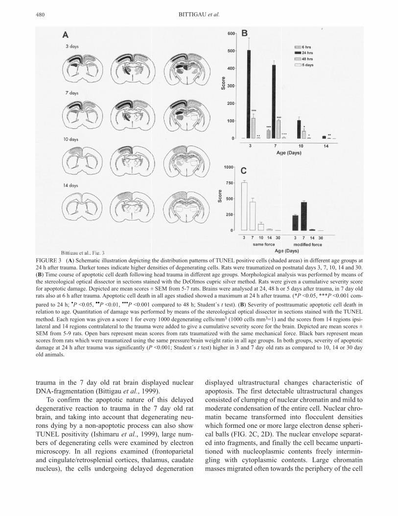
FIGURE 3 (A) Schematic illustration depicting the distribution patterns of TUNEL positive cells (shaded areas) in different age groups at24 h after trauma. Darker tones indicate higher densities of degenerating cells. Rats were traumatized on postnatal days 3, 7, 10, 14 and 30.(B) Time course of apoptotic cell death following head trauma in different age groups. Morphological analysis was performed by means ofthe stereological optical dissector in sections stained with the DeOlmos cupric silver method. Rats were given a cumulative severity scorefor apoptotic damage. Depicted are mean scores ± SEM from 5-7 rats. Brains were analysed at 24, 48 h or 5 days after trauma, in 7 day oldrats also at 6 h after trauma. Apoptotic cell death in all ages studied showed a maximum at 24 h after trauma. (*P <0.05, ***P <0.001 com-
pared to 24 h; •P <0.05, ••P <0.01, •••P <0.001 compared to 48 h; Student´s t test). (B) Severity of posttraumatic apoptotic cell death inrelation to age. Quantitation of damage was performed by means of the stereological optical dissector in sections stained with the TUNELmethod. Each region was given a score 1 for every 1000 degenerating cells/mm3 (1000 cells mm3=1) and the scores from 14 regions ipsi-lateral and 14 regions contralateral to the trauma were added to give a cumulative severity score for the brain. Depicted are mean scores ±SEM from 5-9 rats. Open bars represent mean scores from rats traumatized with the same mechanical force. Black bars represent meanscores from rats which were traumatized using the same pressure/brain weight ratio in all age groups. In both groups, severity of apoptoticdamage at 24 h after trauma was significantly (P <0.001; Student´s t test) higher in 3 and 7 day old rats as compared to 10, 14 or 30 dayold animals.

TRAUMATIC BRAIN INJURY IN DEVELOPING BRAIN 481
and in some cases the cell divided into separate inde-pendent bodies consisting of a contingent of cytoplasmand one or more nuclear chromatin balls. This type andsequence of changes are identical to changes seen inneurons undergoing physiological cell death, a naturalapoptotic process by which redundant or unsuccessfulneurons are deleted from the developing brain(Ishimaru et al., 1999) and meet established criteria fordiagnosing apoptosis (Wyllie et al., 1980). At 24 h aftertrauma, cells in both early and late stages of apoptosiswere detected, indicating that the process of cell sui-cide was still progressing. We did not identify cellsundergoing non-apoptotic degeneration by electronmicroscopy in affected brain regions at 16-24 h aftertrauma, which indicates that, at those times, apoptosisis the predominant form of degeneration, whereas up to6 h after trauma excitotoxic degeneration primarilytakes place in the infant rat brain (Ikonomidou et al.,1996; Bittigau et al., 1999).
These results demonstrate that both acute excitotoxicand slower active or apoptotic cell death occur in thecontext of traumatic brain damage in developing rats.
AGE-DEPENDENT DISTRIBUTION PATTERNSAND SEVERITY OF APOPTOTIC CELL DEATHFOLLOWING TRAUMA
We studied age dependent severity of apoptotic neu-rodegeneration following TBI by performing twoseries of experiments, one in which the brains of rats atall ages (3-30 days old) were subjected to the samemechanical force and one in which the mechanicalforce was modified in order to achieve the same pres-sure/brain weight ratio (Bittigau et al., 1999). In bothseries we detected apoptotic cell death at 24 h afterhead trauma in 3-14 day old rats. Silver and TUNELstains gave similar distribution patterns in all agegroups. The age-dependent distribution patterns areshown in figure 3.
When the same force of 160gcm was used to trauma-tize pups of all age groups, severity of apoptotic celldeath was highest in 3 day old rats and decreased sig-nificantly with increasing age (FIG. 3C). Even whenthe force was adjusted to provide the samepressure/brain weight ratio in all age groups, distantapoptotic damage in older animals remained minimalas compared to 3 and 7 day old rats. Under this exper-imental condition (fixed pressure/brain weight ratio), 7day old rats were most vulnerable to distant apoptoticdamage triggered by mechanical trauma (FIG. 3C).
Apoptotic cell death reached a peak at 24 h followingtrauma in the brains of pups at all ages studied and was
non-detectable at 5 days after the insult (FIG. 3B). In our studies in infant animals, we found that, at a
given developmental age, highest densities of apoptot-ic cells following trauma were detected in areas thatalso displayed highest densities of cells undergoingphysiologic cell death (Bittigau et al., 1999). This pos-sibly indicates that neurons and glia may be most vul-nerable to die via apoptosis when exposed to an exoge-nous insult during a certain period of their maturationand differentiation process. It has been proposed thatthe ratio of pro- versus antiapoptotic factors within acell primarily determines its vulnerability and likeli-hood to undergo active cell death (Kroemer, 1997;Hengartner, 2000). Thus, the ontogenetically regulatedexpression of potential proapoptotic factors early indevelopment, such as c-jun, c-fos, p53 (Ferrer et al.,1996), which, under physiological conditions, promotedifferentiation of immature neurons and glia may,under pathological circumstances, predispose thesesame cells to undergo suicide. P53, for example, pro-motes cell differentiation but initiates apoptotic dele-tion following irradiation (Borovitskaya et al., 1996;Norimura et al., 1996; Almog and Rotter, 1997).Progressive axonal injury and secondary axotomy aftertrauma may be an additional mechanism that promotesneuronal apoptosis, due to deafferentiation and loss oftrophic support (Maxwell et al., 1997).
Why the same traumatic insult that triggers nodetectable apoptotic cell death in the 30-day-old ratbrain gives rise to a massive, disseminated apoptoticresponse in infant rats is unclear. Certainly age-specif-ic differences in the degree of myelination and brainwater content will allow traumatic forces to transmitmore easily to deeper brain structures the more imma-ture the brain is at the time of injury. Interestingly, theinfant rat brain is most sensitive to apoptotic neurode-generation following trauma during the N-methyl-D-aspartate (NMDA) -receptor-hypersensitivity period(McDonald et al., 1988; Ikonomidou et al., 1989), withpeak vulnerability on postnatal day 7. This exactlycoincides with the age at which the infant rat brain ismost sensitive to NMDA-excitotoxicity. In view ofreports that NMDA receptor stimulation may triggerboth excitotoxic and apoptotic neurodegenerationdepending on intensity of stimulation (Bonfoco et al.,1995), the question arises whether stimulation ofNMDA receptors by endogenous glutamate, may pro-mote apoptosis following trauma in infant rats.However, while attempting to block this apoptoticresponse with NMDA antagonists, we observed anunexpected potentiating effect (Pohl et al., 1999), indi-cating that NMDA receptor blockade promotes apopto-

sis and NMDA receptor stimulation may be neuropro-tective against apoptosis. Radical scavengers andantioxidants elicited a protective effect against delayedapoptotic cell death in the infant rat TBI model (Pohl etal., 1999), suggesting contribution of free radicals inpathogenesis of this form of neurodegeneration.
Other factors likely to complicate tissue injury andpotentially further entertain apoptotic deletion are pro-gressive axonal injury and secondary axotomy aftertrauma with resulting deafferentation, decrease in thelevels of neurotrophic factors and loss of trophic sup-port, activation of glial cells and inflammatory path-ways involving death receptors. Our attempts toexplore some of these mechanisms will be illustrated inthe following.
PATHWAYS LEADING TO APOPTOTIC NEURODEGENERATION
Apoptosis can be initiated by diverse signals and exe-cuted via different biochemical pathways (Hengartner,2000). Triggers include growth factor deprivation,DNA damage, cytokine production and activation ofdeath receptors, as well as release of cytochrome cfrom the mitochondria into the cytoplasm. Althoughbiochemical pathways differ considerably, they allconverge upon activation of effector caspases(Krammer, 2000; Meier et al., 2000; Nicholson, 2000;Rich et al., 2000; Savill and Fadok, 2000; Yuan andYankner, 2000).
An intrinsic and extrinsic apoptotic pathway havebeen defined, the first initiated by release ofcytochrome c into the cytoplasm and the second byactivation of death receptors. Cytochrome c releaseleads to activation of effector caspases via recruitmentof caspase-9 (Hengartner, 2000). Aggregation of thedeath receptor Fas (CD95/Apo-1), a member of theTNF-α superfamily, follows Fas ligand binding andleads to formation of a death-inducing signaling com-plex (DISC): Fas itself, an adapter protein named Fasassociated death domain (FADD) and the inactive formof caspase-8 (Martin-Villalba et al., 1999). After for-mation of the DISC, procaspase-8 is proteolyticallycleaved, activated and released from the DISC(Chinnaiyan et al., 1995; Muzio et al., 1996; Medemaet al., 1997; Krammer, 2000). Caspase-8 then activatesdownstream caspases, such as caspase-3, which exe-cute the cell.
Activation of caspases comprises a subsequent criti-cal step within the apoptotic cascade. Caspases con-tribute to cell cleavage via inactivation of nucleaseinhibitors and survival proteins, direct disassembly of
cell structures, destruction of proteins involved incytoskeleton regulation and inactivation of proteinsinvolved in DNA repair and replication (Thornberryand Lazebnik, 1998).
To investigate involvement of the intrinsic apoptoticpathway in the pathogenesis of apoptotic neurodegen-eration following trauma to the developing brain, weanalyzed changes in the expression of antiapoptoticproteins of the bcl-2 group that decrease mitochondri-al membrane permeability, changes in cytochrome cimmunoreactivity in the cytosolic fraction and changesin caspase-9 activity in the infant rat brain traumamodel. To investigate involvement of the extrinsicpathway, Fas-expression and caspase-8 activity inbrain tissue were measured. To investigate the role ofneurotrophins, endogenous mRNA levels for neu-rotrophin-3 (NT-3) and brain derived neurotrophic fac-tor (BDNF) were analyzed. Finally, to test the potentialbenefit of caspase inhibition in TBI to the developingbrain, the pancaspase inhibitor z-VAD.FMK wasadministered to infant rats and neurodegeneration wasquantitated. Our findings indicate that trauma leads toactivation of the intrinsic and the extrinsic apoptoticpathways in the developing rat brain and that inhibitionof effector caspases confers neuroprotection over atime window of at least 8 h after trauma.
TRAUMA-INDUCED CHANGES IN CASPASE-3-LIKE ENZYMATIC ACTIVITY AND DNA-BREAKDOWN IN THE INFANT RAT BRAIN
Head trauma induced significant elevations of caspase3/CPP32-like activity and DNA fragmentation(oligonucleosomes) in extracts from ipsilateral cingu-late and parietal cortex, thalamus, striatum and hip-pocampus as measured at 24 h after trauma (Bittigau etal., 1999).
These findings indicate that activation of CPP32-likecaspases play a critical role in active cell deletion fol-lowing trauma to the immature brain. In contrast to theadult brain, CPP32-like proteolytic activity rose notjust up to 130%, as described in a study by Yakovlev etal. (1997), but up to 2,830% of control values.Together with our histological data, this highlights thedisproportionately large magnitude of the apoptosiscontribution to posttraumatic brain damage in theimmature brain. Using combined optical dissectorstereology and volumetry we calculated that in thebrains of 7 day old rats subjected to head trauma, mil-lions of cells were dying an apoptotic death in the brainat 24 h after trauma as opposed to a few thousand cellsdying an excitotoxic death at 4 h after trauma (Bittigau
BITTIGAU et al.482

TRAUMATIC BRAIN INJURY IN DEVELOPING BRAIN 483
FIGURE 4 (A) Bcl-2 (left) and Fas (right) mRNA expression in right thalamus (ipsilateral to trauma site) in a sham operated rat at 0 hrsafter trauma and in rats subjected to head trauma at 2-120 h after trauma. mRNA was reverse transcribed to cDNA, amplified by polymerasechain reaction using specific primers for bcl-2 and ß-actin and subjected to polyacrylamide gel electrophoresis and silver staining. There isobvious decrease in mRNA levels for bcl-2. There is an increase in mRNA levels for Fas at 4 h, peaking at 12 h after the insult and lastingup to 24 h. These are representative gels from a series performed to analyze bcl-2 and Fas mRNA expression. On the right, Western blotanalysis for Fas and bcl-xL in brain extracts prepared from the thalamus of sham operated rats (C) and rats traumatized on day 7, at 4-24 hafter trauma. Representative blots of a series performed (thalamus, striatum, cortex), demonstrating an increase in Fas and decrease in bcl-xL
protein expression 4 h after trauma in the ipsilateral thalamus. Western blot analysis of cytochrome c immunoreactivity in the cytoplasmicfractions of brain extracts taken from the thalamus of a sham operated rat (C) and rats traumatized on day 7, at 2, 4 and 12 h after traumais also shown. There is an increase in cytochrome c immunoreactivity in the cytosolic fraction by 2 h after trauma. (B) The results of den-sitometric analysis of bcl-2 and bcl-xL specific mRNA-bands on the gels from traumatized rats are presented in reference to ß-actin. Datarepresent the ratio (%) of the density of the bcl-2 or bcl-xL band to the ß-actin band ± SEM. Two way ANOVA revealed that trauma had ahighly significant effect on the mRNA-levels for bcl-2 [F(7,44)=40.9, P<0.001] and bcl-xL [F(7,44)=31.71, P<0.001] in the thalamus.There was a highly significant effect of time (posttraumatic interval) on the mRNA-levels for bcl-2 [F(1,44)=155.1, P<0.001] and bcl-xL
[F(1,44)=648.8] in the thalamus as well, compared to sham operated rats. (C ) The results of densitometric analysis of the Fas specificmRNA bands from the thalamus of traumatized rats are presented in reference to ß-actin. Data represent the ratio (%) of the density of theFas band to the ß-actin band ± SEM. Comparison between sham rats and rats subjected to brain trauma by ANOVA revealed that traumahad a highly significant effect on Fas mRNA levels in the thalamus [F(1,44) = 804.5, P<0.001]. (D) Caspase-9 activity in cytosolic proteinextracts from thalamus of sham rats and rats subjected to head trauma. Specimen were analyzed at 4, 12 and 24 h after trauma or sham sur-gery. Caspase-9-like activity was measured fluorometrically, using the specific substrate LEHD and by determining accumulation of freeaminotrifluoromethyl coumarin (AFC). Data are expressed in relative fluorescent units (RFU) as means ± SEM after subtraction of theappropriate buffer controls. The numbers in parentheses represent the number of specimen in each group. Two way ANOVA revealed thattrauma had a highly significant effect on caspase-9 activity in the thalamus [F(1,30) = 17.56, P<0.001]. The grey column depicts recombi-nant caspase-9 activity which served as control. (E) Caspase-8 activity in cytosolic protein extracts from right thalamus in rats subjected tohead trauma compared to sham rats. Specimens were analyzed at 4, 12 and 24 h after trauma or sham surgery. Caspase-8 like activity wasmeasured fluorometrically using the specific substrate IETD and by determining accumulation of free AFC. Data are expressed in relativefluorescent units (RFU) as the mean ratios ± SEM of signal obtained in specimen from traumatized and sham operated brains after sub-traction of the appropriate buffer controls. The numbers in parentheses represent the number of specimen in each group. Two way ANOVArevealed that trauma had a highly significant effect on caspase-8 activity in the thalamus [F(1,30) = 63.27, P<0.001]. The grey columndepicts recombinant caspase-8 activity which served as control.

et al., 1999). Since apoptotic cells can be detected his-tologically for a few hours (clearance time, 2 h and 20min) (Thomaidou et al., 1997) and apoptosis is occur-ring in the infant rat brain for several days after trauma,the numbers of cells eventually deleted by this mecha-nism are much higher.
ACTIVATION OF THE INTRINSIC APOPTOTIC PATHWAY BY TRAUMA
Downregulation in the Expression of AntiapoptoticGenesThe expression of bcl-2 and bcl-xL, two proteins with
antiapoptotic properties which have been shown todecrease mitochondrial membrane permeability wasfirst investigated at the transcriptional level. Traumatriggered marked and rapid downregulation in theexpression of bcl-2- and bcl-xL-specific mRNA in thal-
amus and cingulate cortex, which was evident within 2h following the insult, persisted up to 48 h and demon-strated a slow, incomplete recovery by 120 h after trau-ma (FIG. 4A, 4B).
Downregulation of bcl-2 family members was con-firmed at the protein level in that immunoreactivity ofbcl-xL was analyzed by Western blotting in brain
extracts from thalamus, striatum and cortex. Decreasedlevels of bcl-xL protein were found in these areas at
various time points after trauma (FIG. 4A).
Cytochrome c Release and Caspase-9 ActivationCytochrome c immunoreactivity was analyzed byWestern blotting in the cytosolic fraction of brainextracts from thalamus, striatum and cortex of 7 dayold rats subjected to head trauma. Cytochrome cimmunoreactivity increased at 2 h after trauma in thecytosolic fraction (FIG. 4A), at a time point when nosigns of delayed neurodegeneration were detectable byhistological techniques.
The activity of the initiator caspase-9 was measuredin thalamic tissue using the specific substrate (LEHD)in sham rats and rats subjected to head trauma.Compared to sham operated rats, there was a signifi-cant increase of caspase-9 activity in the thalamus inrats subjected to head trauma at 12 and 24 h after trau-ma (FIG. 4D).
Activation of the intrinsic apoptotic pathway has pre-viously been reported in in vivo trauma models(Morita-Fujimura et al., 1999; Raghupathi et al. 2000;Keane et al. 2001). How cytochrome c manages tocross the mitochondrial membrane is not understood.In all proposed models (Hengartner, 2000), members
of the bcl-2 family play a key role in that they decreasemitochondrial membrane permeability and preventrelease of cytochrome c into the cytoplasm (Nicholson,2000). In the infant rat brain we demonstrate downreg-ulation of the expression of bcl-2 and bcl-xL following
trauma, which is expected to result in increased per-meability of the mitochondrial membranes. Changes atthe mRNA levels correlated with decreased proteinlevels (Felderhoff-Mueser et al., 2002). Reasons fordecreased expression of antiapoptotic bcl-2 family pro-teins remain unclear. Transcription of antiapoptoticbcl-2 family members is influenced by CREB, whoseactivity level is regulated by growth factors (Xing etal., 1996). It has been shown that release of trophic fac-tors and their trophic effects on developing neuronsdepend upon the level of neuronal activity (McCallister et al., 1996; Liou and Fu, 1997). Spreadingdepression triggered by trauma disrupts physiologicalsynaptic activity. Even in the presence of increasedneurotrophin levels, it is possible that disruption ofphysiological synaptic activity may lead to impairmentof intracellular neurotrophin-initiated signaling path-ways and result in decrease in the transcription of sur-vival genes.
APOPTOSIS BY DEATH RECEPTOR ACTIVA-TION (EXTRINSIC PATHWAY) FOLLOWINGTRAUMA TO THE DEVELOPING BRAIN
Changes in expression of the death receptor Fas fol-lowing trauma to the 7 day old rat brain were deter-mined at defined time points post-injury. Trauma trig-gered increase of Fas-mRNA levels at 4 h after traumawhich lasted up to 24 hours and subsequentlydecreased to pre-trauma levels (FIG. 4A). Increasedprotein levels for Fas were found in the ipsilateral thal-amus, striatum and cortex starting at 4 hours after trau-ma, with this increase being most pronounced at 12and 24 h after trauma (FIG. 4A).
In the intact developing brain there is moderate phys-iological expression of Fas. Fas immunoreactivityincreased particularly in the cortex and in the thalamusipsilateral to the injury after trauma (FIG. 4A, 4C). At48 h, reduction of Fas immunoreactivity occurred inaffected brain regions, possibly reflecting evacuationof cells that overexpressed the receptor (Felderhoff etal., 2002).
To further confirm activation of the extrinsic apop-totic pathway following trauma, caspase-8 activity wasmeasured fluorometrically in the thalamus using thespecific caspase-8 substrate IETD. Trauma had a high-ly significant effect on caspase-8 activity in the thala-
BITTIGAU et al.484
mus after trauma (FIG. 4E). Previous studies havedemonstrated Fas expression in the context of traumato the adult brain and in infant hypoxia-ischemia(Nakashima et al., 1999; Beer et al., 2000; Felderhoff-Mueser et al., 2000). Our findings imply Fas receptorinvolvement in activation of caspase-8 after neonatalbrain trauma but do not exclude the possibility thatother death receptors and their ligands (TNF, TRAIL)may also contribute to activation of this extrinsic apop-totic pathway.
A cross talk between the extrinsic pathway and themitochondria has been postulated (Hengartner, 2000).Activation of caspase-8 leads to proteolysis of theproapoptotic protein bid. Truncated bid enters themitochondria and promotes cytochrome c release.Time course studies suggest that activation of caspase-8 occurs early (within 4 h) after trauma. Therefore, it ispossible that caspase-8 mediated bid cleavage mayconstitute one additional mechanism to facilitaterelease of cytochrome c into the cytoplasm.
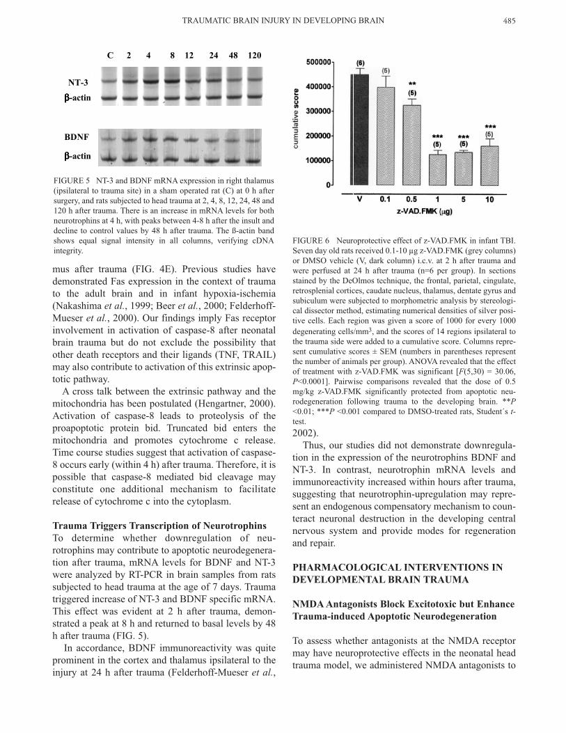
Trauma Triggers Transcription of NeurotrophinsTo determine whether downregulation of neu-rotrophins may contribute to apoptotic neurodegenera-tion after trauma, mRNA levels for BDNF and NT-3were analyzed by RT-PCR in brain samples from ratssubjected to head trauma at the age of 7 days. Traumatriggered increase of NT-3 and BDNF specific mRNA.This effect was evident at 2 h after trauma, demon-strated a peak at 8 h and returned to basal levels by 48h after trauma (FIG. 5).
In accordance, BDNF immunoreactivity was quiteprominent in the cortex and thalamus ipsilateral to theinjury at 24 h after trauma (Felderhoff-Mueser et al.,
2002). Thus, our studies did not demonstrate downregula-
tion in the expression of the neurotrophins BDNF andNT-3. In contrast, neurotrophin mRNA levels andimmunoreactivity increased within hours after trauma,suggesting that neurotrophin-upregulation may repre-sent an endogenous compensatory mechanism to coun-teract neuronal destruction in the developing centralnervous system and provide modes for regenerationand repair.
PHARMACOLOGICAL INTERVENTIONS IN DEVELOPMENTAL BRAIN TRAUMA
NMDA Antagonists Block Excitotoxic but EnhanceTrauma-induced Apoptotic Neurodegeneration
To assess whether antagonists at the NMDA receptormay have neuroprotective effects in the neonatal headtrauma model, we administered NMDA antagonists to
TRAUMATIC BRAIN INJURY IN DEVELOPING BRAIN 485
C 2 4 8 12 24 48 120
NT-3
BDNF
ββββ-actin
ββββ-actin
FIGURE 5 NT-3 and BDNF mRNA expression in right thalamus(ipsilateral to trauma site) in a sham operated rat (C) at 0 h aftersurgery, and rats subjected to head trauma at 2, 4, 8, 12, 24, 48 and120 h after trauma. There is an increase in mRNA levels for bothneurotrophins at 4 h, with peaks between 4-8 h after the insult anddecline to control values by 48 h after trauma. The ß-actin bandshows equal signal intensity in all columns, verifying cDNAintegrity.
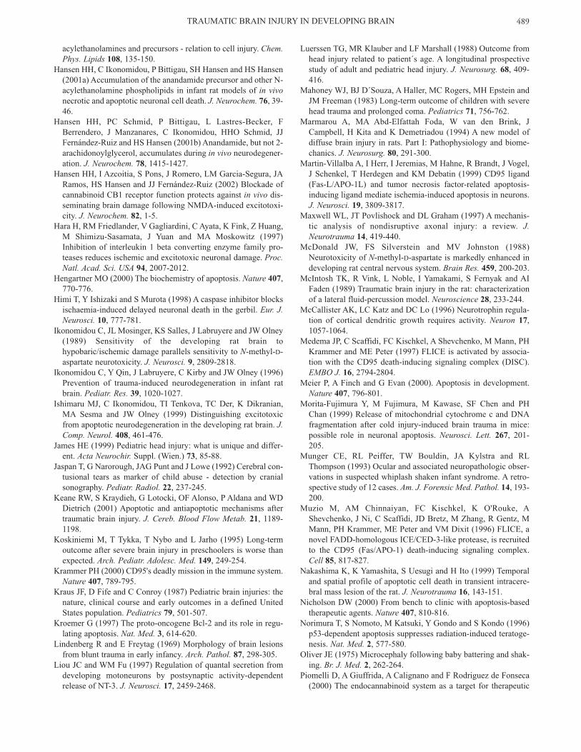
FIGURE 6 Neuroprotective effect of z-VAD.FMK in infant TBI.Seven day old rats received 0.1-10 µg z-VAD.FMK (grey columns)or DMSO vehicle (V, dark column) i.c.v. at 2 h after trauma andwere perfused at 24 h after trauma (n=6 per group). In sectionsstained by the DeOlmos technique, the frontal, parietal, cingulate,retrosplenial cortices, caudate nucleus, thalamus, dentate gyrus andsubiculum were subjected to morphometric analysis by stereologi-cal dissector method, estimating numerical densities of silver posi-tive cells. Each region was given a score of 1000 for every 1000degenerating cells/mm3, and the scores of 14 regions ipsilateral tothe trauma side were added to a cumulative score. Columns repre-sent cumulative scores ± SEM (numbers in parentheses representthe number of animals per group). ANOVA revealed that the effectof treatment with z-VAD.FMK was significant [F(5,30) = 30.06,P<0.0001]. Pairwise comparisons revealed that the dose of 0.5mg/kg z-VAD.FMK significantly protected from apoptotic neu-rodegeneration following trauma to the developing brain. **P<0.01; ***P <0.001 compared to DMSO-treated rats, Student´s t-test.

7 day old rats and evaluated the extent of excitotoxicand apoptotic neurodegeneration following such treat-ment.
Quantitative evaluation 4 h after trauma of the brainsof 7-day-old rats treated with the NMDA antagonistdizocilpine 1 h prior to trauma revealed reduction ofthe extent of excitotoxic damage in the parietal cortex(Ikonomidou et al., 1996; Pohl et al., 1999).
Quantitative evaluation of the brains 24 h after headtrauma however revealed that the apoptotic degenera-tion was more severe in rats treated with dizocilpinethan in those subjected to vehicle (Pohl et al., 1999).NMDA antagonists also gave rise to apoptotic degen-eration in the brains of 7-day-old sham-operated rats,indicating that they promote physiological apoptosis atthis age. In 7-day-old rats subjected to head trauma andsubsequent treatment with NMDA antagonists theseverity of apoptotic cell death was higher (by 27%after treatment with dizocilpine and 37% after treat-ment with CPP) than would be expected if the com-bined effect were due to a simple additive mechanism(Pohl et al., 1999).
These results indicate that NMDA antagonists haveopposite effects on the two components of TBI in thedeveloping brain; they reduce the relatively small exci-totoxic component, while markedly increasing the sub-stantially larger apoptotic component, the net effectbeing increased neurodegeneration. Thus, NMDAantagonists are singularly unsuitable for neuroprotec-tion in TBI to the developing brain.
Treatment with the Pancaspase Inhibitorz-VAD.FMK Blocks Trauma-induced ApoptoticCell Death
To assess the neuroprotective potential of caspase-inhi-bition in TBI in infant rats, the pancaspase inhibitorz-VAD.FMK was administered intracerebroventricu-larly (i.c.v.) to 7 day old rats in doses of 0.1-10 µg at 2h after trauma. Treatment with z-VAD.FMK had a sig-nificant effect on severity of apoptotic brain damagefollowing trauma. Rats receiving a single dose of z-VAD.FMK displayed a reduction in cumulativescores for degenerating cells (FIG. 6) compared to ratssubjected to head trauma and i.c.v. injection of DMSO.
The therapeutic time window for z-VAD.FMK was atleast 8 h, since delayed administration of z-VAD.FMKup to 8 h following trauma resulted in lower scores forapoptotic brain damage compared to vehicle treatedrats (Felderhoff-Mueser et al., 2002).
In order to exclude the possibility that caspase inhi-bition might delay but not inhibit apoptosis, we inject-
ed z-VAD.FMK (1 µg) 2 h after trauma to 7 day oldrats and analyzed their brains at 24, 48 h and 5 daysafter trauma. A protective effect of z-VAD.FMK wasstill evident at 48 h after trauma in comparison to vehi-cle. At 5 days after trauma densities of degeneratingcells in both vehicle and z-VAD.FMK treated rats wereequally low and did not significantly differ betweengroups (Felderhoff et al., 2002)
Finally, to provide additional evidence that treatmentwith z-VAD.FMK offered lasting protection againstTBI, we injected 7 day old rats subjected to brain trau-ma i.c.v. with the protective dose of 1 µg z-VAD.FMKor vehicle at 2 h after trauma. All animals were sacri-ficed 7 days after trauma or sham surgery without tran-scardial perfusion, the forebrains were hemisected andtheir weights monitored. Trauma resulted in significantweight reduction of the right (traumatized) hemispherein vehicle treated rats compared to the non traumatizedleft side (Felderhoff-Mueser et al., 2002). Treatmentwith z-VAD.FMK resulted also in a significant but lesspronounced reduction of right hemispheric weightscompared to vehicle treated traumatized rats and shamoperated rats. These time studies suggest ameliorationof the neurodegenerative response to trauma(Felderhoff et al., 2002).
In adult animal models, caspase inhibition may con-fer neuroprotection in cerebral ischemia (Hara et al.,1997; Endres et al., 1998; Fink et al., 1998; Himi et al.,1998). Furthermore, early treatment of experimentalpneumococcal meningitis with z-VAD.FMK wasshown to have a beneficial effect, whereas delayedapplication of this compound did not result in substan-tial reduction of neuronal loss (Braun et al., 1999). Intraumatic injury to the adult brain, z-VAD.FMK andthe selective caspase-3 inhibitor z-DEVD.FMK canblock neuronal death (Yakovlev et al., 1997; Clark etal., 2000b). In hippocampal and cortical neuronal cul-tures, the cell permeable pancaspase inhibitor boc-aspartyl(OMe)-fluoromethylketone (BAF) and themore selective caspase-8 inhibitor IETD-FMK (IETD)reduced Fas-induced apoptosis (Felderhoff-Mueser etal., 2000). The only existing in vivo study on caspaseinhibition in the immature brain demonstrated neuro-protection with the pancaspase inhibitor BAF in aninfant model of hypoxic-ischemic injury (Cheng et al.,1998). Our data indicate a beneficial effect of caspaseinhibition in brain trauma for neuronal death occurringdistant to the impact site. More importantly, the pro-tective effect could be achieved even when the com-pound was administered in a delayed fashion, indicat-ing relevance in the clinical setting.
BITTIGAU et al.486

TRAUMA ACTIVATES THE ENDOGENOUSCANNABINOID LIGAND-RECEPTOR SYSTEM
Transcriptional mRNA expression and ligand bindingcapacity of cerebral cannabinoid receptors wereassessed at defined time points post-trauma and com-pared to levels of the endogenous cannabinoid receptorligands, anandamide and 2-arachidonoyl glycerol (2-AG). While loss of a neuron-specific mRNA markerwas observed after induction of head trauma, levels ofcannabinoid CB1 receptor mRNA transcription and lig-
and binding capacity were upregulated in the ipsilater-al cerebral cortex. These alterations were most promi-nent in the proximity of the impact site of the contusiveforce (Hansen et al., 2001a). Accumulation of anan-damide and its precursor, but not 2-AG, was apparentin the ipsilateral cortex after induction of head trauma,in particular 24 hours post-injury (Hansen et al.,2001a,b), which suggest an enhanced activity of theendogenous cannabinoid receptor-ligand system in thedeveloping brain as a response to contusive head trau-ma.
Since central presynaptically located cannabinoidreceptors suppress the activity of a range of neuro-transmitter systems, including glutamatergic andGABAergic neurotransmission (Schlicker andKathmann, 2001; Wilson and Nicoll, 2002), andendogenous cannabinoids are found in much higherconcentrations when neurons are in an injured state(Hansen et al., 2000), it is believed that these sub-stances and their receptors may constitute a putativeendogenous response aiming at dampening thedestructive impact of brain insults (Hansen et al., 2000;Piomelli et al., 2000). Paradoxically, exogenouslyinduced overactivation of neuronal cannabinoid recep-tors can also induce apoptotic-like neurodegeneration,presumably depending on the cell type, cell develop-mental state and degree of activation (Downer et al.,2001; Guzmán et al., 2001). Also, blockade of cannabi-noid receptors induces neuroprotection in a develop-mental model of NMDA-induced excitotoxic braindamage (Hansen et al., 2002). It therefore remains tobe established under what circumstances and in whichdirection intrinsically upregulated cannabinoid ligand-receptor function affects pro-survival signaling path-ways in the immature brain and whether the endoge-nous cannabinoid system may represent a target forpharmacotherapy in pediatric TBI.
CONCLUSIONS
Traumatic injury to the developing central nervous sys-
tem has two major components, an acute excitotoxiccomponent at the site of the insult and a delayed apop-totic component affecting the impact site as well asdeeper brain structures. The number of brain cellsaffected by the apoptotic component is disproportion-ally larger than the number of cells degenerating by anexcitotoxic mechanism (Bittigau et al., 1999). Givenour findings, targeting the downstream effectors ofneuronal apoptosis in the acute phase of the insult hastherapeutic potential in the treatment of traumaticinjury to the immature brain. Antiapoptotic therapiesmay give cells enough time to establish intrinsic pro-tection systems and restore cellular homeostasis andfunction (Han and Holtzman, 2000). However, sinceapoptosis is also a physiological process in the devel-oping brain, studies addressing the long-term function-al effects following caspase inhibition appear to bepotential targets for future research (Gillardon et al.,1999).
AcknowledgementsThis work was supported by BMBF grant01KO95151TPA3 and a Rahel-Hirsch award from theHumboldt University.
ReferencesAdelson PD, P Robichaud, RL Hamilton and PM Kochanek (1996)
A model of diffuse traumatic brain injury in the immature rat. J.Neurosurg. 85, 877-884.
Adelson PD, CE Dixon, P Robichaud and PM Kochanek (1997)Motor and cognitive deficits following diffuse traumatic braininjury in the immature rat. J. Neurotrauma 14, 99-108.
Adelson PD and PM Kochanek (1998) Head injury in children. J.Child Neurol. 13, 2-15.
Aldrich EF, HM Eisenberg, C Saydjari, TG Luerssen, MA Foulkes,JA Jane, LF Marshall, A Marmarou and HF Young (1992)Diffuse brain swelling in severely head-injured children. Areport from the NIH Traumatic Coma Data Bank. J. Neurosurg.76, 450-454.
Allen AR (1911) Surgery of experimental lesion of spinal cordequivalent to crush injury of fracture dislocation of spinal col-umn. J. Am. Med. Assoc. 57, 878-880.
Almog N and V Rotter (1997) Involvement of p53 in cell diffenti-ation and development. Biochim. Biophys. Acta 1333, F1-F27.
BarlowKM and RA Minns (2000) Annual incidence of shakenimpact syndrome in young children. Lancet 356, 1571-1572.
Bayir H, VA Kagan, YY Tyurina, V Tyurin, RA Ruppel, PDAdelson, SH Graham, K Janesko, RSB Clark and PM Kochanek(2002) Assessment of antioxidant reserves and oxidative stressin cerebrospinal fluid after severe traumatic brain injury ininfants and children. Pediatric Res. 51, 571-578.
Beer R, G Franz, M Schopf, M Reindl, B Zelger, E Schmutzhard,W Poewe and A Kampfl. (2000) Expression of Fas and Fas lig-and after experimental traumatic brain injury in the rat. J. Cereb.Blood Flow Metab. 20, 669-677.
Bell MJ, PM Kochanek, LA Doughty, JA Carcillo, PD Adelson,RSB Clark, SR Wisniewski, MJ Whalen and ST DeKosky
TRAUMATIC BRAIN INJURY IN DEVELOPING BRAIN 487

(1997) Interleukin-6 and interleukin-10 in cerebrospinal fluidafter severe traumatic brain injury in children. J. Neurotrauma14, 451-457.
Berger RP, MC Pierce, SR Wisniewski, PD Adelson and PMKochanek (2002) Serum S100B concentrations are increasedafter closed head injury in children: a preliminary study. J.Neurotrauma 19, 1405-1409.
Bittigau P, M Sifringer, D Pohl, D Stadthaus, M Ishimaru, HShimizu, M Ikeda, D Lang, A Speer, JW Olney and CIkonomidou (1999) Apoptotic neurodegeneration following trau-ma is markedly enhanced in the immature brain. Ann. Neurol.45, 724-735.
Bonfoco E, D Krainc, M Ankarcron, P Nicotera and SA Lipton(1995) Apoptosis and necrosis: two distinct events induced,respectively, by mild and intense insults with N-methyl-D-aspar-tate or nitric oxide/superoxide in cortical cell cultures. Proc.Natl. Acad. Sci. USA 92, 7162-7166.
Bonnier C, MC Nassogne and P Evrard (1995) Outcome and prog-nosis of whiplash shaken infant syndrome: late consequencesafter a symptom free interval. Dev. Med. Child Neurol. 37, 943-956.
Bonnier C, B Mespiès, S Carpentier, D Henin, and P Gressens(2002) Delayed white matter injury in a murine model of shakenbaby syndrome. Brain Pathol. 12, 320-328.
Borovitskaya AE, VI Evtushenko and SL Sabol (1996) Gamma-radiation-induced cell death in the fetal rat brain possessesmolecular characteristics of apoptosis and is associated with spe-cific messenger RNA elevations. Brain Res. Mol. Brain Res. 35,19-30.
Braun JS, R Novak, KH Herzog, SM Bodner, JL Cleveland and EITuomanen (1999) Neuroprotection by a caspase inhibitor inacute bacterial meningitis. Nat. Med. 5, 298-302.
Brown JK and RA Minns (1993) Non-accidental head injury, withparticular reference to whiplash shaking injury and medico-legalaspects. Dev. Med. Child Neurol. 35, 849-869.
Cheng Y, M Deshmukh, A D'Costa, JA Demaro, JM Gidday, AShah, Y Sun, MF Jacquin, EM Johnson and DM Holtzman(1998) Caspase inhibitor affords neuroprotection with delayedadministration in a rat model of neonatal hypoxic-ischemic braininjury. J. Clin. Invest. 101, 1992-1999.
Chinnaiyan AM, K O´Rourke, M Tewari and VM Dixit (1995)FADD, a novel death domain-containing protein interacts withthe death domain of Fas and initiates apoptosis. Cell 81, 505-512.
Clark RSB, PM Kochanek, PD Adelson, MJ Bell, JA Carcillo, MChen, SR, Wisniewski, K Janesko, MJ Whalen and SH Graham(2000a) Increases in bcl-2 protein in cerebrospinal fluid and evi-dence for programmed cell death in infants and children aftersevere traumatic brain injury. J. Pediatr. 137, 197-204.
Clark RSB, PM Kochanek, SC Watkins, M Chen, CE Dixon, NASeidberg, J Melick, JE Loeffert, PD Nathaniel, KL Jin and SHGraham (2000b) Caspase-3 mediated neuronal death after trau-matic brain injury in rats. J. Neurochem. 74, 740-753.
David TJ (1999) Shaken baby (shaken impact) syndrome: non-accidental head injury in infancy. J. R. Soc. Med. 92, 556-561.
DeOlmos JS and WR Ingram (1971) An improved cupric-silvermethod for impregnation of axonal and terminal degeneration.Brain Res. 33, 523-529.
Diamond PT (1996) Brain injury in the Commonwealth of Virginia:an analysis of Central registry data 1988-1993. Brain Injury 10,414-419.
Dixon CE, GL Clifton, JW Lighthall, A Yaghmai and RL Hayes
(1991) A controlled cortical impact model of traumatic braininjury in the rat. J. Neurosci. Meth. 39, 253-262.
Downer E, B Boland, M Fogarty and V Campbell (2001) Delta9-tetrahydrocannabinol induces the apoptotic pathway in culturedcortical neurones via activation of the CB1 receptor. Neuroreport12, 3973-3978.
Duhaime AC, CW Christian, E Moss and T Seidl (1996) Long-termoutcome in infants with the shaking impact syndrome. Pediatr.Neurosurg. 24, 292-298.
Duhaime AC, CW Christian, LB Rorkea and RA Zimmerman(1998) Nonaccidental head injury in infants - The shaken-babysyndrome. N. Engl. J. Med. 338, 1822-1829.
Endres M, S Namura, M Shimizu-Sasamata, C Weber, L Zhang, TGomez-Isla, BT Hyman and MA Moskowitz (1998) Attenuationof delayed neuronal death after mild focal ischemia in mice byinhibition of the caspase family. J. Cereb. Blood Flow Metab. 18,238-247.
Feeney DM, MG Boyeson, R Linn, HM Murray and WG Dail(1981) Responses to cortical injury: I. Methodology and localeffects of contusions in the rat. Brain Res. 211, 67-77.
Felderhoff-Mueser U, DL Taylor, K Greenwood, M Kozma, DStibenz, UC Joashi, AD Edwards and H Mehmet (2000)Fas/CD95/Apo-1 can function as a death receptor for neuronalcells in vitro and in vivo and is upregulated following cerebralhypoxic-ischemic injury to the developing rat brain. BrainPathol. 10, 17-29.
Felderhoff-Mueser U, M Sifringer, S Pesditschek, H Kuckuck, AMoysich, P Bittigau and C Ikonomidou (2002) Pathways leadingto apoptotic neurodegeneration following trauma to the develop-ing rat brain. Neurobiol. Dis. 11, 231-245.
Ferrer I, M Olivé, J Ribera and AM Planas (1996) Naturally occur-ing (programmed) and radiatio-induced apoptosis are associatedwith selective c-jun expression in the developing rat brain. Eur.J. Neurosci. 8, 1286-1298.
Fink K, J Zhu, S Namura, M Shimizu-Sasamata, M Endres, J Ma,T Dalkara, J Yuan and MA Moskowitz (1998) Prolonged thera-peutic window for ischemic brain damage caused by delayedcaspase activation. J. Cereb. Blood Flow Metab. 18, 1071-1076.
Geddes JF, GH Vowles, JA Nicoll and T Revesz (1999) Neuronalcytoskeletal changes are an early consequence of repetitive headinjury. Acta Neuropathol. (Berl.) 98, 171-178.
Geddes JF, AK Hackshaw, GH Vowles, CD Nickols and HLWhitwell (2001a) Neuropathology of inflicted head injury inchildren I. Patterns of brain damage. Brain 124, 1290-1298.
Geddes JF, GH Vowles, AK Hackshaw, CD Nickols, IS Scott andHL Whitwell (2001b) Neuropathology of inflicted head injury inchildren II. Microscopic brain injury in infants. Brain 124, 1299-1306.
Gillardon F, I Kiprianova, J Sandkuhler, HA Hossmann and MSpranger (1999) Inhibition of caspases prevents cell death ofhippocampal CA1 neurons, but not impairment of hippocampallong-term potentiation following global ischemia. Neuroscience93, 1219-1222.
Goldstein M (1990) Traumatic brain injury: a silent epidemic. Ann.Neurol. 27, 327.
Guzmán M, C Sánchez and I Galve-Roperh (2001) Control of thecell survival/death decision by cannabinoids. J. Mol. Med. 78,613-625.
Han BH and DM Holtzman (2000) BDNF protects the neonatalbrain from hypoxic-ischemic injury in vivo via the ERK path-way. J. Neurosci. 20, 5775-5781.
Hansen HS, B Moesgaard, HH Hansen and G Petersen (2000) N-
BITTIGAU et al.488
acylethanolamines and precursors - relation to cell injury. Chem.Phys. Lipids 108, 135-150.
Hansen HH, C Ikonomidou, P Bittigau, SH Hansen and HS Hansen(2001a) Accumulation of the anandamide precursor and other N-acylethanolamine phospholipids in infant rat models of in vivonecrotic and apoptotic neuronal cell death. J. Neurochem. 76, 39-46.
Hansen HH, PC Schmid, P Bittigau, L Lastres-Becker, FBerrendero, J Manzanares, C Ikonomidou, HHO Schmid, JJFernández-Ruiz and HS Hansen (2001b) Anandamide, but not 2-arachidonoylglycerol, accumulates during in vivo neurodegener-ation. J. Neurochem. 78, 1415-1427.
Hansen HH, I Azcoitia, S Pons, J Romero, LM Garcia-Segura, JARamos, HS Hansen and JJ Fernández-Ruiz (2002) Blockade ofcannabinoid CB1 receptor function protects against in vivo dis-seminating brain damage following NMDA-induced excitotoxi-city. J. Neurochem. 82, 1-5.
Hara H, RM Friedlander, V Gagliardini, C Ayata, K Fink, Z Huang,M Shimizu-Sasamata, J Yuan and MA Moskowitz (1997)Inhibition of interleukin 1 beta converting enzyme family pro-teases reduces ischemic and excitotoxic neuronal damage. Proc.Natl. Acad. Sci. USA 94, 2007-2012.
Hengartner MO (2000) The biochemistry of apoptosis. Nature 407,770-776.
Himi T, Y Ishizaki and S Murota (1998) A caspase inhibitor blocksischaemia-induced delayed neuronal death in the gerbil. Eur. J.Neurosci. 10, 777-781.
Ikonomidou C, JL Mosinger, KS Salles, J Labruyere and JW Olney(1989) Sensitivity of the developing rat brain tohypobaric/ischemic damage parallels sensitivity to N-methyl-D-aspartate neurotoxicity. J. Neurosci. 9, 2809-2818.
Ikonomidou C, Y Qin, J Labruyere, C Kirby and JW Olney (1996)Prevention of trauma-induced neurodegeneration in infant ratbrain. Pediatr. Res. 39, 1020-1027.
Ishimaru MJ, C Ikonomidou, TI Tenkova, TC Der, K Dikranian,MA Sesma and JW Olney (1999) Distinguishing excitotoxicfrom apoptotic neurodegeneration in the developing rat brain. J.Comp. Neurol. 408, 461-476.
James HE (1999) Pediatric head injury: what is unique and differ-ent. Acta Neurochir. Suppl. (Wien.) 73, 85-88.
Jaspan T, G Narorough, JAG Punt and J Lowe (1992) Cerebral con-tusional tears as marker of child abuse - detection by cranialsonography. Pediatr. Radiol. 22, 237-245.
Keane RW, S Kraydieh, G Lotocki, OF Alonso, P Aldana and WDDietrich (2001) Apoptotic and antiapoptotic mechanisms aftertraumatic brain injury. J. Cereb. Blood Flow Metab. 21, 1189-1198.
Koskiniemi M, T Tykka, T Nybo and L Jarho (1995) Long-termoutcome after severe brain injury in preschoolers is worse thanexpected. Arch. Pediatr. Adolesc. Med. 149, 249-254.
Krammer PH (2000) CD95's deadly mission in the immune system.Nature 407, 789-795.
Kraus JF, D Fife and C Conroy (1987) Pediatric brain injuries: thenature, clinical course and early outcomes in a defined UnitedStates population. Pediatrics 79, 501-507.
Kroemer G (1997) The proto-oncogene Bcl-2 and its role in regu-lating apoptosis. Nat. Med. 3, 614-620.
Lindenberg R and E Freytag (1969) Morphology of brain lesionsfrom blunt trauma in early infancy. Arch. Pathol. 87, 298-305.
Liou JC and WM Fu (1997) Regulation of quantal secretion fromdeveloping motoneurons by postsynaptic activity-dependentrelease of NT-3. J. Neurosci. 17, 2459-2468.
Luerssen TG, MR Klauber and LF Marshall (1988) Outcome fromhead injury related to patient´s age. A longitudinal prospectivestudy of adult and pediatric head injury. J. Neurosurg. 68, 409-416.
Mahoney WJ, BJ D´Souza, A Haller, MC Rogers, MH Epstein andJM Freeman (1983) Long-term outcome of children with severehead trauma and prolonged coma. Pediatrics 71, 756-762.
Marmarou A, MA Abd-Elfattah Foda, W van den Brink, JCampbell, H Kita and K Demetriadou (1994) A new model ofdiffuse brain injury in rats. Part I: Pathophysiology and biome-chanics. J. Neurosurg. 80, 291-300.
Martin-Villalba A, I Herr, I Jeremias, M Hahne, R Brandt, J Vogel,J Schenkel, T Herdegen and KM Debatin (1999) CD95 ligand(Fas-L/APO-1L) and tumor necrosis factor-related apoptosis-inducing ligand mediate ischemia-induced apoptosis in neurons.J. Neurosci. 19, 3809-3817.
Maxwell WL, JT Povlishock and DL Graham (1997) A mechanis-tic analysis of nondisruptive axonal injury: a review. J.Neurotrauma 14, 419-440.
McDonald JW, FS Silverstein and MV Johnston (1988)Neurotoxicity of N-methyl-D-aspartate is markedly enhanced indeveloping rat central nervous system. Brain Res. 459, 200-203.
McIntosh TK, R Vink, L Noble, I Yamakami, S Fernyak and AIFaden (1989) Traumatic brain injury in the rat: characterizationof a lateral fluid-percussion model. Neuroscience 28, 233-244.
McCallister AK, LC Katz and DC Lo (1996) Neurotrophin regula-tion of cortical dendritic growth requires activity. Neuron 17,1057-1064.
Medema JP, C Scaffidi, FC Kischkel, A Shevchenko, M Mann, PHKrammer and ME Peter (1997) FLICE is activated by associa-tion with the CD95 death-inducing signaling complex (DISC).EMBO J. 16, 2794-2804.
Meier P, A Finch and G Evan (2000). Apoptosis in development.Nature 407, 796-801.
Morita-Fujimura Y, M Fujimura, M Kawase, SF Chen and PHChan (1999) Release of mitochondrial cytochrome c and DNAfragmentation after cold injury-induced brain trauma in mice:possible role in neuronal apoptosis. Neurosci. Lett. 267, 201-205.
Munger CE, RL Peiffer, TW Bouldin, JA Kylstra and RLThompson (1993) Ocular and associated neuropathologic obser-vations in suspected whiplash shaken infant syndrome. A retro-spective study of 12 cases. Am. J. Forensic Med. Pathol. 14, 193-200.
Muzio M, AM Chinnaiyan, FC Kischkel, K O'Rouke, AShevchenko, J Ni, C Scaffidi, JD Bretz, M Zhang, R Gentz, MMann, PH Krammer, ME Peter and VM Dixit (1996) FLICE, anovel FADD-homologous ICE/CED-3-like protease, is recruitedto the CD95 (Fas/APO-1) death-inducing signaling complex.Cell 85, 817-827.
Nakashima K, K Yamashita, S Uesugi and H Ito (1999) Temporaland spatial profile of apoptotic cell death in transient intracere-bral mass lesion of the rat. J. Neurotrauma 16, 143-151.
Nicholson DW (2000) From bench to clinic with apoptosis-basedtherapeutic agents. Nature 407, 810-816.
Norimura T, S Nomoto, M Katsuki, Y Gondo and S Kondo (1996)p53-dependent apoptosis suppresses radiation-induced teratoge-nesis. Nat. Med. 2, 577-580.
Oliver JE (1975) Microcephaly following baby battering and shak-ing. Br. J. Med. 2, 262-264.
Piomelli D, A Giuffrida, A Calignano and F Rodriguez de Fonseca(2000) The endocannabinoid system as a target for therapeutic
TRAUMATIC BRAIN INJURY IN DEVELOPING BRAIN 489
drugs. Trends Pharmacol. Sci. 21, 218-224.Pohl D, P Bittigau, MJ Ishimaru, D Stadthaus, C Hubner, JW
Olney, L Turski and C Ikonomidou (1999) N-Methyl-D-aspartateantagonists and apoptotic cell death triggered by head trauma indeveloping rat brain. Proc. Natl. Acad. Sci. USA 96, 2508-2513.
Prins ML and D Hovda (1998) Traumatic brain Injury in theDeveloping rat: effects of maturation on Morris Water MazeAcquisition. J. Neurotrauma 15, 799-811.
Prins ML, SM Lee, CL Cheng, DP Becker and DA Hovda (1996)Fluid percussion brain injury in the developing and adult rat: acomparative study of mortality, morphology, intracranial pres-sure and mean arterial blood pressure. Brain Res. Dev. BrainRes. 95, 272-282.
Raghupathi R, DI Graham and TK McIntosh (2000) Apoptosisafter traumatic brain injury. J. Neurotrauma 17, 927-938.
Rich T, RL Allen and AH Wyllie (2000) Defying death after DNAdamage. Nature 407, 777-783.
Robertson CL, MJ Bell, PM Kochanek, PD Adelson, RA Ruppel,JA Carcillo, SR Wisniewski, Z Mi, KL Janeski, RS Clark, DWMarion, SH Graham and EK Jackson (2001) Increased adeno-sine in cerebrospinal fluid after severe traumatic brain injury ininfants and children: association with severity of injury and exci-totoxicity. Crit. Care Med. 29, 2287-2293.
Savill J and V Fadok (2000) Corpse clearance defines the meaningof cell death. Nature 407, 784-788.
Schlicker E and M Kathmann (2001) Modulation of transmitterrelease via presynaptic cannabinoid receptors. TrendsPharmacol. Sci. 22, 565-572.
Shannon P, CR Smith, J Deck, LC Ang, M Ho and L Becker (1998)Axonal injury and the neuropathology of shaken baby syndrome.Acta Neuropathol. (Berl.) 95, 625-31.
Shaver EG, AC Duhaime, M Curtis, LM Gennarelli and R Barret
(1996) Experimental acute subdural hematoma in infant piglets.Pediatr. Neurosurg. 25, 123-129.
Sosin DM, JE Sniezek and RJ Waxweiler (1995) Trends in deathassociated with traumatic brain injury, 1979 through 1992.Success and failure. JAMA 273, 1778-1780.
Thomaidou D, MC Mione, JFR Cavanagh and JG Parnavelas(1997) Apoptosis and its relation to the cell cycle in the devel-oping cerebral cortex. J. Neurosci. 17, 1075-1085.
Thornberry NA and Y Lazebnik (1998) Caspases: enemies within.Science 281, 1312-1316.
Thurman DJ, C Alverson, KA Dunn, J Guerrero and JE Sniezek(1999) Traumatic brain injury in the United States: a publichealth perspective. J. Head Trauma Rehabil. 14, 602-615.
Vowles GH, CL Scholtz and JM Cameron (1987) Diffuse axonalinjury in early infancy. J. Clin. Pathol. 40, 185-189.
Wilson RI and RA Nicoll (2002) Endocannabinoid signaling in thebrain. Science 296, 678-682.
Wyllie AH, JFR Kerr and AR Currie (1980) Cell death: the signif-icance of apoptosis. Int. Rev. Cytol. 68, 251-306.
Xing J, DD Ginty and ME Greenberg (1996) Coupling of the RAS-MAPK pathway to gene activation by RSK2, a growth factor-regulated CREB kinase. Science 273, 959-963.
Yakovlev AG, SM Knoblach, L Fan, GB Fox, R Goodnight and AIFaden (1997) Activation of CPP32-like caspases contributes toneuronal apoptosis and neurological dysfunction after traumaticbrain injury. J. Neurosci. 17, 7415-7424.
Yuan J and BA Yankner (2000) Apoptosis in the nervous system.Nature 407, 802-809.
Zimmerman RA, LT Bilaniuk, D Bruce, I Schut, B Uzzell and HIGoldberg (1979) Computed tomography of craniocerebral injuryin the abused child. Neuroradiology 130, 687-690.
BITTIGAU et al.490
Related Documents