Neurons, Synapses, and Signaling CHAPTER 48 and 50

Neurons, Synapses, and Signaling CHAPTER 48 and 50.
Dec 30, 2015
Welcome message from author
This document is posted to help you gain knowledge. Please leave a comment to let me know what you think about it! Share it to your friends and learn new things together.
Transcript

Neurons, Synapses, and Signaling
CHAPTER 48 and 50
Figure 48.1 Overview of a vertebrate nervous system

NERVOUS SYSTEM
• Central nervous system (CNS) – brain and spinal cord
• Peripheral nervous system (PNS) – nerves that communicate motor and sensory signals between CNS and rest of body

NEURON• Functional unit of nervous system• Relatively large cell body• Processes:
– Dendrites – convey signals from tips to cell body; often branched
– Axons – conduct signals away from body and toward tip; often single
• Myelin sheath – protective, insulating layer that covers many axons in vertebrates– Made by Schwann cells in the PNS– Made by oligodendrocytes in the CNS

• Axon ends at synaptic terminals– Synapse – site of contact between
synaptic terminal and target cell (neuron or effector cell – for example a muscle cell)
– Neurotransmitter – chemical messengers between neurons and other cells

Figure 48.2 Structure of a vertebrate neuron

Figure 48.0 A neuron on a microprocessor

Figure 48.0x1 Aplysia neuron

Figure 48.5 Schwann cells

ORGANIZATION OF NEURONS
• Sensory neurons – communicate sensory information from eyes and other senses and internal conditions– Senses, blood pressure, muscle tension,
CO2 levels)
• Interneurons – integrate sensory input and motor output; communicate only between neurons; make up vast majority of brain neurons
• Motor neurons – convey impulses from CNS to effector cells (muscles and glands)

Figure 48.3 The knee-jerk reflex

MEMBRANE POTENTIAL
• Voltage measured across the membrane (like a battery)
• Inside of cell more negative• Typically –50 to –80 mV (resting
potential)• Sodium-potassium pump keeps ionic
gradient (3Na+ out, 2K+ in)

Figure 8.15 The sodium-potassium pump: a specific case of active transport
Figure 48.6 Measuring membrane potentials
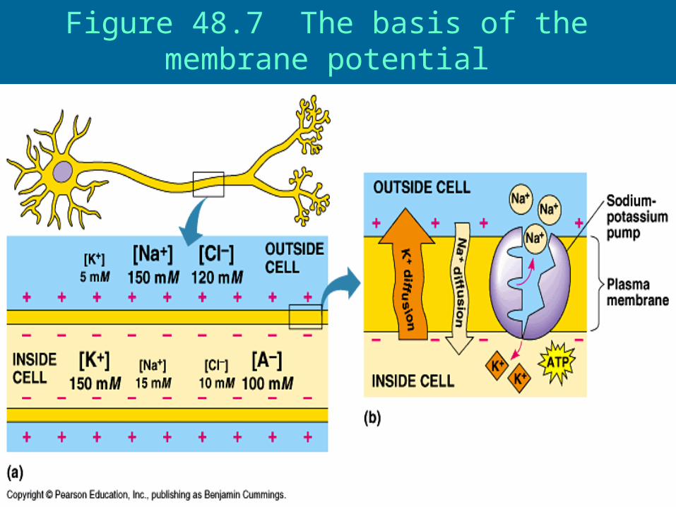
Figure 48.7 The basis of the membrane potential

Charges Across Membranes
• Neurons have ability to generate changes in their membrane potential
• Resting potential – membrane potential of cell at rest (-60mV to -80mV)
• Gated ion channels control membrane potential – open to different stimuli– Hyperpolarization – increase in
electrical gradient•Open K+ channel (K+ moves out)
•Cell becomes more negative•No action potential because it makes it harder to depolarize

– Depolarization – decrease in electrical gradient•Open Na+ channel (Na+ moves in)•Cell becomes more positive•Action potential generated if threshold is reached (-50mV to -55mV)
–Massive change in voltage
•Threshold causes all-or-none event
–Action potential - massive change in membrane voltage that can spread along the membrane

Figure 48.8 Graded potentials and the action potential in a neuron

Figure 48.9 The role of voltage-gated ion channels in the action potential

ROLE OF GATED CHANNELS• Depolarizing – Na+ gates open rapidly so Na+
moves into cell• Repolarizing – K+ gates finally open and K+
moves out; Na+ gates close• Undershoot (Refractory Period) - K+ still open
(they are slower to close) and Na+ still closed so cell becomes even more negative than resting and cannot be depolarized
• Stronger stimuli result in greater frequency of action potentials and NOT from stronger action potentials
• Propagation– Action potentials move in one direction due to
refractory period

Propagation of the action potential
Na+ moves into cell starting action potential.
Depolarization spreads and K+ repolarizes initial area. Prevents action potential on that side.

Figure 48.11 Saltatory conduction• Voltage leaps from node to node

SYNAPSES• Presynaptic cell – transmitting cell• Postsynaptic cell – receiving cell• Two types of synapses
– Electrical •Need gap junctions (channels between
neurons)•No delays
– Chemical•Narrow gap, synaptic cleft, between cells•More common than electrical in vertebrates
and most invertebrates•Require neurotransmitters (chemical
intercellular messengers)

•Depolarization of presynaptic membrane causes influx of Ca2+
•Increased Ca2+ in cell causes synaptic vesicles to fuse to cell membrane and release neurotransmitters via exocytosis
•Neurotransmitters diffuse to postsynaptic cell
•Postsynaptic membrane has gated channels that open when neurotransmitters bond to specific receptors

Figure 48.12 A chemical synapse

• A single neuron may receive many inputs simultaneously
• Neurotransmitters cause 2 different responses depending on the gates that are opened– Inhibitory
•(hyperpolarization)– Excitatory
•(depolarization)• Neurotransmitters are quickly degraded• Excitatory postsynaptic potential
(EPSP) – Na+ in and K+ out = depolarization• Inhibitory postsynaptic potential (IPSP)
- K+ out or CL- in = hyperpolarization

Figure 48.13 Integration of multiple synaptic inputs

Figure 48.14 Summation of postsynaptic potentials

NEUROTRANSMITTERS
• Acetylcholine – one of the most common – can excite skeletal muscle and
inhibit cardiac muscle• Epinephrine and norepinephrine
– also function as hormones

• Dopamine – Usually excitatory– Excess dopamine can cause schizophrenia– Lack of dopamine can cause Parkinson’s
• Sertonin – Usually inhibitory
• Endorphins – Natural painkillers (morphine and opium
mimic endorphins shape)• Nitric Oxide (NO)
– Released during sexual arousal (increasing blood flow)
– Nitroglycerin used to treat chest pain

SKELETAL MUSCLE
•Attached to bones and responsible for their movement
•Consist of bundles of long fibers•Each fiber is a single cell with
many nuclei

Figure 49.31x1 Skeletal muscle

•Each fiber made up of smaller myofibrils
•Myofibrils made of 2 kinds of myofilaments–Thin myofilaments
•2 strand of actin with a regulatory protein (tropomyosin)
–Thick myofilaments•Staggered arrays of myosin

•Striated muscle due to repeating light and dark bands
•Sarcomere – basic unit of muscle
•Contraction of sarcomeres results in muscle contraction.
•Actin and myosin slide pass each other to shorten the sarcomere.

Figure 49.31 The structure of skeletal muscle

Figure 49.32 The sliding-filament model of muscle contraction

Figure 49.33 Myosin-actin interactions generate the force for muscle contraction
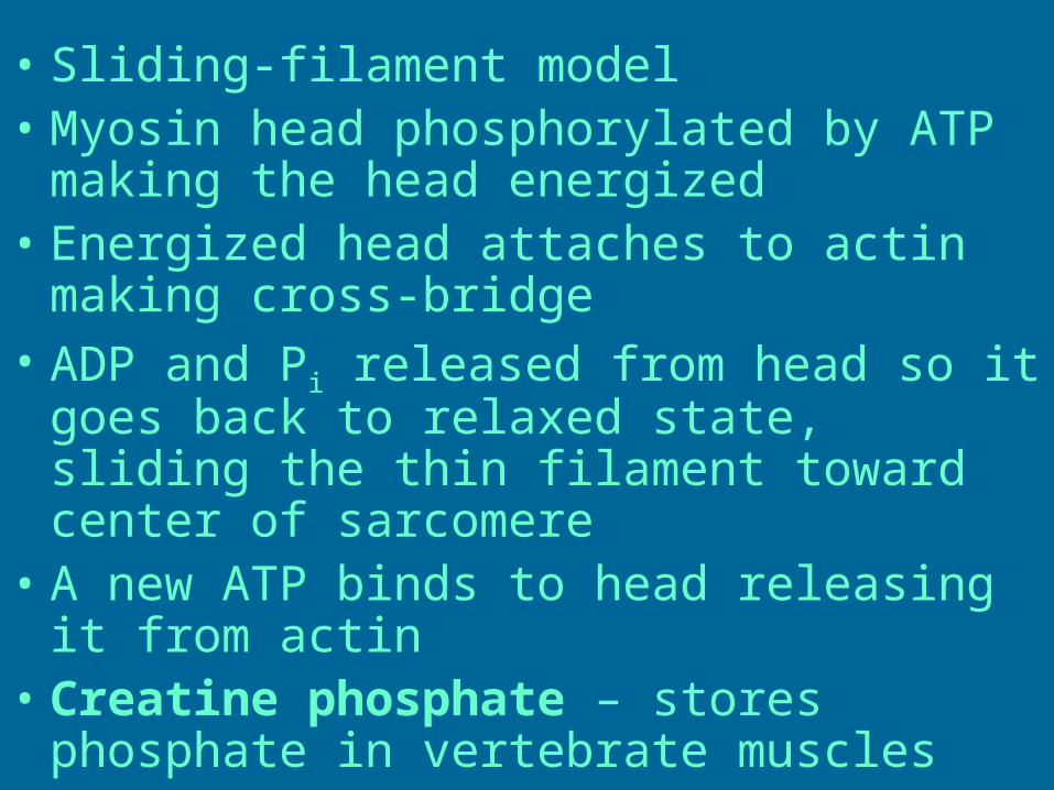
• Sliding-filament model • Myosin head phosphorylated by ATP
making the head energized• Energized head attaches to actin
making cross-bridge• ADP and Pi released from head so it
goes back to relaxed state, sliding the thin filament toward center of sarcomere
• A new ATP binds to head releasing it from actin
• Creatine phosphate – stores phosphate in vertebrate muscles

How is skeletal muscle contraction regulated?
• An action potential begins in the brain and travels via nerve to muscle.
• The action potential causes neuron to release acetylcholine (neurotransmitter). This results in an excitatory response in muscle.

•Acetylcholine triggers action potential in T-tubules within muscle–T-tubules are infoldings of muscle cell’s cell membrane

•T-tubules touch sarcoplasmic reticulum and change is permeability to Ca2+ which means it releases Ca2+ –Sarcoplasmic reticulum – specialized ER that stores Ca2+
•Ca2+ binds to troponin which frees binding site for myosin head

Figure 49.35 The roles of the muscle fiber’s sarcoplasmic reticulum and T tubules in contraction
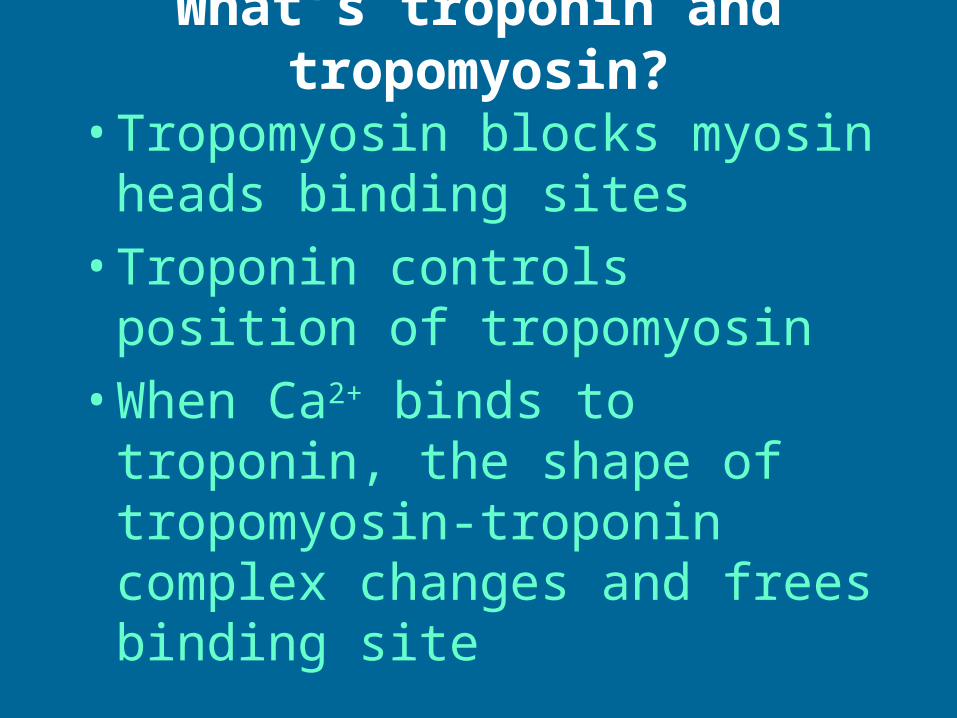
What’s troponin and tropomyosin?
•Tropomyosin blocks myosin heads binding sites
•Troponin controls position of tropomyosin
•When Ca2+ binds to troponin, the shape of tropomyosin-troponin complex changes and frees binding site

Figure 49.34 Hypothetical mechanism for the control of muscle contraction

Figure 49.36 Review of skeletal muscle contraction

•Summation and frequency of action potentials determine muscle tension
•One muscle cell only innervated by one motor neuron, but one motor neuron may innervated many muscle cells
•More cells activated = more tension
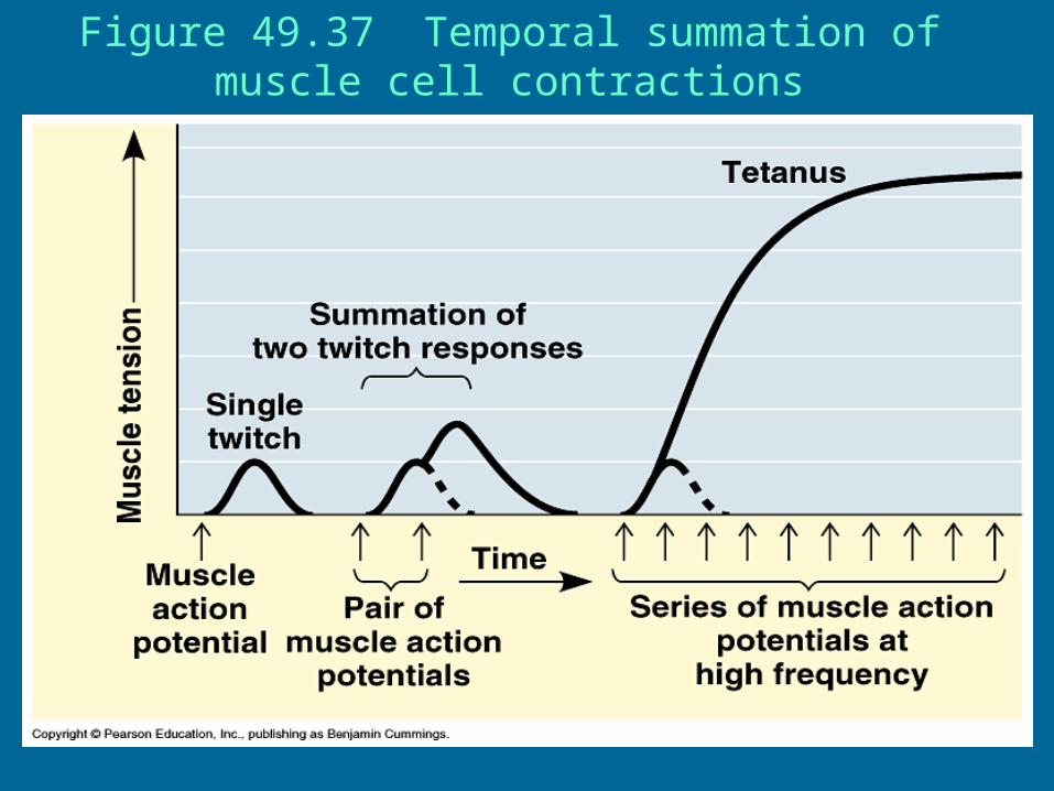
Figure 49.37 Temporal summation of muscle cell contractions
Big Picture – Making a muscle contract
• Action potential generated in brain and travels down nerve
• Action potential causes acetylcholine to diffuse across synapse to muscle
• Acetylcholine causes excitatory responses (action potential) that moves down T-tubules
• Change in membrane potential causes SR to release calcium
• Calcium binds to troponin, which then moves tropomyosin
• ATP used to bind myosin head to actin• Sarcomere contracts and then ATP used to
break bridge
Related Documents