Neuronal Activation by GPI-Linked Neuroligin-1 Displayed in Synthetic Lipid Bilayer Membranes Michael M. Baksh † and Jay T. Groves *,† Department of Chemistry, University of California Berkeley, Berkeley, California 94720 Camin Dean ‡ , Sophie Pautot ‡,§ , Shannon DeMaria ‡ , and Ehud Isacoff ‡ Department of Molecular and Cell Biology, University of California Berkeley, Berkeley, California 94720 Materials Science Division, Lawrence Berkeley National Laboratory, University of California Berkeley, Berkeley, California 94720 Abstract We have characterized, in vitro, interactions between hippocampal neuronal cells and silica microbeads coated with synthetic, fluid, lipid bilayer membranes containing the glycosylphosphatidyl inositol (GPI)-linked extracellular domain of the postsynaptic membrane protein neuroligin-1. These bilayer–neuroligin-1 beads activated neuronal cells to form presynaptic nerve terminals at the point of contact in a manner similar to that observed for live PC12 cells, ectopically expressing the full length neuroligin-1. The synthetic membranes exhibited biological activity at neuroligin-1 densities of ∼1 to 6 proteins/μm 2 . Polyolycarbonate beads with neuroligin-1 covalently attached to the surface failed to activate neurons despite the fact that neuroligin-1 binding activity is preserved. This implies that a lipid membrane environment is likely to be essential for neuroligin-1 activity. This technique allows the study of isolated proteins in an environment that has physical properties resembling those of a cell surface; proteins can diffuse freely within the membrane, retain their in vivo orientations, and are in a nondenatured state. In addition, the synthetic membrane environment affords control over both lipid and protein composition. This technology is easily implemented and can be applied to a wide variety of cellular studies. Introduction The lipid bilayer architecture of cell membranes creates a uniquely complex surface environment, consisting of a nanoscale fluid emulsion of proteins, lipids, and cholesterol. The biological functionality of proteins within membranes is substantially affected by factors influencing both accessibility and spatial organization. For example, interactions between cells and polymer surfaces displaying the RGD peptide motif exhibit a pronounced sensitivity to the nanoscale clustering state of the RGD peptide. 1 Similar effects have been observed through the use of multivalent ligands that, in the solution state, facilitate aggregation of corresponding membrane-associated receptors. 2-7 Indeed, intrinsically bivalent or multivalent cross-linked antibodies to cell surface receptors are widely used to initiate signaling, and the multivalent nature of these binding interactions can be used to explore the role of protein oligomerization in signal transduction. Of relevance to the work described here, this strategy has been employed * To whom correspondence should be addressed. † Department of Chemistry. ‡ Department of Molecular and Cell Biology. § Lawrence Berkeley National Laboratory. Supporting Information Available: Direct measurements of neuroligin-1 mobility using single-particle tracking methods. This material is available free of charge via the Internet at http://pubs.acs.org. NIH Public Access Author Manuscript Langmuir. Author manuscript; available in PMC 2006 April 26. Published in final edited form as: Langmuir. 2005 November 8; 21(23): 10693–10698. NIH-PA Author Manuscript NIH-PA Author Manuscript NIH-PA Author Manuscript

Welcome message from author
This document is posted to help you gain knowledge. Please leave a comment to let me know what you think about it! Share it to your friends and learn new things together.
Transcript
Neuronal Activation by GPI-Linked Neuroligin-1 Displayed inSynthetic Lipid Bilayer Membranes
Michael M. Baksh† and Jay T. Groves*,†Department of Chemistry, University of California Berkeley, Berkeley, California 94720
Camin Dean‡, Sophie Pautot‡,§, Shannon DeMaria‡, and Ehud Isacoff‡Department of Molecular and Cell Biology, University of California Berkeley, Berkeley, California94720
Materials Science Division, Lawrence Berkeley National Laboratory, University of CaliforniaBerkeley, Berkeley, California 94720
AbstractWe have characterized, in vitro, interactions between hippocampal neuronal cells and silicamicrobeads coated with synthetic, fluid, lipid bilayer membranes containing theglycosylphosphatidyl inositol (GPI)-linked extracellular domain of the postsynaptic membraneprotein neuroligin-1. These bilayer–neuroligin-1 beads activated neuronal cells to form presynapticnerve terminals at the point of contact in a manner similar to that observed for live PC12 cells,ectopically expressing the full length neuroligin-1. The synthetic membranes exhibited biologicalactivity at neuroligin-1 densities of ∼1 to 6 proteins/μm2. Polyolycarbonate beads with neuroligin-1covalently attached to the surface failed to activate neurons despite the fact that neuroligin-1 bindingactivity is preserved. This implies that a lipid membrane environment is likely to be essential forneuroligin-1 activity. This technique allows the study of isolated proteins in an environment that hasphysical properties resembling those of a cell surface; proteins can diffuse freely within themembrane, retain their in vivo orientations, and are in a nondenatured state. In addition, the syntheticmembrane environment affords control over both lipid and protein composition. This technology iseasily implemented and can be applied to a wide variety of cellular studies.
IntroductionThe lipid bilayer architecture of cell membranes creates a uniquely complex surfaceenvironment, consisting of a nanoscale fluid emulsion of proteins, lipids, and cholesterol. Thebiological functionality of proteins within membranes is substantially affected by factorsinfluencing both accessibility and spatial organization. For example, interactions between cellsand polymer surfaces displaying the RGD peptide motif exhibit a pronounced sensitivity tothe nanoscale clustering state of the RGD peptide.1 Similar effects have been observed throughthe use of multivalent ligands that, in the solution state, facilitate aggregation of correspondingmembrane-associated receptors.2-7 Indeed, intrinsically bivalent or multivalent cross-linkedantibodies to cell surface receptors are widely used to initiate signaling, and the multivalentnature of these binding interactions can be used to explore the role of protein oligomerizationin signal transduction. Of relevance to the work described here, this strategy has been employed
*To whom correspondence should be addressed.†Department of Chemistry.‡Department of Molecular and Cell Biology.§Lawrence Berkeley National Laboratory.Supporting Information Available: Direct measurements of neuroligin-1 mobility using single-particle tracking methods. This materialis available free of charge via the Internet at http://pubs.acs.org.
NIH Public AccessAuthor ManuscriptLangmuir. Author manuscript; available in PMC 2006 April 26.
Published in final edited form as:Langmuir. 2005 November 8; 21(23): 10693–10698.
NIH
-PA Author Manuscript
NIH
-PA Author Manuscript
NIH
-PA Author Manuscript

to expose a role of receptor clustering in the neurexin–neuroligin signaling system8 and in theephrin/Eph receptor signaling system.9
Neurexins constitute a large family of neuronal cell surface receptors, with β-neurexinsbelieved to be receptors for the postsynaptic cell surface protein, neuroligin-1.8,10Nonneuronal cells (e.g., HEK293 or PC12) ectopically expressing neuroligin-1 (orneuroligin-2) can induce the formation of presynaptic elements in contacting axons, in vitro.Oligomerization of neuroligin is required for this function, and β-neurexin clustering issufficient to recruit synaptic vesicles. Thus, postsynaptic neuroligin oligomers appear to induceclustering of axonal neurexins, and rearrangement of neurexins nucleates assembly of theneuronal intracellular protein complex responsible for neurotransmitter secretion.8
Planar supported membranes have been used extensively as an in vitro method of studyingmolecular interactions at membrane surfaces.11,12 Supported membranes can be formed byspontaneous adsorption and fusion of lipid vesicles with a solid substrate such as silica andvarious polymers.13-15 The process is generally selflimiting at a single bilayer, resulting in asingle, continuous membrane coating on the substrate. A thin layer of water between themembrane and substrate preserves the lateral mobility of both lipids16-18 and membrane-associated proteins.19,20 Supported membranes displaying glycosylphosphatidylinositol(GPI)-linked forms of the major histocompatibility complex (MHC) and intercellular adhesionmolecule (ICAM) have been used as surrogate antigen-presenting cells (APCs) for interactionwith living T cells. A dynamic immunological synapse can form between the T cell and thesupported membrane involving micrometer-scale spatial rearrangement of both supportedmembrane and T cell surface proteins.21 This hallmark example of a functional interactionbetween a living cell and a supported membrane clearly illustrates the dramatic extent to whichthe lateral mobility of proteins can be coupled to activity.
Here we describe functional interactions between a GPI-linked form of neuroligin-1 insupported membranes and primary neuronal cells in culture. Silica beads, typically 5 μm indiameter, were used as the supported membrane substrate. One reason for this choice ofsubstrate configuration is the ease with which beads can be added to a preexisting cell culture.Membranes were formed on the silica beads by a process analogous to that used to form planarsupported membranes.16,22,23 Beads coated with neuroligin-1-containing membranesactivated neurons in a manner similar to live PC12 cells expressing neuroligin-1. Neuroligin-1was active at densities of ∼1 to 6 proteins/μm2 and appeared to require a membraneenvironment for functionality; naked polystyrene beads to which neuroligin-1 was covalentlyattached failed to activate neuronal cells. This suggests that the lipid bilayer bead is superiorto established methods of attaching proteins to beads for a number of possible reasons; theorientation and configuration of the protein is preserved by the bilayer, the amount of proteinpresent on the beads can be quantitated directly, and the membrane environment preserves afluid environment similar to that of a cell membrane, in which proteins can diffuse freely.
Results and DiscussionThe aim of the experiments described below is to determine if a neuron can be induced to formsynapses when brought into contact with a GPI-linked form of the postsynaptic adhesionprotein, neuroligin-1, incorporated into synthetic lipid bilayers that are coated onto silica beads(Figure 1A). The GPI-linked neuroligin-1 binds to β-neurexin present in the membrane of theaxons of neurons in culture. This binding interaction induces the recruitment of synapticvesicles to the site of neuroliginneurexin interaction and the formation of a functionalpresynaptic terminal at the site. Figure 1B shows a bright field image of silica microbeads,coated with lipid bilayers containing GPI-linked neuroligin-1, contacting a neuron in culture.
Baksh et al. Page 2
Langmuir. Author manuscript; available in PMC 2006 April 26.
NIH
-PA Author Manuscript
NIH
-PA Author Manuscript
NIH
-PA Author Manuscript

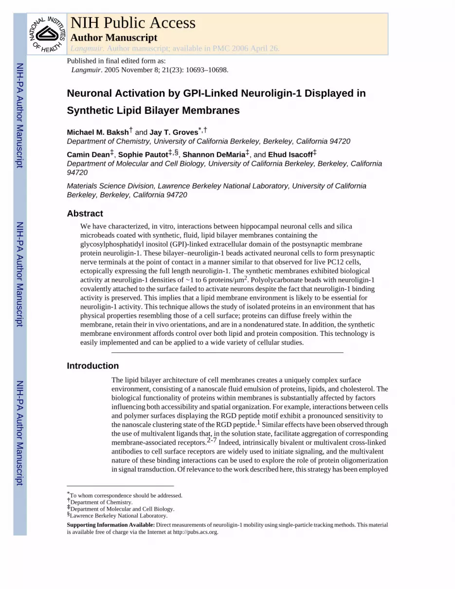
GPI-linked proteins can be incorporated into supported membranes, preserving both lateralmobility and biological functionality.11,19-21,24,25 Here, an engineered GPI-linked form ofneuroligin-1 was incorporated into artificial bilayers on silica beads, which were thenmanipulated into contact with neuronal cells containing endogenous neurexin (Figure 1).Unlike more conventional methods, such as covalent binding, which can cause indiscriminatebinding of proteins to surfaces, the GPI linkage favors insertion into the membrane in such away that a proper orientation is maintained. The GPI-linked neuroligin-1 was expressed inHEK293 cells and purified by affinity chromatography. The purified protein was reconstitutedinto lipid vesicles via detergent dialysis, and these proteoliposomes were used to createsupported membranes on 5 μm diameter silica beads.16,22,23,26 These proteomembrane-coated beads were stored in 1× PBS at 4 °C for up to 4 h. A silver-stained protein gel showsneuroligin-1 recovered from a preparation of proteoliposome-coated beads (Figure 2A). Thisillustrates another advantage of using membrane-coated beads; proteins incorporated ontomembrane-coated beads can be easily removed to quantitate the amount of protein present andensure that it is in a nondenatured state.
Membrane fluidity is another distinct advantage of using membrane-coated beads. Membrane-associated proteins and adhesion molecules often undergo dynamic, two-dimensionalreorientation in response to binding to extracellular partners. Such molecules may require amembrane-like environment in which fluidity is preserved in order to retain in vivofunctionality. Membrane fluidity of proteomembrane-coated beads was monitored in twoways: by fluorescence recovery after photobleaching (FRAP) studies of a lipid-linkedfluorescent probe, Texas red-DPPE or NBD-PC, and electric field mobility observations ofplanar supported membranes (Figure 2). The similar diffusion characteristics of these probesin bead-supported membranes suggest that they are physically equivalent to the well-studiedplanar supported membranes.12,14,15
Dissociated embryonic rat hippocampal neurons were plated on polylysine-coated glasscoverslips and cultured in Neurobasal medium for 10–12 days until use.27,28 For theexperiment, membrane-coated beads were added to the medium and allowed to settle by gravityonto the hippocampal neurons. After incubation for an additional 24 h, both cells and beadswere fixed and stained with fluorescently labeled antibodies. Interactions between the cellsand beads were then analyzed using standard fluorescence microscopy techniques.
Neuronal cells can be activated through contact with cells displaying various cognate synapticmolecules on their surface, mimicking a postsynaptic neuronal partner.8,10 This is illustratedin Figure 3A, in which PC12 cells expressing the full-length neuroligin-1 are shown to triggerpresynaptic differentiation in hippocampal neuronal cells. Activation consists of localclustering of neuroligin-1 on the surface of the PC12 cell at sites of contact with neuronal cellsand both clustering of β-neurexin on the surface of an axonal process of the neuron andaccumulation of synaptic vesicles inside the axon at the site of PC12–axon contact. PC12 cellswithout neuroligin-1 exhibit no discernible activating effect (not shown).
The activation of hippocampal neuronal cells by full-length neuroligin-1-expressing PC12 cellswas mimicked by 5 μm diameter silica microbeads, coated with synthetic membranescontaining GPI-linked neuroligin-1 external domain (Figure 3B). Neuroligin-1 on the beadcoclustered with β-neurexin on the surface of the axon and synaptic vesicles inside the axonat the site of contact with the bead. Neuroligin-1 displayed on membrane-coated beads wasequally effective at activating hippocampal neuronal cells as when the protein is expressed onthe surface of PC12 cells. Silica glass beads coated with plain synthetic membranes withoutneuroligin-1 (Figure 3C), beads coated with synthetic membranes containing alkalinephosphatase, another GPI-linked protein that does not bind to proteins on neurons (Figure 3D),
Baksh et al. Page 3
Langmuir. Author manuscript; available in PMC 2006 April 26.
NIH
-PA Author Manuscript
NIH
-PA Author Manuscript
NIH
-PA Author Manuscript

or bare silica glass beads (Figure 3E), all failed to activate the hippocampal neuronal cells. Theeffect is specific to neuroligin-1.
Polymer beads, such as polystyrene, are commonly employed to display cell surface proteinsfor various assays and interactions with cultured cells. This technique was tested as an alternatemethod to display neuroligin-1 to hippocampal neuronal cells. For this comparison,neuroligin-1 was covalently attached to 6 μm diameter polystyrene beads by means of acarbodiimide coupling reaction.29 On the basis of established data of the minimal effectivenessof this reaction, this chemical coupling strategy routinely achieved protein densitiesapproximately 10 times greater on the polystyrene bead surface than was achieved with themembrane. Additionally, the polystyrene–carbodiimide–neuroligin-1 beads were found toadhere to HEK293 cells expressing β-neurexin (Figure 4, parts C and D) but not to cells lackingneurexin (not shown). Despite the fact that the neuroligin-1 covalently attached to the beadswas capable of specific adhesion, its ability to activate the presynaptic differentiation of anaxon was drastically reduced compared with that of the membrane coatings (Figure 3F). Thisobservation illustrates the significance of a membrane environment for the natural activity ofneuroligin-1.
The density of GPI-linked neuroligin-1 on the surface of the membrane-coated silica beadswas determined to be between ∼1 and 6 molecules/μm2 by quantitative fluorescence. Adetermined number of membrane-coated beads displaying neuroligin-1, displaying acorrespondingly determined total membrane surface area, were incubated with fluorescent anti-HA antibodies, which bound to an engineered HA tag on the neuroligin-1. Subsequently, themembranes containing both neuroligin-1 and the bound antibody were stripped off of the beadsusing a nonionic detergent, Triton X-100. The fluorescence intensity of the antibody in theresulting solution was then measured and the values compared with those from a reference setof data to determine the total amount of neuroligin-1-engaged antibody (Figures 4 and 5). Fromthe known membrane surface area, the range of between 1 and 6 molecules/μm2 of neuroligin-1in the membranes was determined. Given our observation that neuroligin-1 concentrates at thesite of the neuronal synapse, the functional signaling area likely contains a higher proteindensity.
SignificanceAmong artificial materials, supported membranes can most closely mimic the fluidcharacteristic of a live cell surface. Such mimicry allows the retention of the functional aspectsof natural protein behavior in vitro. This is illustrated by the functional neuroligin-1reconstitution described here as well as previous work with immune cell surface proteins.11In comparison to proteins displayed by direct coupling to solid surfaces, the supportedmembrane provides a drastically different environment. Orientation and conformation ofincorporated proteins is maintained due to direct engineering of the GPI link into the proteinof interest and the natural processes involved in self-assembly of the membrane bilayer. Inaddition, the fluidity of the membrane is likely to be an important factor in retaining in vivofunctionality of membrane-associated proteins and adhesion molecules. Long-range lateralrearrangements of GPI-linked proteins in supported membranes can lead to substantialenrichments in the contact zone with a living cell. This type of reaction–diffusion process iscommon at cell–cell contacts. At a more subtle level, the molecular scale freedom of motionafforded within a supported membrane enables proteins to adopt differential configurationsand orientations on the membrane surface. This orientational freedom may further facilitateformation of functional interactions with cognate proteins on an apposing live cell surface. Thephysical advantages provided by supported membranes are magnified by the general successof the GPI linkage strategy as a method of displaying cell surface proteins within an artificialmembrane system. Lastly, the ease of manipulation of membrane-coated microbeads should
Baksh et al. Page 4
Langmuir. Author manuscript; available in PMC 2006 April 26.
NIH
-PA Author Manuscript
NIH
-PA Author Manuscript
NIH
-PA Author Manuscript

further facilitate usage of supported membrane display strategies in a variety of live cellexperiments.
Experimental SectionMaterials
Lipids were obtained from Avanti Polar Lipids (Alabaster, AL). L-α egg phosphatidylcholinewas received in chloroform and stored at −20 °C for up to 2 weeks. The fluorescent probe N-(Texas red sulfonyl)-1,2-dipalmitoyl-sn-glycero-3-phosphoethanolamine, triethylammoniumsalt (Texas red-DPPE) was purchased from Molecular Probes (Eugene, OR) in powder formand dissolved in chloroform before use. The fluorescent probe 1-palmitoyl-2-[12-[(7-nitro-2-1,3-benzoxadiazol-4-yl)amino]dodecanoyl]-sn-glycero-3-phosphocholine (NBD-PC)was purchased from Avanti Polar Lipids in chloroform and stored at −20 °C until used. Rabbitantisynapsin antibodies were purchased from Chemicon (Temecula, CA), rat anti-HA IgGfragment epitope tag 3F10 was purchased from Roche (Indianapolis, IN), and mouseantialkaline phosphatase was purchased from Sigma Chemicals (St. Louis, MO); all three werestored at 4 °C prior to use. Secondary antibodies were purchased from Jackson Immunologicals(West Grove, PA) and stored at 4 °C. Silica glass microspheres, 5 μm mean diameter, wereobtained from Bangs laboratories (Fishers, IN) and stored at 4 °C under deionized H2O.Polystyrene beads, 6 μm diameter, and a carbodiimide protein-coupling kit were obtained fromPolySciences Inc. (Warrington, PA) and stored at 4 °C. NTA-agarose was obtained fromQiagen (Valencia, CA).
Purification and Isolation of ProteinRecombinant HA/His-tagged GPI-anchored neuroligin-1 was expressed in HEK293 cells andisolated from cell lysates by chromatography on NTA-agarose. The protein bound to thecolumn in phosphate-buffered saline containing NaCl (350 mM), Triton X-100 (0.5%), andimidazole (6 mM). Nonspecifically bound proteins were removed by extensive washing withPBS containing NaCl (1 M) and Triton X-100 (0.1%). The detergent was then switched to n-octyl-glucopyranoside (25 mM), and bound protein was eluted with an imidazole gradient (10–200 mM). The eluted material revealed a single protein band at the predicted size for GPI-neuroligin-1. Bands were resolved by silver staining and showed immune reactivity withspecific antibodies directed against the HA tag.
Preparation of Polystyrene BeadsBeads were washed twice with carbonate buffer (0.1 M) and phosphate buffer (0.02 M),respectively, and then incubated in a carbodiimide solution (2%) for4hin order to activatesurface carboxyl groups. Activated beads were washed 3 times in borate buffer (0.2 M) andmixed overnight in borate buffer (0.2 M) containing 5 μg of purified GPI-neuroligin-1. Thenext day, beads were incubated for 30 min in a solution of borate buffer and ethanolamine andincubated in BSA for 30 min to block unreacted sites and nonspecific binding.
Synthetic MembranesSupported bilayers were formed by the fusion of small unilamellar vesicles (SUVs) onto cleanglass microspheres. For SUV preparation, a lipid solution in chloroform was evaporated ontosmall round-bottomed flasks and hydrated for 60 min at 4 °C in 18.2 MΩ-cm water to a finalconcentration of ∼3.3 mg/mL. The lipids were probe-sonicated at 25 W for 90 s, (5 s pulseswith 1 s intervals) in an ice water bath and then ultracentrifuged for 2 hat 160 000g and 4 °C.The supernatant was stored at 4 °C for up to 1 week. Proteoliposomes were formed by mixingpeak fractions of purified GPI-linked neuroligin-1 in 15 mM n-octyl-glucopyranoside withpreformed SUVs. The detergent was removed by dialysis against three changes of 1× PBS over
Baksh et al. Page 5
Langmuir. Author manuscript; available in PMC 2006 April 26.
NIH
-PA Author Manuscript
NIH
-PA Author Manuscript
NIH
-PA Author Manuscript

the course of 18 h. The resulting proteoliposomes were stored for up to 2 days in 1× PBS at 4°C. Bilayers were allowed to self-assemble on the microbead surface by mixing equal amounts(100 μL) of spreading solution (1:1 SUV/PBS) and microbead suspension (10% solids indeionized water) together in a 1.5 mL centrifuge tube. Excess vesicles were removed bypelleting the microbeads via pulse centrifugation and removal of the supernatant. Then, 1 mLof 18.2 MΩ-cm water was added to the pelleted microbeads and the entire mixture vortexedto allow resuspension.
Quantitation of Neuroligin-1For neuroligin-1 quantitation, proteoliposomes were used to deposit supported membranes on5.08 μm silica beads as described above. A typical sample contained about 7.0 ± 0.35 × 10⁷
beads, corresponding to a total bilayer area of about 5.7 × 109 μm2. To prevent nonspecificbinding of the HA antibody, the bead surface was blocked by incubation for 30 min using a 1mg/mL solution of BSA in 1× PBS. After incubation, the beads were incubated with an excessof FITC-labeled HA antibody for another 30 min. All incubations were carried out at roomtemperature, and the sample tubes were placed on a rotator to prevent beads from settling andaggregating, which creates defects in the membrane. To remove all unbound antibody, thebeads were washed several times by pulse centrifugation, removal of the supernatant, andresuspension in fresh 1× PBS buffer. After the last wash, the beads were resuspended in 250μL of 1× PBS, and 2% Triton X-100 was added to remove the bilayer from the beads. After10 min, the beads were spun down and the fluorescence corresponding to the antibody in thesupernatant was measured to determine the amount of neuroligin-1 incorporated into thebilayer. Fluorescence measurements were performed on a fluorescence spectrometer (Jasco,model FP-750) with the excitation wavelength fixed at λexc = 491 nm with a 5 nm slit. Theintensity of the emitted light at λem = 522 nm was detected with a slit of 10 nm for concentrationsof FITC-labeled, anti-HA antibody ranging from 1 to 50 ng/mL.
Hippocampal CulturesHippocampi were removed from embryonic day 18 rats and treated with trypsin for 20 min at37 °C, followed by washing and trituration. Dissociated cells were plated at 100 000 cells/cm2 on polylysine-coated glass coverslips and cultured in Neurobasal medium supplementedwith Glutamax (2 mM) and B-27 (2%).
Polystyrene Bead ControlHEK293 cells were plated on glass coverslips and transfected with β-neurexin at an efficiencyof ∼50%. Both transfected and nontransfected cells adhered strongly to the glass. Coverslipswith cells were placed in a closed chamber, and polystyrene beads, to which neuroligin-1 wascovalently coupled with a carboiimide linkage, were washed into the chamber and allowed tosettle onto the cells. Following 20 min of bead–cell contact, the chamber was inverted, and thebehavior of the beads and cells was observed.
ImagingImages were taken with a Zeiss LSM510 confocal microscope. Laser power andphotomultipliers were set such that no detectable bleed-through occurred between differentchannels. Eight to ten sections were taken from top to bottom of the specimen, and brightestpoint projections were made. Images were processed using Adobe Photoshop software (AdobeSystems, CA).
Supplementary MaterialRefer to Web version on PubMed Central for supplementary material.
Baksh et al. Page 6
Langmuir. Author manuscript; available in PMC 2006 April 26.
NIH
-PA Author Manuscript
NIH
-PA Author Manuscript
NIH
-PA Author Manuscript

Acknowledgment
We thank Jeffrey Nye for providing the single-particle tracking measurements of neuroligin-1. This work wassupported by the Chemical Sciences, Geosciences, and Biosciences Division, Office of Basic Energy Sciences, of theU.S. Department of Energy under Contract No. DE-AC03-76SF00098.
References1. Maheswari G, et al. Cell adhesion and motility depend on nanoscale RGD clustering. J. Cell Science
2000;113:1677–1686. [PubMed: 10769199]2. Mammen M, Choi S-K, Whitesides GM. Polyvalent interactions in biological systems: implications
for design and use of multivalent ligands and inhibitors. Angew. Chem. Int. Ed 1998;37:2754–2794.3. He X-S, et al. Quantitative analysis of hepatitis C virus-specific CD8+ T cells in peripheral blood and
liver using peptide-MHC tetramers. Proc. Natl. Acad. Sci. U.S.A 1999;96:5692–5697. [PubMed:10318946]
4. Cochran JR, Cameron TO, Stern LJ. The relationship of MHC-peptide binding and T cell activationusing chemically defined MHC class II oligomers. Immunity 2000;12:241–250. [PubMed: 10755611]
5. Kiessling LL, Gestwicki JE, Strong L. Synthetic multivalent ligands in the exploration of cell-surfaceinteractions. Curr. Opin. Chem. Biol 2000;4:696–703. [PubMed: 11102876]
6. Gestwicki JE, Kiessling LL. Inter-receptor communication through arrays of bacterial chemoreceptors.Nature 2002;415(3):81–84. [PubMed: 11780121]
7. Bray D, Levin MD, Morton-Firth CJ. Receptor clustering as a cellular mechanism to control sensitivity.Nature 1998;393(7):85–88. [PubMed: 9590695]
8. Dean C, et al. Neurexin mediates the assembly of presynaptic terminals. Nat. Neurosci 2003;6(7):708–716. [PubMed: 12796785]
9. Stein E, et al. Eph receptors discriminate specific ligand oligomers to determine alternative signalingcomplexes, attachment and assembly receptors. Genes Dev 1998;12:667–678. [PubMed: 9499402]
10. Scheiffele P, et al. Neuroligin expressed in nonneuronal cells triggers presynaptic development incontacting axons. Cell 2000;101:657–669. [PubMed: 10892652]
11. Groves JT, Dustin ML. Supported planar bilayers in studies on immune cell adhesion andcommunication. J. Immunol. Methods 2003;278:19–32. [PubMed: 12957393]
12. Sackmann E. Supported membranes: Scientific and practical applications. Science 1996;271:43–48.[PubMed: 8539599]
13. Sackmann E, Tanaka M. Supported membranes on soft polymer cushions: fabrication,characterization and applications. Trends Biotechnol 2000;18:58–64. [PubMed: 10652510]
14. Groves JT. Membrane array technology for drug discovery. Curr. Opin. Drug Discovery Dev 2002;5(4):606–612.
15. Groves JT, Boxer SG. Micropattern formation in supported lipid membranes. Acc. Chem. Res2002;35:149–157. [PubMed: 11900518]
16. Bayerl TM, Bloom M. Physical properties of single phospholipid bilayers adsorbed to micro glassbeads. Biophys. J 1990;58:357–362. [PubMed: 2207243]
17. Koenig BW, et al. Neutron reflectivity and atomic force microscopy studies of a lipid bilayer in wateradsorbed to the surface of a silicon single crystal. Langmuir 1996;12(5):1343–1350.
18. Johnson SJ, et al. Structure of an adsorbed dimyristoylphosphatidylcholine bilayer measured withspecular reflection of neutrons. Biophys. J 1991;59:289–294. [PubMed: 2009353]
19. Chan P-Y, et al. Influence of receptor lateral mobility on adhesion strengthening between membranescontaining LFA-3 and CD2. J. Cell Biol 1991;115(1):245–255. [PubMed: 1717480]
20. Groves JT, Wülfing C, Boxer SG. Electrical manipulation of glycanphosphatidylinisitol-tetheredproteins in supported lipid bilayers. Biophys. J 1996;71:2716–2723. [PubMed: 8913608]
21. Grakoui A, et al. The immunological synapse: A molecular machine controlling T cell activation.Science 1999;285:221–227. [PubMed: 10398592]
22. Buranda T, et al. Biomimetic molecular assemblies on glass and mesoporous silica microbeads forbiotechnology. Langmuir 2003;19(5):1654–1663.
Baksh et al. Page 7
Langmuir. Author manuscript; available in PMC 2006 April 26.
NIH
-PA Author Manuscript
NIH
-PA Author Manuscript
NIH
-PA Author Manuscript

23. Baksh MM, Jaros M, Groves JT. Detection of molecular interactions at membrane surfaces throughcolloid phase transitions. Nature 2004;427:139–141. [PubMed: 14712272]
24. Tözeren A, et al. Micromanipulation of adhesion of a jurkat cell to a planar bilayer membranecontaining lymphocyte function-associated antigen 3 molecules. J. Cell Biol 1992;116(4):997–1006.[PubMed: 1370839]
25. Dustin ML, et al. Visualization of CD2 interaction with LFA-3 and determination of the two-dimensional dissociation constant for adhesion receptors in a contact area. J. Cell Biol 1996;132(3):465–474. [PubMed: 8636222]
26. Sapuri AR, Baksh MM, Groves JT. Electrostatically targeted intermembrane lipid exchange withmicropatterned supported membranes. Langmuir 2003;19(5):1606–1610.
27. Banker GA, Cowan WM. Rat hippocampal neurons in dispersed cell culture. Brain Res1977;126:397–342. [PubMed: 861729]
28. Brewer GJ, et al. Optimized survival of hippocampal neurons in B-27 supplemented Neurobasal, anew serum-free medium combination. J. Neurosci. Res 1993;35:567–576. [PubMed: 8377226]
29. Gallo G, Lecfort FB, Letourneau PC. The trkA receptor mediates growth cone turning toward alocalized source of nerve growth factor. J. Neurosci 1997;17(14):5445–5454. [PubMed: 9204927]
Baksh et al. Page 8
Langmuir. Author manuscript; available in PMC 2006 April 26.
NIH
-PA Author Manuscript
NIH
-PA Author Manuscript
NIH
-PA Author Manuscript

Figure 1.(A) Schematic diagram of the experiment: GPI-linked neuroligin-1 (red) incorporated into asynthetic lipid bilayer on a silica microbead binds to β-neurexin (green) expressed in a neuron.Formation of the neuroligin–neurexin complex results in the eventual recruitment ofscaffolding proteins (grey) and exocytosis-competent synaptic vesicles (blue). (B) Membrane-coated beads were added to hippocampal cell cultures, allowed to settle by gravity, andincubated for 24 h. Transmission image of beads coated with synthetic lipid bilayers containingGPI-linked neuroligin-1, interacting with a hippocampal neuron in culture.
Baksh et al. Page 9
Langmuir. Author manuscript; available in PMC 2006 April 26.
NIH
-PA Author Manuscript
NIH
-PA Author Manuscript
NIH
-PA Author Manuscript
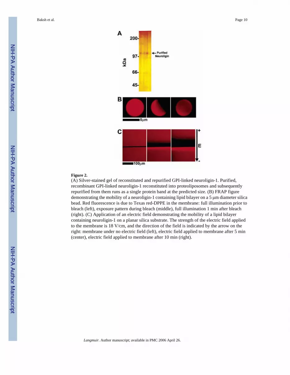
Figure 2.(A) Silver-stained gel of reconstituted and repurified GPI-linked neuroligin-1. Purified,recombinant GPI-linked neuroligin-1 reconstituted into proteoliposomes and subsequentlyrepurified from them runs as a single protein band at the predicted size. (B) FRAP figuredemonstrating the mobility of a neuroligin-1 containing lipid bilayer on a 5 μm diameter silicabead. Red fluorescence is due to Texas red-DPPE in the membrane: full illumination prior tobleach (left), exposure pattern during bleach (middle), full illumination 1 min after bleach(right). (C) Application of an electric field demonstrating the mobility of a lipid bilayercontaining neuroligin-1 on a planar silica substrate. The strength of the electric field appliedto the membrane is 18 V/cm, and the direction of the field is indicated by the arrow on theright: membrane under no electric field (left), electric field applied to membrane after 5 min(center), electric field applied to membrane after 10 min (right).
Baksh et al. Page 10
Langmuir. Author manuscript; available in PMC 2006 April 26.
NIH
-PA Author Manuscript
NIH
-PA Author Manuscript
NIH
-PA Author Manuscript
Figure 3.Presynaptic differentiation triggered by neuroligin-1 on the surface of cells or on lipid bilayer-coated membranes. (A) PC12 cells transfected with neuroligin-1–IRES–GFP (pseudo-coloredred for consistency with other panels) were added to a 12 day old hippocampal culture,incubated overnight, then fixed and stained with antisynapsin (green in all panels) to markpresynaptic synaptic vesicles in the axons of the neuron. Synaptic vesicles accumulate, inpuncta, at the site of contact with the PC12 cell. (B–F) Twelve day old hippocampal culturesinteracting with beads coated with (B) synthetic lipid bilayers containing GPI-linkedneuroligin-1 (neuroligin-1 stained in red), (C) synthetic lipid bilayers with no added protein(plain membranes in red), (D) synthetic lipid bilayers containing GPI-linked alkalinephosphatase (GPI-linked alkaline phosphatase in red), (E) neither synthetic lipid bilayer norprotein, bare silica beads (middle panel is DIC image of beads and neurons), and (F) withchemically coupled neuroligin-1 on polystyrene beads (neuroligin-1 in red). Cultures werefixed 24 h following introduction of beads and were stained with anti-HA antibodies for eitherneuroligin-1 (red), GPI-linked alkaline phosphatase (red), or synapsin (green). Microbeadscoated only with synthetic lipid bilayers included 1% Texas red–DPPE in the bilayer forfluorescence imaging. Only beads with synthetic lipid bilayers containing GPI-linkedneuroligin-1 induced presynaptic differentiation of the neurons, as indicated by synapsinaccumulation in axons contacting the beads.
Baksh et al. Page 11
Langmuir. Author manuscript; available in PMC 2006 April 26.
NIH
-PA Author Manuscript
NIH
-PA Author Manuscript
NIH
-PA Author Manuscript

Figure 4.Adherence of HEK293 cells to polystyrene beads covered with chemically coupledneuroligin-1. White circles denote the positions of polystyrene beads in fluorescence images.(A, B) HEK293 cells not transfected with β-neurexin; no beads are observed to adhere to thecells and have fallen away from the coverslip after inversion of the chamber. (C) Bright fieldimage of polystyrene beads, coupled to neuroligin-1, adhering to HEK293 cells expressing β-neurexin. In all cases, both transfected and nontransfected cells remained adhered to the glasscoverslip. (D) Fluorescence image of polystyrene beads, coupled to neuroligin-1, and adheringto β-neurexin-expressing HEK293 cells.
Baksh et al. Page 12
Langmuir. Author manuscript; available in PMC 2006 April 26.
NIH
-PA Author Manuscript
NIH
-PA Author Manuscript
NIH
-PA Author Manuscript

Figure 5.Reference curve of the intensity of emitted light at a wavelength of 522 nm for differentconcentrations of FITC HA antibody in a 1× PBS solution with 2 vol % Triton X-100 (squares).The plain line is a linear fit to the data. Dashed lines indicate the intensity values measured forthe antibody bound to neuroligin-1 incorporated in the synthetic membrane. Three sampleswere prepared using three different preparations of neuroligin-1, and the range ofcorresponding concentration of bound antibody is assumed to be directly proportional to therange of neuroligin-1 present in the synthetic membrane.
Baksh et al. Page 13
Langmuir. Author manuscript; available in PMC 2006 April 26.
NIH
-PA Author Manuscript
NIH
-PA Author Manuscript
NIH
-PA Author Manuscript
Related Documents