PLEASE SCROLL DOWN FOR ARTICLE This article was downloaded by: [Ecco, Roselene] On: 17 April 2011 Access details: Access Details: [subscription number 936484245] Publisher Taylor & Francis Informa Ltd Registered in England and Wales Registered Number: 1072954 Registered office: Mortimer House, 37- 41 Mortimer Street, London W1T 3JH, UK Avian Pathology Publication details, including instructions for authors and subscription information: http://www.informaworld.com/smpp/title~content=t713405810 Neurological lesions in chickens experimentally infected with virulent Newcastle disease virus isolates Roselene Ecco a ; Leonardo Susta a ; Claudio L. Afonso b ; Patti J. Miller b ; Corrie Brown a a Department of Pathology, College of Veterinary Medicine, The University of Georgia, Athens, GA, USA b Southeast Poultry Research Laboratory, Athens, GA, USA Online publication date: 15 April 2011 To cite this Article Ecco, Roselene , Susta, Leonardo , Afonso, Claudio L. , Miller, Patti J. and Brown, Corrie(2011) 'Neurological lesions in chickens experimentally infected with virulent Newcastle disease virus isolates', Avian Pathology, 40: 2, 145 — 152 To link to this Article: DOI: 10.1080/03079457.2010.544289 URL: http://dx.doi.org/10.1080/03079457.2010.544289 Full terms and conditions of use: http://www.informaworld.com/terms-and-conditions-of-access.pdf This article may be used for research, teaching and private study purposes. Any substantial or systematic reproduction, re-distribution, re-selling, loan or sub-licensing, systematic supply or distribution in any form to anyone is expressly forbidden. The publisher does not give any warranty express or implied or make any representation that the contents will be complete or accurate or up to date. The accuracy of any instructions, formulae and drug doses should be independently verified with primary sources. The publisher shall not be liable for any loss, actions, claims, proceedings, demand or costs or damages whatsoever or howsoever caused arising directly or indirectly in connection with or arising out of the use of this material.

Welcome message from author
This document is posted to help you gain knowledge. Please leave a comment to let me know what you think about it! Share it to your friends and learn new things together.
Transcript
PLEASE SCROLL DOWN FOR ARTICLE
This article was downloaded by: [Ecco, Roselene]On: 17 April 2011Access details: Access Details: [subscription number 936484245]Publisher Taylor & FrancisInforma Ltd Registered in England and Wales Registered Number: 1072954 Registered office: Mortimer House, 37-41 Mortimer Street, London W1T 3JH, UK
Avian PathologyPublication details, including instructions for authors and subscription information:http://www.informaworld.com/smpp/title~content=t713405810
Neurological lesions in chickens experimentally infected with virulentNewcastle disease virus isolatesRoselene Eccoa; Leonardo Sustaa; Claudio L. Afonsob; Patti J. Millerb; Corrie Browna
a Department of Pathology, College of Veterinary Medicine, The University of Georgia, Athens, GA,USA b Southeast Poultry Research Laboratory, Athens, GA, USA
Online publication date: 15 April 2011
To cite this Article Ecco, Roselene , Susta, Leonardo , Afonso, Claudio L. , Miller, Patti J. and Brown, Corrie(2011)'Neurological lesions in chickens experimentally infected with virulent Newcastle disease virus isolates', AvianPathology, 40: 2, 145 — 152To link to this Article: DOI: 10.1080/03079457.2010.544289URL: http://dx.doi.org/10.1080/03079457.2010.544289
Full terms and conditions of use: http://www.informaworld.com/terms-and-conditions-of-access.pdf
This article may be used for research, teaching and private study purposes. Any substantial orsystematic reproduction, re-distribution, re-selling, loan or sub-licensing, systematic supply ordistribution in any form to anyone is expressly forbidden.
The publisher does not give any warranty express or implied or make any representation that the contentswill be complete or accurate or up to date. The accuracy of any instructions, formulae and drug dosesshould be independently verified with primary sources. The publisher shall not be liable for any loss,actions, claims, proceedings, demand or costs or damages whatsoever or howsoever caused arising directlyor indirectly in connection with or arising out of the use of this material.

Neurological lesions in chickens experimentally infectedwith virulent Newcastle disease virus isolates
Roselene Ecco1, Leonardo Susta1, Claudio L. Afonso2, Patti J. Miller2 and Corrie Brown1*
1Department of Pathology, College of Veterinary Medicine, The University of Georgia, 501 D. W. Brooks Drive, Athens,GA 30602-7388, USA, and 2Southeast Poultry Research Laboratory, ARS-USDA, SAA, 934 College Station Road,Athens, GA 30605, USA
Distribution, character, and severity of lesions were evaluated in tissues from the central nervous system ofchickens inoculated with 10 different Newcastle disease virus (NDV) isolates: CA 1083, Korea 97-147,Australia (all velogenic viscerotropic), Texas GB and Turkey North Dakota (both velogenic neurotropic),Nevada cormorant, Anhinga and Roakin (all mesogenic), and B1 and QV4 (lentogenic). Tissues for thepresent study included archived formalin-fixed, paraffin-embedded brain (all strains) plus spinal cord (twostrains). Encephalitis was observed in all velogenic viscerotropic and velogenic neurotropic strains, and insome mesogenic strains. In general, the encephalitic lesions began at 5 days post infection, with more severelesions occurring around 10 days post infection. At this time point, especially in the grey matter of the brain,cerebellum and spinal cord, there were neuronal necrosis, neuronal phagocytosis, and clusters of cells withmicroglial morphology. Axonal degeneration and demyelination was also observed. Immunohistochemistry(IHC) for viral nucleoprotein confirmed the presence of virus. In the areas of encephalomyelitis, IHC forCD3 revealed that many of the inflammatory cells were T lymphocytes. IHC using an antibody for glialfibrillar acid protein showed reactive astrogliosis, which was most pronounced at the later time points.
Introduction
Newcastle disease (ND) causes considerable impact onthe poultry industry worldwide, with significant morbid-ity and mortality, and high economic losses. Occurrenceof the disease requires reporting to the World Organiza-tion for Animal Health (Office International des Epi-zooties) and subsequent trade restrictions (Alexander &Senne, 2008). ND is caused by different strains of avianparamyxovirus-1 (APMV-1), Avulavirus genus, Paramyx-
oviridae family. Newcastle disease virus (NDV) has anegative-sense, single-stranded, filamentous RNA gen-ome and a glycoprotein/lipid membrane (Phillips et al.,1998; Wise et al., 2004).
Classification of ND strains has been implementedthrough the use of pathogenicity indices, mainly themean death time in embryonated eggs and the intracer-ebral pathogenicity index (ICPI). The mean death timeconsists of inoculation via the chorioallantoic route of10-day-old embryonated chicken eggs, and subsequentscoring of the time to embryo death (Alexander & Senne,2008; Office International des Epizooties, 2008). TheICPI involves intracerebral inoculation of 1-day-oldchicks, followed by numerical scoring of the clinicalsigns over an 8-day period. The ICPI is the internation-ally recognized method for the classification of thedifferent NDV isolates, and strains that score �0.7 areconsidered reportable to the international community.Another internationally recognized method for classifi-cation is the amino acid sequence of the fusion proteincleavage site. Strains with at least three arginine or lysine
residues between positions 113 and 116 and a phenyla-lanine residue at position 117 are considered virulent andare therefore notifiable (Alexander & Senne, 2008).
The virulence of NDV varies greatly among thedifferent isolates. The clinical signs, lesions and severitydepend on the virulence of the virus (Bhaiyat et al. 1995;Alexander & Senne, 2008), species, immune status, ageand concurrent diseases (Alexander & Senne, 2008;Farkas et al., 2009). Because of the wide span ofvirulence, NDV isolates have been divided into threemajor groups, from the most to the least virulent:velogenic, mesogenic and lentogenic. The velogens havebeen further divided into viscerotropic (VVND) andneurotropic (VNND) based on behaviour in adultchickens subsequent to cloacal inoculation. Velogenicviscerotropic NDV strains inoculated in this mannerproduce a high mortality rate (approaching 100%) andhaemorrhagic intestinal lesions centred over lymphoidareas; neurotropic velogenic NDV have a lower mortal-ity rate (50%) and cause mainly nervous signs. Themesogenic viruses in general are associated with a lowmortality rate, and cause mild respiratory or nervoussigns; lentogenic viruses cause minimal or subclinicalinfection (Alexander & Senne, 2008).
In the present study, we describe and compare thelesions and the reaction of the neuropil and glial cells inthe central nervous system (CNS) in chickens inoculatedwith 10 different NDV isolates. Also, 12 regions of thebrain using the new anatomical nomenclature (Reiner
*To whom correspondence should be addressed. E-mail: [email protected]
Avian Pathology (April 2011) 40(2), 145�152
Received 26 October 2010
ISSN 0307-9457 (print)/ISSN 1465-3338 (online)/11/020145-08 # 2011 Houghton Trust LtdDOI: 10.1080/03079457.2010.544289
Downloaded By: [Ecco, Roselene] At: 01:50 17 April 2011

et al., 2004) were analysed, considering the intensity andspatial distribution of the lesions. Using an antibodydirected against the viral nucleoprotein, the distributionand relative amounts of the virus were determined. Inaddition, the character of the inflammation and glialreaction was partially assessed through immunohisto-chemical identification of lymphocytes and astrocytes.
Materials and Methods
Tissue samples. Tissues for the present study included archived
formalin-fixed, paraffin-embedded brain without (eight cases) or with
(two strains; Turkey ND and Anhinga) spinal cord from experimentally
infected male and female 4-week-old White Leghorn specific pathogen
free chickens. All of the tissues had been harvested in an identical
manner from several different pathogenesis experiments (Brown et al.,
1999; Susta et al., 2011). Every experiment was approved by the
Institutional Animal Care and Use Committee. In all cases, birds were
infected with approximately 105 TCID50 units of virus via eye-drop
instillation. Birds were sampled at 2, 5, and 10 days post infection
(d.p.i.) or if severely ill. For each time period and strain, two chickens
were euthanized and tissues harvested. Birds were euthanized with
intravenous injection of pentobarbital and tissues collected into 10%
neutral buffered formalin immediately post mortem. After 52 to 54 h in
formalin, tissues were processed for histology. Archived paraffin blocks
were stored at room temperature. All chickens for all these studies were
from the same source flock kept at the Southeast Poultry Research
Laboratory. All the tissues in this study were from these experiments. A
list of the viruses used, their pathotype and the initial publication for
each study is presented in Table 1.
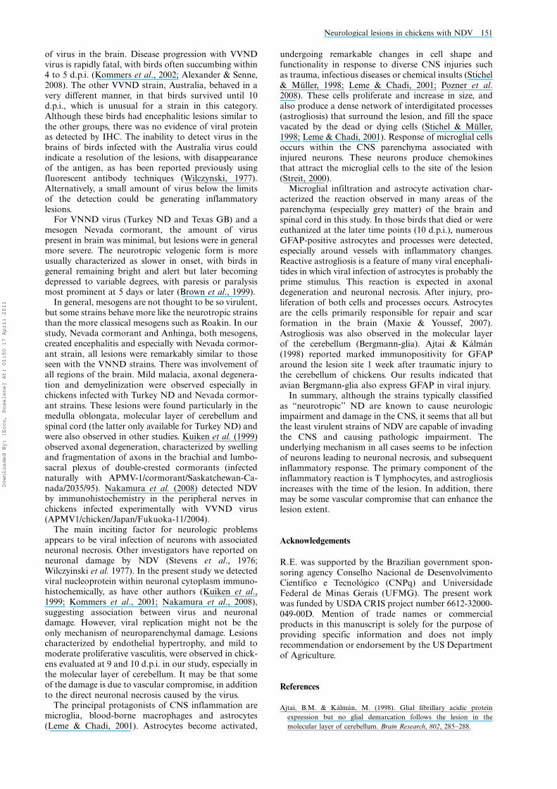
Histopathology. For the present study, brain tissues were examined
histologically de novo. All the brains consisted of sagittal sections (taken
from the median plane). These sections allowed analysis of the
telencephalon (cerebrum), cerebellum, thalamus, midbrain and hind-
brain (medulla oblongata). Figure 1 illustrates the location of these
brain regions. For those strains having spinal cord available (two strains:
Turkey ND and Anhinga), sections were longitudinal (cervical, thoracic
and lumbar) from two strains. A list of all lesions seen by light
microscopy in these sections was prepared. The lesions observed in the
brains and spinal cords were graded according to the lesion severity and
character. Specific lesions included: inflammatory cell infiltration,
endothelial hypertrophy, vasculitis, increase of the cellularity of the
neuropil (microgliosis and astrogliosis), chromatolysis, neuronal necro-
sis and neuronophagy, axonal degeneration and demyelinization. Each
anatomic region of the brain was examined for the presence of these
lesions.
Immunohistochemistry. For Newcastle disease virus. Brains from all of
the birds*and in addition, for two of the strains, spinal cords*were
available, and were processed via immunohistochemistry (IHC) to
detect viral nucleoprotein. Unstained sections were subjected to heat-
mediated antigen retrieval by microwaving for 20 min at minimum
power (approximately 600 W) in unmasking solution (Vector Labora-
tories, Burlingame, California, USA). Following antigen retrieval,
blocking was done with universal blocking reagent (Biogenex, San
Ramon, California, USA) as recommended by the manufacturer. The
primary antibody, made in rabbits, was an antipeptide, against the
nucleoprotein, used at 1:8000 dilution. The slides were incubated with
biotinylated goat anti-rabbit antibody and then with avidin�biotin
alkaline phosphatase (Vector Laboratories) at room temperature. The
reaction was revealed using a naphthol-based dye (Fast Red; DAKO,
Carpinteria, California, USA). Negative controls consisted of the brains
of age-matched, non-infected chickens.
For T lymphocytes and astrocytes. Selected sections of nervous system
showing inflammation were processed via IHC with polyclonal rabbit
anti-human CD3 (A 0452-DAKO) and monoclonal mouse anti-glial
fibrillar acid protein (GFAP) (MU020-UC; Biogenex).
For CD3, unstained sections were subjected to antigen retrieval by
microwaving for 20 min at minimum power in unmasking solution
(Vector Laboratories). Following antigen retrieval, blocking was done
with universal blocking reagent (Biogenex) and then incubated with the
primary anti-CD3 antibody (polyclonal rabbit) at a 1:100 dilution, at
48C overnight. After incubation, the slides were washed and incubated
with an alkaline phosphatase-linked polymer system (Labvision Cor-
poration, Fremont, California, USA) for 30 min at room temperature.
As a positive control, the spleen of a specific pathogen free chicken was
used. The reaction was revealed using Vector Red (Vector Laboratories).
Selected sections of the brain that had inflammation and increased
cellularity in the neuropil were examined by IHC for the presence of
GFAP using an automated method (DAKO autostainer). Slides were
subjected to antigen retrieval using citrate buffer with pH 6.0 (HK086-
9K; Biogenex) followed by heat. Endogenous peroxidase was blocked
using 3% hydrogen peroxide (H312-500; Fischer Scientific, Fair Lawn,
New Jersey, USA). The slides were incubated with the primary antibody
(monoclonal mouse anti-GFAP, MU020-UC; Biogenex) for 60 min at a
1:600 dilution. The secondary antibody used was a biotinylated horse
anti-mouse IgG (BA-2001; Vector Laboratories). The detection system
Table 1. Viruses in archived tissue used in the present study.
Strain
designation Full name of strain
Classification of
pathotype Reference
CA 1083 Chicken/US/CA
1083 (Fontana)/72
VVND Brown
et al. (1999)
Korea
97-147
Korea 97-147 VVND Lee et al.
(2004)
Australia Chicken /Australia/
9809-19-1107/1998
VVND Susta et al.
(2011)
Texas GB Chicken/US/Texas
GB/48
VNND Brown
et al. (1999)
Turkey ND Chicken/US/Turkey
ND/43084/92
VNND Brown
et al. (1999)
Nevada
cormorant
Double crested
cormorant/US/
Nevada/19529-04/
2005
Mesogen Susta et al.
(2011)
Anhinga Chicken/US[FL]/
Anhinga/93 44083/
93
Mesogen Brown
et al. (1999)
Roakin Chicken/US/
Roakin/48
Mesogen Brown
et al. (1999)
B1 Chicken/US/B1/48 Lentogen Brown
et al. (1999)
QV4 Chicken /Australia /
Queensland (QV4)/
31
Lentogen Brown
et al. (1999)
Figure 1. Schematic diagram showing view of the avian brain
according to the actual names. Diagram adapted from Reiner
et al. (2004). LSt, lateral striatum; MSt, medial striatum; Nc,
nuclei cerebelaris; Mo, medulla oblongata.
146 R. Ecco et al.
Downloaded By: [Ecco, Roselene] At: 01:50 17 April 2011
used was the labelled streptavidin�biotin streptavidin�horseradish
peroxidase (K1016; DAKO). The reaction was revealed by 3,3’-
diaminobenzidine tetrahydrochloride (K3466; DAKO) chromogen to
yield a brown staining. The cerebellum of a non-infected specific
pathogen free chicken was used as a negative control.
In all procedures, tissue sections were counterstained with Gills II
haematoxylin, dehydrated through graded alcohol, cleared in xylene,
and cover slipped with Permount.
Results
Clinical disease. The clinical findings have been reportedin previous publications for each strain. For purposes ofclarity, some are repeated here. As described in previouspublications, birds infected with a viscerotropic velogenicstrain (CA 1083) were severely depressed at 2 d.p.i. anddied or were euthanized when moribund between 4 and 5d.p.i. (Brown et al., 1999). With another viscerotropicvelogen, Korea 97-147, birds died between 4 and 5 d.p.i.with mild neurologic signs consisting of head twitch andsevere depression (data not published). Birds infectedwith a third viscerotropic velogenic strain (Australia)demonstrated depression between 3 and 4 d.p.i., andmild neurologic signs including depression and headtremors between 4 and 9 d.p.i. but at 10 d.p.i., they wereclinically normal and were euthanized (Susta et al.,2011). With the Texas GB strain (neurotropic velogen),birds were bright and alert at 2 d.p.i. but were in lateralrecumbency and unable to right themselves by 5 d.p.i.Chickens inoculated with the second velogenic neuro-tropic strain, Turkey ND, were clinically normal at 5d.p.i. but by 10 d.p.i. were depressed, with unilateral legparesis. Birds inoculated with mesogenic (Roakin andAnhinga) and lentogenic (B1 and QV4) isolates demon-strated no signs of clinical disease (Brown et al., 1999).However, birds inoculated with another mesogenic strain(Nevada cormorant) had severe neurologic signs(recumbency and paralysis) between 4 and 10 d.p.i.(Susta et al., 2011).
Histopathology. Histologic changes were seen in thebrains from birds in all strains associated with neurolo-gic signs, which includes the VVND strains (CA 1083,Korea 97-147, Australia) the VNND strains (TurkeyND, Texas GB), and the Nevada cormorant strain(mesogenic). In addition, histologic changes were seenin the brains of birds infected with Anhinga virus(mesogenic strain), even though no overt neurologicsigns were recorded for these birds. The brains of birdsfrom chickens infected with Roakin (mesogen) and thetwo lentogens (B1, QV4) were histologically unremark-able.
For those strains that produced encephalitis and forwhich there was more than one sampling period in whichthe encephalitis was present, inevitably the lesion severitywas more severe at the later time point. The degree ofpathologic changes in the various regions of the brainfrom the one or two time periods available for each ofthe strains is presented in Table 2. In general, whenmultiple brain sections were available from one strain ata certain time point, there was good similarity of lesiondistribution and severity.
In all cases where lesions were observed, it waspossible to observe some degree of encephalitis (orencephalomyelitis) and microgliosis. Microglial cellsoccurred predominantly around vessels or surroun-
ding necrotic neurons. Additional alterations such asvasculitis, vascular proliferation, axonal degeneration,demyelination and astrogliosis were present in somestrains. Findings for each strain are described in moredetail below.
For the VVND strains (Korea 97-147 and Australia),the inflammatory lesions consisted of one to two layersof lymphocytes in and around vascular walls. Encepha-litis was observed in the nuclei cerebellaris, mesopallium,nidopallium, hyperpallium and meninges correspondingto these regions. In these areas there was mild micro-gliosis. The inflammatory lesions had moderate intensityand multifocal distribution. Chromatolysis and somenecrotic neurons were also observed. In birds infectedwith VVND CA 1083 strain, in the midbrain, nucleicerebellaris and medulla oblongata there was mildencephalitis, moderate multifocal haemorrhage andoedema around vessels.
For the VNND strains (Turkey ND and Texas GB),inflammatory lesions were initially observed at 5 d.p.i.Inflammatory lesions in general were more pronounced,especially with Turkey ND, and in addition to theperivascular cuffs of lymphocytes there was widespreadchromatolysis, axonal degeneration and necrotic neu-rons. In birds infected with the Texas GB strain,moderate microgliosis was also observed at 10 d.p.i. Atthis time point, all these changes were most prominent inthe striatum and medulla oblongata. In the birdsinfected with Turkey ND, lesions became progressivelymore severe at around 10 d.p.i. and were distributedextensively throughout all regions of brain, includingtelencephalon. The inflammation was characterized asthree to four layers of lymphocytes and fewer plasmacells in the walls and around vessels (Figure 2). In somevenules, numerous endothelial cells were hypertrophic,and had large nuclei with vesiculated chromatin pro-truding into the vascular lumen. Multifocal vascularproliferation was observed in the molecular layer of thecerebellum and medulla oblongata. Multifocal and mildto moderate inflammation was observed in the subar-achnoid spaces in the midbrain and cerebellum, andoccasionally in the cerebrum. Neuronal necrosis asso-ciated with neuronophagia and microgliosis (Figure 3),occasional mitosis, vacuolization of neuropil and astro-cytosis were observed in the grey matter. Axonaldegeneration (Figure 4) was present in the white matterand grey matter of the medulla oblongata and spinalcord. In the cerebellar folia (junction between granularand molecular layers) there was multifocal loss ofPurkinje cells associated with areas of inflammationand vascular proliferation (Figure 5). All the lesions hada multifocal to coalescing distribution.
In birds infected with one of the mesogenic strains,Nevada cormorant, less severe lesions were observed at 5d.p.i., becoming progressively more severe at 6 and 9d.p.i. The lesions were similar to those described forTurkey ND strain. However, the intensity was less in thecerebrum and there was slightly less neuronal necrosis.The regions affected most intensely were the medullaoblongata, midbrain, thalamus and molecular layerof cerebellum. In these areas, axonal spheroids werepresent.
For another mesogenic strain (Anhinga) evaluated at10 d.p.i., the inflammatory lesions were moderate andmultifocal, and distributed in the striatum, midbrain,medulla oblongata, molecular layer of cerebellum, nuclei
Neurological lesions in chickens with NDV 147
Downloaded By: [Ecco, Roselene] At: 01:50 17 April 2011

Table 2. Distribution and intensity of histological lesions and stain for virus in different areas of the nervous systema in chickens experimentally infected with different NDV isolates.
Strain Pathotype d.p.i. Stain Me H M N Hp St Th Mb Cc Nc Mo Sc
CA 1083 (Fontana) VVND 5 HE � � � � � � � � � � � NE
IHC � � � �� � � � � � � � NE
Korea 97-147 VVND 5 HE � � �� �� �� � � �� � �� �� NE
IHC � �� ��� � � � � ��� �� � � NE
Australia VVND 10 HE � � �� �� � �� �� � � � �� NE
IHC � � � � � � � � � � � NE
Texas GB VNND 5 HE � � � � � � � �� � �� �� NE
IHC � � � � � � � � �� � �� NE
Texas GB VNND 10 HE � � � � � ��� � � � �� �� NE
IHC � � � � � � � � � � � NE
Turkey ND VNND 5 HE � � � � � � � �� �� �� �� ��IHC � ��� � � � �� �� ��� �� � �� �
Turkey ND VNND 10 HE �� ��� ��� ��� ��� �� �� �� ��� ��� ��� ���IHC � �� �� �� � �� �� � � � � ��
Nevada cormorant Mesogen 6 HE � � � � � � � �� � �� �� NE
IHC � �� � �� � �� � � �� � �� NE
Nevada cormorant Mesogen 9 HE � � �� �� � �� ��� ��� ��� ��� ��� NE
IHC � �� � � � � �� � � � � NE
Anhinga Mesogen 10 HE � � � � � �� � � � �� �� ���IHC � � � � � � � � � � � �
Roakin Mesogen 5;10 HE � � � � � � � � � � � NE
IHC � � � � � � � � � � � NE
B1 Lentogen 10 HE � � � � � � � � � � � NE
IHC � � � � � � � � � � � NE
QV4 Lentogen 10 HE � � � � � � � � � � � NE
IHC � � � � � � � � � � � NE
�, no lesions/no virus signal; �, mild lesions/virus signal; ��, moderate lesions/virus signal; ���, marked lesions/virus signal; NE, not evaluated; HE, haematoxylin and eosin; Me, meninges; H,
hyperpallium; M, mesopallium; N, nidopallium; Hp, hippocampus; St, striatum; Th, thalamus; Mb, midbrain; Cc, cerebellar cortex; Nc, nuclei cerebelaris; Mo, medulla oblongata; Sc, spinal cord. aThe
nomenclature of the avian brain, and abbreviations, was described according to Reiner et al. (2004) and www.avianbrain.org.
148
R.Ecco
eta
l.
Downloaded By: [Ecco, Roselene] At: 01:50 17 April 2011

cerebellaris, and grey matter of the spinal cord. Thelesions were most intense in the spinal cord. Theinflammatory lesions were characterized by one to threelayers of lymphocytes and some plasma cells in andaround vascular walls. These lesions were associated withmild to moderate microgliosis, axonal degeneration andmild demyelinization (particularly in the medulla ob-longata and spinal cord). Lesions were not observed inthe brain and spinal cord of the birds at 5 d.p.i.
In chickens infected with the Roakin (mesogen) strainand the two lentogenic strains (B1 and QV4), all of thebrains at 2, 5, and 10 d.p.i were examined. There wasnever any inflammation detected in the nervous tissue.
Immunohistochemistry. For Newcastle disease virus. Re-sults are presented in Table 2. In general, immunohisto-chemical signals were seen in multiple regions of thebrain. They were always present as multiple granulesidentifiable in the cytoplasm of neurons, some glial cellsand rarely endothelial cells. In general, the viral nucleo-protein signal was more intense and distributed in
multifocal areas at 5 d.p.i. In those birds with lesionsevaluated around 10 d.p.i, virus was identifiable by IHCin lesser amounts in the brain, often associated withlesions. However, in many cases, there would be noassociation between degree or location of inflammationand presence of moderate IHC signal (Figure 6). Viralnucleoprotein was not detected in the brains of chickensinfected with the Australia strain (VVND virus). Simi-larly, virus was not detected in any of the birds infectedwith strains without encephalitis (Roakin, a mesogen,and B1, QV4, lentogens).
For T lymphocytes and astrocytes. Results are presentedin Table 3. Many inflammatory cells in the wall andaround vessels were positive by IHC-CD3 (T lympho-cytes) in the brains of all chickens that presentedencephalitis. In the brains with marked inflammationexamined at 10 d.p.i. (Turkey ND) and 9 d.p.i. (Nevadacormorant), some CD3-positive cells were also present in
Figure 4. Cerebellum, 4-week-old chicken, experimentally in-
fected with Turkey ND strain. 10 d.p.i. The molecular layer (M)
has focal encephalitis and vascular proliferation. Some Purkinje
cells in the subjacent Purkinje cell layer (P) are missing. In the
granular layer (G), there are no changes. Haematoxylin and
eosin. Bar � 50 mm.
Figure 2. Cerebrum (mesopallium), 4-week-old chicken, ex-
perimentally infected with Turkey ND strain. 10 d.p.i. Marked
infiltration of lymphocytes and plasma cells in and around the
vascular wall. Haematoxylin and eosin. Bar � 50 mm.
Figure 3. Medulla oblongata, 4-week-old chicken, experimen-
tally infected with Nevada cormorant strain. 9 d.p.i. Necrotic
neurons undergoing phagocytosis (arrow) by many microglial
cells. Haematoxylin and eosin. Bar � 50 mm.
Figure 5. Medulla oblongata, 4-week-old chicken, experimen-
tally infected with Turkey ND strain. Spongy changes (*),
axonal spheroids (arrow head) and hypertrophy of the nuclei of
astrocytes. Haematoxylin and eosin. Bar � 50 mm.
Neurological lesions in chickens with NDV 149
Downloaded By: [Ecco, Roselene] At: 01:50 17 April 2011

the neuropil around vessels. For GFAP, the signalrevealed an increased number of astrocytes, as well asincreased thickness of the astrocyte processes (astro-gliosis), in the parenchyma of the CNS in chickens withencephalitis that survived to 9 or 10 d.p.i. An exceptionwas the chickens infected with the Australia strain(VVND) in which birds were sampled until 10 d.p.i.,but still there was no increase in GFAP noted. Inchickens infected with Anhinga strain (mesogen), thereaction was minimal. Astrogliosis was strongest in thosechickens infected with Turkey ND and Nevada cormor-ant, and was especially prominent in the medullaoblongata, hyperpallium (Figure 7) and molecular layerof cerebellum. These are also the two strains for whichspinal cord was also available, and it was apparent thatthis reaction extended into this region as well (Figure 8).The astrocytic reaction was more prominent surround-ing areas of intense perivascular cuffs.
Discussion
The present study showed the distribution and extent ofneurologic lesions in chickens infected with differentstrains of NDV. With the lentogens, there was never anyevidence of encephalitis or presence of viral nucleopro-
tein in the brain as detected by IHC. Similarly, with one
of the mesogens, Roakin, encephalitis was not a feature
and there was no virus present in the brain. For all of the
other strains, distinct encephalitic lesions in birds
infected with Texas GB, Turkey ND and Nevada
cormorant strains became increasingly severe over
time. In all these brains, the predominance of lesions
was observed especially in the grey matter. The inflam-
matory lesions were similar in character but differed
somewhat in severity in all strains that had encephalitis.For those two strains that caused death by 5 d.p.i. (CA
1083 and Korea 97-147, both VVND viruses), the
inflammation in the brain was mild or moderate,
respectively, and multifocal. The amount of virus seen
in the brain, as determined by IHC, was mild and
multifocal for CA 1083, suggesting that the virus had not
replicated extensively in the nervous tissue. For the
Korea strain, a moderate to marked amount of viral
protein IHC signal indicated a more extensive presence
Figure 6. Hyperpallium, 4-week-old chicken, experimentally
infected with Korea 97-147 strain. 5 d.p.i. Immunohistochemistry
for viral nucleoprotein (NDV). Multiple granules are identifiable
in the cytoplasm of neurons. Bar � 50 mm.
Table 3. Greatest histologic severity lesions, degree of inflam-
mation, signal for CD3 and intensity of signal for GFAP.
Strain Pathotype d.p.i. Severity CD3 GFAP
CA 1083
(Fontana)
VVND 5 � Positive Negative
Korea 97-147 VVND 5 �� Positive Negative
Australia VVND 10 �� Positive Negative
Texas GB VNND 10 �� Positive 1
Turkey ND VNND 5 �� Positive Negative
Turkey ND VNND 10 ��� Positive 3
Nevada
cormorant
Mesogen 6 �� Positive Negative
Nevada
cormorant
Mesogen 9 ��� Positive 2
Anhinga Mesogen 10 �� Positive 1
�, mild lesions; ��, moderate lesions; ���, marked
lesions; 1, mild; 2, moderate; 3, marked.
Figure 7. Cerebrum (hyperpallium), 4-week-old chicken, ex-
perimentally infected with Turkey ND strain. 10 d.p.i. Immuno-
histochemistry for GFAP. Many GFAP-positive astrocytes can be
observed in the areas of inflammation. Bar � 50 mm.
Figure 8. Spinal cord, 4-week-old chicken, experimentally
infected with Turkey ND strain. 10 d.p.i. Immunohistochemistry
for GFAP. Grey matter and a focal area under meninges with
inflammatory changes and a moderate increase of GFAP expres-
sion around. Bar � 50 mm.
150 R. Ecco et al.
Downloaded By: [Ecco, Roselene] At: 01:50 17 April 2011
of virus in the brain. Disease progression with VVNDvirus is rapidly fatal, with birds often succumbing within4 to 5 d.p.i. (Kommers et al., 2002; Alexander & Senne,2008). The other VVND strain, Australia, behaved in avery different manner, in that birds survived until 10d.p.i., which is unusual for a strain in this category.Although these birds had encephalitic lesions similar tothe other groups, there was no evidence of viral proteinas detected by IHC. The inability to detect virus in thebrains of birds infected with the Australia virus couldindicate a resolution of the lesions, with disappearanceof the antigen, as has been reported previously usingfluorescent antibody techniques (Wilczynski, 1977).Alternatively, a small amount of virus below the limitsof the detection could be generating inflammatorylesions.
For VNND virus (Turkey ND and Texas GB) and amesogen Nevada cormorant, the amount of viruspresent in brain was minimal, but lesions were in generalmore severe. The neurotropic velogenic form is moreusually characterized as slower in onset, with birds ingeneral remaining bright and alert but later becomingdepressed to variable degrees, with paresis or paralysismost prominent at 5 days or later (Brown et al., 1999).
In general, mesogens are not thought to be so virulent,but some strains behave more like the neurotropic strainsthan the more classical mesogens such as Roakin. In ourstudy, Nevada cormorant and Anhinga, both mesogens,created encephalitis and especially with Nevada cormor-ant strain, all lesions were remarkably similar to thoseseen with the VNND strains. There was involvement ofall regions of the brain. Mild malacia, axonal degenera-tion and demyelinization were observed especially inchickens infected with Turkey ND and Nevada cormor-ant strains. These lesions were found particularly in themedulla oblongata, molecular layer of cerebellum andspinal cord (the latter only available for Turkey ND) andwere also observed in other studies. Kuiken et al. (1999)observed axonal degeneration, characterized by swellingand fragmentation of axons in the brachial and lumbo-sacral plexus of double-crested cormorants (infectednaturally with APMV-1/cormorant/Saskatchewan-Ca-nada/2035/95). Nakamura et al. (2008) detected NDVby immunohistochemistry in the peripheral nerves inchickens infected experimentally with VVND virus(APMV1/chicken/Japan/Fukuoka-11/2004).
The main inciting factor for neurologic problemsappears to be viral infection of neurons with associatedneuronal necrosis. Other investigators have reported onneuronal damage by NDV (Stevens et al., 1976;Wilczyinski et al. 1977). In the present study we detectedviral nucleoprotein within neuronal cytoplasm immuno-histochemically, as have other authors (Kuiken et al.,1999; Kommers et al., 2001; Nakamura et al., 2008),suggesting association between virus and neuronaldamage. However, viral replication might not be theonly mechanism of neuroparenchymal damage. Lesionscharacterized by endothelial hypertrophy, and mild tomoderate proliferative vasculitis, were observed in chick-ens evaluated at 9 and 10 d.p.i. in our study, especially inthe molecular layer of cerebellum. It may be that someof the damage is due to vascular compromise, in additionto the direct neuronal necrosis caused by the virus.
The principal protagonists of CNS inflammation aremicroglia, blood-borne macrophages and astrocytes(Leme & Chadi, 2001). Astrocytes become activated,
undergoing remarkable changes in cell shape andfunctionality in response to diverse CNS injuries suchas trauma, infectious diseases or chemical insults (Stichel& Muller, 1998; Leme & Chadi, 2001; Pozner et al.2008). These cells proliferate and increase in size, andalso produce a dense network of interdigitated processes(astrogliosis) that surround the lesion, and fill the spacevacated by the dead or dying cells (Stichel & Muller,1998; Leme & Chadi, 2001). Response of microglial cellsoccurs within the CNS parenchyma associated withinjured neurons. These neurons produce chemokinesthat attract the microglial cells to the site of the lesion(Streit, 2000).
Microglial infiltration and astrocyte activation char-acterized the reaction observed in many areas of theparenchyma (especially grey matter) of the brain andspinal cord in this study. In those birds that died or wereeuthanized at the later time points (10 d.p.i.), numerousGFAP-positive astrocytes and processes were detected,especially around vessels with inflammatory changes.Reactive astrogliosis is a feature of many viral encephali-tides in which viral infection of astrocytes is probably theprime stimulus. This reaction is expected in axonaldegeneration and neuronal necrosis. After injury, pro-liferation of both cells and processes occurs. Astrocytesare the cells primarily responsible for repair and scarformation in the brain (Maxie & Youssef, 2007).Astrogliosis was also observed in the molecular layerof the cerebellum (Bergmann-glia). Ajtai & Kalman(1998) reported marked immunopositivity for GFAParound the lesion site 1 week after traumatic injury tothe cerebellum of chickens. Our results indicated thatavian Bergmann-glia also express GFAP in viral injury.
In summary, although the strains typically classifiedas ‘‘neurotropic’’ ND are known to cause neurologicimpairment and damage in the CNS, it seems that all butthe least virulent strains of NDV are capable of invadingthe CNS and causing pathologic impairment. Theunderlying mechanism in all cases seems to be infectionof neurons leading to neuronal necrosis, and subsequentinflammatory response. The primary component of theinflammatory reaction is T lymphocytes, and astrogliosisincreases with the time of the lesion. In addition, theremay be some vascular compromise that can enhance thelesion extent.
Acknowledgements
R.E. was supported by the Brazilian government spon-soring agency Conselho Nacional de DesenvolvimentoCientıfico e Tecnologico (CNPq) and UniversidadeFederal de Minas Gerais (UFMG). The present workwas funded by USDA CRIS project number 6612-32000-049-00D. Mention of trade names or commercialproducts in this manuscript is solely for the purpose ofproviding specific information and does not implyrecommendation or endorsement by the US Departmentof Agriculture.
References
Ajtai, B.M. & Kalman, M. (1998). Glial fibrillary acidic protein
expression but no glial demarcation follows the lesion in the
molecular layer of cerebellum. Brain Research, 802, 285�288.
Neurological lesions in chickens with NDV 151
Downloaded By: [Ecco, Roselene] At: 01:50 17 April 2011
Alexander, D.J. & Senne, D.A. (2008). Newcastle disease. In Y.M. Saif,
A.M. Fadly, J.R. Glisson, L.R. McDougald, L.K. Nolan & D.E.
Swayne, (Eds.). Diseases of Poultry 12th edn (pp. 75�115). Ames,
Iowa: Blackwell Publishing.
Bhaiyat, M.I., Kobayashi, Y., Itakura, C., Islam, M.A. & Kida, H.
(1995). Brain lesions in chickens experimentally infected with a
neuroadapted strain of mesogenic Newcastle disease virus. Journal of
Veterinary Medical Science, 57, 237�244.
Brown, C., King, D.J. & Seal, B.S. (1999). Pathogenesis of Newcastle
disease in chickens experimentally infected with viruses of different
virulence. Veterinary Pathology, 36, 125�132.
Farkas, T., Szekely, E., Belak, S. & Kiss, I. (2009). Real-time PCR-
based pathotyping of Newcastle disease virus by use of TaqMan
minor groove binder probes. Journal Clinical Microbiology, 47, 2114�2123.
Kommers, G.D., King, D.J., Seal, B.S. & Brown, C.C. (2001). Virulence
of pigeon-origin Newcastle disease virus isolates for domestic
chickens. Avian Diseases, 45, 906�921.
Kommers, G.D., King, D.J., Seal, B.S., Carmichael, K.P. & Brown, C.C.
(2002). Pathogenesis of six pigeon-origin of Newcastle disease virus
for domestic chickens. Veterinary Pathology, 39, 353�362.
Kuiken, T., Wobeser, G., Leighton, F.A., Haines, D.M., Chelack, B.,
Bogdan, J., et al. (1999). Pathology of Newcastle disease in double-
crested cormorants from Saskatchewan, with comparison of diag-
nostic methods. Journal Wildlife Diseases, 35, 8�23.
Lee, Y.J., Sung, H.W., Choi, J.G., Kim, J.H. & Song, C.S. (2004).
Molecular epidemiology of Newcastle disease viruses isolated
in South Korea using sequencing of the fusion protein cleavage
site region and phylogenetic relationships. Avian Pathology, 33, 482�491.
Leme, R.J. & Chadi, G. (2001). Distant microglial and astroglial
activation secondary to experimental spinal cord lesion. Arquivos de
Neuro-psiquiatria, 59, 483�492.
Maxie, M.G. & Youssef, S. (2007). Nervous system. In M.G. Maxie,
(Ed.). Jubb, Kennedy, and Palmer’s Pathology of Domestic Animals
Vol. 1, 5th edn (pp. 281�457). Toronto: Elsevier.
Nakamura, K., Ohtsu, N., Nakamura, T., Yamamoto, Y., Yamada, M.,
Mase, M. & Imai, K. (2008). Pathologic and immunohistochemical
studies of Newcastle disease (ND) in broiler chickens vaccinated with
ND: severe nonpurulent encephalitis and necrotizing pancreatitis.
Veterinary Pathology, 45, 928�933.
Office International des Epizootes. (2008). Manual of Diagnostic Tests
and Vaccines for Terrestrial Animals. Paris: World Organization for
Animal Health.
Phillips, R.J., Samson, A.C.R. & Emmerson, P.T. (1998). Nucleotide
sequence of the 5?-terminus of Newcastle disease virus and assembly
of the complete genomic sequence: agreement with the ‘‘rule of six’’.
Archives of Virology Journal, 143, 1993�2002.
Pozner, R.G., Collado, S., de Giusti, C.J., Ure, A.E., Biedma, M.E.,
Romanowski, V., et al. (2008). Astrocyte response to Junin virus
infection. Neuroscience Letters, 445, 31�35.
Reiner, A., Perkel, D.J., Mello, C.V. & Jarvis, E.D. (2004). Songbirds
and the revised avian brain nomenclature. Annals of the New York
Academy of Sciences, 1016, 77�108.
Stevens, J.G., Nakamura, R.M., Cook, M.L. & Wilczynski, S.P. (1976).
Newcastle disease as a model for paramyxovirus-induced neurologi-
cal syndromes: pathogenesis of the respiratory disease and prelimin-
ary characterization of the ensuing encephalitis. Infection and
Immunity, 13, 590�599.
Stichel, C.C. & Muller, H.W. (1998). The CNS lesion scar: new vistas on
an old regeneration barrier. Cell and Tissue Research, 294, 1�9.
Streit, W.J. (2000). Microglial response to brain injury: a brief synopsis.
Toxicologic Pathology, 28, 28�30.
Susta, L., Miller, P.J., Afonso, C.L., Brown, C.C. (2011). Clinicopatho-
logical characterization in poultry of three strains of Newcastle
disease virus isolated from recent outbreaks. Veterinary Pathology, 48.
Wilczynski, S.P., Cook, M.L. & Stevens, J.G. (1977). Newcastle disease
as a model for paramyxovirus-induced neurologic syndromes. II.
Detailed characterization of the encephalitis. American Journal of
Pathology, 89, 649�666.
Wise, M.G., Sellers, H.S., Alvarez, R. & Seal, B.S. (2004). RNA-
dependent RNA polymerase gene analysis of worldwide Newcastle
disease virus isolates representing different virulence types and their
phylogenetic relationship with other members of the paramyxovir-
idae. Virus Research, 104, 71�80.
152 R. Ecco et al.
Downloaded By: [Ecco, Roselene] At: 01:50 17 April 2011
Related Documents