Neurologic Manifestations Associated with an Outbreak of Typhoid Fever, Malawi - Mozambique, 2009: An Epidemiologic Investigation James Sejvar 1 *, Emily Lutterloh 2,3 , Jeremias Naiene 4 , Andrew Likaka 5 , Robert Manda 5 , Benjamin Nygren 6 , Stephan Monroe 1 , Tadala Khaila 5 , Sara A. Lowther 2 , Linda Capewell 2 , Kashmira Date 2 , David Townes 2 , Yanique Redwood 2 , Joshua Schier 7 , Beth Tippett Barr 8 , Austin Demby 8 , Macpherson Mallewa 9 , Sam Kampondeni 9 , Ben Blount 7 , Michael Humphrys 6 , Deborah Talkington 6 , Gregory L. Armstrong 10 , Eric Mintz 6 1 Division of High-Consequence Pathogens and Pathology, National Center for Emerging and Zoonotic Infectious Diseases (NCEZID), Centers for Disease Control and Prevention (CDC), Atlanta, Georgia, United States of America, 2 Scientific Education and Professional Development Program Office, Epidemic Intelligence Service, CDC, Atlanta, Georgia, United States of America, 3 Current Position, New York State Department of Health, Albany, New York, United States of America, 4 Ministry of Health, Maputo, Mozambique, 5 Ministry of Health, Lilongwe, Malawi, 6 Division of Foodborne, Waterborne, and Environmental Infectious Diseases, NCEZID, CDC, Atlanta, Georgia, United States of America, 7 Division of Environmental Hazards and Health Effects, National Center for Environmental Health (NCEH), CDC, Atlanta, Georgia, United States of America, 8 Global AIDS Program, CDC, Lilongwe, Malawi, 9 Malawi-Liverpool-Wellcome Trust, College of Medicine, Blantyre, Malawi, 10 Division of Viral Diseases, National Center for Immunization and Respiratory Diseases, CDC, Atlanta, Georgia, United States of America Abstract Background: The bacterium Salmonella enterica serovar Typhi causes typhoid fever, which is typically associated with fever and abdominal pain. An outbreak of typhoid fever in Malawi-Mozambique in 2009 was notable for a high proportion of neurologic illness. Objective: Describe neurologic features complicating typhoid fever during an outbreak in Malawi-Mozambique Methods: Persons meeting a clinical case definition were identified through surveillance, with laboratory confirmation of typhoid by antibody testing or blood/stool culture. We gathered demographic and clinical information, examined patients, and evaluated a subset of patients 11 months after onset. A sample of persons with and without neurologic signs was tested for vitamin B6 and B12 levels and urinary thiocyanate. Results: Between March – November 2009, 303 cases of typhoid fever were identified. Forty (13%) persons had objective neurologic findings, including 14 confirmed by culture/serology; 27 (68%) were hospitalized, and 5 (13%) died. Seventeen (43%) had a constellation of upper motor neuron findings, including hyperreflexia, spasticity, or sustained ankle clonus. Other neurologic features included ataxia (22, 55%), parkinsonism (8, 20%), and tremors (4, 10%). Brain MRI of 3 (ages 5, 7, and 18 years) demonstrated cerebral atrophy but no other abnormalities. Of 13 patients re-evaluated 11 months later, 11 recovered completely, and 2 had persistent hyperreflexia and ataxia. Vitamin B6 levels were markedly low in typhoid fever patients both with and without neurologic signs. Conclusions: Neurologic signs may complicate typhoid fever, and the diagnosis should be considered in persons with acute febrile neurologic illness in endemic areas. Citation: Sejvar J, Lutterloh E, Naiene J, Likaka A, Manda R, et al. (2012) Neurologic Manifestations Associated with an Outbreak of Typhoid Fever, Malawi - Mozambique, 2009: An Epidemiologic Investigation. PLoS ONE 7(12): e46099. doi:10.1371/journal.pone.0046099 Editor: Martyn Kirk, The Australian National University, Australia Received February 1, 2012; Accepted August 28, 2012; Published December 3, 2012 This is an open-access article, free of all copyright, and may be freely reproduced, distributed, transmitted, modified, built upon, or otherwise used by anyone for any lawful purpose. The work is made available under the Creative Commons CC0 public domain dedication. Funding: U.S. government funding, Centers for Disease Control and Prevention. The funders had no role in study design, data collection and analysis, decision to publish, or preparation of the manuscript. Competing Interests: The authors have declared that no competing interests exist. * E-mail: [email protected] Introduction Typhoid fever is a bacterial disease caused by infection with Salmonella enterica serovar Typhi (Salmonella Typhi). It is transmitted through the fecal-oral route, generally by contaminated water or food. Typically, it presents as an acute febrile illness often accompanied by signs and symptoms such as headache, abdominal pain, diarrhea or constipation, and malaise [1]. Other, more severe complications of typhoid fever include intestinal perfora- tion, hepatitis, pneumonia, and tissue abscesses [1,2]. Neurologic illness has also been described, most frequently as acute encephalopathy or meningitis [3]. A variety of objective neurologic signs have been documented, including acute neuropsychiatric illness [4,5,6], spasticity and clonus [4,7], ataxia [8,9,10,11,12,13], PLOS ONE | www.plosone.org 1 December 2012 | Volume 7 | Issue 12 | e46099

Welcome message from author
This document is posted to help you gain knowledge. Please leave a comment to let me know what you think about it! Share it to your friends and learn new things together.
Transcript
Neurologic Manifestations Associated with an Outbreakof Typhoid Fever, Malawi - Mozambique, 2009: AnEpidemiologic InvestigationJames Sejvar1*, Emily Lutterloh2,3, Jeremias Naiene4, Andrew Likaka5, Robert Manda5,
Benjamin Nygren6, Stephan Monroe1, Tadala Khaila5, Sara A. Lowther2, Linda Capewell2,
Kashmira Date2, David Townes2, Yanique Redwood2, Joshua Schier7, Beth Tippett Barr8, Austin Demby8,
Macpherson Mallewa9, Sam Kampondeni9, Ben Blount7, Michael Humphrys6, Deborah Talkington6,
Gregory L. Armstrong10, Eric Mintz6
1 Division of High-Consequence Pathogens and Pathology, National Center for Emerging and Zoonotic Infectious Diseases (NCEZID), Centers for Disease Control and
Prevention (CDC), Atlanta, Georgia, United States of America, 2 Scientific Education and Professional Development Program Office, Epidemic Intelligence Service, CDC,
Atlanta, Georgia, United States of America, 3 Current Position, New York State Department of Health, Albany, New York, United States of America, 4 Ministry of Health,
Maputo, Mozambique, 5 Ministry of Health, Lilongwe, Malawi, 6 Division of Foodborne, Waterborne, and Environmental Infectious Diseases, NCEZID, CDC, Atlanta,
Georgia, United States of America, 7 Division of Environmental Hazards and Health Effects, National Center for Environmental Health (NCEH), CDC, Atlanta, Georgia, United
States of America, 8 Global AIDS Program, CDC, Lilongwe, Malawi, 9 Malawi-Liverpool-Wellcome Trust, College of Medicine, Blantyre, Malawi, 10 Division of Viral Diseases,
National Center for Immunization and Respiratory Diseases, CDC, Atlanta, Georgia, United States of America
Abstract
Background: The bacterium Salmonella enterica serovar Typhi causes typhoid fever, which is typically associated with feverand abdominal pain. An outbreak of typhoid fever in Malawi-Mozambique in 2009 was notable for a high proportion ofneurologic illness.
Objective: Describe neurologic features complicating typhoid fever during an outbreak in Malawi-Mozambique
Methods: Persons meeting a clinical case definition were identified through surveillance, with laboratory confirmation oftyphoid by antibody testing or blood/stool culture. We gathered demographic and clinical information, examined patients,and evaluated a subset of patients 11 months after onset. A sample of persons with and without neurologic signs wastested for vitamin B6 and B12 levels and urinary thiocyanate.
Results: Between March – November 2009, 303 cases of typhoid fever were identified. Forty (13%) persons had objectiveneurologic findings, including 14 confirmed by culture/serology; 27 (68%) were hospitalized, and 5 (13%) died. Seventeen(43%) had a constellation of upper motor neuron findings, including hyperreflexia, spasticity, or sustained ankle clonus.Other neurologic features included ataxia (22, 55%), parkinsonism (8, 20%), and tremors (4, 10%). Brain MRI of 3 (ages 5, 7,and 18 years) demonstrated cerebral atrophy but no other abnormalities. Of 13 patients re-evaluated 11 months later, 11recovered completely, and 2 had persistent hyperreflexia and ataxia. Vitamin B6 levels were markedly low in typhoid feverpatients both with and without neurologic signs.
Conclusions: Neurologic signs may complicate typhoid fever, and the diagnosis should be considered in persons with acutefebrile neurologic illness in endemic areas.
Citation: Sejvar J, Lutterloh E, Naiene J, Likaka A, Manda R, et al. (2012) Neurologic Manifestations Associated with an Outbreak of Typhoid Fever, Malawi -Mozambique, 2009: An Epidemiologic Investigation. PLoS ONE 7(12): e46099. doi:10.1371/journal.pone.0046099
Editor: Martyn Kirk, The Australian National University, Australia
Received February 1, 2012; Accepted August 28, 2012; Published December 3, 2012
This is an open-access article, free of all copyright, and may be freely reproduced, distributed, transmitted, modified, built upon, or otherwise used by anyone forany lawful purpose. The work is made available under the Creative Commons CC0 public domain dedication.
Funding: U.S. government funding, Centers for Disease Control and Prevention. The funders had no role in study design, data collection and analysis, decision topublish, or preparation of the manuscript.
Competing Interests: The authors have declared that no competing interests exist.
* E-mail: [email protected]
Introduction
Typhoid fever is a bacterial disease caused by infection with
Salmonella enterica serovar Typhi (Salmonella Typhi). It is transmitted
through the fecal-oral route, generally by contaminated water or
food. Typically, it presents as an acute febrile illness often
accompanied by signs and symptoms such as headache, abdominal
pain, diarrhea or constipation, and malaise [1]. Other, more
severe complications of typhoid fever include intestinal perfora-
tion, hepatitis, pneumonia, and tissue abscesses [1,2]. Neurologic
illness has also been described, most frequently as acute
encephalopathy or meningitis [3]. A variety of objective neurologic
signs have been documented, including acute neuropsychiatric
illness [4,5,6], spasticity and clonus [4,7], ataxia [8,9,10,11,12,13],
PLOS ONE | www.plosone.org 1 December 2012 | Volume 7 | Issue 12 | e46099
aphasia [14,15,16], and cerebritis [3,17]. However, these findings
have generally appeared as case reports or small case series.
Beginning in June 2009, an outbreak of unexplained febrile
illness occurred in villages along the border region between
southern Malawi and western Mozambique. This area was known
to have a high rate of general mild malnutrition, with most diets
high in consumption of wheat, corn, and leafy vegetables. Cassava
is consumed, but infrequently. Initial reports described many
persons who presented with acute neurologic illness including
mental status changes, headache, ‘‘difficulty walking’’, dysarthria,
and hyperreflexia. Other neurologic features including seizures
and neck stiffness were also described. Gastrointestinal complaints
were not prominent among patients early in the outbreak. The
investigators initially suspected common etiologies of such
neurologic abnormalities in sub-Saharan Africa such as acute
encephalitis or heavy metal toxicity, as well as less common
etiologies such as neurolathyrism and konzo. However, subsequent
investigation revealed the outbreak to be caused by typhoid fever,
and after the etiology was determined, persons with signs and
symptoms more typical of typhoid fever were increasingly
recognized.
We describe the results of an investigation into the clinical,
neurologic and laboratory features of persons with typhoid fever
during this outbreak. Our investigation suggests that signs of upper
motor neuron dysfunction were predominant, neurologic features
were generally a later manifestation of typhoid fever, and outcome
was generally favorable.
Methods
Patient IdentificationThe outbreak was first noted in June 2009 by health personnel
in Neno District, Malawi, who observed an increase in patients
hospitalized at Neno District Hospital with fever and neurologic
illness. Ill patients were from villages in Neno District and
neighboring Tsangano District, Mozambique. The outbreak
occurred in a remote location; the closest health center, Nsambe
Health Centre, is approximately 8.5 km away by dirt road over
rough terrain. As cases continued, a larger investigation was
initiated by the Malawi Ministry of Health (MOH).
Between July and November, 2009, an epidemiologic investi-
gation was conducted [18] that included structured retrospective
interviews of previously ill individuals to determine initial signs and
symptoms, risk factors, and possible exposures; retrospective
hospital record review; structured interview and clinical examina-
tion of acutely ill individuals; and verbal autopsy for deceased
patients. Questions included specific assessment of dietary habits,
sources of drinking water, and other possible exposures that might
lead to an infectious or toxic etiology or result in a nutritional
deficiency, Based upon initial clinical findings and laboratory
results among outbreak patients, a case definition was established
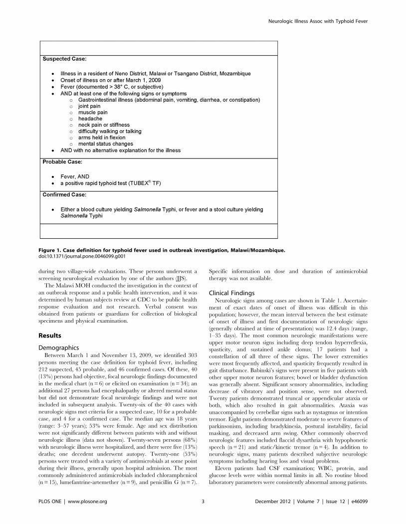
for suspected, probable, and confirmed cases [18] (Figure 1).
Active surveillance was implemented in affected villages to identify
possible patients presenting early in the course of illness.
We identified patients with neurologic illness by several
methods. We reviewed medical records at Neno District Hospital
for admissions during March through November 2009 for
descriptions of objective neurologic findings. From July 22 through
November 13, 2009, we prospectively gathered data on persons
presenting with illness, and hospitalized patients meeting the case
definition underwent neurologic evaluation by one of the authors,
a U.S. board-certified neurologist (JJS). When possible, suspected
patients meeting clinical case criteria but not hospitalized were
evaluated in their villages. When possible, hospitalized patients
were serially re-evaluated in order to document progression of
illness; a subset of patients underwent re-evaluation approximately
11 months after acute illness to detect the presence of long-term
neurologic sequelae.
Laboratory TestingCerebrospinal fluid (CSF) white blood cell count (WBC),
glucose, and protein level results were reviewed when available.
Clinical specimens obtained from acutely ill patients, including
serum, blood, urine, and CSF, initially underwent testing for a
number of infectious agents including various viruses, bacteria,
parasites, and rickettsiae, as well as various toxins at laboratories at
CDC (Table S1). Typhoid-specific testing in the field was
performed using a rapid diagnostic assay (TUBEXH TF, IDL
Biotech, Bromma, Sweden) that detects IgM antibodies against the
O9 lipopolysaccharide antigen of Salmonella Typhi. Following the
establishment of field capacity to collect and transport specimens
for culture, confirmatory testing for typhoid was performed by
blood or stool culture.
Because some of the neurologic features observed in the patients
were similar to those seen in some micronutrient abnormalities, we
tested serum specimens on a subset of patients with and without
neurologic illness for vitamin B6 and B12 concentrations. Serum
B12 concentrations were assessed by electrochemiluminescence
immunoassay (ECLIA; Roche Diagnostics Modular Analytics
E170) at CDC. Serum B6 concentrations [pyridoxal 59-phosphate
(PLP) and 4-pyridoxic acid (4PA)] were assessed by high-
performance liquid chromatography with fluorometric detection
[19]. PLP is indicative of longer-term vitamin B6 status, while 4PA
is indicative of short-term vitamin B6 intake. Since the neurologic
presentation including spasticity and clonus appeared similar to
konzo, an illness due to cyanogen toxicity from consumption of
improperly cooked bitter cassava, we also tested urine specimens
from a subset of patients with and without neurologic illness for
urinary thiocyanate levels, a marker for cyanogenic compound
exposure. Urine thiocyanate was quantified using isotope dilution
tandem mass spectrometry at CDC [20].
Neurodiagnostic TestingThree patients with neurologic illness underwent brain and
spinal cord magnetic resonance imaging (MRI) at the Malawi
MRI Facility in Blantyre. Autopsy was performed on one decedent
with neurologic illness; tissue from central nervous system,
meninges, lung, spleen, kidney, and liver was assessed by routine
histology at University of Malawi Medical School, and immuno-
histochemical staining for leptospira and flaviviruses at CDC.
Data AnalysisData were entered into an AccessTM database. Mean and
median concentrations of vitamins B6 and B12 and urinary
thiocyanate were calculated; comparisons between patients with
and without neurologic illness were made using Wilcoxon rank-
sum analysis. SAS software version 9.2 (SAS Institute Inc, Cary,
NC) was used for analyses.
Determination of Possible Subclinical CommunityNeurologic Findings
To determine the possible presence of subclinical or mildly
clinical upper motor neuron findings among village populations,
persons in two affected villages, who were approximately
representative of age group and sex distribution of typhoid cases
were evaluated for the presence of objective neurologic findings
Neurologic Illness Assoc with Typhoid Fever
PLOS ONE | www.plosone.org 2 December 2012 | Volume 7 | Issue 12 | e46099
during two village-wide evaluations. These persons underwent a
screening neurological evaluation by one of the authors (JJS).
The Malawi MOH conducted the investigation in the context of
an outbreak response and a public health intervention, and it was
determined by human subjects review at CDC to be public health
response evaluation and not research. Verbal consent was
obtained from patients or guardians for collection of biological
specimens and physical examination.
Results
DemographicsBetween March 1 and November 13, 2009, we identified 303
persons meeting the case definition for typhoid fever, including
212 suspected, 45 probable, and 46 confirmed cases. Of these, 40
(13%) persons had objective, focal neurologic findings documented
in the medical chart (n = 6) or elicited on examination (n = 34); an
additional 27 persons had encephalopathy or altered mental status
but did not demonstrate focal neurologic findings and were not
included in subsequent analysis. Twenty-six of the 40 cases with
neurologic signs met criteria for a suspected case, 10 for a probable
case, and 4 for a confirmed case. The median age was 18 years
(range: 3–57 years); 53% were female. Age and sex distribution
were not significantly different between patients with and without
neurologic illness (data not shown). Twenty-seven persons (68%)
with neurologic illness were hospitalized, and there were five (13%)
deaths; one decedent underwent autopsy. Twenty-one (53%)
persons were treated with a variety of antimicrobials at some point
during their illness, generally upon hospital admission. The most
commonly administered antimicrobials included chloramphenicol
(n = 15), lumefantrine-artemether (n = 9), and penicillin G (n = 7).
Specific information on dose and duration of antimicrobial
therapy was not available.
Clinical FindingsNeurologic signs among cases are shown in Table 1. Ascertain-
ment of exact dates of onset of illness was difficult in this
population; however, the mean interval between the best estimate
of onset of illness and first documentation of neurologic signs
(generally obtained at time of presentation) was 12.4 days (range,
1–35 days). The most common neurologic manifestations were
upper motor neuron signs including deep tendon hyperreflexia,
spasticity, and sustained ankle clonus; 17 patients had a
constellation of all three of these signs. The lower extremities
were most frequently affected, and spasticity frequently resulted in
gait disturbance. Babinski’s signs were present in five patients with
other upper motor neuron features; bowel or bladder dysfunction
was generally absent. Significant sensory abnormalities, including
decrease of vibratory and position sense, were not observed.
Twenty patients demonstrated truncal or appendicular ataxia or
both, which also resulted in gait abnormalities. Ataxia was
unaccompanied by cerebellar signs such as nystagmus or intention
tremor. Eight patients demonstrated moderate to severe features of
parkinsonism, including bradykinesia, postural instability, facial
masking, and decreased arm swing. Other commonly observed
neurologic features included flaccid dysarthria with hypophonetic
speech (n = 21) and static/kinetic tremor (n = 4). In addition to
neurologic signs, many patients described subjective neurologic
symptoms including hearing loss and visual problems.
Eleven patients had CSF examination; WBC, protein, and
glucose levels were within normal limits in all. No routine blood
laboratory parameters were consistently abnormal among patients.
Figure 1. Case definition for typhoid fever used in outbreak investigation, Malawi/Mozambique.doi:10.1371/journal.pone.0046099.g001
Neurologic Illness Assoc with Typhoid Fever
PLOS ONE | www.plosone.org 3 December 2012 | Volume 7 | Issue 12 | e46099
Extensive diagnostic testing for other viral, bacterial, parasitic, and
rickettsial pathogens, including broad-spectrum polymerase-chain
reaction (PCR) testing and random-primer sequencing, was
performed on 16 of the 303 patients overall and included four
patients with neurologic illness. Viral cultures, serologic assays for
infectious agents, and PCR for pathogen-specific nucleic acid
sequences were negative, and random-primer PCR assays in
serum were unremarkable or nonspecific. Autopsy specimens from
one decedent with focal neurologic findings, including ataxia,
spasticity, and clonus, showed patchy necrosis in the liver;
histopathology of cerebral cortex, cerebellum, pons, and medulla
were unremarkable and without perivascular cuffing or other signs
of acute inflammation. Immunohistochemical assays for leptospira
and flaviviruses in all tissues were negative.
Dietary Findings and Laboratory ResultsAlthough chronic malnutrition was present in this poor and
rural area, there had been no acute changes in food availability or
food type consumption reported by villagers. Cassava consump-
tion was reported in all affected areas, including both bitter and
sweet cultivars, but no recent changes in cassava processing were
reported. We were unable to elicit a history of pea or legume
consumption, or other plants that would be suggestive of Lathyrus
sativus. Serum vitamin B12 concentrations were assessed in 13
patients with and 10 patients without neurologic signs. The
distribution of the 23 levels (range: 175–1540 pg/mL) was mainly
within the central 95 percent reference interval generated from a
sample of presumably healthy U.S. residents, and the distributions
of the two groups were not significantly different from each other
(Table 2). Only one patient (without neurologic signs) had a serum
vitamin B12 concentration ,200 pg/mL, a cutoff value often used
to indicate B12 deficiency [21]. Serum PLP and 4-PA concentra-
tions were assessed in eight persons with and nine without
neurologic illness. The distribution of these 17 PLP concentrations
(range: 0.7–60.4 nmol/L) was lower than the central 95 percent
reference interval generated from a U.S. population [22], while
the 4PA concentrations (range: 7.4–456 nmo/L) were mainly
within this referent range. Fourteen (82%) of the samples had low
PLP values (,20 nmol/L) indicative of B6 deficiency, a higher
percentage than that seen in the U.S. population (25% of non-
supplement users) [22]. The PLP and 4PA concentrations in the
two groups were characterized by a large variance, making it
difficult to assess statistical differences. However, the group with
neurologic signs did not appear to have lower PLP and 4PA values
than the group without neurologic signs. Urinary thiocyanate
levels among 16 persons without neurologic illness were signifi-
cantly higher than in 5 persons with neurologic illness (p = 0.004,
Table 2); however, concentrations in all but one (without
neurologic signs) were within the established reference range
[23], and no urinary thiocyanate concentrations were above those
previously associated with health effects [24].
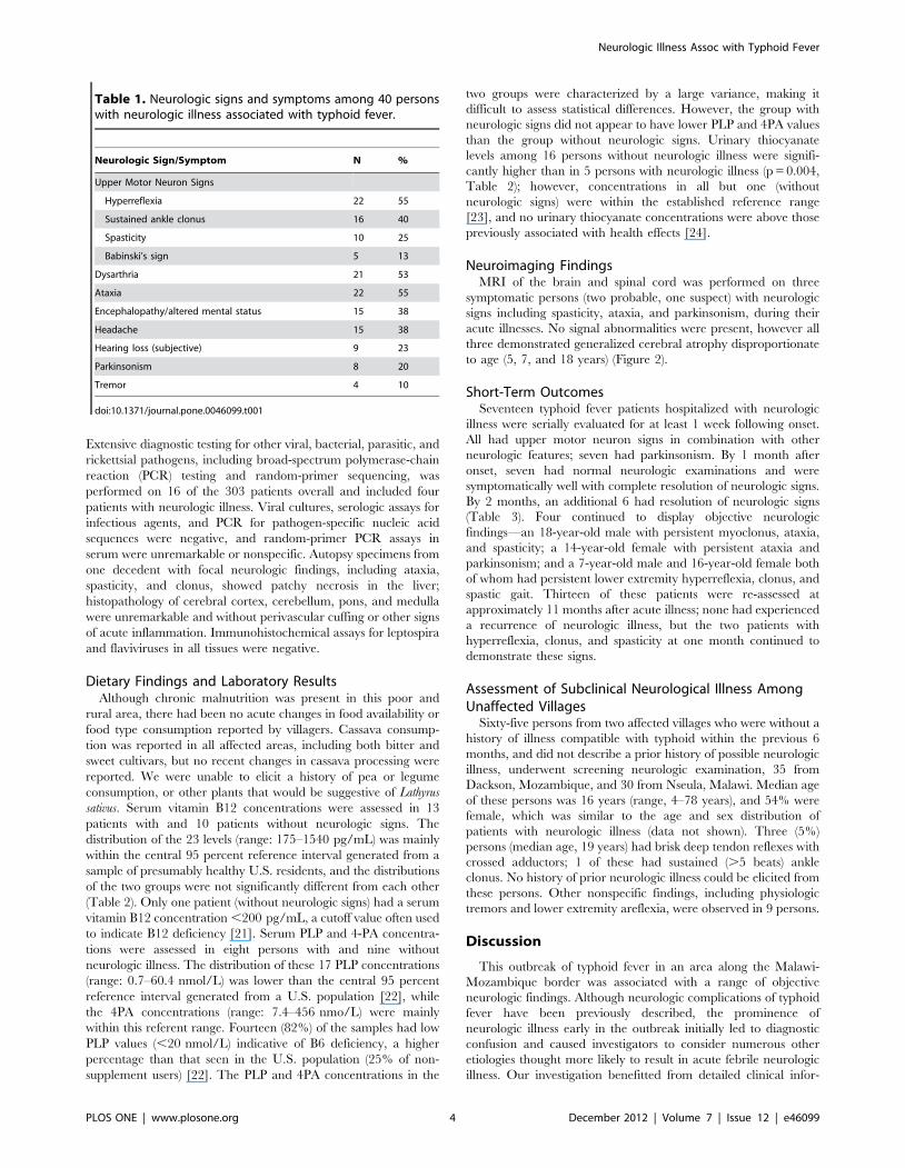
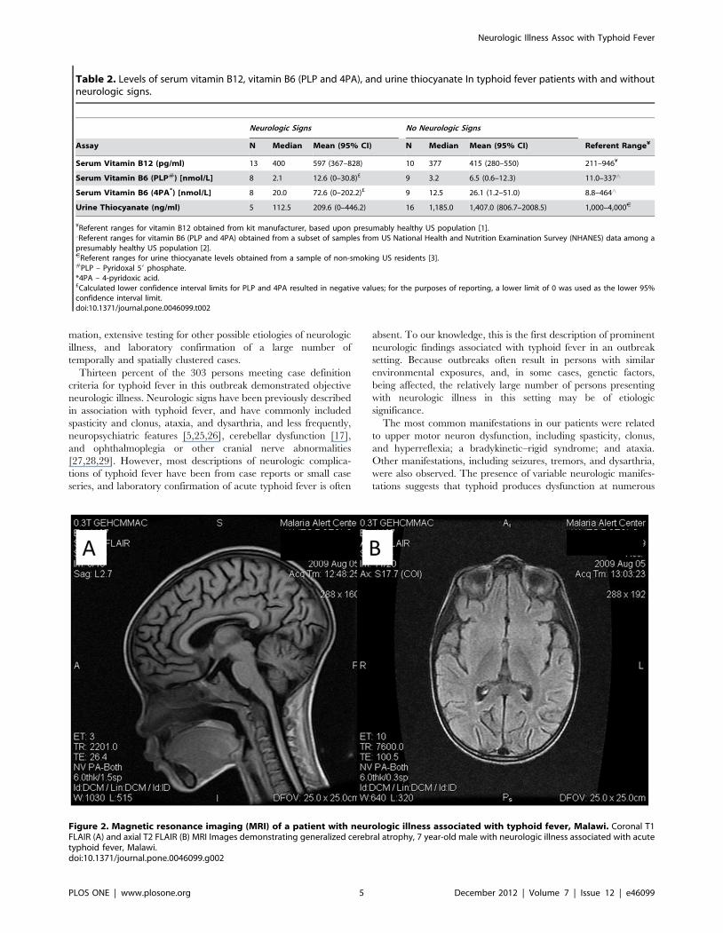
Neuroimaging FindingsMRI of the brain and spinal cord was performed on three
symptomatic persons (two probable, one suspect) with neurologic
signs including spasticity, ataxia, and parkinsonism, during their
acute illnesses. No signal abnormalities were present, however all
three demonstrated generalized cerebral atrophy disproportionate
to age (5, 7, and 18 years) (Figure 2).
Short-Term OutcomesSeventeen typhoid fever patients hospitalized with neurologic
illness were serially evaluated for at least 1 week following onset.
All had upper motor neuron signs in combination with other
neurologic features; seven had parkinsonism. By 1 month after
onset, seven had normal neurologic examinations and were
symptomatically well with complete resolution of neurologic signs.
By 2 months, an additional 6 had resolution of neurologic signs
(Table 3). Four continued to display objective neurologic
findings—an 18-year-old male with persistent myoclonus, ataxia,
and spasticity; a 14-year-old female with persistent ataxia and
parkinsonism; and a 7-year-old male and 16-year-old female both
of whom had persistent lower extremity hyperreflexia, clonus, and
spastic gait. Thirteen of these patients were re-assessed at
approximately 11 months after acute illness; none had experienced
a recurrence of neurologic illness, but the two patients with
hyperreflexia, clonus, and spasticity at one month continued to
demonstrate these signs.
Assessment of Subclinical Neurological Illness AmongUnaffected Villages
Sixty-five persons from two affected villages who were without a
history of illness compatible with typhoid within the previous 6
months, and did not describe a prior history of possible neurologic
illness, underwent screening neurologic examination, 35 from
Dackson, Mozambique, and 30 from Nseula, Malawi. Median age
of these persons was 16 years (range, 4–78 years), and 54% were
female, which was similar to the age and sex distribution of
patients with neurologic illness (data not shown). Three (5%)
persons (median age, 19 years) had brisk deep tendon reflexes with
crossed adductors; 1 of these had sustained (.5 beats) ankle
clonus. No history of prior neurologic illness could be elicited from
these persons. Other nonspecific findings, including physiologic
tremors and lower extremity areflexia, were observed in 9 persons.
Discussion
This outbreak of typhoid fever in an area along the Malawi-
Mozambique border was associated with a range of objective
neurologic findings. Although neurologic complications of typhoid
fever have been previously described, the prominence of
neurologic illness early in the outbreak initially led to diagnostic
confusion and caused investigators to consider numerous other
etiologies thought more likely to result in acute febrile neurologic
illness. Our investigation benefitted from detailed clinical infor-
Table 1. Neurologic signs and symptoms among 40 personswith neurologic illness associated with typhoid fever.
Neurologic Sign/Symptom N %
Upper Motor Neuron Signs
Hyperreflexia 22 55
Sustained ankle clonus 16 40
Spasticity 10 25
Babinski’s sign 5 13
Dysarthria 21 53
Ataxia 22 55
Encephalopathy/altered mental status 15 38
Headache 15 38
Hearing loss (subjective) 9 23
Parkinsonism 8 20
Tremor 4 10
doi:10.1371/journal.pone.0046099.t001
Neurologic Illness Assoc with Typhoid Fever
PLOS ONE | www.plosone.org 4 December 2012 | Volume 7 | Issue 12 | e46099
mation, extensive testing for other possible etiologies of neurologic
illness, and laboratory confirmation of a large number of
temporally and spatially clustered cases.
Thirteen percent of the 303 persons meeting case definition
criteria for typhoid fever in this outbreak demonstrated objective
neurologic illness. Neurologic signs have been previously described
in association with typhoid fever, and have commonly included
spasticity and clonus, ataxia, and dysarthria, and less frequently,
neuropsychiatric features [5,25,26], cerebellar dysfunction [17],
and ophthalmoplegia or other cranial nerve abnormalities
[27,28,29]. However, most descriptions of neurologic complica-
tions of typhoid fever have been from case reports or small case
series, and laboratory confirmation of acute typhoid fever is often
absent. To our knowledge, this is the first description of prominent
neurologic findings associated with typhoid fever in an outbreak
setting. Because outbreaks often result in persons with similar
environmental exposures, and, in some cases, genetic factors,
being affected, the relatively large number of persons presenting
with neurologic illness in this setting may be of etiologic
significance.
The most common manifestations in our patients were related
to upper motor neuron dysfunction, including spasticity, clonus,
and hyperreflexia; a bradykinetic–rigid syndrome; and ataxia.
Other manifestations, including seizures, tremors, and dysarthria,
were also observed. The presence of variable neurologic manifes-
tations suggests that typhoid produces dysfunction at numerous
Table 2. Levels of serum vitamin B12, vitamin B6 (PLP and 4PA), and urine thiocyanate In typhoid fever patients with and withoutneurologic signs.
Neurologic Signs No Neurologic Signs
Assay N Median Mean (95% CI) N Median Mean (95% CI) Referent Range¥
Serum Vitamin B12 (pg/ml) 13 400 597 (367–828) 10 377 415 (280–550) 211–946¥
Serum Vitamin B6 (PLP#) [nmol/L] 8 2.1 12.6 (0–30.8)£ 9 3.2 6.5 (0.6–12.3) 11.0–337‘
Serum Vitamin B6 (4PA*) [nmol/L] 8 20.0 72.6 (0–202.2)£ 9 12.5 26.1 (1.2–51.0) 8.8–464‘
Urine Thiocyanate (ng/ml) 5 112.5 209.6 (0–446.2) 16 1,185.0 1,407.0 (806.7–2008.5) 1,000–4,000J
¥Referent ranges for vitamin B12 obtained from kit manufacturer, based upon presumably healthy US population [1].‘Referent ranges for vitamin B6 (PLP and 4PA) obtained from a subset of samples from US National Health and Nutrition Examination Survey (NHANES) data among apresumably healthy US population [2].JReferent ranges for urine thiocyanate levels obtained from a sample of non-smoking US residents [3].#PLP – Pyridoxal 59 phosphate.*4PA – 4-pyridoxic acid.£Calculated lower confidence interval limits for PLP and 4PA resulted in negative values; for the purposes of reporting, a lower limit of 0 was used as the lower 95%confidence interval limit.doi:10.1371/journal.pone.0046099.t002
Figure 2. Magnetic resonance imaging (MRI) of a patient with neurologic illness associated with typhoid fever, Malawi. Coronal T1FLAIR (A) and axial T2 FLAIR (B) MRI Images demonstrating generalized cerebral atrophy, 7 year-old male with neurologic illness associated with acutetyphoid fever, Malawi.doi:10.1371/journal.pone.0046099.g002
Neurologic Illness Assoc with Typhoid Fever
PLOS ONE | www.plosone.org 5 December 2012 | Volume 7 | Issue 12 | e46099
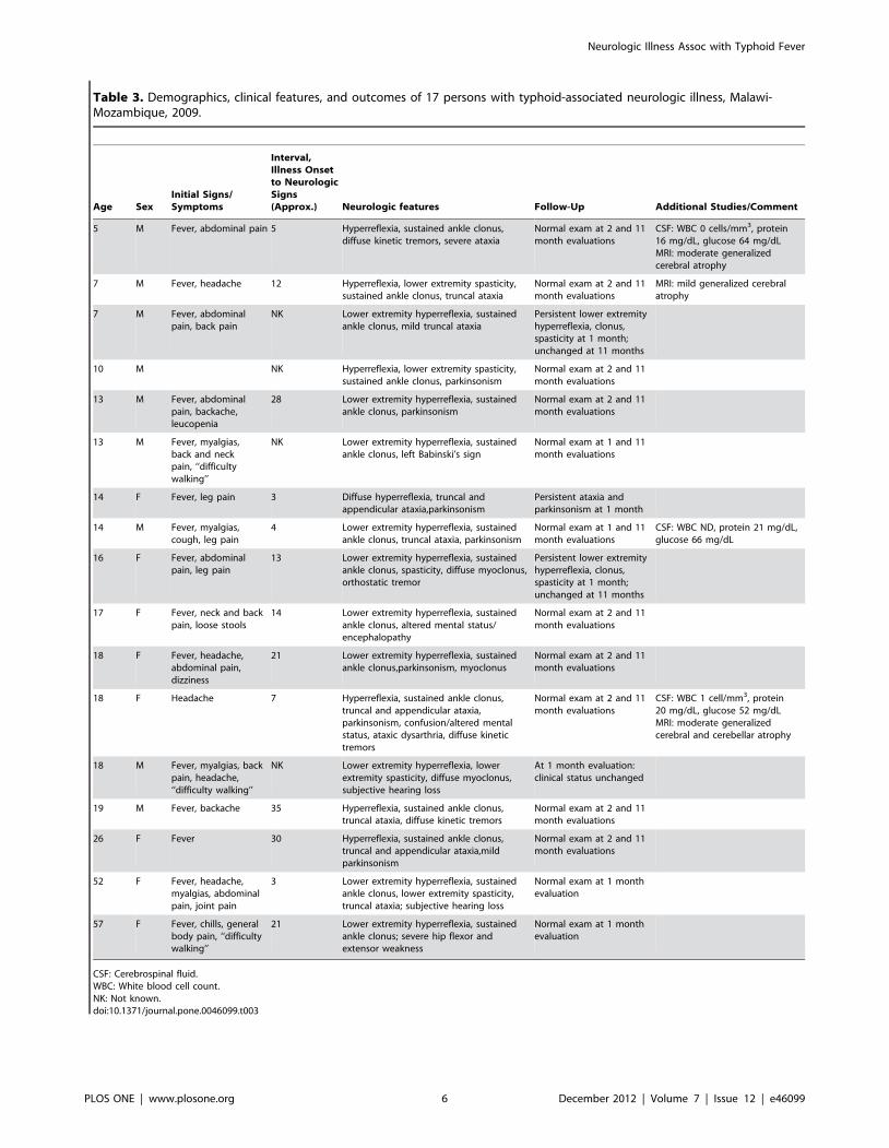
Table 3. Demographics, clinical features, and outcomes of 17 persons with typhoid-associated neurologic illness, Malawi-Mozambique, 2009.
Age SexInitial Signs/Symptoms
Interval,Illness Onsetto NeurologicSigns(Approx.) Neurologic features Follow-Up Additional Studies/Comment
5 M Fever, abdominal pain 5 Hyperreflexia, sustained ankle clonus,diffuse kinetic tremors, severe ataxia
Normal exam at 2 and 11month evaluations
CSF: WBC 0 cells/mm3, protein16 mg/dL, glucose 64 mg/dLMRI: moderate generalizedcerebral atrophy
7 M Fever, headache 12 Hyperreflexia, lower extremity spasticity,sustained ankle clonus, truncal ataxia
Normal exam at 2 and 11month evaluations
MRI: mild generalized cerebralatrophy
7 M Fever, abdominalpain, back pain
NK Lower extremity hyperreflexia, sustainedankle clonus, mild truncal ataxia
Persistent lower extremityhyperreflexia, clonus,spasticity at 1 month;unchanged at 11 months
10 M NK Hyperreflexia, lower extremity spasticity,sustained ankle clonus, parkinsonism
Normal exam at 2 and 11month evaluations
13 M Fever, abdominalpain, backache,leucopenia
28 Lower extremity hyperreflexia, sustainedankle clonus, parkinsonism
Normal exam at 2 and 11month evaluations
13 M Fever, myalgias,back and neckpain, ‘‘difficultywalking’’
NK Lower extremity hyperreflexia, sustainedankle clonus, left Babinski’s sign
Normal exam at 1 and 11month evaluations
14 F Fever, leg pain 3 Diffuse hyperreflexia, truncal andappendicular ataxia,parkinsonism
Persistent ataxia andparkinsonism at 1 month
14 M Fever, myalgias,cough, leg pain
4 Lower extremity hyperreflexia, sustainedankle clonus, truncal ataxia, parkinsonism
Normal exam at 1 and 11month evaluations
CSF: WBC ND, protein 21 mg/dL,glucose 66 mg/dL
16 F Fever, abdominalpain, leg pain
13 Lower extremity hyperreflexia, sustainedankle clonus, spasticity, diffuse myoclonus,orthostatic tremor
Persistent lower extremityhyperreflexia, clonus,spasticity at 1 month;unchanged at 11 months
17 F Fever, neck and backpain, loose stools
14 Lower extremity hyperreflexia, sustainedankle clonus, altered mental status/encephalopathy
Normal exam at 2 and 11month evaluations
18 F Fever, headache,abdominal pain,dizziness
21 Lower extremity hyperreflexia, sustainedankle clonus,parkinsonism, myoclonus
Normal exam at 2 and 11month evaluations
18 F Headache 7 Hyperreflexia, sustained ankle clonus,truncal and appendicular ataxia,parkinsonism, confusion/altered mentalstatus, ataxic dysarthria, diffuse kinetictremors
Normal exam at 2 and 11month evaluations
CSF: WBC 1 cell/mm3, protein20 mg/dL, glucose 52 mg/dLMRI: moderate generalizedcerebral and cerebellar atrophy
18 M Fever, myalgias, backpain, headache,‘‘difficulty walking’’
NK Lower extremity hyperreflexia, lowerextremity spasticity, diffuse myoclonus,subjective hearing loss
At 1 month evaluation:clinical status unchanged
19 M Fever, backache 35 Hyperreflexia, sustained ankle clonus,truncal ataxia, diffuse kinetic tremors
Normal exam at 2 and 11month evaluations
26 F Fever 30 Hyperreflexia, sustained ankle clonus,truncal and appendicular ataxia,mildparkinsonism
Normal exam at 2 and 11month evaluations
52 F Fever, headache,myalgias, abdominalpain, joint pain
3 Lower extremity hyperreflexia, sustainedankle clonus, lower extremity spasticity,truncal ataxia; subjective hearing loss
Normal exam at 1 monthevaluation
57 F Fever, chills, generalbody pain, ‘‘difficultywalking’’
21 Lower extremity hyperreflexia, sustainedankle clonus; severe hip flexor andextensor weakness
Normal exam at 1 monthevaluation
CSF: Cerebrospinal fluid.WBC: White blood cell count.NK: Not known.doi:10.1371/journal.pone.0046099.t003
Neurologic Illness Assoc with Typhoid Fever
PLOS ONE | www.plosone.org 6 December 2012 | Volume 7 | Issue 12 | e46099
sites within the nervous system. Many patients presented with
neurologic findings in the absence of encephalopathy or other
alteration in mental status, indicating that typhoid may produce
focal, as well as generalized, neurologic dysfunction. With few
exceptions, the neurologic findings in these subjects resolved over
time, sometimes within weeks of acute illness, and long-term or
recurrent neurologic sequelae were largely absent among a subset
of persons we were able to assess in extended follow-up. Notably,
we did not observe some of the other neurologic manifestations
that have been frequently mentioned in the setting of typhoid
fever, such as acute psychosis [6,25], acute inflammatory
polyradiculoneuropathy [15,30], or focal cortical signs [14,15,16].
The reason for the high proportion of cases with neurologic
illness during this outbreak is unclear, but there are several
possibilities. Surveillance bias is possible; early surveillance and
case detection efforts focused on those persons hospitalized with
neurologic features. Following recognition of typhoid as the cause
of the outbreak, more persons with features typical of typhoid
fever, including abdominal pain and other gastrointestinal
symptoms, were detected. The involvement of neurologists in
the outbreak investigation possibly led to detection of neurologic
features that might not be typically assessed or noted by other
clinicians. Neurologic manifestations of typhoid have been
described as a late manifestation of illness [5,31,32], and the
median interval between symptom onset and documentation of
neurologic signs in our patients was 12 days. Several factors,
including delayed presentation to clinical care and ineffective
antimicrobial treatment early in the outbreak because of multi-
drug resistance of the causative Salmonella Typhi strain [18] may
have led to a prolonged course of illness early in the outbreak,
resulting in a greater prevalence of neurologic signs. Importantly,
following implementation of early diagnostic capabilities and
appropriate definitive antimicrobial treatment of typhoid fever
with ciprofloxacin, the number of persons presenting with
neurologic illness appeared to decrease, suggesting that prompt
treatment may avert the onset of neurologic illness.
The mechanism by which typhoid fever may produce neuro-
logic illness is unknown. Rare cases of Salmonella Typhi bacterial
meningitis, meningo-encephalitis, and intracranial abscesses have
been reported both in children and adults [3,33,34]. However, a
neuroinvasive bacterial process appears unlikely in our patients;
CSF was generally unremarkable and without pleocytosis or
protein elevation, features of meningismus were generally absent,
and CNS tissue from one confirmed case with neurologic illness, as
well as brain and spinal cord MRI on 3 acutely ill patients, did not
demonstrate signs of inflammation. This is consistent with prior
reports in which neurologic manifestations have largely been
unassociated with evidence of CNS inflammation. For these same
reasons, a para- or post-infectious immune-mediated inflammato-
ry process in the majority of cases would seem unlikely.
An underlying host factor or environmental exposure that may
predispose persons to develop neurologic illness in the setting of
severe systemic infection due to typhoid is possible. Many of the
predominant signs and symptoms observed in these patients,
including spasticity, clonus, hyperreflexia, and ataxia, may be seen
with micronutrient abnormalities including vitamin B6 toxicity/
deficiency and B12 deficiency [35,36,37,38]. We assessed these
micronutrient levels in a subset of persons with and without
neurologic signs and did not detect significant differences;
however, our sample size was small and variability in the data
made it difficult to assess statistical differences. The upper motor
neuron findings in this population initially appeared similar to
konzo, a neurologic illness seen in tropical areas and associated
with thiocyanate toxicity due to consumption of inadequately
cooked bitter cassava [39,40,41]. The ataxia demonstrated by
some of our patients resembled tropical ataxic neuropathy, also
related to the dietary use of large quantities of cassava over long
periods of time [42,43]. Although we considered these etiologies
because cassava was part of the local diet, the often dramatic and
complete resolution of neurologic signs in our patients is
inconsistent with konzo or tropical ataxic neuropathy, and
measurement of urinary thiocyanate levels did not demonstrate
evidence of acute or chronic cyanogen toxicity. Similarly,
neurolathyrism seemed unlikely due to improvement in neurologic
signs, and we could not obtain a history of consumption of peas or
legumes [44,45].
Production of a bacterial toxin may lead to neurologic illness,
with toxins produced by Clostridium botulinum and Corynebacterium
diphtheriae being fundamental examples [46]. The diffuse nature of
neurologic involvement observed with typhoid-associated disease,
and the apparent reversibility of these signs may be suggestive of a
bacterial toxic etiology. Salmonella Typhi produces a cytolethal
toxin, but the role of this toxin in the pathogenesis of typhoid fever
is unknown [47,48]. Isolates of Salmonella Typhi obtained from
cases in this outbreak, including persons with neurologic illness,
did not demonstrate significant differences in genetic or bacteri-
ologic properties from other isolates in central Africa [18]; further
investigations into the possible presence of a Salmonella Typhi-
produced neurotoxin are ongoing. Some viral and bacterial
infections have been proposed to result in a ‘‘cytokinemia’’ in
which hyper-reactive pro- and anti-inflammatory cytokines result
in alterations of CNS function with encephalopathy and neuro-
logic illness [49,50,51]. While our investigation did not include
assessments of cytokine function, such an indirect effect of
Salmonella Typhi on the nervous system should be explored in
future studies.
Our study has limitations. We did not perform neurologic
examinations on all outbreak patients, and the number of cases of
neurologic illness may have been underestimated or otherwise
biased. Not all patients with suspected illness were positive for or
underwent testing for Salmonella Typhi infection, and misclassifi-
cation of some cases is possible. While we initially screened a large
number of cases for numerous other infectious and toxic etiologies
of neurologic illness, other alternative or concomitant causes of
febrile neurologic illness in these persons cannot be entirely
excluded. HIV, which could certainly be a contributory factor in
neurologic illness occurring in this population, was not routinely
tested for. Baseline neurologic status on these patients was
unknown, and some may have demonstrated neurologic findings
unrelated to their typhoid illness. Specifically, the presence of
disproportionate cerebral atrophy on MRI in 3 cases suggests that
other host factors, such as nutritional deficiencies, prior cerebral
infections, antenatal/perinatal insults, or other factors may result
in a background level of mild neurologic illness in this population.
It is possible that severe systemic illness caused by typhoid fever, or
a Salmonella Typhi-specific factor, may exacerbate mild or
subclinical neurologic deficits.
Our study demonstrates that persons with typhoid fever may
develop acute and severe neurologic illness. The underlying
pathophysiological mechanisms producing these features remain
unknown. The varying neurologic manifestations observed in this
group of patients with typhoid-associated neurologic illness suggest
involvement of multiple nervous system localizations. Neurologic
illness associated with typhoid fever appears to resolve over time,
with few ongoing sequelae, a feature that is important in the
prognostic assessment of cases. Acute infection with Salmonella
Typhi should be included in the differential diagnosis of persons
originating or traveling from a typhoid-endemic area with acute
Neurologic Illness Assoc with Typhoid Fever
PLOS ONE | www.plosone.org 7 December 2012 | Volume 7 | Issue 12 | e46099
febrile neurologic illness, particularly if viral and bacterial
etiologies more typically associated with neurologic illness are
not apparent. A better understanding of the underlying patho-
physiologic mechanisms associated with neurologic illness in
typhoid fever is needed.
Supporting Information
Table S1 Initial pathogen testing among ill personsduring outbreak of typhoid fever, Malawi – Mozam-bique, 2009(DOC)
Video S1 18-year old female with typhoid-associatedneurologic illness demonstrating severe truncal andappendicular ataxia and facial masking.(WMV)
Acknowledgments
The authors would like to thank the following individuals for their valuable
contributions to this manuscript: Malawi Ministry of Health: Secretary
Chris Kang’ombe, Ben Chilima, Kundai Moyo, and Kapangaza Ntonya;
the Neno District Rapid Response Team; Partners in Health: Keith Joseph
and Annemarie Ackerman; World Health Organization: F. R. Zawaira,
Kelias Msyamboza, and Reggis Kasande; Population Services Interna-
tional; United Nations International Children’s Fund; United States
Agency for International Development; CDC-Malawi: Dr. Thomas
Warne, Rankin Thamanda, Laston Thamangira, Victor Samidu, Ethel
Mpagaja, CDC-Mozambique: Amy DuBois, Lisa Nelson; CDC-Nutrition-
al Biomarkers Branch: Christine Pfeiffer, Michael Ryback, Usha Mandava,
Huiping Chen, Donna LaVoie, Usha Mandava, Christine Pfeiffer, Daniel
Rabinowitz, Michael Rybak, Rosemary Schleicher, Mary Xu, Mindy
Zhang; CDC-DASH Laboratory: Nicole Burcher, Michael Dillon; CDC-
NCEH Laboratory: Charles Dodson; CDC-Global Disease Detection: Ray
Arthur, Rohit Chitale, Kira Christian; CDC-NCEZID: Cheryl Bopp,
Michele Parsons, Ermias Belay, Katie Schilling, Sherif Zaki, Wun-Ju
Shieh; CDC-NCIRD: Lauren Stockman; and the CDC Emergency
Operations Center staff.
The findings and conclusions in this report are those of the authors and
do not necessarily represent the views of the U.S. Centers for Disease
Control and Prevention/Agency for Toxic Substances and Disease
Registry.
Author Contributions
Conceived and designed the experiments: J. Sejvar EL SM J. Schier B.
Barr MH GA. Performed the experiments: J. Sevjar EL JN AL RM SM SL
LC KD DT YR J. Schier B. Barr SK B. Blount MH. Analyzed the data: J.
Sejvar EL BN J. Schier MH GA EM. Contributed reagents/materials/
analysis tools: BN SK B. Blount MH DT GA EM. Wrote the paper: J.
Sejvar EL JN AL RM BN SM TK SL LC KD DT YR J. Schier B. Barr
AD MM SK B. Blount MH DT EM.
References
1. Parry CM, Hien TT, Dougan G, White NJ, Farrar JJ (2002) Typhoid fever.
N Engl J Med 347: 1770–1782.
2. Crum NF (2003) Current trends in typhoid Fever. Curr Gastroenterol Rep 5:
279–286.
3. Rajeshwari K, Yadav S, Puri RK, Khanijo CM, Sethi Y (1995) Cerebritis in
typhoid fever. Indian Pediatr 32: 1305–1307.
4. Osuntokun BO, Bademosi O, Ogunremi K, Wright SG (1972) Neuropsychiatricmanifestations of typhoid fever in 959 patients. Arch Neurol 27: 7–13.
5. Venkatesh S, Grell GA (1989) Neuropsychiatric manifestations of typhoid fever.West Indian Med J 38: 137–141.
6. (1973) Psychiatric symptoms in typhoid fever. Br Med J 2: 436–437.
7. Osuntokun BO, Adeuja AO, Bademosi O (1974) The prognosis of motor neurondisease in Nigerian africans. A prospective study of 92 patients. Brain 97: 385–
394.
8. Sachdev HS, Puri MP, Mohan M (1982) Acute cerebellar ataxia in typhoid
fever. Indian Pediatr 19: 639–640.
9. Sawhney IM, Prabhakar S, Dhand UK, Chopra JS (1986) Acute cerebellar
ataxia in enteric fever. Trans R Soc Trop Med Hyg 80: 85–86.
10. Chand G, Singh K (1988) Acute cerebellar ataxia–a rare complication of entericfever. J Assoc Physicians India 36: 741.
11. Cheong BM (2008) Typhoid fever presenting as acute cerebellar ataxia andsevere thrombocytopenia. Med J Malaysia 63: 77–78.
12. David CB, Tolaymat A (1978) Typhoid fever: unusual presentation. J Pediatr 93:
533.
13. Dewan P, Pooniya V, Kaushik JS, Gomber S, Singhal S (2009) Isolated
cerebellar ataxia: an early neurological complication of enteric fever. Ann TropPaediatr 29: 217–219.
14. Singh S, Gupta A, Marwaha RK (1993) Wenckebach phenomenon and motoraphasia in enteric fever. Indian J Pediatr 60: 147–149.
15. Adehossi E, Parola P, Brouqui P (2003) Febrile Broca’s aphasia: a rare
presentation of typhoid fever. J Travel Med 10: 192–193.
16. Bansal AS, Venkatesh S, Jones SR, Williams W (1995) Acute aphasia
complicating typhoid fever in an adult. J Trop Med Hyg 98: 392–394.
17. Misra GC, Singh SP, Mohapatra MK, Mohapatra RK, Prusty PK, et al. (1985)
Typhoid cerebellitis. J Indian Med Assoc 83: 352–353.
18. Lutterloh E, Likaka A, Sejvar J, Manda R, Naiene J, et al. (2012) Multidrug-resistant typhoid fever with neurologic findings on the Malawi-Mozambique
border. Clin Infect Dis 54: 1100–1106.
19. Rybak ME, Pfeiffer CM (2004) Clinical analysis of vitamin B(6): determination
of pyridoxal 59-phosphate and 4-pyridoxic acid in human serum by reversed-phase high-performance liquid chromatography with chlorite postcolumn
derivatization. Anal Biochem 333: 336–344.
20. Valentin-Blasini L, Blount BC, Delinsky A (2007) Quantification of iodide and
sodium-iodide symporter inhibitors in human urine using ion chromatographytandem mass spectrometry. J Chromatogr A 1155: 40–46.
21. Thorpe SJ, Heath A, Blackmore S, Lee A, Hamilton M, et al. (2007)International Standard for serum vitamin B(12) and serum folate: international
collaborative study to evaluate a batch of lyophilised serum for B(12) and folatecontent. Clin Chem Lab Med 45: 380–386.
22. Morris MS, Picciano MF, Jacques PF, Selhub J (2008) Plasma pyridoxal 59-
phosphate in the US population: the National Health and Nutrition
Examination Survey, 2003–2004. Am J Clin Nutr 87: 1446–1454.
23. Blount BC, Pirkle JL, Osterloh JD, Valentin-Blasini L, Caldwell KL (2006)
Urinary perchlorate and thyroid hormone levels in adolescent and adult men
and women living in the United States. Environ Health Perspect 114: 1865–
1871.
24. Cliff J, Nicala D, Saute F, Givragy R, Azambuja G, et al. (1999) Ankle clonus
and thiocyanate, linamarin, and inorganic sulphate excretion in school children
in communities with Konzo, Mozambique. J Trop Pediatr 45: 139–142.
25. Khosla SN, Srivastava SC, Gupta S (1977) Neuro-psychiatric manifestations of
typhoid. J Trop Med Hyg 80: 95–98.
26. Ali G, Rashid S, Kamli MA, Shah PA, Allaqaband GQ (1997) Spectrum of
neuropsychiatric complications in 791 cases of typhoid fever. Trop Med Int
Health 2: 314–318.
27. Ghosh JB (1995) Pharyngeal palsy in enteric fever. Indian J Pediatr 62: 627–628.
28. Kamala CS, Manimegalai S, Kumar S (1991) Palatal paralysis in enteric fever.
Indian Pediatr 28: 1213–1214.
29. Nahata MC (1961) Ophthalmoplegia following enteric fever. J Indian Med
Assoc 37: 134–135.
30. Ozen H, Cemeroglu P, Ecevit Z, Secmeer G, Kanra G (1993) Unusual
neurologic complications of typhoid fever (aphasia, mononeuritis multiplex, and
Guillain-Barre syndrome): a report of two cases. Turk J Pediatr 35: 141–144.
31. Chikanza IC, Latif AS, Neill P, Mason P, Olweny CL (1986) Unusual
complications of typhoid fever. Cent Afr J Med 32: 31–34.
32. Kanwar K (1962) Symptomatology and treatment of typhoid paratyphoid fevers
with special reference to the neurologic manifestations and liver damage.
Indian J Child Health 11: 525–539.
33. Suri S, Bhasin A, Srivastava VK (1992) Salmonella typhi meningitis with facial
nerve palsy. Indian Pediatr 29: 901–902.
34. Maheshwari VD, Jain MK, Karant VN (2001) Intracerebral haemorrhage as a
complication of enteric fever. J Assoc Physicians India 49: 1035.
35. Gdynia HJ, Muller T, Sperfeld AD, Kuhnlein P, Otto M, et al. (2008) Severe
sensorimotor neuropathy after intake of highest dosages of vitamin B6.
Neuromuscul Disord 18: 156–158.
36. Incecik F, Herguner MO, Altunbasak S, Leblebisatan G (2010) Neurologic
findings of nutritional vitamin B12 deficiency in children. Turk J Pediatr 52: 17–
21.
37. Mankad K, Kullmann DM, Davagnanam I (2010) Neurological manifestation of
vitamin B12 deficiency. Am J Med 123: e1–2.
38. Senol MG, Sonmez G, Ozdag F, Saracoglu M (2008) Reversible myelopathy
with vitamin B12 deficiency. Singapore Med J 49: e330–332.
39. Cliff J, Muquingue H, Nhassico D, Nzwalo H, Bradbury JH (2010) Konzo and
continuing cyanide intoxication from cassava in Mozambique. Food Chem
Toxicol.
40. Cliff J, Nicala D (1997) Long-term follow-up of konzo patients. Trans R Soc
Trop Med Hyg 91: 447–449.
Neurologic Illness Assoc with Typhoid Fever
PLOS ONE | www.plosone.org 8 December 2012 | Volume 7 | Issue 12 | e46099
41. Tylleskar T, Howlett WP, Rwiza HT, Aquilonius SM, Stalberg E, et al. (1993)
Konzo: a distinct disease entity with selective upper motor neuron damage.
J Neurol Neurosurg Psychiatry 56: 638–643.
42. Njoh J (1990) Tropical ataxic neuropathy in Liberians. Trop Geogr Med 42: 92–
94.
43. Madhusudanan M, Menon MK, Ummer K, Radhakrishnanan K (2008)
Clinical and etiological profile of tropical ataxic neuropathy in Kerala, South
India. Eur Neurol 60: 21–26.
44. Barceloux DG (2009) Grass pea and neurolathyrism (Lathyrus sativus L.). Dis
Mon 55: 365–372.
45. Bradbury JH, Lambein F (2011) Konzo and neurolathyrism: similarities and
dissimilarities between these crippling neurodegenerative diseases of the poor.
Food Chem Toxicol 49: 537–538.
46. Henkel JS, Baldwin MR, Barbieri JT (2010) Toxins from bacteria. EXS 100: 1–
29.
47. von Rhein C, Bauer S, Lopez Sanjurjo EJ, Benz R, Goebel W, et al. (2009) ClyA
cytolysin from Salmonella: distribution within the genus, regulation of expressionby SlyA, and pore-forming characteristics. Int J Med Microbiol 299: 21–35.
48. von Rhein C, Hunfeld KP, Ludwig A (2006) Serologic evidence for effective
production of cytolysin A in Salmonella enterica serovars typhi and paratyphi Aduring human infection. Infect Immun 74: 6505–6508.
49. Kalita J, Srivastava R, Mishra MK, Basu A, Misra UK (2010) Cytokines andchemokines in viral encephalitis: a clinicoradiological correlation. Neurosci Lett
473: 48–51.
50. Ichiyama T, Morishima T, Isumi H, Matsufuji H, Matsubara T, et al. (2004)Analysis of cytokine levels and NF-kappaB activation in peripheral blood
mononuclear cells in influenza virus-associated encephalopathy. Cytokine 27:31–37.
51. Ichiyama T, Isumi H, Ozawa H, Matsubara T, Morishima T, et al. (2003)Cerebrospinal fluid and serum levels of cytokines and soluble tumor necrosis
factor receptor in influenza virus-associated encephalopathy. Scand J Infect Dis
35: 59–61.
Neurologic Illness Assoc with Typhoid Fever
PLOS ONE | www.plosone.org 9 December 2012 | Volume 7 | Issue 12 | e46099
Related Documents