Research Paper Mediators of Inflammation 3, 189-197 (1994) CAPSAICIN, substance P, and ovalbumin, instilled into the bladders of naive and ovalbumin (OVA) sensitized guinea- pigs caused inflammation, as indicated by increased vas- cular permeability. Histological changes after exposure to these compounds progressed with time from intense vasodilatation to marginalization of granulocytes fol- lowed by interstitial migration of leukocytes. In vitro incubation of guinea-pig bladder tissue with substance P and ovalbumin stimulated release of prostaglandin D and leukotrienes. In vitro incubation of bladder tissue with capsaicin, OVA, prostaglandin Da, leukotriene C4, histamine, or calcium ionophore A-23587 all stimulated substance P release. These data suggest that bladder in- flammation initiated by a variety of stimuli could lead to a cyclic pattern of release of inflammatory mediators and neuropeptides, which could result in amplification and persistence of cystitis after the inciting cause has sub- sided. Neurogenic inflammation of guinea-pig bladder D. E. Bjorling, c* M. R. Saban and R. Saban Smooth Muscle Laboratory, Department of Surgical Sciences, School of Veterinary Medicine, University of Wisconsin, Madison, WI, USA CA Corresponding Author Key words: Antigen challenge, Bladder, Cystitis, Guinea-pig, Neuropeptides Introduction Apparently spontaneous inflammation of the uri- nary bladder in the absence of infection has been reported in humans and cats. 1,2 In humans, the most widely recognized form of cystitis in the absence of infection is interstitial cystitis (IC), a chronic inflam- matory disease of the lower urinary tract. Clinical symptoms commonly associated with IC include in- creased urinary frequency and urgency, nocturia, suprapubic pressure and pain. 1, Although numerous theories regarding the aetiology of IC have been proposed, the pathogenesis remains unknown. Traumatic injury, lymphatic obstruction and autoimmune disease are among the suggested causes. Regardless of the cause(s) of IC and other forms of non-infectious cystitis, a number of reports suggest that the nervous system is intimately involved in the development and persistence of non-infectious cystitis. 5-7 Neurogenic inflammation affecting various organs, including the skin, lungs, airways, gut, eye and joints, has been reported in several species, including humans. -1 A variety of stimuli, such as antigens, heat, cold, bacterial or viral infection, and direct stimulation of sensory nerves, can trigger the onset of neurogenic inflammation. -1 Neuropeptides such as substance P (SP), neurokinin A (NKA), calcitonin gene-related peptide (CGRP) and vasoactive intesti- nal peptide (VIP) are located in capsaicin sensitive primary afferent nerves in the bladders of humans and animals. 11,12 The release of SP, NKA and CGRP has been correlated with smooth muscle contraction, ( 1994 Rapid Communications of Oxford Ltd vasodilatation, increased vascular permeability and facilitated neurotransmitter release from intramural nerves. 1 SP, CGRP and VIP also mediate the sensory function of capsaicin sensitive nervous control of the micturition threshold and may facilitate spontaneous bladder contraction. 14 Experimentally, antidromic electrical stimulation of unmyelinated afferent nerve fibres causes extravasation of Evans blue dye in the urinary bladder of guinea-pigs. 15 Of the neuropeptides present in the afferent sensory nerve fibres, SP is thought to be the primary neurotrans- matter responsible for initiation of neurogenic inflam- mation, and CGRP is believed to play an important role in facilitating and mediating the effects of SP. 16 Intravenous administration of SP also causes extravasation of Evans blue dye and accumulation of leukocytes in the bladder wall of experimental ani- mals, and intravesical application of SP has a similar effect.7. 8 Capsaicin, an extract of hot pepper, is a neurotoxin for unmyelinated sensory nerve fibres which selec- tively depletes SP and other neuropeptides from the nerves at low doses and causes degeneration of these nerves at high doses. Pretreatment of rats with capsaicin significantly reduced the severity of xylene induced cystitis, strongly suggesting the participation of SP in the pathogenesis of this model of cystitis. 19 It has been reported that SP stimulates histamine release from various tissues and isolated mast cells, but this effect appears to be specific to the tissue source of the mast cells and the species from which these tissues and mast cells are obtained, a Prelimi- nary studies in our laboratory have indicated that Mediators of Inflammation. Vo13. 1994 189

Welcome message from author
This document is posted to help you gain knowledge. Please leave a comment to let me know what you think about it! Share it to your friends and learn new things together.
Transcript
Research Paper
Mediators of Inflammation 3, 189-197 (1994)
CAPSAICIN, substance P, and ovalbumin, instilled into thebladders of naive and ovalbumin (OVA) sensitized guinea-pigs caused inflammation, as indicated by increased vas-cular permeability. Histological changes after exposure tothese compounds progressed with time from intensevasodilatation to marginalization of granulocytes fol-lowed by interstitial migration of leukocytes. In vitroincubation of guinea-pig bladder tissue with substance Pand ovalbumin stimulated release of prostaglandin Dand leukotrienes. In vitro incubation of bladder tissuewith capsaicin, OVA, prostaglandin Da, leukotriene C4,histamine, or calcium ionophore A-23587 all stimulatedsubstance P release. These data suggest that bladder in-flammation initiated by a variety of stimuli could lead toa cyclic pattern of release of inflammatory mediators andneuropeptides, which could result in amplification andpersistence of cystitis after the inciting cause has sub-sided.
Neurogenic inflammation ofguinea-pig bladder
D. E. Bjorling,c* M. R. Saban and R. Saban
Smooth Muscle Laboratory, Department ofSurgical Sciences, School of VeterinaryMedicine, University of Wisconsin, Madison,WI, USA
CA Corresponding Author
Key words: Antigen challenge, Bladder, Cystitis, Guinea-pig,Neuropeptides
Introduction
Apparently spontaneous inflammation of the uri-nary bladder in the absence of infection has beenreported in humans and cats. 1,2 In humans, the most
widely recognized form of cystitis in the absence ofinfection is interstitial cystitis (IC), a chronic inflam-matory disease of the lower urinary tract. Clinicalsymptoms commonly associated with IC include in-creased urinary frequency and urgency, nocturia,suprapubic pressure and pain. 1,
Although numerous theories regarding theaetiology of IC have been proposed, thepathogenesis remains unknown. Traumatic injury,lymphatic obstruction and autoimmune disease areamong the suggested causes. Regardless of thecause(s) of IC and other forms of non-infectiouscystitis, a number of reports suggest that the nervoussystem is intimately involved in the development andpersistence of non-infectious cystitis. 5-7
Neurogenic inflammation affecting various organs,including the skin, lungs, airways, gut, eye andjoints, has been reported in several species, includinghumans.-1 A variety of stimuli, such as antigens,heat, cold, bacterial or viral infection, and directstimulation of sensory nerves, can trigger the onsetof neurogenic inflammation.-1 Neuropeptides suchas substance P (SP), neurokinin A (NKA), calcitoningene-related peptide (CGRP) and vasoactive intesti-nal peptide (VIP) are located in capsaicin sensitiveprimary afferent nerves in the bladders of humansand animals. 11,12 The release of SP, NKA and CGRPhas been correlated with smooth muscle contraction,
( 1994 Rapid Communications of Oxford Ltd
vasodilatation, increased vascular permeability andfacilitated neurotransmitter release from intramuralnerves.1 SP, CGRP and VIP also mediate the sensoryfunction of capsaicin sensitive nervous control of themicturition threshold and may facilitate spontaneousbladder contraction. 14 Experimentally, antidromicelectrical stimulation of unmyelinated afferent nervefibres causes extravasation of Evans blue dye in theurinary bladder of guinea-pigs. 15 Of theneuropeptides present in the afferent sensory nervefibres, SP is thought to be the primary neurotrans-matter responsible for initiation of neurogenic inflam-mation, and CGRP is believed to play an importantrole in facilitating and mediating the effects of SP. 16
Intravenous administration of SP also causesextravasation of Evans blue dye and accumulation ofleukocytes in the bladder wall of experimental ani-
mals, and intravesical application of SP has a similareffect.7.8
Capsaicin, an extract of hot pepper, is a neurotoxinfor unmyelinated sensory nerve fibres which selec-tively depletes SP and other neuropeptides from thenerves at low doses and causes degeneration ofthese nerves at high doses. Pretreatment of rats withcapsaicin significantly reduced the severity of xyleneinduced cystitis, strongly suggesting the participationof SP in the pathogenesis of this model of cystitis. 19
It has been reported that SP stimulates histaminerelease from various tissues and isolated mast cells,but this effect appears to be specific to the tissuesource of the mast cells and the species from whichthese tissues and mast cells are obtained,a Prelimi-nary studies in our laboratory have indicated that
Mediators of Inflammation. Vo13. 1994 189
D. E. Bjorling, M. R. Saban and R. Saban
isolated guinea-pig bladder tissue releases histaminein response to exposure to capsaicin or SP. We havepreviously described the results of investigationsusing a guinea-pig model for IC in which cystitisdevelops in response to antigen sensitization andchallenge.21,22 Antigen challenge of sensitized blad-der tissue in this model causes release of histamine,prostaglandins and leukotrienes, and this model ofcystitis was used to test the hypothesis that antigenchallenge of sensitized guinea-pig urinary bladdertissue would stimulate SP release from sensory affer-ent nerves and that exposure of guinea-pig bladdertissue to inflammatory mediators would also stimu-late release of SP from sensory afferent nerves, sug-gesting a positive feedback mechanism for the ampli-fication of cystitis.
Materials and Methods
Female, albino guinea-pigs (450-600 g) were usedin these experiments. Sensitized animals were ac-tively sensitized to ovalbumin (OVA; chicken eggalbumin, ovalbumin grade V; Sigma Chemical Com-pany, St Louis, MO) by administering three intraperi-toneal injections of OVA (10 mg/kg) at 48 h intervals.Control animals received injections of the same vol-ume of saline at the same time intervals. Twenty-onedays after the last injection, animals were sacrificedfor in vitro studies with sodium pentobarbital(100 mg/kg, i.p.) or anaesthetized for in vivo studiesas described subsequently. For in vitro studies, uri-nary bladders were removed and placed in physio-logic salt solution (PSS) of the following composition(mM): NaC1, 119; NaHiPO4, 1; KC1, 4.7; CaCl2, 2.5;MgCl2, 0.5; NaHCO3, 25; and glucose, 11. The PSSwas maintained at 37C and aerated continuouslywith a mixture of 95% O2 and 5% CO2 (pH 7.4).
Intravesical instillation of capsaicin, SP and OVA:Guinea-pigs were anaesthetized with ketamine HC1(40 mg/kg, i.p.) and xylazine (2.5 mg/kg, i.m.). Apolypropylene catheter with a closed end and sideopenings (31/2 French Tom Cat Catheter, SherwoodMedical, St Louis, MO) was introduced transureth-rally into the bladder. The catheter was advanceduntil the first drop of urine appeared. Urine wasdrained from the bladder by applying light pressureon the abdomen, and 3 ml of 0.9% saline, 3 l.tMcapsaicin, 100 btM SP, or OVA (1 btg/ml) was infused.To ensure consistent contact of the bladder withsaline or the test substances, the animals received atotal of four instillations during a 2 h period. Someguinea-pigs were sacrificed by pentobarbital over-dose 30 min after the first instillation, and the remain-der were sacrificed 2, 4, 8 and 20 h after the firstinstillation. Bladders were removed and processedfor histology.
Tissues were fixed in neutral buffered formalin,
and 5m thick sections were stained withhaematoxylin and eosin or Giemsa. Stained tissuesections were viewed and photographed using lightmicroscopy. Microscopic images were scanned(CoolscanTM, Nikon Electronic Images, Melville, NY)using Adobe PhotoshopTM 2.5.1 software (AdobeSystems, Inc., Mountain View, CA) and printed witha postscript imager (LynotronicTM Model 300,Lynotype, Hell, Germany) at 2 540 dots per inch.
Measurement of plasma extravasation: Plasmaextravasation was quantified using a standard Evansblue dye technique. 15 Briefly, Evans blue dye (SigmaChemical Company, St Louis, MO) was diluted in
0.9% saline (30 mg/ml), filtered with a 5.0 l.tm filter,and injected (30 mg/kg) into a jugular vein 15 minbefore intravesical instillation of saline, capsaicin, SPor OVA. Guinea-pigs were sacrificed 4 or 18 to 20 hafter intravesical instillation of saline, capsaicin, SP orOVA for determination of plasma extravasation. Im-mediately prior to sacrifice, the thorax was opened,and a blunt 13-gauge needle was passed into theaorta via a left ventriculotomy. The right atrium wasincised to allow outflow of perfusate, and the animalwas perfused with 100ml of 0.9% saline at100 mmHg pressure to remove intravascular dye. Theurinary bladder was removed, blotted three times onfilter paper and weighed. Tissues were incubated in2 ml of 100% formamide (Sigma Chemical Company,St Louis, MO) at 37 C for 20 h to extract Evans bluedye, and the dye concentration was determinedspectrophotometrically by light absorbance at620nm. Tissue concentrations of dye were ex-pressed as ng dye per mg of wet tissue weight.
Release of inflammatory mediatorsfrom bladder tis-sue: Tissues were placed in individual test tubescontaining 2 ml of PSS (37 C) aerated with a mixtureof 95% O2 and 5% CO2. The PSS bathing the bladdertissues in the test tubes was replaced with freshsolution at 15 min intervals for 60 min. After 60 min,tissues were incubated in fresh PSS for an additional15 min, and this fluid was saved for subsequentdetermination of spontaneous release of leuko-trienes, prostaglandin D (POD2) or SP.
Leukotriene and PGD2 release from bladder tissueincubated with SP was compared with release ofthese compounds stimulated by sensitized tissuesexposed to OVA. Bladder tissue was incubated withOVA (1 l.tg/ml) or SP (100 btM) for 30 min, and thebath solution was collected for determination ofconcentrations of PGD2 and leukotrienes.To compare the capacity of various substances to
stimulate SP release from the bladder, other tissueswere challenged with capsaicin (3 btM), leukotrieneC (LTC4, 1001ttM), PGD (100btM), histamine(100 l-tM), OVA (1 g/ml), or calcium ionophoreA23587 (100 btM) for 30 min, after which the bath
190 Mediators of Inflammation Vol 3. 1994
Neurogenic inflammation ofguinea-pig bladder
solution was collected for subsequent determinationof SP release.
PGDe radioimmunoassay: PGD2 content was deter-mined by radioimmunoassay as described previ-ously. 23 This assay is sensitive to about 0.03 pmol/0.1 ml.
vented by addition of a combination of peptidaseinhibitors, including captopril (5 x 10⁻⁶ M), thiorphan(1 10-6 M), and phosphoramidon (5 x 10.6 M).SP was prepared in sodium metabisulphate (0.05%in saline) containing these inhibitors. In experi-ments investigating OVA-induced SP release, the PSSused contained these compounds.
Leukotriene radioimmunoassay: The amount ofleukotriene released was measured by radioimmuno-assay. 24 The limit of sensitivity of this assay is ap-proximately 0.03 pmol as defined by that amountrequired to inhibit [3H]-LTC binding by 10%. Theanti-peptidoleukotriene antibody is highly selectivewith little affinity (cross-reactivity < 1%) for a varietyof heterologous eicosanoids. The antibody does not,however, distinguish markedly between LTC4, LTDand LTE4. Therefore, the results are presented asleukotriene-like immunoreactivity (LLI).
Substance P radioimmunoassay: SP release was de-termined by radioimmunoassay (RIA). Five l.tg ofsynthetic [Tyr-O]-substance P was dissolved in 50 10.1 M phosphate buffer, pH 7.5. One mCi (5 t.tl) ofhigh specific activity 125I and 20 1 (20 t-tg) chlora-mine-T was added, and the mixture was agitated for45 s. The reaction was stopped by addition of 200 btlsaturated L-tyrosine. The labelled peptide was puri-fied by gel filtration on a 0.9 30 cm column of G-35 Sephadex (fine) eluted with 0.02 M phosphatebuffered saline, pH 7.5. [125I]-O-Tyr-SP was nearly100% bindable with excess antibody. Standards (0 to2 000 pg/ml) or sample (200 1) were added to 500 1of anti-substance P (produced by G. P. Kozlowski;1:8 000 initial dilution) in 0.1 M tris, pH 7.5 contain-
ing 0.5% normal rabbit serum. Tracer SP (100 1containing 10000 cpm) was added to each tube andmixed. Tubes containing only tracer, as well as non-specific binding tubes containing PBS-NRS bufferwithout antibody, were also prepared at this time.
Samples were mixed and incubated for 24 h at 4 %.Bound tracer was precipitated by addition of 100 btlof 1:4 sheep anti-rabbit IgG (produced in our labo-ratory) and 1 000 btl of 3.3% polyethylene glycol inPBS, and samples were centrifuged at 3000xgfor 20 min. The supernatant was discarded, andbound radioactivity was counted in the pellet with agamma counter equipped with RIA data reduction(Gammatrac Model 1290, Tm Analytic, Inc., ElkGrove Village, IL). Characteristics of this assay in-clude a minimum detectable dose less than 10 pg/tube at ECs0, intra-assay variation less than 5%, andinter-assay variation of approximately 8%. Cross-re-activity studies (at EC50) showed 100% cross-reactivityfor SP, SP (4-11) and physalaemin, and less than0.01% for metenkephalin and somatostatin. Cross-reactivity of other peptides is being evaluated.Substance P degradation in samples was pre-
Statistical analysis: Statistical analysis of the resultswas performed with MinitabTM 8.0 (Minitab Inc.,Rosemont, PA) software using analysis of varianceand Student’s t-test for paired or unpaired data.25
The n values refer to one strip of tissue per ex-perimental animal. All values were expressed asmean + standard error of the mean (S.E.M.), and avalue of p < 0.05 was considered significant.
Results
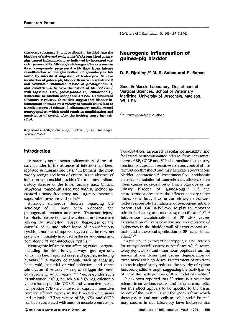
Plasma extravasation: Intravesical instillation of sa-line in control and sensitized animals, and OVA innon-sensitized animals, failed to stimulate detectableEvans blue dye extravasation. Four h after intra-vesical instillation of capsaicin (3 M), SP (100 btM),or OVA (1 btg/ml, in sensitized animals), Evans bluedye extravasation had occurred (Fig. 1), and Evansblue dye extravasation was not increased whenmeasured 18 to 20 h after intravesical instillationof these compounds. In preliminary studies, extra-vasation of Evans blue dye in response to 1 btg/mlOVA (the concentration of OVA used consistently inthis model of cystitis21,22) instillation into sensitizedbladders was determined, and extravasation of Evansblue dye in response to various concentrations ofintravesical capsaicin and SP was investigated. Theconcentrations of capsaicin (3 btM) and SP (100 btM)
60
50
40
" 30
lO
FIG. 1. Evans blue dye extravasation in guinea-pig bladders 4 h after in vivointravesical infusion of ovalbumin (1 lg/ml, sensitized animals only), sub-stance P (100 IM), or capsaicin (3 IM). Bladders in sensitized and naiveguinea-pigs infused with saline contained 2-5 lg Evans blue dye/g tissue(n 12; data not shown). Asterisk indicates significant difference (p < 0.05)between the effects of capsaicin (n= 10) and substance P (n= 8) orovalbumin (n=8). , ovalbumin, lg/ml; J, substance P, 1001M;r-I, capsaicin, 3 M.
Mediators of Inflammation. Vol 3. 1994 191
D. E. Bjorling, M. R. Sabar and R. Saban
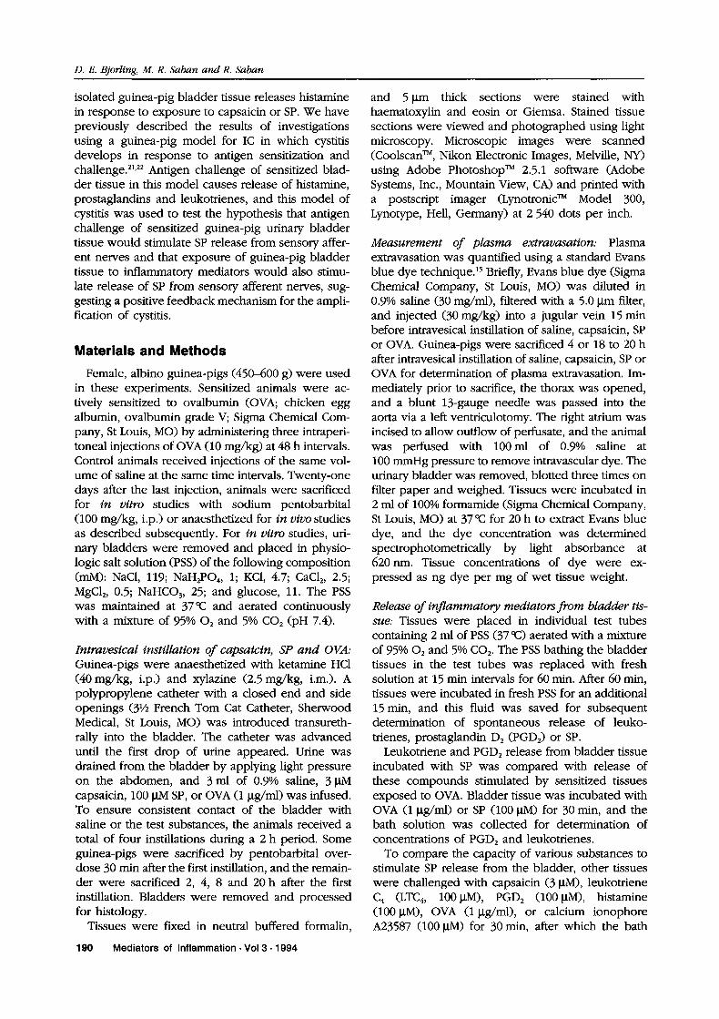
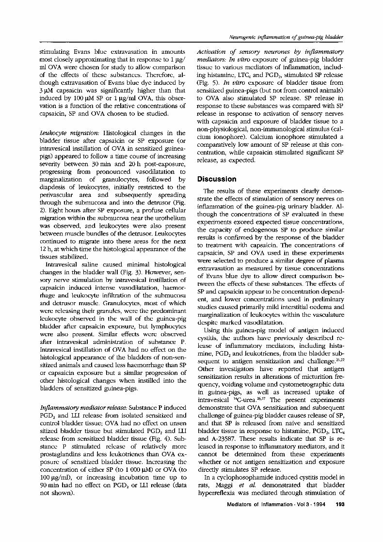
FIG. 2. These photomicrographs illustrate the progression of histological changes in the bladder wall after intravesical instillation of capsaicin 3 #M, SP100 #M or ovalbumin pg/ml (sensitized animals only). The progression of increasing severity of cellular infiltration was similar for these three groups, butcapsaicin-treated bladders exhibited significantly more haemorrhage. Examples of OVA-treated bladders from sensitized animals are shown, because thechanges are less obscured by haemorrhage. A, 30 min after challenge (approximately 200 x); B, 8 h (approximately 400 x); C, 12 h (approximately 600 x);and D, 20 h after challenge (approximately 100 x). Arrows indicate urothelium (U) or blood vessels (BV).
192 Mediators of Inflammation Vol 3 1994
Neurogenic inflammation ofguinea-pig bladder
stimulating Evans blue extravasation in amountsmost closely approximating that in response to 1 lttg/ml OVA were chosen for study to allow comparisonof the effects of these substances. Therefore, al-though extravasation of Evans blue dye induced by3 M capsaicin was significantly higher than thatinduced by 100 btM SP or 1 btg/ml OVA, this obser-vation is a function of the relative concentrations ofcapsaicin, SP and OVA chosen to be studied.
Leukocyte migration: Histological changes in thebladder tissue after capsaicin or SP exposure (orintravesical instillation of OVA in sensitized guinea-pigs) appeared to follow a time course of increasingseverity between 30 min and 20 h post-exposure,progressing from pronounced vasodilatation tomarginalization of granulocytes, followed bydiapdesis of leukocytes, initially restricted to theperivascular area and subsequently spreadingthrough the submucosa and into the detrusor (Fig.2). Eight hours after SP exposure, a profuse cellularmigration within the submucosa near the urotheliumwas observed, and leukocytes were also presentbetween muscle bundles of the detrusor. Leukocytescontinued to migrate into these areas for the next12 h, at which time the histological appearance of thetissues stabilized.
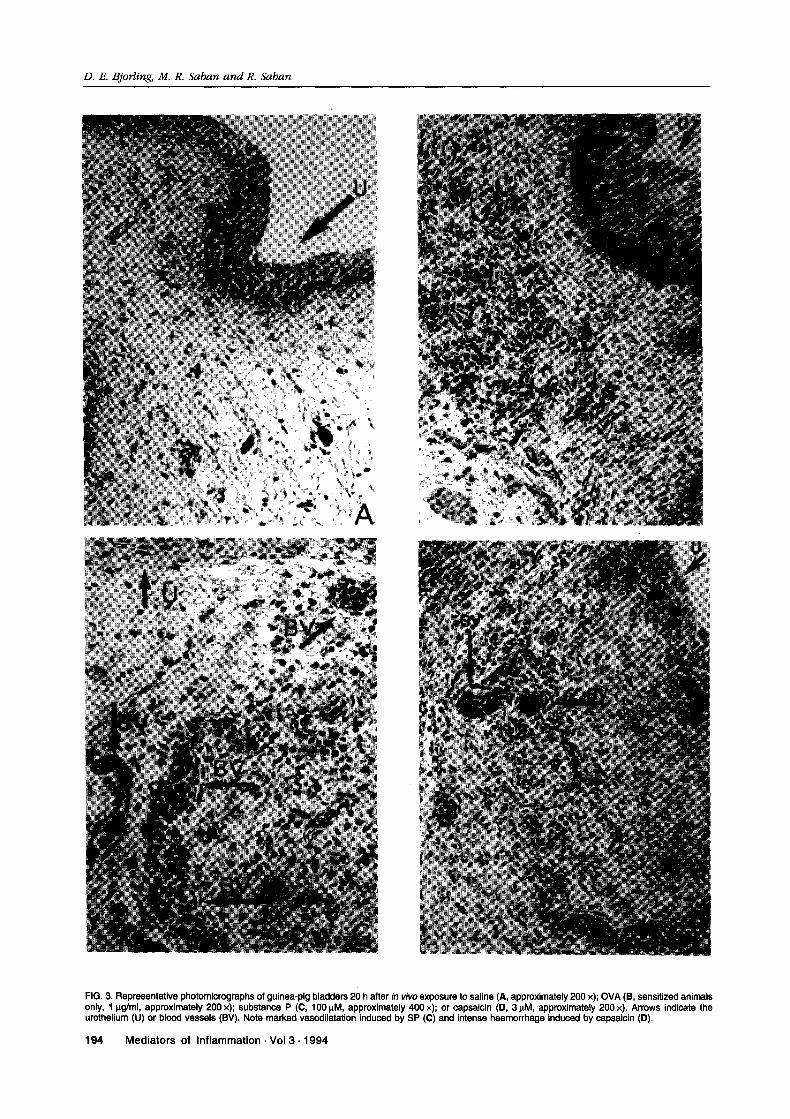
Intravesical saline caused minimal histologicalchanges in the bladder wall (Fig. 3). However, sen-sory nerve stimulation by intravesical instillation ofcapsaicin induced intense vasodilatation, haemor-rhage and leukocyte infiltration of the submucosaand detrusor muscle. Granulocytes, most of whichwere releasing their granules, were the predominantleukocyte observed in the wall of the guinea-pigbladder after capsaicin exposure, but lymphocyteswere also present. Similar effects were observedafter intravesical administration of substance P.Intravesical instillation of OVA had no effect on thehistological appearance of the bladders of non-sen-sitized animals and caused less haemorrhage than SPor capsaicin exposure but a similar progression ofother histological changes when instilled into thebladders of sensitized guinea-pigs.
Inflammatory mediator release: Substance P induced
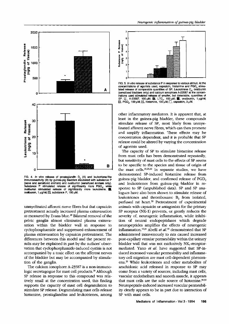
PGD2 and LLI release from isolated sensitized andcontrol bladder tissue; OVA had no effect on unsensitized bladder tissue but stimulated PGD and LLIrelease from sensitized bladder tissue (Fig. 4). Sub-stance P stimulated release of relatively moreprostaglandins and less leukotrienes than OVA ex-posure of sensitized bladder tissue. Increasing theconcentration of either SP (to 1 000 M) or OVA (to100btg/ml), or increasing incubation time up to
90 min had no effect on PGD2 or LLI release (datanot shown).
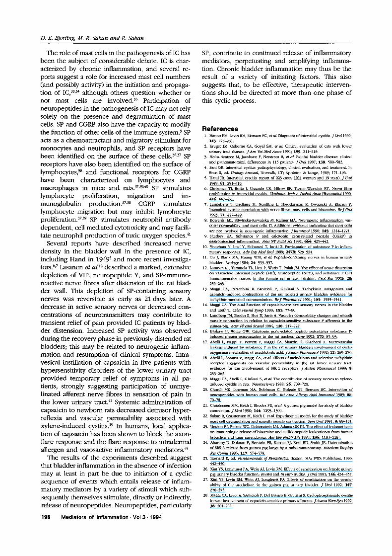
Activation of sensory neurones by inflammatorymediators: In vitro exposure of guinea-pig bladdertissue to various mediators of inflammation, includ-ing histamine, LTC and PGD2, stimulated SP release(Fig. 5). In vitro exposure of bladder tissue fromsensitized guinea-pigs (but not from control animals)to OVA also stimulated SP release. SP release inresponse to these substances was compared with SPrelease in response to activation of sensory nerveswith capsaicin and exposure of bladder tissue to a
non-physiological, non-immunological stimulus (cal-cium ionophore). Calcium ionophore stimulated acomparatively low amount of SP release at this con-centration, while capsaicin stimulated significant SPrelease, as expected.
Discussion
The results of these experiments clearly demon-strate the effects of stimulation of sensory nerves oninflammation of the guinea-pig urinary bladder. Al-though the concentrations of SP evaluated in theseexperiments exceed expected tissue concentrations,the capacity of endogenous SP to produce similarresults is confirmed by the response of the bladderto treatment with capsaicin. The concentrations ofcapsaicin, SP and OVA used in these experimentswere selected to produce a similar degree of plasmaextravasation as measured by tissue concentrationsof Evans blue dye to allow direct comparison be-tween the effects of these substances. The effects ofSP and capsaicin appear to be concentration depend-ent, and lower concentrations used in preliminarystudies caused primarily mild interstitial oedema andmarginalization of leukocytes within the vasculaturedespite marked vasodilatation.Using this guinea-pig model of antigen induced
cystitis, the authors have previously described re-lease of inflammatory mediators, including hista-mine, PGD and leukotrienes, from the bladder sub-sequent to antigen sensitization and challenge. 21,22
Other investigators have reported that antigensensitization results in alterations of micturition fre-quency, voiding volume and cystometrographic datain guinea-pigs, as well as increased uptake ofintravesical 14C-urea.26,27 The present experimentsdemonstrate that OVA sensitization and subsequentchallenge of guinea-pig bladder causes release of SP,and that SP is released from naive and sensitizedbladder tissue in response to histamine, PGD2, LTC4and A-23587. These results indicate that SP is re-leased in response to inflammatory mediators, and itcannot be determined from these experimentswhether or not antigen sensitization and exposuredirectly stimulates SP release.
In a cyclophosophamide induced cystitis model inrats, Maggi et al. demonstrated that bladderhyperreflexia was mediated through stimulation of
Mediators of Inflammation Vol 3 1994 193
D. E. Bjorling, M. R. Saban anvl R. Saban
FIG. 3. Representative photomicrographs of guinea-pig bladders 20 h after in vivo exposure to saline (., approximately 200 x); OVA (1, sensitized animalsonly, g/ml, approximately 200 x); substance P ((3, 100 IM, approximately 400 x)" or capsaicin (13, 3 IM, approximately 200 x). Arrows indicate theurothelium (U) or blood vessels (BV). Note marked vasodilatation induced by SP (C) and intense haemorrhage induced by capsaicin (13).
194 Mediators of Inflammation Vol 3. 1994
Neurogenic inflammation ofguinea-pig bladder
2000
1500
1000
500
A
40
(R) 30
="6 20
(R) 0
FIG. 4. In vitro release of prostaglandin D (A) and leukotriene-likeimmunoreactivity (B) by guinea-pig bladders incubated with substance P(naive and sensitized animals) and ovalbumin (sensitized animals only).Substance P stimulated release of significantly more PGD2, whileovalbumin stimulated release of significantly more leukotriene..,ovalbumin, lg/ml; I, substance P, 100 M.
unmyelinated afferent nerve fibres but that capsaicinpretreatment actually increased plasma extravasationas measured by Evans blue.’* Bilateral removal of thepelvic ganglia almost eliminated plasma extrava-sation within the bladder wall in response tocyclophosphamide and suppressed enhancement ofplasma extravasation by capsaicin pretreatment. Thedifferences between this model and the present re-suits may be explained in part by the authors’ obser-vation that cyclophosphamide-induced cystitis is notaccompanied by a toxic effect on the afferent nervesof the bladder but may be accompanied by stimula-tion of the ganglia.The calcium ionophore A-23587 is a non-immuno-
logic secretagogue for mast cell products.29 AlthoughSP release in response to this compound was rela-tively small at the concentration used, this findingsupports the capacity of mast cell degranulation tostimulate SP release. Degranulating mast cells releasehistamine, prostaglandins and leukotrienes, among
6
._0’$’5
.’ 4
2
FIG. 5. In vitro release of substance P in response to various stimuli. At theconcentrations of agonists used, capsaicin, histamine and PGD stimu-lated release of comparable quantities of SP. Leukotriene C4, ovalbumin(sensitized bladders only) and calcium ionophore A-23587 at the concen-trations used stimulated release of smaller, but detectable, quantities ofSP. rll], A-23587, 100M; I1, LTC,, 1001M; ,, ovalbumin, lg/ml;I, PGD, 100 IM; [], histamine, 100 IM; r-l, capsaicin, 3 M.
other inflammatory mediators. It is apparent that, atleast in the guinea-pig bladder, these compoundsstimulate release of SP, most likely from unmye-linated afferent nerve fibres, which can then promoteand amplify inflammation. These effects may beconcentration dependent, and it is probable that SPrelease could be altered by varying the concentrationof agonists used.The capacity of SP to stimulate histamine release
from mast cells has been demonstrated repeatedly,but sensitivity of mast cells to the effects of SP seemsto be specific to the species and tissue of origin ofthe mast cells.2,23,3 In separate studies, we havedemonstrated SP-induced histamine release fromguinea-pig bladder, and confirmed release of PGD2and leukotrienes from guinea-pig bladder in re-sponse to SP (unpublished data). SP and SP ana-logues have also been shown to stimulate release ofleukotrienes and thromboxane B from isolated,perfused rat heart.3 Pretreatment of experimentalanimals with capsaicin or antagonists for the primarySP receptor (NK-1) prevents, or greatly reduces theseverity of, neurogenic inflammation, while inhibi-tion of neutral endopeptidases which degradeneuropeptides amplifies the effects of neurogenicinflammation.1,1 Abelli et al. TM demonstrated that SPadministered intravenously to rats caused increasedpost-capillary venular permeability within the urinarybladder wall that was not exclusively NKl-receptor-mediated. Yano et al. have suggested that SP-in-duced increased vascular permeability and inflamma-tory cell migration are mast cell-dependent phenom-ena.32 While leukotrienes and other metabolites ofarachidonic acid released in response to SP maycome from a variety of sources, including mast cells,vascular endothelium and smooth muscle, it appearsthat mast cells are the sole source of histamine.,"
Neuropeptide-induced increased vascular permeabil-ity clearly appears to be in part due to interaction ofSP with mast cells.
Mediators of Inflammation. Vol 3. 1994 195
D. E. Bjorling, M. R. Saban and R. Saban
The role of mast cells in the pathogenesis of IC hasbeen the subject of considerable debate. IC is char-acterized by chronic inflammation, and several re-ports suggest a role for increased mast cell numbers(and possibly activity) in the initiation and propaga-tion of IC,33’34 although others question whether ornot mast cells are involved.35 Participation ofneuropeptides in the pathogenesis of IC may not relysolely on the presence and degranulation of mastcells. SP and CGRP also have the capacity to modifythe function of other cells of the immune system. SPacts as a chemoattractant and migratory stimulant formonocytes and neutrophils, and SP receptors havebeen identified on the surface of these cells. 36,7 SPreceptors have also been identified on the surface oflymphocytes,8 and functional receptors for CGRPhave been characterized on lymphocytes andmacrophages in mice and rats.7,9,4 SP stimulateslymphocyte proliferation, migration and im-munoglobulin production.7,8 CGRP stimulateslymphocyte migration but may inhibit lymphocyteproliferation.37,9 SP stimulates neutrophil antibodydependent, cell mediated cytotoxicity and may facili-tate neutrophil production of toxic oxygen species.41
Several reports have described increased nervedensity in the bladder wall in the presence of IC,including Hand in 1949 and more recent investiga-tors.<7 Lasanen et a1.12 described a marked, extensive
depletion of VIP, neuropeptide Y, and SP-immuno-reactive nerve fibres after distension of the rat blad-der wall. This depletion of SP-containing sensorynerves was reversible as early as 21 days later. Adecrease in active sensory nerves or decreased con-centrations of neurotransmitters may contribute totransient relief of pain provided IC patients by blad-der distension. Increased SP activity was observedduring the recovery phase in previously distended ratbladders; this may be related to neurogenic inflam-mation and resumption of clinical symptoms. Intra-vesical instillation of capsaicin in five patients withhypersensitivity disorders of the lower urinary tractprovided temporary relief of symptoms in all pa-tients, strongly suggesting participation of unmye-linated afferent nerve fibres in sensation of pain inthe lower urinary tract.42 Systemic administration ofcapsaicin to newborn rats decreased detrusor hyper-reflexi and vascular permeability associated withxylene-induced cystitis. 19 In humans, local applica-tion of capsaicin has been shown to block the axon-flare response and the flare response to intradermalallergen and vasosactive inflammatory mediators.4
The results of the experiments described suggestthat bladder inflammation in the absence of infectionmay at least in part be due to initiation of a cyclicsequence of events which entails release of inflam-matory mediators by a variety of stimuli which sub-sequently themselves stimulate, directly or indirectly,release of neuropeptides. Neuropeptides, particularly
SP, contribute to continued release of inflammatorymediators, perpetuating and amplifying inflamma-tion. Chronic bladder inflammation may thus be theresult of a variety of initiating factors. This alsosuggests that, to be effective, therapeutic interven-tions should be directed at more than one phase ofthis cyclic process.
References1. Hanno PM, Levin RM, Monson FC, et al. Diagnosis of interstitial cystitis. J Uro11990;
143: 278-281.2. Kruger JM, Osborne CA, Goyal SM, et al. Clinical evaluation of cats with lower
urinary tract disease. JAm Vet Med Assoc 1991; 199: 211-216.3. Holm-Bentzen M, Jacobsen F, Nerstrom B, et al. Painful bladder disease: clinical
and pathoanatomical differences in 115 patients. J Urol 1987; 138: 500-502.4. Sant GR. Interstitial cystitis: pathophysiology, clinical evaluation, and treatment. In
Rous S, ed. Urology Annual, Norwalk, CT; Appleton & Lange, 1989; 171-196.5. Hand JR. Interstitial cystitis: report of 223 (204 and 19 men). J Urol
1949; 61: 291-310.6. Christmas TJ, Rode J, Chapple CR, Milroy EF, Turner-Warwick RT. Nerve fibre
proliferation in interstitial cystitis. Virchows Arch A Pathol Anat Histopathol 1990;416: 447-451.
7. Lundeberg T, Liedberg H, Nordling L, Theodorsson E, Owzarski A, Ekman P.
Interstitial cystitis: correlation with fibres, mast cells and histamine. BrJ Urol
1993; "/1: 427-429.8. Kowalski ML, Sliwinska-Kowalska M, Kaliner MA. Neurogenic inflammation,
cular permeability, and mast cells. II. Additional evidence indicating that mast cellsnot involved in neurogenic inflammation. Jlmmunol 1990; 145: 1214-1221.
9. Sharkey KA. Substance P and calcitonin gene-related peptide (CGRP) in
gastrointestinal inflammation. Ann NYAcad Sci 1992; 664: 425-442.10. Yonehara N, Imai Y, Shibutani T, Inoki R. Participation of substance P in inflam-
matory responses. Adv Exp Med Biol 1989; 24-/: 529-534.11. Gu J, Blank MA, Huang WM, et al. Peptide-containing in human urinary
bladder. Urology 1984; 24: 353-357.12. Lasanen LT, Tammela TL, Liesi P, Waris T, Polak JM. The effect of acute distension
vasoactive intestinal peptide (VIP), neuropeptide (NPY), and substance P (SP)
immunoreactive in the female rat urinary bladder. Urol Res 1992; 2{}:
259-263.13. Maggi CA, Patacchini R, Sanicioli P, Giuliani S. Tachykinin antagonists and
capsaicin-induced contractions of the rat isolated urinary bladder: evidence for
tachykinin-mediated cotransmission. BrJ Pharmacol 1991; 1{}3:1535-1541.14. Maggi CA. The dual function of capsaicin-sensitive sensory in the bladder
and urethra. Ciba Found Syrup 1990; 151: 77-90.15. Lundberg JM, Brodin E, Hua X, Saria A. Vascular permeability changes and smooth
muscle contraction in relation to capsaicin-sensitive substance P afferents in the
guinea-pig. Acta Physiol Scand 1984; 12{}: 217-227.16. Brokaw JJ, White GW. Calcitonin gene-related peptide potentiates substance P-
induced plasma extravasation in the rat trachea. Lung 1992; 1"/{}: 85-93.17. Abelli L, Nappi F, Perretti F, Maggi CA, Manzini S, Giachetti A. Microvascular
leakage induced by substance P in the rat urinary bladder: involvement of cyclo-oxygenase metabolites of arachidonic acid. JAuton Pharmaco11992; 12: 269-276.
18. Abelli L, Somma V, Maggi CA, et al. Effects of tachykinins and selective tachykininreceptor antagonists vascular permeability in the rat lower urinary tract:
evidence for the involvement of NK-1 receptors. J Auton Pharmacol 1989; 9:253-263.
19. Maggi CA, Abelli L, Giuliani S, et al. The contribution of sensory to xylene-induced cystitis in rats. Neuroscience 1988; 26: 709-723.
20. Church MK, Lowman MA, Robinson C, Holgate ST, Benyon RC. Interaction ofneuropeptides with human mast cells. Int Arch Allergy Appl Immunol 1989; 88:
70-78.21. Christensen MM, Keith I, Rhodes PR, etal. A guinea pig model for study of bladder
contraction. J Uro11990; 144: 1293-1300.22. Saban R, Christensen M, Keith I, et al. Experimental model for the study of bladder
mast cell degranulation and smooth muscle contraction. Sem Uro11991; 9: 88-101.
23. Undem BJ, Pickett WC, Lichtenstein LM, Adams GK III. The effect of indomethacin
immunologic release of histamine and sulfidopeptide leukotrienes from humanbronchus and lung parenchyma. Am Rev RespirDis 1987; 136: 1183-1187.
24. Aharony D, Dobson P, Berstein PR, Kusner EJ, Krell RD, Smith JB. Determination
of SRS-A release from guinea pig lungs by radioimmunoassay. Biochem BiophysRes Comm 1983; 11"/: 574-579.
25. Bernard R, ed. Fundamentals of Biostatistics. Boston, MA: PWS Publishers, 1990;442-450.
26. Kim YS, Longhurst PA, Wein AJ, Levin RM. Effects of sensitization female guineapig urinary bladder function: in vivo and in vitro studies. J Uro11991; 146: 454-457.
27. Kim YS, Levin RM, Wein AJ, Longhurst PA. Effects of sensitization the perme-ability of the urothelium in the guinea pig urinary bladder. J Urol 1992; 14"/:
270-273.28. Maggi CA, Lecci A, Santicioli P, Del Bianco E, Giuliani S. Cyclophosphamide cystitis
in rats: involvement of capsaicin-sensitive primary afferents. JAuton Nerv Syst 1992;38: 201-208.
196 Mediators of Inflammation Vol 3 1994
Neurogenic inflammation ofguinea-pig bladder
29. Vliagoftis H, Dimitriadou V, Boucher W, et al. Estradiol augments while tamoxifeninhibits rat mast cell secretion. Int Arch Allergy Immunol 1992; 98: 398-409.
30. Assem ESK, Ghanem NS, Abdullah NA, Repke H, Foreman JC, Hayes NA. SubstanceP and arg-pro-lys-pro-NH-C12-H25-induced mediator release from different mast cellsubtypes of rat and guinea-pig. Immunopharmacology 1989; 1"/: 119-128.
31. Nadel JA. Neurogenic inflammation in airways and its modulation by peptidases.Ann NYAcad Sci 1992; 664: 408-414.
32. Yano H, Wershil BK, Arizono N, Galli SJ. Substance P-induced augmentation ofcutaneous vascular permeability and granulocyte infiltration in mice is mast celldependent. J Clin Invest 1989; 84: 1276-1286.
33. Nielsen KK, Kroman-Andersen B, Steven K, Hald T. Failure of combinedsupratrigonal cystectomy and Mainz ileocecocystoplasty in intractible interstitial
cystitis: is histology and mast cell count reliable predictor for the outcome of
surgery? J Urol 1990; 144: 255-258.34. Theoharides TC, Flarris N, Cronin CT, Ucci A, Meares E. Mast cell activation in
sterile bladder and prostate inflammation. Int Arch Allergy Appl Immunol 1990; 92:281-286.
35. MacDermott JP, Charpied GL, Tesluk H, Stone AR. Can histological assessment
predict the outcome in interstitial cystitis? BrJ Urol 1991; {"/: 44-47.36. Haines KA, Kolasinski SL, Cronstein BN, Reibman J, Gold LI, Weissmann G.
Chemoattraction of neutrophils by substance P and transforming growth factor-betais inadequately explained by current models of lipid remodeling. JImmuno11993;
151: 1491-1499.37. McGillis JP, Mitsuhashi M, Payan DG. Immunomodulation by tachykinin
neuropeptides. Ann NYAcad Sci 1990; 594: 85-94.38. Pascual DW, Bost KL, Xu-Amano J, Kiyono H, McGhee JR. The cytokine-like action
of substance P upon B cell differentiation. Reg Immunol 1992; 4: 100-104.
39. Umeda Y, Arisawa M. Characterization of the calcitonin gene-related peptidereceptor in T lymphocytes. Neuropeptides 1989; 14: 237-242.
40. Abello J, Kaiserlian D, Cuber JC, Revillard JP, Chayvialle JA. Characterization ofcalcitonin gene-related peptide receptors and adenylate cyclase response in themurine macrophage cell line P388 D1. Neuropeptides 1991; 19: 43-49.
41. Wozniak A, McLennan G, Betts WH, Murphy GA, Scicchitano R. Activation of
human neutrophils by substance P: effect FMLP-stimulated oxidative andarachidonic acid metabolism and antibody-dependent cell-mediated
cytotoxicity. Immunology 1989; {8: 359-364.42. Maggi CA, Barbanti G, Santicioli P, et al. Cystometric evidence that capsaicin-
sensitive modulate the afferent branch of micturition reflex in humans.
J Urol 1989; 142: 150-154.43. Lundblad L, Lundberg L, Anggard A, Zetterstrom O. Capsaicin pretreatment inhibits
the flare component of the cutaneous allergic reaction in EurJ Pharmacol1985; 113: 461-462.
ACKNOWLEDGEMENTS. This work supported by grant #1R01DK42897-CIA2 fromNational Institutes of Health.
The authors acknowledge the contribution of Joel Armstrong from The Radio-
nuclide Laboratory, School of Veterinary Medicine, University of Wisconsin-
Madison for assistance in the substance P radioimmunoassays and Kermit Groothuisfor assistance with tissues processing for histology.
Received 17 January 1994;accepted in revised form 15 February 1994
Mediators of Inflammation. Vol 3. 1994 197
Related Documents


![Neurogenic bladder [Dr. Edmond Wong]](https://static.cupdf.com/doc/110x72/554af038b4c90559058b4779/neurogenic-bladder-dr-edmond-wong.jpg)


