Electrophysiological and morphological changes in colonic myenteric neurons from chemotherapy-treated patients: a pilot study S. E. CARBONE,* V. JOVANOVSKA,* S. J. H. BROOKES† & K. NURGALI * *Centre for Chronic Disease, College of Health and Biomedicine, Victoria University, Melbourne, VIC, Australia †Discipline of Human Physiology and Centre for Neuroscience, Flinders University, Adelaide, SA, Australia Key Points • This is the first electrophysiological study of human enteric neurons in a pathological condition. • This study aimed to investigate the effects of anticancer chemotherapy on functional and morphological properties of human myenteric neurons. • Intracellular electrophysiology combined with morphological identification of recorded neurons and immuno- histochemistry were used to characterize myenteric neurons in fresh colon specimens from colorectal cancer patients treated and untreated with chemotherapeutic agents. • The results of this study demonstrated hyperexcitability of myenteric S neurons, increase in the number of neurons with translocation of Hu protein from the cytoplasm to the nucleus, and increase in the soma size of neuronal nitric oxide synthase-immunoreactive neurons from chemotherapy-treated patients. Abstract Background Patients receiving anticancer chemother- apy experience a multitude of gastrointestinal side- effects. However, the causes of these symptoms are uncertain and whether these therapeutics directly affect the enteric nervous system is unknown. Our aim was to determine whether the function and morphology of myenteric neurons are altered in spec- imens of the colon from chemotherapy-treated patients. Methods Colon specimens were compared from chemotherapy-treated and non-treated patients following colorectal resections for removal of carci- noma. Intracellular electrophysiological recordings from myenteric neurons and immunohistochemistry were performed in whole mount preparations. Key Results Myenteric S neurons from chemother- apy-treated patients were hyperexcitable; more action potentials (11.4 9.4, p < 0.05) were fired in response to depolarising current pulses than in non-treated patients (1.4 0.5). The rheobase and the threshold to evoke action potentials were significantly lower for neurons from chemotherapy-treated patients com- pared to neurons from non-treated patients (p < 0.01). Fast excitatory postsynaptic potential reversal potential was more positive in neurons from chemotherapy-treated patients (p < 0.05). An increase in the number of neurons with translocation of Hu protein from the cytoplasm to the nucleus was observed in specimens from chemotherapy-treated patients (103 25 neurons/mm 2 , 37.2 7.0%, n = 8) compared to non-treated (26 5 neurons/ mm 2 , 11.9 2.7%, n = 12, p < 0.01). An increase in the soma size of neuronal nitric oxide synthase- immunoreactive neurons was also observed in these specimens. Conclusions & Inferences This is the first study suggesting functional and structural changes in human myenteric neurons in specimens of colon from patients receiving anticancer chemotherapy. These Address for Correspondence Dr Kulmira Nurgali, Centre for Chronic Disease, College of Health and Biomedicine, Victoria University, McKechnie St, St Albans, VIC 3021, Australia. Tel: +61 3 8395 8223; e-mail: [email protected] Received: 8 September 2015 Accepted for publication: 14 January 2016 © 2016 The Authors. Neurogastroenterology & Motility Published by John Wiley & Sons Ltd. This is an open access article under the terms of the Creative Commons Attribution License, which permits use, distribution and reproduction in any medium, provided the original work is properly cited. 975 Neurogastroenterol Motil (2016) 28, 975–984 doi: 10.1111/nmo.12795 Neurogastroenterology & Motility

Welcome message from author
This document is posted to help you gain knowledge. Please leave a comment to let me know what you think about it! Share it to your friends and learn new things together.
Transcript
Electrophysiological and morphological changes in
colonic myenteric neurons from chemotherapy-treated
patients: a pilot study
S. E. CARBONE,* V. JOVANOVSKA,* S. J. H. BROOKES† & K. NURGALI*
*Centre for Chronic Disease, College of Health and Biomedicine, Victoria University, Melbourne, VIC, Australia
†Discipline of Human Physiology and Centre for Neuroscience, Flinders University, Adelaide, SA, Australia
Key Points
• This is the first electrophysiological study of human enteric neurons in a pathological condition.
• This study aimed to investigate the effects of anticancer chemotherapy on functional and morphological
properties of human myenteric neurons.
• Intracellular electrophysiology combined with morphological identification of recorded neurons and immuno-
histochemistry were used to characterize myenteric neurons in fresh colon specimens from colorectal cancer
patients treated and untreated with chemotherapeutic agents.
• The results of this study demonstrated hyperexcitability of myenteric S neurons, increase in the number
of neurons with translocation of Hu protein from the cytoplasm to the nucleus, and increase in the
soma size of neuronal nitric oxide synthase-immunoreactive neurons from chemotherapy-treated
patients.
Abstract
Background Patients receiving anticancer chemother-
apy experience a multitude of gastrointestinal side-
effects. However, the causes of these symptoms are
uncertain and whether these therapeutics directly
affect the enteric nervous system is unknown. Our
aim was to determine whether the function and
morphology of myenteric neurons are altered in spec-
imens of the colon from chemotherapy-treated
patients. Methods Colon specimens were compared
from chemotherapy-treated and non-treated patients
following colorectal resections for removal of carci-
noma. Intracellular electrophysiological recordings
from myenteric neurons and immunohistochemistry
were performed in whole mount preparations.
Key Results Myenteric S neurons from chemother-
apy-treated patients were hyperexcitable; more action
potentials (11.4 � 9.4, p < 0.05) were fired in response
to depolarising current pulses than in non-treated
patients (1.4 � 0.5). The rheobase and the threshold to
evoke action potentials were significantly lower for
neurons from chemotherapy-treated patients com-
pared to neurons from non-treated patients
(p < 0.01). Fast excitatory postsynaptic potential
reversal potential was more positive in neurons from
chemotherapy-treated patients (p < 0.05). An increase
in the number of neurons with translocation of Hu
protein from the cytoplasm to the nucleus was
observed in specimens from chemotherapy-treated
patients (103 � 25 neurons/mm2, 37.2 � 7.0%,
n = 8) compared to non-treated (26 � 5 neurons/
mm2, 11.9 � 2.7%, n = 12, p < 0.01). An increase in
the soma size of neuronal nitric oxide synthase-
immunoreactive neurons was also observed in these
specimens. Conclusions & Inferences This is the first
study suggesting functional and structural changes in
human myenteric neurons in specimens of colon from
patients receiving anticancer chemotherapy. These
Address for Correspondence
Dr Kulmira Nurgali, Centre for Chronic Disease, College ofHealth and Biomedicine, Victoria University, McKechnie St,St Albans, VIC 3021, Australia.Tel: +61 3 8395 8223;e-mail: [email protected]: 8 September 2015Accepted for publication: 14 January 2016
© 2016 The Authors.Neurogastroenterology & Motility Published by John Wiley & Sons Ltd.This is an open access article under the terms of the Creative Commons Attribution License,which permits use, distribution and reproduction in any medium, provided the original work is properly cited.
975
Neurogastroenterol Motil (2016) 28, 975–984 doi: 10.1111/nmo.12795
Neurogastroenterology & Motility
changes may contribute to the causation of gastroin-
testinal symptoms experienced by chemotherapy-
treated patients.
Keywords chemotherapy, colorectal cancer, electro-
physiology, enteric neurons, human.
INTRODUCTION
Colorectal cancer (CRC) is the second most commonly
diagnosed cancer and is a major cause of cancer-related
deaths worldwide. Chemotherapy alone, or in combi-
nation with radiotherapy, is given before or after
surgery to most patients. Currently, the standard
first-line chemotherapy of metastatic CRC is 5-fluor-
ouracil (5-FU) combined with folinic acid plus either
oxaliplatin (FOLFOX) or irinotecan (FOLFIRI).1
Although these drugs increase survival rate and reduce
the risk of disease progression, both have acute and
long-term toxicities leading to a wide spectrum of side-
effects. Symptoms such as pain, paresthesia (tingling or
numbness), cold-induced dysesthesia (burning sensa-
tions), and a general loss of sensation2–6 have been
attributed to peripheral neuropathies resulting from
the neurotoxic effects of chemotherapeutic drugs.
Severe gastrointestinal side-effects include nausea,
vomiting, constipation, and diarrhoea.7–9 Gastroin-
testinal toxicity is one of the main reasons for dose
limitation of chemotherapy, often reducing the efficacy
of anticancer treatment. Chronic gastrointestinal side-
effects can persist for more than 10 years post treat-
ment, greatly affecting patients’ quality of life.10 The
traditional view is that gastrointestinal side-effects of
ant-cancer drugs are due to mucosal damage.11
Although mucosal damage undoubtedly plays a signif-
icant role in the acute symptoms associated with
chemotherapeutic treatment, the persistence of gas-
trointestinal symptoms suggests that chemotherapy
may damage the gastrointestinal innervation. The
enteric nervous system controls many major functions
of the gut, including motility, blood flow, secretion and
absorption of nutrients, electrolytes, and water.12 The
morphology and functions of enteric neurons are
compromised in various gastrointestinal pathologies.13
Because anticancer chemotherapeutics cause wide-
spread peripheral neuropathy, we hypothesized that
gastrointestinal side-effects associated with
chemotherapy may result from damage to the enteric
nervous system.
Few studies have examined the effects of anticancer
chemotherapies on the enteric nervous system. We
have previously shown changes in colonic motility and
reduction in the total number of neurons within the
myenteric plexus in mice treated in vivo with the
anticancer chemotherapeutic oxaliplatin.14 Similar
results were found in the colon of rats treated with
another platinum-based chemotherapeutic agent, cis-
platin.15 In addition, cisplatin-treated rats had reduced
gastric motility.15–17
Immunohistochemical methods have been exten-
sively used to document changes in the neurochemical
coding and morphology of human enteric neurons
under a variety of conditions.18–21 Electrophysiological
recordings of human enteric neurons in freshly dis-
sected preparations reveal functional properties, but
have only been reported in two published studies to
date, both from specimens of the colon.22,23 Studies
based on these techniques can demonstrate changes in
certain conditions, such as in inflammation, where
enteric neurons become hyperexcitable.24,25
This study aimed to investigate the effects of
anticancer chemotherapeutics on human myenteric
neurons. Using electrophysiology combined with
immunohistochemistry, the functional properties of
morphologically identified myenteric neurons
were compared in colon specimens from chemother-
apy-treated versus non-treated patients for the first
time.
MATERIALS AND METHODS
Specimens of the human colon were provided by the VictorianCancer Biobank (number of individual patients N = 13) andFlinders Medical Centre (N = 8). All studies were approved bythe Victoria University Human Research Ethics and SouthernAdelaide Clinical Research Ethics Committees and have beenperformed in accordance with the ethical standards laid down inthe 1964 Helsinki Declaration and its later amendments. Prior tosurgical removal of non-obstructive carcinoma, written informedconsent was obtained from all patients. Fresh specimens weredelivered after the surgery in Roswell Park Memorial Institute(RPMI) culture medium or Krebs solution at 4 °C. Of all 21specimens, nine were from patients who had received chemother-apeutic treatments. Due to the limited number of samplesavailable, all specimens from chemotherapy-treated patients werecombined in a single chemotherapy-treated group. Patientsreceived 5-Fluorouracil alone (5-FU, N = 1), combined FOLFOXregimen (Folinic acid, 5-FU, and oxaliplatin, N = 4) for 6 cyclesand neoadjuvant 5-FU in combination with radiotherapy treat-ment (N = 4). Control specimens were obtained from patientswho had not received chemotherapy or radiotherapy prior tosurgery (N = 12, termed non-treated patients). The age of patientsat the time of surgery ranged between 39 and 89 years. Non-treated patients averaged 72.9 � 3.0 years (10 male, 2 female) andchemotherapy-treated patients averaged 57.4 � 4.7 years (7 male,2 female). Specimens from non-treated patients (distal colon: 7,proximal colon: 5) as well as specimens from chemotherapy-treated patients (distal colon: 8, proximal colon: 1) were predom-inantly from the distal colon. Aside from two specimens (bothfrom non-treated patients), all segments of the colon were from
© 2016 The Authors.Neurogastroenterology & Motility Published by John Wiley & Sons Ltd.
976
S. E. Carbone et al. Neurogastroenterology and Motility
regions distal to the tumor, but the distances from the tumor werenot made available.
The methods used in this study have been described previ-ously.22 Briefly, full thickness specimens were placed in Krebssolution at room temperature (mM: NaCl 118, KCl 4.6, CaCl2 3.5,MgSO4 1.2, NaH2PO4 1, NaHCO3 25, D-Glucose 11, bubbled with95% O2 and 5% CO2) and pinned in a Sylgard-lined Petri dish(Dow Corning, Midland, MI, USA) to make a flat sheet prepara-tion for dissection. The serosa, fat, mesentery, mucosa, andsubmucosa were removed using sharp dissection techniques andthe myenteric plexus was exposed by peeling away the circularmuscle layer with forceps. The Krebs solution was changed atleast every 15 min.
Intracellular recording
Nine specimens were used for intracellular electrophysiologyanalysis (non-treated: five specimens, chemotherapy-treated: fourspecimens). Specimens were repinned in a recording chamberwith 50 lm gold plated, tungsten pins. Myenteric ganglia wereidentified by creamy-white pigmentation. Extra pins (20 lmdiameter wire) were added nearby to stabilize ganglia for record-ing. The chamber was mounted on a Zeiss Axiovert-200 invertedmicroscope (Zeiss, Oberkochen, Germany). To recover fromdissection, the preparation was superfused with Krebs solutioncontaining 1 lM atropine and 1 lM nicardipine (35 °C) for 1 h.26
Conventional glass micropipettes were used to impale neurons.They contained 5% 5,6-carboxyfluorescein in 20 mM Tris bufferin 1 M KCl (pH 7.0)26 and had resistances of 100–150 MΩ. AnAxoclamp 2B amplifier (Axon Instruments, Foster City, CA, USA)was used for recordings; signals were digitized at 1–10 kHz by aDigidata 1440A interface (Molecular Devices, Sunnyvale, CAUSA) and stored on a computer using PClamp 10.0 (MolecularDevices). To identify the morphology of the impaled cell,carboxyfluorescein was injected by iontophoresis using hyperpo-larizing current pulses (0.5 nA, 0.2 s duration at 2.5 Hz) for 2 min.Cells labeled in this way could be visualized in situ usingfluorescence.22,26 Only neurons that were adequately filled withcarboxyfluorescein and had resting membrane potentials (RMPs)more negative than �40 mV were analyzed. Input resistance (Rin)was calculated from intracellular hyperpolarizing current pulses(500 ms, 100–500 pA). A tungsten stimulating electrode (10–50 lm tip diameter), connected to an ISO-Flex stimulator (AMPI,Jerusalem, Israel), was either positioned between identified gan-glia and 1 mm circumferential to the impaled cell. Axon tractswere stimulated by a single-shot stimulus (0.4 ms; 10–60 V), sothat fast excitatory postsynaptic potentials (fEPSPs) could berecorded in the impaled cell. Slow excitatory postsynaptic poten-tials were rarely evoked even by trains of stimuli and weretherefore not studied. Axograph X software was used to analyzedata. Following recording, preparations were fixed in Zamboni’sfixative (2% formaldehyde and 0.2% picric acid) overnight at 4 °Cand processed for immunohistochemistry.
Immunohistochemistry
Preparations were similarly dissected for immunohistochemistry.They were stretched to a maximum length (nicardipine [3 lM]was added to the Krebs solution to facilitate maximal stretching),then fixed in Zamboni’s fixative (4 °C for 48 h), cleared usingdimethylsulfoxide (3 times for 10 min), then washed with phos-phate buffered saline (PBS, 3 times for 10 min). Specimens wereincubated in primary antibodies: goat antineuronal nitric oxidesynthase (nNOS, 1 : 500, NB100-858; Novus Biologicals, Little-
ton, CO, USA), mouse anti-Hu (1 : 500, A21271; MolecularProbes, Eugene, OR, USA) for 48 h, rinsed in PBS and incubatedin secondary antibodies: DyLight 405 donkey antigoat andantimouse Alexa Fluor 594 (1 : 200; Jackson ImmunoresearchLaboratories, West Grove, PA, USA) for 4 h. Tissues were viewedon an IX71 Olympus microscope (Olympus, Tokyo, Japan) or anEclipse Ti confocal microscope (Nikon, Tokyo, Japan).
Images of four randomly selected ganglia, showing myentericneurons immunoreactive (IR) for Hu and nNOS, were captured byconfocal microscopy at 209 magnification. The number ofneurons within each image was counted to calculate the totalnumber of neurons/mm2. The soma-dendritic area (lm2) of nNOS-IR neurons was measured by tracing neuronal profiles, usingImage J software (NIH, Bethesda, MD, USA). The average size ofneurons was calculated from 10 cells per image.
Drugs
All drugs used in this study and 5,6-carboxyfluorescein werepurchased from Sigma-Aldrich (Castle Hill, NSW, Australia). Bothnicardipine and atropine were dissolved in sterile water and storedat 10�2 M.
Statistics
For electrophysiology results, where the properties of neuronswere compared, n is the number of cells and N is the number oftissue samples, which was equivalent to the number of patients.In some instances, more than one neuron was recorded from asingle patient; values are presented as means for all cells � SD.For immunohistochemistry results, n and N are the same; valuesare represented as means � SEM to demonstrate how the samplemean represents the population mean. Results were comparedusing an unpaired t-test without variance correction. Differenceswere considered statistically significant at p < 0.05. All authorshad access to the study data and had reviewed and approved thefinal manuscript.
RESULTS
The effects of chemotherapeutic treatment onthe electrophysiological properties of colonicmyenteric neurons
Enteric neurons can be classified as S or AH cells based
on their electrophysiological properties.12 S cells had
no visible inflection on the falling phase of their action
potentials, lacked a long after-hyperpolarization fol-
lowing a single action potential and showed fast EPSPs
in response to focal electrical stimulation of nearby
nerve tracts. A long after-hyperpolarization was
recorded in only one neuron; this neuron also had an
inflection on the falling phase of its action potential;
features identifying it as an AH type II enteric neurons.
Three additional neurons had inflections on the falling
phase of their action potentials but lacked a long after-
hyperpolarization. These neurons were multiaxonal
dendritic Dogiel type II neurons. Due to the scarcity of
© 2016 The Authors.Neurogastroenterology & Motility Published by John Wiley & Sons Ltd.
977
Volume 28, Number 7, July 2016 Chemotherapy and human enteric neurons
AH neurons, only S neurons confirmed by the presence
of a fast EPSP and uniaxonal morphology were ana-
lyzed in this study.
The RMP and Rin of S type I myenteric neurons in
colonic specimens from non-treated (RMP:
�59.7 � 11.5 mV, Rin: 100.5 � 78.2 MΩ, n = 5, N = 5)
or chemotherapy-treated (RMP: �57.3 � 1.6 mV; Rin:
132.7 � 125.6 MΩ, n = 5, N = 4) patients were not
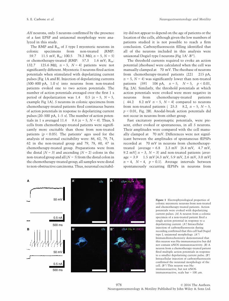
significantly different. Myenteric S neurons fired action
potentials when stimulated with depolarizing current
pulses (Fig 1A and B). Injection of depolarizing currents
(500–800 pA, 1.0 s) into neurons from non-treated
patients evoked one to two action potentials. The
number of action potentials averaged over the first 1 s
period of depolarization was 1.4 � 0.5 (n = 5, N = 5,
example Fig 1A). S neurons in colonic specimens from
chemotherapy-treated patients fired continuous bursts
of action potentials in response to depolarizing current
pulses (20–500 pA, 1–5 s). The number of action poten-
tials in 1 s averaged 11.4 � 9.4 (n = 5, N = 4). Thus, S
cells from chemotherapy-treated patients were signifi-
cantly more excitable than those from non-treated
patients (p < 0.05). The patients’ ages used for the
analysis of neuronal excitability were: 86, 62, 79, 74,
62 in the non-treated group and 79, 78, 60, 47 in
chemotherapy-treated group. Preparations were from
the distal (N = 3) and ascending (N = 2) colons in the
non-treated group and all (N = 5) from the distal colon in
the chemotherapy-treated group; all sampleswere distal
to non-obstructive carcinoma.Thus, neuronal excitabil-
ity did not appear to depend on the age of patients or the
location of the cells, although given the low numbers of
patients studied it is not possible to reach a firm
conclusion. Carboxyfluorescein filling identified that
all of the neurons included in this analysis were
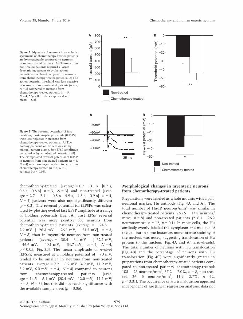
uniaxonal Dogiel type I neurons (Fig 1A’–B”).The threshold currents required to evoke an action
potential (rheobase) were calculated when the cell was
manually clamped at�70 mV. The rheobase of neurons
from chemotherapy-treated patients (221 � 215 pA,
n = 5, N = 4) was significantly lower than non-treated
patients (591 � 106 pA, n = 5, N = 5, p < 0.01,
Fig. 2A). Similarly, the threshold potentials at which
action potentials were evoked were more negative in
neurons from chemotherapy-treated patients
(�44.2 � 8.2 mV n = 5, N = 4) compared to neurons
from non-treated patients (�25.3 � 8.2, n = 5, N = 5,
p < 0.01, Fig. 2B). Anodal-break action potentials did
not occur in neurons from either group.
Fast excitatory postsynaptic potentials, were pre-
sent, either evoked or spontaneous, in all S neurons.
Their amplitudes were compared with the cell manu-
ally clamped at �70 mV. Differences were not signif-
icant between the amplitudes of spontaneous fEPSPs
recorded at �70 mV in neurons from chemotherapy-
treated (average = 6.8 � 2.3 mV [6.4 mV, 4.7 mV,
9.2 mV] n = 3, N = 3) and non-treated patients (aver-
age = 3.9 � 1.5 mV [4.3 mV, 5.9 mV, 2.6 mV, 3.0 mV]
n = 4, N = 4, p = 0.1). Average intervals between
spontaneously occurring fEPSPs in neurons from
50 mV
0.5 nA
500 ms
50 mV
0.5 nA
500 ms
cfs cfs+ Hu + nNOS
cfs cfs+ Hu + nNOS
B′B B′′
A′A A′′
Figure 1 Electrophysiological properties of
colonic myenteric neurons from non-treated
and chemotherapy-treated patients. Action
potentials were evoked with depolarizing
current pulses. (A) A neuron from a colonic
specimen of a non-treated patient fired a
single action potential in response to a
depolarizing current. (A’) Intracellular
injection of carboxyfluorescein during
recording confirmed that this cell had Dogiel
type I, uniaxonal morphology. (A”)
Immunohistochemistry demonstrated that
this neuron was Hu-immunoreactive but did
not contain nNOS immunoreactivity. (B) A
neuron from a chemotherapy-treated patient
fired multiple action potentials in response
to a smaller depolarising current pulse. (B’)
Intracellular injection of carboxyfluorescein
confirmed the neuronal morphology of the
cell. (B”) This neuron was Hu-
immunoreactive, but not nNOS-
immunoreactive, scale bar = 100 lm.
© 2016 The Authors.Neurogastroenterology & Motility Published by John Wiley & Sons Ltd.
978
S. E. Carbone et al. Neurogastroenterology and Motility
chemotherapy-treated (average = 0.7 � 0.1 s [0.7 s,
0.6 s, 0.8 s] n = 3, N = 3) and non-treated (aver-
age = 2.7 � 2.4 s [0.5 s, 4.9 s, 4.6 s, 0.9 s] n = 4,
N = 4) patients were also not significantly different
(p = 0.2). The reversal potential for fEPSPs was calcu-
lated by plotting evoked fast EPSP amplitude at a range
of holding potentials (Fig. 3A). Fast EPSP reversal
potential was more positive for neurons from
chemotherapy-treated patients (average = �24.5 �2.9 mV [�26.3 mV, �26.1 mV, �21.2 mV], n = 3,
N = 3) than in myenteric neurons from non-treated
patients (average = �38.4 � 6.4 mV [�32.1 mV,
�46.6 mV, �40.1 mV, �34.7 mV], n = 4, N = 4,
p < 0.05, Fig. 3B). The mean amplitude of evoked
fEPSPs, measured at a holding potential of �70 mV,
tended to be smaller in neurons from non-treated
patients (average = 7.2 � 3.2 mV [4.9 mV, 11.9 mV,
5.9 mV, 6.0 mV] n = 4, N = 4) compared to neurons
from chemotherapy-treated patients (aver-
age = 14.5 � 5.1 mV [20.4 mV, 12.0 mV, 11.1 mV]
n = 3, N = 3), but this did not reach significance with
the available sample sizes (p = 0.06).
Morphological changes in myenteric neuronsfrom chemotherapy-treated patients
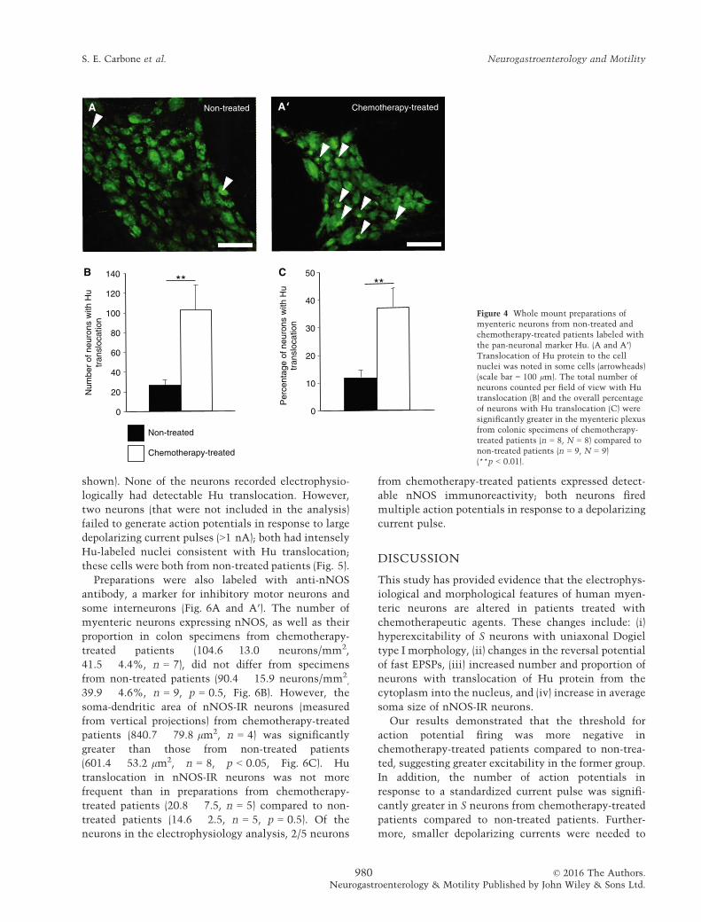
Preparations were labeled as whole mounts with a pan-
neuronal marker, Hu antibody (Fig. 4A and A’). The
total number of Hu-IR neurons/mm2 was similar in
chemotherapy-treated patients (263.6 � 17.8 neurons/
mm2, n = 8) and non-treated patients (216.1 � 26.2
neurons/mm2, n = 12, p = 0.1). In most cells, the Hu
antibody evenly labeled the cytoplasm and nucleus of
the cell but in some instances more intense staining of
the nucleus was noted, suggesting translocation of Hu
protein to the nucleus (Fig. 4A and A’, arrowheads).
The total number of neurons with Hu translocation
(Fig. 4B) and the percentage of neurons with Hu
translocation (Fig. 4C) were significantly greater in
preparations from chemotherapy-treated patients com-
pared to non-treated patients (chemotherapy-treated:
103 � 25 neurons/mm2, 37.2 � 7.0%, n = 8; non-trea-
ted: 26 � 5 neurons/mm2, 11.9 � 2.7%, n = 12,
p < 0.01). The occurrence of Hu translocation appeared
independent of age (linear regression analysis, data not
A B
–60
–50
–40
–30
–20
–10
0
Thr
esho
ld p
oten
tial (
mV
)
0
100
200
300
400
500
600
700
800
Thr
esho
ld c
urre
nt (
pA)
**
**Non-treated
Chemotherapy-treated
Figure 2 Myenteric S neurons from colonic
specimens of chemotherapy-treated patients
are hyperexcitable compared to neurons
from non-treated patients. (A) Neurons from
non-treated patients required a larger
depolarizing current to evoke action
potentials (rheobase) compared to neurons
from chemotherapy-treated patients. (B) The
action potential threshold was less negative
in neurons from non-treated patients (n = 5,
N = 5) compared to neurons from
chemotherapy-treated patients (n = 5,
N = 4, **p < 0.01, data expressed as
mean � SD).
Rev
ersa
l pot
entia
l (m
V)
*25 ms
–80mV
–70mV
–60mV
–50mV
A B
–50
–40
–30
–20
–10
0
Non-treated
Chemotherapy-treated
Figure 3 The reversal potentials of fast
excitatory postsynaptic potentials (fEPSPs)
were less negative in neurons from
chemotherapy-treated patients. (A) The
holding potential of the cell was set by
manual current clamp; fast EPSP amplitude
increased at hyperpolarized potentials. (B)
The extrapolated reversal potential of fEPSP
in neurons from non-treated patients (n = 4,
N = 4) was more negative than in cells from
chemotherapy-treated (n = 3, N = 3)
patients (*p < 0.05).
© 2016 The Authors.Neurogastroenterology & Motility Published by John Wiley & Sons Ltd.
979
Volume 28, Number 7, July 2016 Chemotherapy and human enteric neurons
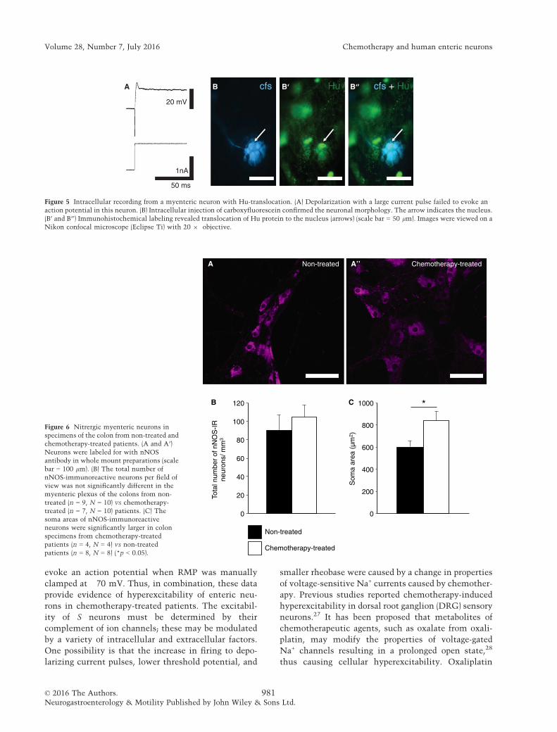
shown). None of the neurons recorded electrophysio-
logically had detectable Hu translocation. However,
two neurons (that were not included in the analysis)
failed to generate action potentials in response to large
depolarizing current pulses (>1 nA); both had intensely
Hu-labeled nuclei consistent with Hu translocation;
these cells were both from non-treated patients (Fig. 5).
Preparations were also labeled with anti-nNOS
antibody, a marker for inhibitory motor neurons and
some interneurons (Fig. 6A and A’). The number of
myenteric neurons expressing nNOS, as well as their
proportion in colon specimens from chemotherapy-
treated patients (104.6 � 13.0 neurons/mm2,
41.5 � 4.4%, n = 7), did not differ from specimens
from non-treated patients (90.4 � 15.9 neurons/mm2,
39.9 � 4.6%, n = 9, p = 0.5, Fig. 6B). However, the
soma-dendritic area of nNOS-IR neurons (measured
from vertical projections) from chemotherapy-treated
patients (840.7 � 79.8 lm2, n = 4) was significantly
greater than those from non-treated patients
(601.4 � 53.2 lm2, n = 8, p < 0.05, Fig. 6C). Hu
translocation in nNOS-IR neurons was not more
frequent than in preparations from chemotherapy-
treated patients (20.8 � 7.5, n = 5) compared to non-
treated patients (14.6 � 2.5, n = 5, p = 0.5). Of the
neurons in the electrophysiology analysis, 2/5 neurons
from chemotherapy-treated patients expressed detect-
able nNOS immunoreactivity; both neurons fired
multiple action potentials in response to a depolarizing
current pulse.
DISCUSSION
This study has provided evidence that the electrophys-
iological and morphological features of human myen-
teric neurons are altered in patients treated with
chemotherapeutic agents. These changes include: (i)
hyperexcitability of S neurons with uniaxonal Dogiel
type I morphology, (ii) changes in the reversal potential
of fast EPSPs, (iii) increased number and proportion of
neurons with translocation of Hu protein from the
cytoplasm into the nucleus, and (iv) increase in average
soma size of nNOS-IR neurons.
Our results demonstrated that the threshold for
action potential firing was more negative in
chemotherapy-treated patients compared to non-trea-
ted, suggesting greater excitability in the former group.
In addition, the number of action potentials in
response to a standardized current pulse was signifi-
cantly greater in S neurons from chemotherapy-treated
patients compared to non-treated patients. Further-
more, smaller depolarizing currents were needed to
B C
0
20
40
60
80
100
120
140
Num
ber
of n
euro
ns w
ith H
u tr
ansl
ocat
ion
0
10
20
30
40
50P
erce
ntag
e of
neu
rons
with
Hu
tran
sloc
atio
n
Non-treated
Chemotherapy-treated
** **
Non-treated Chemotherapy-treatedA A‘
Figure 4 Whole mount preparations of
myenteric neurons from non-treated and
chemotherapy-treated patients labeled with
the pan-neuronal marker Hu. (A and A’)
Translocation of Hu protein to the cell
nuclei was noted in some cells (arrowheads)
(scale bar = 100 lm). The total number of
neurons counted per field of view with Hu
translocation (B) and the overall percentage
of neurons with Hu translocation (C) were
significantly greater in the myenteric plexus
from colonic specimens of chemotherapy-
treated patients (n = 8, N = 8) compared to
non-treated patients (n = 9, N = 9)
(**p < 0.01).
© 2016 The Authors.Neurogastroenterology & Motility Published by John Wiley & Sons Ltd.
980
S. E. Carbone et al. Neurogastroenterology and Motility
evoke an action potential when RMP was manually
clamped at �70 mV. Thus, in combination, these data
provide evidence of hyperexcitability of enteric neu-
rons in chemotherapy-treated patients. The excitabil-
ity of S neurons must be determined by their
complement of ion channels; these may be modulated
by a variety of intracellular and extracellular factors.
One possibility is that the increase in firing to depo-
larizing current pulses, lower threshold potential, and
smaller rheobase were caused by a change in properties
of voltage-sensitive Na+ currents caused by chemother-
apy. Previous studies reported chemotherapy-induced
hyperexcitability in dorsal root ganglion (DRG) sensory
neurons.27 It has been proposed that metabolites of
chemotherapeutic agents, such as oxalate from oxali-
platin, may modify the properties of voltage-gated
Na+ channels resulting in a prolonged open state,28
thus causing cellular hyperexcitability. Oxaliplatin
50 ms
1nA
20 mV
cfs Hu cfs + HuA B B‘ B‘’
Figure 5 Intracellular recording from a myenteric neuron with Hu-translocation. (A) Depolarization with a large current pulse failed to evoke an
action potential in this neuron. (B) Intracellular injection of carboxyfluorescein confirmed the neuronal morphology. The arrow indicates the nucleus.
(B’ and B”) Immunohistochemical labeling revealed translocation of Hu protein to the nucleus (arrows) (scale bar = 50 lm). Images were viewed on a
Nikon confocal microscope (Eclipse Ti) with 20 9 objective.
A A’’
B C
0
20
40
60
80
100
120
Tota
l num
ber
of n
NO
S-I
R
neur
ons/
mm
3
*
Non-treated Chemotherapy-treated
0
200
400
600
800
1000
Som
a ar
ea (
µm2 )
Non-treated
Chemotherapy-treated
Figure 6 Nitrergic myenteric neurons in
specimens of the colon from non-treated and
chemotherapy-treated patients. (A and A’)
Neurons were labeled for with nNOS
antibody in whole mount preparations (scale
bar = 100 lm). (B) The total number of
nNOS-immunoreactive neurons per field of
view was not significantly different in the
myenteric plexus of the colons from non-
treated (n = 9, N = 10) vs chemotherapy-
treated (n = 7, N = 10) patients. (C) The
soma areas of nNOS-immunoreactive
neurons were significantly larger in colon
specimens from chemotherapy-treated
patients (n = 4, N = 4) vs non-treated
patients (n = 8, N = 8) (*p < 0.05).
© 2016 The Authors.Neurogastroenterology & Motility Published by John Wiley & Sons Ltd.
981
Volume 28, Number 7, July 2016 Chemotherapy and human enteric neurons
modulates Na+ channel properties in nerve conduction
studies especially in sensory nerve fibers in patients
suffering from post-chemotherapy neuropathy,
although it tends to decrease excitability after long-
term exposure in contrast to this study where
increased excitability of enteric neurons was evi-
dent.29,30 In clinical studies, abnormalities in Na+
currents were detected in 78% of patients with chronic
symptoms of chemotherapy-induced peripheral neu-
ropathy.30 While there is an evidence of hyperexcitabil-
ity of sensory neurons and peripheral nerves after
treatment with platinum-based chemotherapy,2
peripheral sensory neuropathy associated with 5-FU
treatment is rare31 and has not been extensively
studied. However, gastrointestinal dysmotility induced
by 5-FU treatment outlasts mucosal inflammation.32
Our study is the first demonstrating hyperexcitability
of enteric neurons from patients treated with 5-FU
alone or in combination with oxaliplatin or radiother-
apy. Enteric neuronal hyperexcitability, leading to
long-term intestinal dysfunction, has been demon-
strated in animal models of intestinal inflamma-
tion.24,25,33 The hyperexcitability of enteric neurons
observed in our study may contribute to the intestinal
dysfunctions associated with chemotherapy. Approxi-
mately, 40% of patients receiving standard-dose
chemotherapy and 100% of patients receiving high-
dose chemotherapy for gastrointestinal cancers experi-
ence pain, bloating, diarrhea and/or constipation
throughout the course of treatment.34 Gastrointestinal
side-effects are a predominant reason for dose limita-
tion, presenting a constant challenge for efficient and
tolerable treatment of CRC.8 Chronic gastrointestinal
side-effects can persist for more than 10 years after
treatment is stopped, greatly affecting patients’ quality
of life.10
Our study also suggested that fast EPSPs in myen-
teric neurons from chemotherapy-treated patients tend
to be slightly larger in amplitude (although this was
not significant with the small sample size). The
reversal potential of electrically-evoked fEPSPs was
less negative in neurons from chemotherapy-treated
patients compared to controls. Both of these changes to
cholinergic neurotransmission would be expected to
increase excitability of enteric neural circuits. They are
comparable with previously reported ectopic activity
observed at motor and autonomic neuromuscular
junctions after chemotherapy, which were attributed
to the changes in Na+ channel properties.28 The
mechanisms underlying changes in fast synaptic trans-
mission in enteric neurons remain to be elucidated.
They may result from direct effects of oxaliplatin or its
metabolites on ion channels, or be indirectly mediated
via other cells or mechanisms such as oxidative stress
or inflammation. In the case of peripheral neuropathy,
chemotherapeutics have multiple effects on neu-
rons.9,35
Hu immunolabeling has been used as a pan-neuronal
marker of the enteric nervous system in laboratory
animals36,37 and humans.38,39 Typically, the cytoplasm
and nucleus of neurons are uniformly labeled, with
little or no staining of axons. In our study, Hu
translocation from the cell body to the nucleus was
observed as increased intensity of nuclear immunoflu-
orescence. This phenomenon occurred in a higher
proportion of neurons in the colon of chemotherapy-
treated patients than non-treated patients. Hu proteins
are a family of RNA-binding proteins, expressed from
the Elavl (Elav-like) genes. Elavl1, which encodes HuR
protein is expressed in a wide range of tissues,40 but
Elavl2, Elavl3, and Elavl4 (which encode HuB, HuC,
and HuD proteins) are exclusively expressed in neu-
rons.41 Hu proteins regulate post-transcriptional gene
expression through alternate splicing and RNA edit-
ing42, and stabilize mRNA by binding to uracil-rich
RNA sequences (AU and GU). Continuous shuttling of
Hu proteins between the nucleus and cytoplasm has
been implicated in their critical roles in control of
nuclear export, stabilization, and cell survival.43 Their
role in post-transcriptional regulation adapts the pro-
teome to changes in environmental conditions. Like
other RNA-binding proteins, Hu proteins change their
expression level and subcellular location in response to
stressful stimuli. Toxic neuropathy, associated with
antiretroviral treatment, induced overexpression of
HuD in DRG and spinal cord neurons.44 Altered
intracellular localization of Hu proteins has been
reported in response to DNA damaging agents such
as ultraviolet light, heat shock, and actinomycin
D.45,46 Increased translocation of neuronal Hu proteins
from the cytoplasm to the nucleus was observed in
myenteric neurons after intestinal ischemia/reperfu-
sion damage.47
Studies in the central nervous system demonstrated
that Hu proteins promote the synthesis of the excita-
tory neurotransmitter glutamate48 and play important
roles in neural plasticity, maintenance, and survival.41
In our study, myenteric neurons with Hu translocation
to the nuclei failed to evoke action potentials, suggest-
ing that intracellular localization of Hu proteins plays
important role in cellular functioning.
In this study, nNOS-IR cells in the colon samples
from chemotherapy-treated patients were larger than
those in non-treated patients. This is similar to our
previous observations in chemotherapy-treated mice.14
It was notable that Hu translocation, induced by
© 2016 The Authors.Neurogastroenterology & Motility Published by John Wiley & Sons Ltd.
982
S. E. Carbone et al. Neurogastroenterology and Motility
chemotherapy, was less marked in nNOS-IR enteric
neurons than in cells that lacked nNOS. This suggests
that nNOS-expressing cells may be differentially
affected by oxaliplatin treatment, with a tendency to
increase in size rather than translocate Hu. Moreover,
functional properties of these neurons were different.
Unlike neurons with Hu translocation, nNOS-IR neu-
rons were hyperexcitable. In our recent study of mice
treated with oxaliplatin, nNOS-IR cells increased as a
proportion of all neurons, reflecting either selective
survival or changes in gene expression.14
CONCLUSIONS
Intracellular recording from human enteric neurons is
technically challenging; the number of cells that could
be recorded in this study was correspondingly small.
The technical difficulty may be why relatively few
studies have been published using this approach.22,23,49
Direct investigation of human enteric neurons,
in vitro, is invaluable as a tool to translate discoveries
in laboratory animals to human patients.50,51 To our
knowledge, this is the first paper to suggest functional
changes to human enteric neurons in a pathological
condition, using this direct recording approach. The
mechanisms underlying chemotherapy-induced
changes are still to be identified. Future studies in
animal models will be invaluable in the attempt to
understand the effects of chemotherapy on bowel
function.
FUNDING
This study is supported by Australian National Health & MedicalResearch Council Project grant 1032414.
DISCLOSURE
The authors do not have any potential conflicts to disclose.
AUTHOR CONTRIBUTION
SEC, SJHB, and KN were responsible for experimental design; SECand VJ performed all experiments and analysis; SEC drafted themanuscript; KN and SJHB obtained funding and supervised thestudy. The manuscript was edited and reviewed by all authors.
REFERENCES
1 Goodwin RA, Asmis TR. Overview ofsystemic therapy for colorectal cancer.ClinColonRectal Surg 2009; 22: 251–6.
2 Lehky TJ, Leonard GD, Wilson RH,Grem JL, Floeter MK. Oxaliplatin-induced neurotoxicity: acute hyperex-citability and chronic neuropathy.Muscle Nerve 2004; 29: 387–92.
3 Miltenburg NC, Boogerd W. Che-motherapy-induced neuropathy: acomprehensive survey. Cancer Treat
Rev 2014; 40: 872–82.4 Ewertz M, Qvortrup C, Eckhoff L.
Chemotherapy-induced peripheralneuropathy in patients treated withtaxanes and platinum derivatives.Acta Oncol 2015; 54: 587–91.
5 Argyriou AA, Cavaletti G, Briani C,Velasco R, Bruna J, Campagnolo M,Alberti P, Bergamo F et al. Clinicalpattern and associations of oxaliplatinacute neurotoxicity. Cancer 2012;119: 438–44.
6 Lucchetta M, Lonardi S, Bergamo F,Alberti P, Velasco R, Argyriou AA,Briani C, Bruna J et al. Incidence ofatypical acute nerve hyperexcitabilitysymptoms in oxaliplatin-treatedpatients with colorectal cancer. Can-
cer Chemother Pharmacol 2012; 70:889–902.
7 Stringer AM, Gibson RJ, Bowen JM,Logan RM, Ashton K, Yeoh ASJ, Al-Dasooqi N, Keefe DMK. Irinotecan-inducedmucositismanifesting as diar-rhoea corresponds with an amendedintestinal flora andmucin profile. Int JExp Pathol 2009; 90: 489–99.
8 Mcquade RM, Bornstein JC, NurgaliK. Anti-colorectal cancer chemother-apy-induced diarrhoea: current treat-ments and side-effects. IJCM 2014; 5:393–406.
9 Stojanovska V, Sakkal S, Nurgali K.Platinum-based chemotherapy: gas-trointestinal immunomodulation andenteric nervous system toxicity. Am JPhysiol Gastrointest Liver Physiol
2015; 308: G223–32.10 Denlinger CS, Barsevick AM. The
challenges of colorectal cancer sur-vivorship. J Natl Compr Canc Netw
2009; 7: 883–93.11 Keefe DMK. Gastrointestinal mucosi-
tis: a new biological model. SupportCare Cancer 2004; 12: 6–9.
12 Furness JB. The enteric nervous sys-tem and neurogastroenterology. Nat-ure 2012; 9: 286–94.
13 De Giorgio R, Barbara G, Furness JB,Tonini M. Novel therapeutic targetsfor enteric nervous system disorders.Trends Pharmacol Sci 2007; 28: 473–81.
14 Wafai L, Taher M, Jovanovska V,Bornstein JC, Dass CR, Nurgali K.Effects of oxaliplatin on mouse myen-teric neurons and colonic motility.Front Neurosci 2013; 7: 30. doi:10.3389/fnins.2013.00030.
15 Vera G, Castillo M, Cabezos PA,Chiarlone A, Mart�ın MI, Gori A,Pasquinelli G, Barbara G et al.
Enteric neuropathy evoked byrepeated cisplatin in the rat. Neuro-
gastroenterol Motil 2011; 23: 370–e163.
16 Vera G, L�opez-P�erez AE, Mart�ınez-Villaluenga M, Cabezos PA, Abalo R.X-ray analysis of the effect of the 5-HT3 receptor antagonist granisetronon gastrointestinal motility in ratsrepeatedly treated with the antitu-moral drug cisplatin. Exp Brain Res2014; 232: 2601–12.
17 OzakiA, SukamotoT. Improvement ofcisplatin-induced emesis and delayedgastric emptying by KB-R6933, a novel5-HT 3 receptor antagonist. Gen Phar-
macol 1999; 33: 283–8.18 Kurian SS, Ferri GL, De Mey J, Polak
JM. Immunocytochemistry of sero-tonin-containing nerves in thehuman gut. Histochemistry 1983;78: 523–9.
19 De Giorgio R, Bovara M, Barbara G,Canossa M, Sarnelli G, De Ponti F,
© 2016 The Authors.Neurogastroenterology & Motility Published by John Wiley & Sons Ltd.
983
Volume 28, Number 7, July 2016 Chemotherapy and human enteric neurons
Stanghellini V, Tonini M et al. Anti-HuD-induced neuronal apoptosisunderlying paraneoplastic gut dys-motility. Gastroenterology 2003;125: 70–9.
20 Wattchow D, Brookes S, Murphy E,Carbone S, De Fontgalland D, CostaM. Regional variation in the neuro-chemical coding of the myentericplexus of the human colon andchanges in patients with slow transitconstipation. NeurogastroenterolMotil 2008; 20: 1298–305.
21 Beyer J, Jabari S, Rau TT, NeuhuberW, Brehmer A. Substance P- andcholine acetyltransferase immunore-activities in somatostatin-containing,human submucosal neurons. His-
tochem Cell Biol 2013; 140: 157–67.22 Carbone SE, Jovanovska V, Nurgali K,
Brookes SJH. Human enteric neurons:morphological, electrophysiological,and neurochemical identification.Neurogastroenterol Motil 2014; 26:1812–6.
23 Brookes SJ, Ewart WR, Wingate DL.Intracellular recordings from myen-teric neurones in the human colon. JPhysiol 1987; 390: 305–18.
24 Linden DR, Sharkey KA, Mawe GM.Enhanced excitability of myentericAH neurones in the inflamed gui-nea-pig distal colon. J Physiol 2003;547: 589–601.
25 Nurgali K, Qu Z, Hunne B, ThackerM, Pontell L, Furness JB. Morpholog-ical and functional changes in guinea-pig neurons projecting to the ilealmucosa at early stages after inflam-matory damage. J Physiol 2011; 589:325–39.
26 Carbone SE, Wattchow DA, SpencerNJ, Brookes SJH. Loss of responsive-ness of circular smooth muscle cellsfrom the guinea pig ileum is associ-ated with changes in gap junctioncoupling. Am J Physiol Gastrointest
Liver Physiol 2012; 302: G1434–44.27 Adelsberger H, Quasthoff S, Gros-
skreutz J, Lepier A, Eckel F, LerschC. The chemotherapeutic oxaliplatinalters voltage-gated Na(+) channelkinetics on rat sensory neurons. EurJ Pharmacol 2000; 406: 25–32.
28 Webster RG, Brain KL, Wilson RH,Grem JL, Vincent A. Oxaliplatininduces hyperexcitability at motorand autonomic neuromuscular junc-tions through effects on voltage-gatedsodium channels. Br J Pharmacol2005; 146: 1027–39.
29 Park SB, Goldstein D, Lin CSY,Krishnan AV, Friedlander ML, Kier-
nan MC. Acute abnormalities of sen-sory nerve function associated withoxaliplatin-induced neurotoxicity. J
Clin Oncol 2009; 27: 1243–9.30 Krishnan AV, Goldstein D, Friedlan-
der M, Kiernan MC. Oxaliplatin-induced neurotoxicity and the devel-opment of neuropathy. Muscle Nerve2005; 32: 51–60.
31 Stein ME, Drumea K, Yarnitsky D,Benny A. A rare event of 5-fluorour-acil-associated peripheral neuropa-thy: a report of two patients. Am J
Clin Oncol 1998; 21: 248–9.32 Soares PMG, Mota JMSC, Gomes AS,
Oliveira RB, Assreuy AMS, BritoGAC, Santos AA, Ribeiro RA et al.
Gastrointestinal dysmotility in 5-fluorouracil-induced intestinalmucositis outlasts inflammatory pro-cess resolution. Cancer Chemother
Pharmacol 2008; 63: 91–8.33 Lomax AE, Mawe GM, Sharkey KA.
Synaptic facilitation and enhancedneuronal excitability in the submu-cosal plexus during experimental col-itis in guinea-pig. J Physiol 2005; 564:863–75.
34 Muss HB, Bynum DL. Adjuvantchemotherapy in older patients withstage III colon cancer: an underusedlifesaving treatment. J Clin Oncol
2012; 30: 2576–8.35 Jaggi AS, Singh N. Mechanisms in
cancer-chemotherapeutic drugs-ind-uced peripheral neuropathy. Toxicol-ogy 2012; 291: 1–9.
36 Lin Z, Gao N, Hu HZ, Liu S, Gao C,Kim G, Ren J, Xia Y et al. Immunore-activity ofHuproteins facilitates iden-tification of myenteric neurones inguinea-pig small intestine. Neurogas-troenterol Motil 2002; 14: 197–204.
37 Qu Z-D, Thacker M, Castelucci P,Bagy�anszki M, Epstein ML, FurnessJB. Immunohistochemical analysis ofneuron types in the mouse smallintestine. Cell Tissue Res 2008; 334:147–61.
38 Ganns D, Schr€odl F, Neuhuber W,Brehmer A. Investigation of generaland cytoskeletal markers to estimatenumbers and proportions of neuronsin the human intestine. Histol Histo-
pathol 2006; 21: 41–51.39 Murphy EMA, Defontgalland D,
Costa M, Brookes SJH, WattchowDA. Quantification of subclasses ofhuman colonic myenteric neurons byimmunoreactivity to Hu, cholineacetyltransferase and nitric oxide syn-thase. Neurogastroenterol Motil
2007; 19: 126–34.
40 Gorospe M. HuR in the mammaliangenotoxic response: post-transcrip-tional multitasking. Cell Cycle
2003; 2: 412–4.41 Doxakis E. RNA binding proteins: a
common denominator of neuronalfunction and dysfunction. Neurosci
Bull 2014; 30: 610–26.42 Hinman MN, Zhou H-L, Sharma A,
Lou H. All three RNA recognitionmotifs and the hinge region of HuCplay distinct roles in the regulation ofalternative splicing. Nucleic Acids
Res 2013; 41: 5049–61.43 Colombrita C, Silani V, Ratti A.
ELAV proteins along evolution: backto the nucleus? Mol Cell Neurosci
2013; 56: 447–55.44 Sanna MD, Quattrone A, Mello T,
Ghelardini C, Galeotti N. The RNA-binding protein HuD promotes spinalGAP43 overexpression in antiretrovi-ral-induced neuropathy. Exp Neurol2014; 261: 343–53.
45 Wang W, Furneaux H, Cheng H,Caldwell MC, Hutter D, Liu Y, Hol-brook N, Gorospe M et al. HuR reg-ulates p21 mRNA stabilization by UVlight. Mol Cell Biol 2000; 20: 760–9.
46 Mazan-Mamczarz K, Galb�an S, L�opezde Silanes I, Martindale JL, Atasoy U,Keene JD, Gorospe M. RNA-bindingprotein HuR enhances p53 transla-tion in response to ultraviolet lightirradiation. Proc Natl Acad Sci USA
2003; 100: 8354–9.47 Rivera LR, Thacker M, Pontell L, Cho
H-J, Furness JB. Deleterious effectsof intestinal ischemia/reperfusioninjury in the mouse enteric nervoussystem are associated with proteinnitrosylation. Cell Tissue Res 2011;344: 111–23.
48 Ince-Dunn G, Okano HJ, Jensen KB,Park WY, Zhong R, Ule J, Mele A, FakJJ et al. Neuronal Elav-like (Hu) pro-teins regulate RNA splicing andabundance to control glutamatelevels and neuronal excitability. Neu-ron 2012; 75: 1067–80.
49 Maruyama T. Two types of spikegeneration of human Auerbach’splexus cells in culture. Neurosci Lett1981; 25: 143–8.
50 Sanger GJ, Broad J, Kung V, KnowlesCH. Translational neuropharmacol-ogy: the use of human isolated gas-trointestinal tissues. Br J Pharmacol
2012; 168: 28–43.51 Mawe GM. Colitis-induced neuro-
plasticity disrupts motility in theinflamed and post-inflamed colon. J
Clin Invest 2015; 125: 949–55.
S. E. Carbone et al. Neurogastroenterology and Motility
© 2016 The Authors.Neurogastroenterology & Motility Published by John Wiley & Sons Ltd.
984
Related Documents