REVIEW ARTICLE Open Access Neurobiology and sleep disorders in cluster headache Mads Christian Johannes Barloese 1,2 Abstract Cluster headache is characterized by unilateral attacks of severe pain accompanied by cranial autonomic features. Apart from these there are also sleep-related complaints and strong chronobiological features. The interaction between sleep and headache is complex at any level and evidence suggests that it may be of critical importance in our understanding of primary headache disorders. In cluster headache several interactions between sleep and the severe pain attacks have already been proposed. Supported by endocrinological and radiological findings as well as the chronobiological features, predominant theories revolve around central pathology of the hypothalamus. We aimed to investigate the clinical presentation of chronobiological features, the presence of concurrent sleep disorders and the relationship with particular sleep phases or phenomena, the possible role of hypocretin as well as the possible involvement of cardiac autonomic control. We conducted a questionnaire survey on 275 cluster headache patients and 145 controls as well an in-patient sleep study including 40 CH-patients and 25 healthy controls. The findings include: A distinct circannual connection between cluster occurrence and the amount of daylight, substantially poorer sleep quality in patients compared to controls which was present not only inside the clusters but also outside, affected REM-sleep in patients without a particular temporal connection to nocturnal attacks, equal prevalence of sleep apnea in both patient and control groups, reduced levels of hypocretin-1 in the cerebrospinal fluid of patients and finally a blunted response to the change from supine to tilted position in the head-up tilt table test indicating a weakened sympathoexcitatory or stronger parasympathetic drive. Overall, these findings support a theory of involvement of dysregulation in hypothalamic and brainstem nuclei in cluster headache pathology. Further, it is made plausible that the headache attacks are but one aspect of a more complex syndrome of central dysregulation manifesting as sleep-related complaints, sub-clinical autonomic dysregulation and of course the severe attacks of unilateral headache. Future endeavors should focus on pathological changes which persist in the attack-free periods but also heed the possibility of long-lived, cluster-induced pathology. Introduction One of the earliest descriptions of cluster headache (CH) in medical literature is provided by 17th century Dutch surgeon and mayor of Amsterdam Nicolaes Tulp (Famously depicted in Rembrandt’ s The Anatomy Lesson of Dr Nicolaes Tulp.) [1]: “. . . in the beginning of the summer season, [Isaak van Halmaal] was afflicted with a very severe headache, oc- curring and disappearing daily on fixed hours, with such an intensity that he often assured me that he could not bear the pain anymore or he would succumb shortly. For rarely it lasted longer than two hours. And the rest of the day there was no fever, nor indisposition of the urine, nor any infirmity of the pulse. But this recurring pain lasted until the fourteenth day . . .” There can be little doubt that the patient described was suffering from arguably the most painful condition known in medicine [2]. With its stereotypical presenta- tion and the ease with which the diagnosis can be made (even based on a short historical account), it may seem puzzling how CH can remain underdiagnosed, under- treated and so enigmatic in nature. Accounts of the pre- dictable attack patterns and how oxygen, almost by miracle, alleviates the headache have fascinated many, but true progress in our understanding of the disorder is lacking. Correspondence: [email protected] 1 Danish Headache Center, Glostrup Hospital, Nordre Ringvej 57, Glostrup DK-2600, Denmark 2 Department of Clinical Physiology, Frederiksberg and Bispebjerg Hospitals, Nordre Fasanvej 57, DK-2000 Frederiksberg, Denmark © 2015 Barloese. Open Access This article is distributed under the terms of the Creative Commons Attribution 4.0 International License (http://creativecommons.org/licenses/by/4.0/), which permits unrestricted use, distribution, and reproduction in any medium, provided you give appropriate credit to the original author(s) and the source, provide a link to the Creative Commons license, and indicate if changes were made. Barloese The Journal of Headache and Pain (2015) 16:78 DOI 10.1186/s10194-015-0562-0

Welcome message from author
This document is posted to help you gain knowledge. Please leave a comment to let me know what you think about it! Share it to your friends and learn new things together.
Transcript

REVIEW ARTICLE Open Access
Neurobiology and sleep disorders in clusterheadacheMads Christian Johannes Barloese1,2
Abstract
Cluster headache is characterized by unilateral attacks of severe pain accompanied by cranial autonomic features.Apart from these there are also sleep-related complaints and strong chronobiological features. The interactionbetween sleep and headache is complex at any level and evidence suggests that it may be of critical importance inour understanding of primary headache disorders. In cluster headache several interactions between sleep and thesevere pain attacks have already been proposed. Supported by endocrinological and radiological findings as well asthe chronobiological features, predominant theories revolve around central pathology of the hypothalamus. Weaimed to investigate the clinical presentation of chronobiological features, the presence of concurrent sleepdisorders and the relationship with particular sleep phases or phenomena, the possible role of hypocretin as well asthe possible involvement of cardiac autonomic control. We conducted a questionnaire survey on 275 clusterheadache patients and 145 controls as well an in-patient sleep study including 40 CH-patients and 25 healthycontrols. The findings include: A distinct circannual connection between cluster occurrence and the amount ofdaylight, substantially poorer sleep quality in patients compared to controls which was present not only inside theclusters but also outside, affected REM-sleep in patients without a particular temporal connection to nocturnalattacks, equal prevalence of sleep apnea in both patient and control groups, reduced levels of hypocretin-1 in thecerebrospinal fluid of patients and finally a blunted response to the change from supine to tilted position in thehead-up tilt table test indicating a weakened sympathoexcitatory or stronger parasympathetic drive. Overall, thesefindings support a theory of involvement of dysregulation in hypothalamic and brainstem nuclei in clusterheadache pathology. Further, it is made plausible that the headache attacks are but one aspect of a more complexsyndrome of central dysregulation manifesting as sleep-related complaints, sub-clinical autonomic dysregulationand of course the severe attacks of unilateral headache. Future endeavors should focus on pathological changeswhich persist in the attack-free periods but also heed the possibility of long-lived, cluster-induced pathology.
IntroductionOne of the earliest descriptions of cluster headache(CH) in medical literature is provided by 17th centuryDutch surgeon and mayor of Amsterdam NicolaesTulp (Famously depicted in Rembrandt’s The AnatomyLesson of Dr Nicolaes Tulp.) [1]:“. . . in the beginning of the summer season, [Isaak van
Halmaal] was afflicted with a very severe headache, oc-curring and disappearing daily on fixed hours, with suchan intensity that he often assured me that he could notbear the pain anymore or he would succumb shortly.
For rarely it lasted longer than two hours. And the restof the day there was no fever, nor indisposition of theurine, nor any infirmity of the pulse. But this recurringpain lasted until the fourteenth day . . .”There can be little doubt that the patient described
was suffering from arguably the most painful conditionknown in medicine [2]. With its stereotypical presenta-tion and the ease with which the diagnosis can be made(even based on a short historical account), it may seempuzzling how CH can remain underdiagnosed, under-treated and so enigmatic in nature. Accounts of the pre-dictable attack patterns and how oxygen, almost bymiracle, alleviates the headache have fascinated many,but true progress in our understanding of the disorder islacking.
Correspondence: [email protected] Headache Center, Glostrup Hospital, Nordre Ringvej 57, GlostrupDK-2600, Denmark2Department of Clinical Physiology, Frederiksberg and Bispebjerg Hospitals,Nordre Fasanvej 57, DK-2000 Frederiksberg, Denmark
© 2015 Barloese. Open Access This article is distributed under the terms of the Creative Commons Attribution 4.0International License (http://creativecommons.org/licenses/by/4.0/), which permits unrestricted use, distribution, andreproduction in any medium, provided you give appropriate credit to the original author(s) and the source, provide alink to the Creative Commons license, and indicate if changes were made.
Barloese The Journal of Headache and Pain (2015) 16:78 DOI 10.1186/s10194-015-0562-0
CH is a primary headache disorder belonging to thediagnostic group known as the trigeminal autonomiccephalalgias (TAC’s) [2]. These headaches share the com-mon features of short-lasting, severe, strictly unilateralpain in the distribution of the first division of the trigemi-nal cranial nerve. Attacks last from 15 to 180 min andsimultaneous activation of the trigeminal autonomic reflexproduces the characteristic cranial, autonomic, accom-panying symptoms (Table 1) [2]. Systemic manifestationsare also present as patients almost universally become agi-tated, restless and, as opposed to migraineurs, do not ex-perience worsening of the pain during movement, perhapsrather relief. Uniquely, CH patients describe their attacksto exhibit remarkable circadian and annual periodicity .The attacks are described to strike at predictable times ofthe day and the clusters (bouts) of these at specific timesof the year. However, there is no consensus as no twostudies have ever reached identical conclusions concern-ing these patterns [3–7].Diagnostically, CH exists in two forms: an episodic
(ECH) and a chronic variant (CCH), distinguished bythe duration of the attack-free periods [8]. This divisionis diagnostic and there is no evidence for a clear reflec-tion of prognosis, response to treatment or causativepathology. The social impact of CH is considerable [9]and it is associated with sizeable direct and indirect eco-nomic consequences [10]. Consequently, endeavors to
uncover the pathological mechanisms behind this disab-ling headache have intensified over the past two decades;regrettably, the challenges associated with investigatingthe transient, severe pain attacks accompanied by agita-tion have slowed progress. Firstly, it is difficult to cap-ture the attacks and clusters, secondly it may be difficultfor the patients to remain still, complicating measure-ments. Lastly, provoking the attacks may cause changesobscuring the findings themselves.
The chronobiological nature of cluster headacheChronobiology is the study of biological rhythms. Inhumans, by far the most noticeable rhythm is the diur-nal sleep-wake rhythm which roughly follows a 24-h pat-tern [11]. This, along with a multitude of other changesin the organism, is evoked by the light and dark periodsof day and night – a consequence of the 24 h. rotationof the earth around its own axis. These periods andother Zeitgebers entrain the master circadian clock lo-cated in the hypothalamic suprachiasmatic nucleus(SCN) [11]. Through the release of melatonin from thepineal gland the SCN commands the overall rhythm ofthe organism.CH is arguably the headache disorder which demon-
strates the strongest chronobiological characteristics.The attacks themselves have been described to be mostlyrelated to (nocturnal) sleep and to follow specific rhythmsthat often provide a high degree of predictability [7, 12].Nonetheless, numerous unresolved issues in our under-standing of the pathophysiology and the relation to circa-dian and sleep-wake regulation remain. The pioneers ofCH chronobiology described the circadian and annualrhythmicity [5, 6], but it is unclear whether the biphasicsignal of cluster occurrence in ECH is a function of thesolstices, the equinoxes or something else [3, 7]. Further, itappears that the circadian periodicity may be influencedby cultural factors but the precise mechanisms remainobscured [3, 6, 7].
Cluster headache is a sleep-related headacheFor reasons which are partly unknown headache andsleep share an especially close relationship, as evidencedby a dense anatomical and physiological overlap in thecentral nervous system (CNS) [13] but also by a high de-gree of co-occurrence of sleep problems and headache[14]. To reflect this interesting, yet poorly understoodinteraction, migraine, hypnic headache, chronic paroxys-mal hemicranias and CH are all classified as “sleep-relatedheadaches” in the International Classification of SleepDisorders [15].Inspired by anecdotal and clinical experience, sleep
studies in CH have been conducted but results are diver-ging [12]. Anecdotally, patients awakened by nocturnalattacks often lucidly recall dreams and typically report
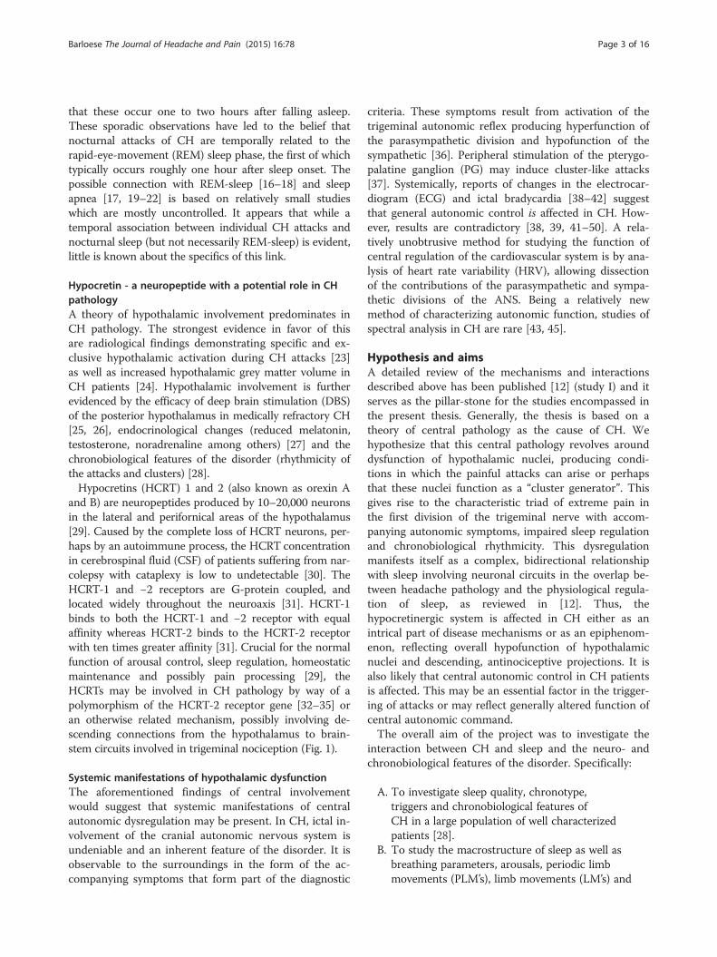
Table 1 Diagnostic criteria for cluster headache according tothe ICHD-2 [2]
Cluster headache
A. At least 5 attacks fulfilling criteria B-D
B. Severe or very severe unilateral orbital, supraorbital and/or temporalpain lasting 15–180 min if untreated
C. Headache is accompanied by at least one of the following:
1. Ipsilateral conjunctival injection and/or lacrimation
2. Ipsilateral nasal congestion and/or rhinorrhoea
3. Ipsilateral eyelid oedema
4. Ipsilateral forehead and facial sweating
5. Ipsilateral miosis and/or ptosis
6. A sense of restlessness or agitation
D. Attacks have a frequency from one every other day to 8 per day
E. Not attributed to another disorder
Episodic cluster headache
A. Diagnostic criteria A-E for cluster headache
B. At least two cluster periods lasting 7–365 days and separated bypain-free remission periods of≥ 1 month
Chronic cluster headache
A. Diagnostic criteria A-E for cluster headache
B. Attacks recur over > 1 year without remission periods or withremission periods lasting < 1 month.
Barloese The Journal of Headache and Pain (2015) 16:78 Page 2 of 16
that these occur one to two hours after falling asleep.These sporadic observations have led to the belief thatnocturnal attacks of CH are temporally related to therapid-eye-movement (REM) sleep phase, the first of whichtypically occurs roughly one hour after sleep onset. Thepossible connection with REM-sleep [16–18] and sleepapnea [17, 19–22] is based on relatively small studieswhich are mostly uncontrolled. It appears that while atemporal association between individual CH attacks andnocturnal sleep (but not necessarily REM-sleep) is evident,little is known about the specifics of this link.
Hypocretin - a neuropeptide with a potential role in CHpathologyA theory of hypothalamic involvement predominates inCH pathology. The strongest evidence in favor of thisare radiological findings demonstrating specific and ex-clusive hypothalamic activation during CH attacks [23]as well as increased hypothalamic grey matter volume inCH patients [24]. Hypothalamic involvement is furtherevidenced by the efficacy of deep brain stimulation (DBS)of the posterior hypothalamus in medically refractory CH[25, 26], endocrinological changes (reduced melatonin,testosterone, noradrenaline among others) [27] and thechronobiological features of the disorder (rhythmicity ofthe attacks and clusters) [28].Hypocretins (HCRT) 1 and 2 (also known as orexin A
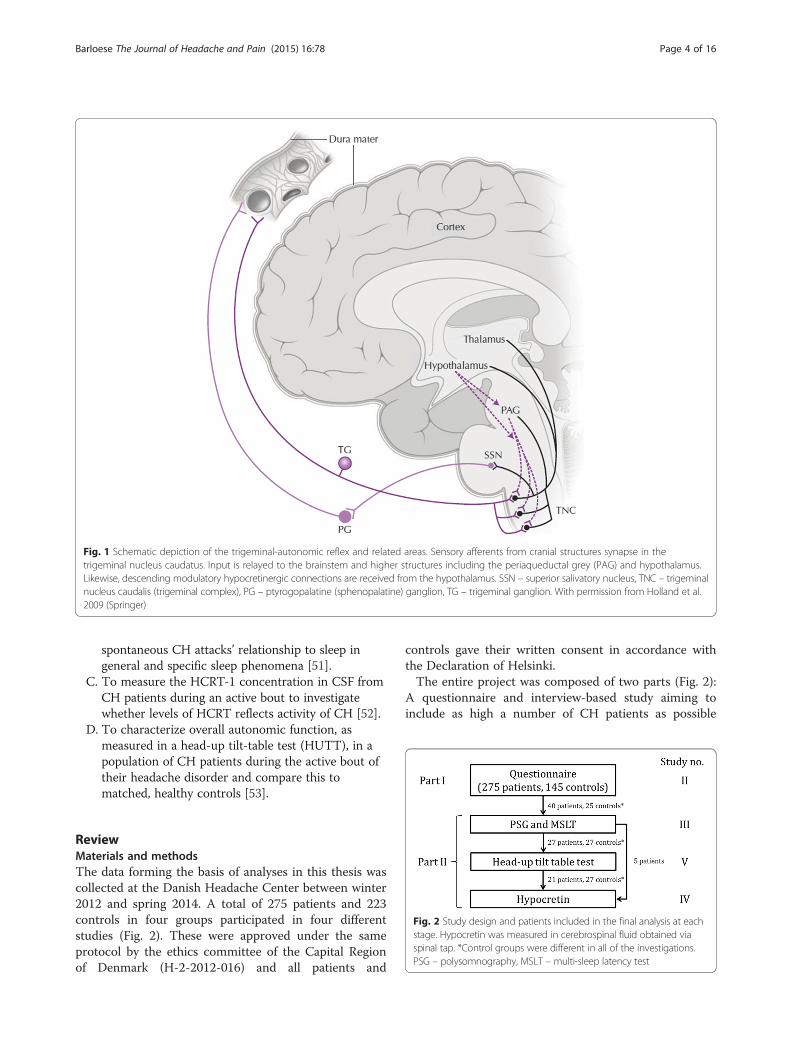
and B) are neuropeptides produced by 10–20,000 neuronsin the lateral and perifornical areas of the hypothalamus[29]. Caused by the complete loss of HCRT neurons, per-haps by an autoimmune process, the HCRT concentrationin cerebrospinal fluid (CSF) of patients suffering from nar-colepsy with cataplexy is low to undetectable [30]. TheHCRT-1 and −2 receptors are G-protein coupled, andlocated widely throughout the neuroaxis [31]. HCRT-1binds to both the HCRT-1 and −2 receptor with equalaffinity whereas HCRT-2 binds to the HCRT-2 receptorwith ten times greater affinity [31]. Crucial for the normalfunction of arousal control, sleep regulation, homeostaticmaintenance and possibly pain processing [29], theHCRTs may be involved in CH pathology by way of apolymorphism of the HCRT-2 receptor gene [32–35] oran otherwise related mechanism, possibly involving de-scending connections from the hypothalamus to brain-stem circuits involved in trigeminal nociception (Fig. 1).
Systemic manifestations of hypothalamic dysfunctionThe aforementioned findings of central involvementwould suggest that systemic manifestations of centralautonomic dysregulation may be present. In CH, ictal in-volvement of the cranial autonomic nervous system isundeniable and an inherent feature of the disorder. It isobservable to the surroundings in the form of the ac-companying symptoms that form part of the diagnostic
criteria. These symptoms result from activation of thetrigeminal autonomic reflex producing hyperfunction ofthe parasympathetic division and hypofunction of thesympathetic [36]. Peripheral stimulation of the pterygo-palatine ganglion (PG) may induce cluster-like attacks[37]. Systemically, reports of changes in the electrocar-diogram (ECG) and ictal bradycardia [38–42] suggestthat general autonomic control is affected in CH. How-ever, results are contradictory [38, 39, 41–50]. A rela-tively unobtrusive method for studying the function ofcentral regulation of the cardiovascular system is by ana-lysis of heart rate variability (HRV), allowing dissectionof the contributions of the parasympathetic and sympa-thetic divisions of the ANS. Being a relatively newmethod of characterizing autonomic function, studies ofspectral analysis in CH are rare [43, 45].
Hypothesis and aimsA detailed review of the mechanisms and interactionsdescribed above has been published [12] (study I) and itserves as the pillar-stone for the studies encompassed inthe present thesis. Generally, the thesis is based on atheory of central pathology as the cause of CH. Wehypothesize that this central pathology revolves arounddysfunction of hypothalamic nuclei, producing condi-tions in which the painful attacks can arise or perhapsthat these nuclei function as a “cluster generator”. Thisgives rise to the characteristic triad of extreme pain inthe first division of the trigeminal nerve with accom-panying autonomic symptoms, impaired sleep regulationand chronobiological rhythmicity. This dysregulationmanifests itself as a complex, bidirectional relationshipwith sleep involving neuronal circuits in the overlap be-tween headache pathology and the physiological regula-tion of sleep, as reviewed in [12]. Thus, thehypocretinergic system is affected in CH either as anintrical part of disease mechanisms or as an epiphenom-enon, reflecting overall hypofunction of hypothalamicnuclei and descending, antinociceptive projections. It isalso likely that central autonomic control in CH patientsis affected. This may be an essential factor in the trigger-ing of attacks or may reflect generally altered function ofcentral autonomic command.The overall aim of the project was to investigate the
interaction between CH and sleep and the neuro- andchronobiological features of the disorder. Specifically:
A. To investigate sleep quality, chronotype,triggers and chronobiological features ofCH in a large population of well characterizedpatients [28].
B. To study the macrostructure of sleep as well asbreathing parameters, arousals, periodic limbmovements (PLM’s), limb movements (LM’s) and
Barloese The Journal of Headache and Pain (2015) 16:78 Page 3 of 16
spontaneous CH attacks’ relationship to sleep ingeneral and specific sleep phenomena [51].
C. To measure the HCRT-1 concentration in CSF fromCH patients during an active bout to investigatewhether levels of HCRT reflects activity of CH [52].
D. To characterize overall autonomic function, asmeasured in a head-up tilt-table test (HUTT), in apopulation of CH patients during the active bout oftheir headache disorder and compare this tomatched, healthy controls [53].
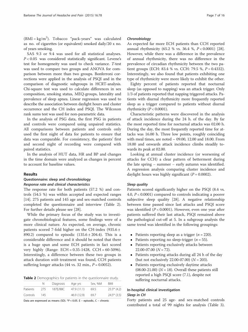
ReviewMaterials and methodsThe data forming the basis of analyses in this thesis wascollected at the Danish Headache Center between winter2012 and spring 2014. A total of 275 patients and 223controls in four groups participated in four differentstudies (Fig. 2). These were approved under the sameprotocol by the ethics committee of the Capital Regionof Denmark (H-2-2012-016) and all patients and
controls gave their written consent in accordance withthe Declaration of Helsinki.The entire project was composed of two parts (Fig. 2):
A questionnaire and interview-based study aiming toinclude as high a number of CH patients as possible
Fig. 2 Study design and patients included in the final analysis at eachstage. Hypocretin was measured in cerebrospinal fluid obtained viaspinal tap. *Control groups were different in all of the investigations.PSG – polysomnography, MSLT – multi-sleep latency test
Fig. 1 Schematic depiction of the trigeminal-autonomic reflex and related areas. Sensory afferents from cranial structures synapse in thetrigeminal nucleus caudatus. Input is relayed to the brainstem and higher structures including the periaqueductal grey (PAG) and hypothalamus.Likewise, descending modulatory hypocretinergic connections are received from the hypothalamus. SSN – superior salivatory nucleus, TNC – trigeminalnucleus caudalis (trigeminal complex), PG – ptyrogopalatine (sphenopalatine) ganglion, TG – trigeminal ganglion. With permission from Holland et al.2009 (Springer)
Barloese The Journal of Headache and Pain (2015) 16:78 Page 4 of 16

(part 1) and an in-hospital clinical investigation includ-ing 40 CH patients (part 2). All patients completed thesame questionnaire and interview which also providedthe clinical characterization of the 40 patients participat-ing in part 2.
Part one: Cross-sectional questionnaire and interviewPatientsPatients diagnosed with ECH or CCH were recruited froma register of present and former contacts at the DanishHeadache Center. Additionally, a notice was posted in thenewsletter and on the website of the Danish patientorganization for CH. Other headache clinics in Denmarkwere contacted and patients receiving treatment at theseclinics were referred to the study if they were interested.Patients were included if they were between 18 and 65 yearsold, had been diagnosed with ECH or CCH (ICHD-IIcriteria) and were able to tell CH-attacks apart from othertypes of headache. Patients were excluded if they had beendiagnosed with another primary or secondary chronicheadache (≥14 days/month) or did not speak and under-stand Danish. All patients’ diagnoses were verified by head-ache specialists at the Danish Headache Center.
ControlsControls were recruited via notices posted on the internetand in work places and sport clubs in the Capital Regionof Denmark. Controls were matched for sex and age andwere required to be between 18 and 65 years old andheadache-free (≤1 day of headache/month). They wereexcluded if they had severe symptoms of sleep disorders.
QuestionnaireA questionnaire composed of already validated sections(Pittsburgh Sleep Quality Index (PSQI), Morningness-Eveningness Questionnaire (MEQ)) and new questionsconcerning headache characteristics, life style, impair-ment and others developed by MB and RJ was sent topatients and controls. In the final phase of developmentthe questionnaire was assessed for content and face val-idity by an expert panel comprised of 10 senior headacheexperts (unpublished data). They were asked to evaluatethe questions: Are the instructions clear, is the question-naire coherent, are the questions relevant, is the ques-tionnaire logically divided into parts, are the questionsformulated clearly. The questionnaire was also testedwith a randomly selected population of 10 CH patientsrepresenting different ages, sexes and diagnoses (ECHand CCH) and comments and suggestions were imple-mented. The paper questionnaire contained 362 questionsin 7 sections: Headache diagnosis, headache burden, treat-ment, sleep, work, lifestyle habits, and physical activity. Itwas estimated that it took the patients roughly one hourto complete the questionnaire. Following completion of
the questionnaire, answers were verified and ambiguitiesruled out by a structured interview conducted by a phys-ician or a trained medical student. If the patient or controlinvited to participate did not respond within 21 days theywere contacted again by letter and/or telephone and en-couraged to complete the questionnaire.The PSQI [54] is a validated [55] 19-item measure of
subjective sleep quality during the past month. The sumof seven component scores provides a global score, ahigher value reflecting poorer sleep quality. A global scoregreater than 5 yields a high sensitivity and specificity indistinguishing “good and poor sleepers” [54].The MEQ [56] uses 19 multiple-choice items to chrono-
type patients into five categories: Definite morning, mod-erate morning, intermediate, moderate evening, definiteevening. The questionnaire has been validated in middleaged populations [57]. Chronotype reflects at what time ofthe day a person is active or inactive, often reduced tosleeping habits only, i.e. “lark” or “owl”.To characterize rhythmicity of attacks and clusters,
patients were asked to report the hours of the day or themonths of the year where these were most likely tooccur. They also had the option to report no rhythmi-city. For further details please see [28].
Part two: In-hospital investigation of sleep, hypocretinand autonomic functionForty patients underwent clinical examination, routineblood sampling, polysomnography (PSG) and multi-sleeplatency test (MSLT). 29 of the patients completed the tilt-table test and 27 the spinal tap procedure. All investi-gations were done at Glostrup Hospital, Departments ofNeurology, Neurophysiology and Diagnostics, and at theCoordinating Research Centre, Dept. of Clinical Physiologyand Nuclear Medicine, Frederiksberg Hospital.
PatientsPatients for part 2 were recruited as described above andwere investigated during the active cycle of the headachedisorder (1–8 attacks/day over the past week) and atleast two weeks into the bout. Exclusion criteria wereother chronic primary or secondary headaches and ser-ious somatic or psychiatric illness. If patients were onprophylactic medication this was kept stable for at leastseven days prior to investigation. Patients would attemptto treat their attacks using oxygen but were allowed touse other medication such as injectable or nasal triptans,as a rescue.
ControlsThree separate populations of controls were used:
Sleep investigation For study III 25 controls with an age,sex and BMI-makeup similar to the patients were included.
Barloese The Journal of Headache and Pain (2015) 16:78 Page 5 of 16

Controls were recruited through www.forsoegsperson.dk,a website for healthy volunteers, and did not suffer fromchronic headaches, sleep disorders or any other healthproblem (one control had mild, controlled hypertensionand hypercholesterolemia) as concluded by interview,examination and questionnaires. For further details pleasesee [51].
Hypocretin samples For study IV 27 healthy controlswithout headache-, sleep- or other neurological disor-ders were included from a prior study [58]. Twelve ofthe subjects were recruited through advertisement forhealthy volunteers (www.forsoegsperson.dk) and clinicaland neurological examination by a physician was con-ducted. The remaining 15 subjects were referred for sub-jective sleep-related complaints but were found healthy byexperienced sleep specialists by interviews, normal clinicaland neurological findings, PSG and MSLT. Controls werenot matched according to age or sex as these factors maynot influence HCRT-levels [59].
Head-up tilt table test For study V 27 controls matchedaccording to age, sex, BMI were included. All controlswere interviewed to ensure they were healthy and did notsuffer from disorders including primary headaches. Forfurther details please see [53].
MethodsPolysomnographic recordingsRecordings took place during admission at the DanishCenter for Sleep Medicine and Department of Neurologyat Glostrup Hospital, Denmark. PSG recordings wereperformed and scored in accordance with the AASMstandard [15]. Final assessment and possible sleep dis-order diagnoses were made by senior doctors, special-izing in sleep medicine according to the ICSD-2 [15].Two nights of PSG-recordings were made for patientsand one night for controls. MSLT was done after thelast night of sleep recording. Additionally, for detailedmethods please see [51].
Measurement of cerebrospinal fluid hypocretin-110 mL CSF was collected by the spinal tap procedure inthe attack-free state between 08.00 and 12:00. Hypocretin-1 was analyzed in crude CSF by radioimmunoassay fromPhoenix Pharmaceuticals (Belmont, CA, USA). We usedthe same methodology as in a previous study [58]. Assayquality was monitored by the internal positive controlsample included in the assay kit. Previously used group-ings of CSF intervals for HCRT-1 concentrations (low(≤110 pg/ml), intermediate (>110 ≤ 200 pg/ml), and nor-mal (>200 pg/ml)) were not strictly applied in this sampleas these are most relevant in the diagnosis of narcolepsy[30, 60, 61]. For detailed methods please see [52].
Head-up tilt table testAll tests were performed in the fasting state between08.00 and 14.00 h. at standard room temperature. After10 min. of supine rest, baseline data were acquired duringa further 10 min. of rest, where the subjects refrained fromspeaking and from unnecessary movements. The subjectswere then loosely strapped to an electrically driven tilttable and tilted to a 60° head-up position (HUT) within10 s. and stayed in this position for at least 10 min.if intolerable symptoms did not appear. RR-intervalsand blood pressure (BP) were measured continuouslyfrom a bipolar 2-channel ECG and by Finometer equip-ment (Finapres Medical Systems BV, Amsterdam, TheNetherlands), respectively. Baseline values of HR, SBP anddiastolic blood pressure (DBP) were calculated as meanvalues from the 30 s. preceding tilt in the supine pos-ition and during 30 s. obtained in the 5th and 10thmin. of HUT. Analysis of HRV was performed according tocurrent guidelines [62] using share-ware (Kubios, vers.2.0, kubios.uef.fi). For detailed methods please see[53].
Data and statisticsCharacterization of the patients for all four studies wasbased on the questionnaire and interview. A CH indexwas calculated by the following equation: Attacks perday x hours per attack x days per cluster x clusters peryear and is a measure of the total time/year that the pa-tient has cluster headache. In this specific calculation,for CCH patients, number of clusters/year was set atone and cluster duration was set at 365 days. The motiv-ation for calculating this index is to provide a singlenumber which reflects the amount of headache, as thisis sometimes not completely clear, taking into consider-ation clusters, cluster duration, attacks and attack dur-ation. Further, we wanted to characterize the patientsbeyond the dichotomy of episodic-chronic as some epi-sodic patients in fact may experience far more headachethan some chronic.Apart from diagnosis (ECH, CCH) and sex, the
patients were stratified based on self-reported character-istics including: “Annual rhythmicity” – clusters occur atthe same time each year, “diurnal rhythmicity” – attacksoccur at the same time each day, “sleep attacks mostly” –the patient reports that the majority of attacks occur dur-ing sleep, “sleep and awake or mostly awake” – the patientreports no predominance of attacks during sleep, attacksduring PSG monitoring and whether the patients primar-ily suffered attacks during sleep or both during sleep andwake. The data gathered in the questionnaire andinterview study allowed for even finer distinction ofpatients and an analysis of the exact times patientsreported attacks was made: 08.00-21.00, 22.00-07.00. BMIwas calculated from self-reported height and weight
Barloese The Journal of Headache and Pain (2015) 16:78 Page 6 of 16
(BMI = kg/m2). Tobacco “pack-years” was calculatedas no. of cigarettes (or equivalent) smoked daily/20 x no.of years smoking.SAS 9.3 or 9.4 was used for all statistical analyses.
P < 0.05 was considered statistically significant. Levene’stest for homogeneity was used to check variance. T-testwas used to compare two groups and ANOVA for com-parison between more than two groups. Bonferroni cor-rections were applied in the analysis of PSQI and in thecomparison of diagnostic subgroups in HCRT-analysis.Chi-square test was used to calculate differences in sexcomposition, smoking status, MEQ-groups, laterality andprevalence of sleep apnea. Linear regression was used todescribe the association between daylight hours and clusteroccurrence and the CH index and PSQI. The Wilcoxonrank sums test was used for non-parametric data.In the analysis of PSG data, the first PSG in patients
and controls were compared using unpaired statistics.All comparisons between patients and controls onlyused the first night of data for patients to ensure thatdata was comparable. For consistency, the patients’ firstand second night of recording were compared withpaired statistics.In the analysis of HUT data, HR and BP and changes
in the time domain were analyzed as changes in percentto account for baseline values.
ResultsQuestionnaire: sleep and chronobiologyResponse rate and clinical characteristicsThe response rate for both patients (57.2 %) and con-trols (54.5 %) was within accepted and expected ranges[14]. 275 patients and 145 age and sex-matched controlscompleted the questionnaire and interview (Table 2).For further details please see [28].While the primary focus of the study was to investi-
gate chronobiological features, some findings were of amore clinical nature. As expected, on average, chronicpatients scored 7-fold higher on the CH-index (935.6 ±890.2) compared to episodic (135.6 ± 204.4). This is aconsiderable difference and it should be noted that thereis a huge span and some ECH patients in fact scoredvery highly (Range: ECH = 0.35-1428, CCH = 60-5096).Interestingly, a difference between these two groups inattack duration with treatment was found, CCH patientssuffering longer attacks (44 vs. 32 min., P = 0.0052).
ChronobiologyAs expected far more ECH patients than CCH reportedannual rhythmicity (65.2 % vs. 36.4 %, P < 0.0001) [28].However, while there was a difference in the prevalenceof annual rhythmicity, there was no difference in theprevalence of circadian rhythmicity between the two pa-tient groups (ECH: 83.4 % vs. CCH: 79.5 %, P = 0.4332).Interestingly, we also found that patients exhibiting onetype of rhythmicity were more likely to exhibit the other.Eighty percent of patients reported that nocturnal
sleep (as opposed to napping) was an attack trigger. Only1/3 of patients reported that napping triggered attacks. Pa-tients with diurnal rhythmicity more frequently reportedsleep as a trigger compared to patients without diurnalrhythmicity (P < 0.0001).Characteristic patterns were discovered in the analysis
of attack incidence during the 24 h. of the day. By farthe most reported time for nocturnal attacks was 02.00 h.During the day, the most frequently reported time for at-tacks was 16.00 h. Three low points, roughly coincidingwith meal times, are noted – 09.00, 12.00 and 18.00. From18.00 and onwards attack incidence climbs steadily to-wards its peak at 02.00.Looking at annual cluster incidence (or worsening of
attacks for CCH) a clear pattern of betterment duringthe late spring – summer – early autumn was identified.A regression analysis comparing cluster incidence anddaylight hours was highly significant (P = 0.0002).
Sleep qualityPatients scored significantly higher on the PSQI (8.4 vs.4.0, P < 0.0001) compared to controls indicating a poorersubjective sleep quality [28]. A negative relationshipbetween time passed since last attacks and PSQI scorewas identified (P < 0.0001). However, even one year afterpatients suffered their last attack, PSQI remained abovethe pathological cut-off at 5. In a subgroup analysis thesame trend was identified in the following groupings:
� Patients reporting sleep as a trigger (n = 220).� Patients reporting no sleep-trigger (n = 55).� Patients reporting exclusively attacks between
22.00-07.00 (N = 72).� Patients reporting attacks during all 24 h of the day
(but not exclusively 22.00-07.00) (N = 203).� Patients reporting exclusively daytime attacks
(08.00-21.00) (N = 18). Overall these patients stillreported a high PSQI score (7.1), despite notsuffering nocturnal attacks.
In-hospital clinical investigationSleep in CHForty patients and 25 age- and sex-matched controlscontributed a total of 99 nights for analysis (Table 3).
Table 2 Demographics for patients in the questionnaire study.
N Diagnosis Age yrs Sex, %M BMI
Patients 275 187E/88C 47.9 (11.1) 69.5 25.5* (4.2)
Controls 145 46.9 (12.9) 69.7 24.5* (3.5)
Data are expressed as means (SD). *P < 0.05. E – episodic, C – chronic
Barloese The Journal of Headache and Pain (2015) 16:78 Page 7 of 16

Findings included lower REM-density (17.3 vs. 23.0 %,P = 0.0037) and latency (2.0 vs. 1.2 h., P = 0.0012) inpatients compared to controls [51]. Apart from theaffected REM-sleep, macrostructurally there was nodifference between patients and controls except forlower efficiency (77 vs. 87 %, P = 0.0026) and longer sleeplatency (0.56 vs. 0.18 h, P = 0.0057). There was no tem-poral association between REM-sleep, any other sleepstage or particular sleep-related events, including apneasand desaturations and observed, spontaneous nocturnalattacks (N = 45). Furthermore, patients had far fewerarousals (7.3 vs. 14.1, P = 0.0030). As opposed to all previ-ous studies no difference in prevalence of sleep apnea inpatients vs. matched controls (38 vs. 34 %, P = 0.64) wasidentified. However, average AHI in patients was numeric-ally higher (AHI 10.75 vs. 4.93).In one patient a striking rhythmicity of nocturnal at-
tacks was noticed (Fig. 3). This patient suffered a total of
nine attacks during the two nights of recording. Therewas no relation with any particular sleep stage, ratherthe attacks occurred roughly every 90 min. During night1, at 02.00, the patient wakes up but without an attack.
HypocretinIn total 26 patient and 27 control samples of CSF wereincluded in the HCRT-1 analysis (Table 3). A highlysignificant reduction of HCRT-1 levels in patientscompared to controls was identified (382 vs. 431 pg/ml,P = 0.0004) [52]. Both subgroups of CH patients (ECH(375 pg/ml, P = 0.0005) and CCH (389 pg/ml, P = 0.0221))were significantly reduced compared to controls (Fig. 4).There was no difference between the two subgroups ofpatients. However, on average, CCH patients had higherconcentrations and the range and standard deviation wasgreater (CCH: 291-480 ± 60 vs. ECH: 312-426 ± 38). Therewere no significant differences in HCRT-1 concentrations
Table 3 Clinical characteristics. Attack duration is for treated attacks. Bout duration and bouts/year only includes ECH
N Diag. Sex %M Age (yrs.) BMI CH duration(yrs.)
Attack duration(min.)
Attacks/day Bout duration(wks.)
Bouts/year
PSG & MSLT Patients 40 21E 19C 73 44.2 (11.2) 25.7 (4.0) 11.2 (7.4) 35.0 (33.1) 4.6 (1.9) 12.3 (20.3) 1.7 (1.1)
Controls 25 64 47.6 (12.1) 24.2 (3.4)
HCRT Patients 26 14E 12C 66 44.3 (11.5) 24.9 (3.8) 10.3 (5.9) 27.2 (18.2) 4.3 (1.8) 14.2 (8.8) 1.6 (0.9)
Controls 27 44 32.4 (9.5)
HUT Patients 27 14E 13C 70 44.9 (11.3) 25.4 (3.8) 11.3 (7.8) 37.2 (37.8) 4.4 (1.8) 12.7 (9.2) 1.7 (1.2)
Controls 27 70 49.0 (8.3) 24.4 (2.7)
Data is presented as means (SD). PSG – polysomnography, MSLT – multi-sleep latency test, HCRT – hypocretin, HUT – head-up tilt table test, E – episodic, C – chronic
Fig. 3 Hypnograms from night 1 (top) and 2 (bottom) from a patient suffering nine spontaneous CH attacks (arrows) during recordings. As isseen, the attacks occur in stages W, REM, N2 and N3 at remarkably regular intervals. With permission from Barloese et al. 2014 (Wiley) [51]
Barloese The Journal of Headache and Pain (2015) 16:78 Page 8 of 16

between patients dichotomized according to rhythmicityand sleep attacks.
Autonomic functionThere were no differences between patients and controlsin standard cardiovascular responses to tilt including HRand BP. However, patients did present a higher BPthroughout the test (Patients: 121–128/79–88 mmHg,controls: 107–115/56–68 mmHg, p < 0.01–0.0001) [63].In both the non-linear and frequency domain analyses,patients presented a blunted response to tilt. These dif-ferences were significant in the analysis of normalizedunits (HF n.u. and LF n.u.) and the LF/HF ratio as wellas the SD1/SD2 ratio. Within the CH population therewere no differences when dichotomizing according tosubdiagnosis, rhythmicity and sleep- or sleep and wakeattacks [63].
DiscussionIn the investigations made over the past 30 years severaltheories about sleep and nocturnal CH attacks have beenproposed including a strictly temporal connection withthe REM phase of sleep [17, 64] and association betweensleep apnea and CH [65]. While some case reports mayseem to provide evidence of such a direct, causal rela-tionships [66–68], it is likely that the interaction be-tween sleep and CH is more complex, multifaceted andindirect in nature.
Cluster headache is a chronobiological disorderCluster headache provides a unique opportunity to studya disorder which is present at very specific times of the
year and then spontaneously remits and is, at least withregards to the headache, completely absent for longperiods. Few other disorders show this kind of strongchronobiological features, and those that do have someinteresting similarities with CH, such as the rare lithiumresponsive [69], recurrent hypersomnia Kleine-Levinsyndrome [70]. While it is obvious and easy to establishthat the headache attacks completely remit outside ofthe cluster period in the episodic subforms, it is un-known whether underlying pathology remains present,perhaps manifesting in unknown ways.The present results suggest that there may be a long-
lived dysfunction present, which the majority of the timemanifests as poor sleep quality and at specific times ofthe year perhaps when entrainment by natural lightcues is weaker, results in periodic disinhibition of thetrigeminal autonomic reflex. The anatomical substratefor this interaction may be the complex brainstemand hypothalamic circuits where an overlap betweenheadache and sleep exists [13]. In susceptible individ-uals, at times of the year when Zeitgebers change orare weaker, resultant cluster penetration may involvemelatonin metabolism. Melatonin concentrations havebeen shown to be lowered and to exhibit a bluntednocturnal peak in CH patients regardless of clusterpresence [71–74], and when administered prophylac-tically it may have a slight, positive effect on the clus-ters [75]. The strong association between clusteroccurrence and the amount of daylight demonstratedin this study further strengthens a hypothesis of anintricate relationship between melatonin metabolismand CH. A possible connection with testosterone has
Fig. 4 Hypocretin-1 levels in patients and controls. ECH – episodic cluster headache, CCH – chronic cluster headache, CTRL – control, HCRT-1 –hypocretin 1
Barloese The Journal of Headache and Pain (2015) 16:78 Page 9 of 16

also been suggested which may be especially interest-ing considering REM-sleep’s effect on this hormone[76] and recent findings suggesting it may positivelyaffect antinociceptive signaling in the trigeminal com-plex [77].A PSQI score above 5 gives a high sensitivity and spe-
cificity in distinguishing “good and poor sleepers” [54].In the results presented here, sleep quality improves astime passes since the last attack but remains abnormaleven one year after the last attack, implying a permanentor long-lived dysfunction in sleep regulatory mecha-nisms. It also means that it is not only the nocturnalattacks themselves that in a direct manner disturb thesleep of CH patients, since sleep quality remains pooreven in the attack-free state. This is further evidenced bythe fact that the 18 patients reporting exclusively day-time attacks also reported poor sleep quality. Subjectivesleep quality may thus be an indirect measure of thepresence of an underlying pathological mechanism, andshould be investigated further. A systematic, detailed re-cording of sleep history may be useful in the clinicalevaluation of patients and sleep quality could be used toevaluate this sub-threshold presence of the cluster.The classification of patients as episodic or chronic
presents some problems unique to CH. For example,current diagnostic criteria [2, 8] do not specify whetherthe patient should be completely off preventive medica-tion to be classified as episodic. Further, many patientsclassified as episodic may be significantly more affectedby the headache than some patients fulfilling the criteriafor chronic CH. For research purposes, it seems that theusefulness of dichotomizing according to subdiagnosismay not be universal. These arguments were the mainreason for the development of the CH-index which re-flects the total time the patient has suffered from head-ache over the past year. As can be seen, the range ishuge and there is a significant overlap between ECH andCCH. Although the calculation is subject to recall bias,the index still provides one number reflecting theamount of headache the patient has, taking into accountattack frequency, attack duration and cluster duration.When based on prospective recordings the index mayprecisely and in a comparable manner reflect total head-ache burden.
A complex, bi-directional relationship with REM-sleepThe connection between CH and REM-sleep is the sub-ject of a long-running debate. Early studies indicated atemporal relationship which was supported by observa-tions that CH attacks typically occur 60–90 min. afterfalling asleep, coinciding with the first REM phase. Sev-eral reports, including the present results, now showthat there is no relation with REM-sleep for any of thesubdiagnoses [78, 79]. However, it is clear that REM-
sleep is affected in CH patients but the cause remainsunclear. Firstly, it must be taken into consideration thatthe homeostatic pressure for sleep and REM-sleep ismost likely affected in this patient group as a result ofnocturnal awakenings. Secondly, considering the de-scribed overlap of sleep and headache, it seems likelythat changes in hypothalamic and brainstem nuclei maydirectly or indirectly affect REM sleep. The ventrolateralgrey and lateral pontine tegmentum, receives hypocre-tinergic input, and may be an area of interest in thisregard [13]. Further, in the present results there was nodifference in the macrostructural composition of sleepbetween patients and controls apart from a lower REMdensity. Serotonergic and noradrenergic activity leads tosuppression of REM sleep [80, 81] and the noradrenergiclocus coeruleus and the serotonergic dorsal raphe nu-cleus are areas of direct anatomical overlap betweensleep regulation and headache [13]. With this in mind,the challenge is to dissect the contributions from achange in homeostatic pressure and a change in functionof the hypothalamic and brainstem nuclei involved inthe regulation of sleep and pathology of headache. Atpresent, our understanding of these circuits and interac-tions is not sufficient to make precise conclusions. Asingle case report hints at a fascinating change in sleeppatterns taking place before the cluster [82]. This find-ing, and the fact that subjective sleep quality is worseeven outside of clusters in ECH patients suggests thatCH is a syndrome in which changes first manifest asdysregulation of sleep and secondly as a destabilizationof trigeminal nociceptive processing.
Sleep disordered breathing is a common finding in menUp to 24 % of middle-aged males exhibit sleep disor-dered breathing (AHI ≥ 5) [83] and the studies suggest-ing an increased prevalence of sleep apnea in CH areuncontrolled [17, 20, 22] except two [19, 65]. Further, inearlier studies it is not always clear whether patientswere in active bout during investigation and one studyinvestigating the same patients inside and outside ofbout does not specify how many patients were investi-gated outside of bout and for how long the patients hadbeen attack-free [65]. All the patients included in thepresent study [51] were in active bout and we did notfind a significant difference between prevalence of sleepapnea in patients compared with controls. However, pa-tients did present more severe cases and a numericallyhigher apnea-hypopnea index (AHI), but it is entirelyconceivable that this may have been caused by themassive over-representation of smokers in the patientgroup. Our patients were only investigated during theactive part of the disorder and the possibility thathypothalamic dysregulation produces sleep apnea dur-ing the cluster period cannot be excluded. However,
Barloese The Journal of Headache and Pain (2015) 16:78 Page 10 of 16

it seems unlikely that the apnea event itself is thetrigger of nocturnal CH attacks, as no connection be-tween the apnea events and nocturnal attacks wereidentified. Further anecdotal evidence provides no in-dication that treatment of the apnea alleviates the head-aches [51, 65].
Fewer arousals is a common finding in headache disordersThe role arousals play in healthy sleep is not completelyclear but by definition they signify cortical activation. Ithas been suggested that they ensure the reversibility ofthe sleep stage and connect the sleeper with the (dangersof the) outside world [84]. They may represent a disrupt-ing feature of sleep but may also be necessary for nor-mal, healthy sleep and are associated with autonomicactivation [84]. They are the result of changes in activat-ing systems located in lower brain centers and the find-ing of reduced arousals – hypoarousal – in CH patientssuggests reduced activity of ascending projections fromthe thalamus, hypothalamus and brainstem to the cor-tex. Hypoarousal has been identified before in otherheadache diagnoses [85–90] but the significance re-mains unknown. In the present study it is particularlysurprising that the patients have fewer arousals, sincethey, in parallel with this finding, present with ahigher AHI, traditionally associated with a tendencytowards a higher number of (pathological) arousals.As with sleep in general, arousals may be affected byhomeostatic pressure, and in this patient group it isunknown which role is played by underlying patho-physiological mechanisms and the direct influence ofnocturnal CH attacks.
Reduced hypocretin and trigeminal pain processingInvestigation into the physiological actions of HCRT isan area of active research, there are many unknowns anda discussion hereof remains somewhat hypothetical. Ithas, however, become clear that the phenotype exhib-ited in narcolepsy with cataplexy, brought about by acomplete loss of hypocretinergic signaling, does notreveal all of HCRT’s functions. HCRT modulates thefunction of dopaminergic, histaminergic, noradrener-gic and serotonergic neurons [91–93] in a complexmanner and project widely in the CNS including tothe LC, raphe nuclei, hypothalamic nuclei, spinally tothe dorsal and ventral horns, to motor nuclei andlimbic regions as well as the neocortex [94]. The dra-matic effect of the complete loss of hypocretinergicsignaling demonstrated in narcolepsy with cataplexyhas given rise to a theory of a switching or gatingfunction of HCRT in neuronal, including trigeminal,processing [93]. That HCRT plays a role in the modula-tion of trigeminal pain processing is supported by fourlines of evidence:
1. Studies show an increased prevalence of migraine innarcolepsy patients [95, 96]. Further, the fact thatdual HCRT antagonists frequently produce headache(although not migraine or CH-like pain) as a sideeffect [97, 98] have provided indirect evidence thathypofunction of HCRT signaling may destabilizetrigeminal nociceptive processing resulting inheadache.
2. Animal studies demonstrate that administeredHCRT-1 produces anti-nociceptive results andHCRT-2 pro-nociceptive results [99]. Further, ratstreated systemically with HCRT-1 have inhibitednociceptive responses of TNC neurons in responseto electrical stimulation of the dura mater [100].
3. The HCRT-1 concentration has been shown to beincreased in MOH and chronic migraine comparedwith healthy controls [101]. In CH, we founddecreased levels of HCRT-1 (but within normallevels) which is in agreement with a prior smallstudy which found numerically decreased levels inECH but not CCH [102].
4. Genetic studies implicate the HCRT-system inCH; studies show that a particular polymorphism inthe HCRT-2 receptor gene may increase the risk ofCH [32, 34, 103], although not entirely consistently[35, 104].
Thus, clinical and animal studies suggest that HCRTmay indeed play a fundamental role in the way the CNSprocesses pain, particularly cranial nociception. Whetherthe observed reduced levels of HCRT-1 levels in the CSFof CH patients truly reflects a hypofunction of hypothal-amic descending antinociceptive signaling, or is simplyan epiphenomenon, perhaps reflecting overall hypothal-amic hypofunction, remains to be elucidated in futurestudies. HCRT-1 concentrations may be influenced bysleep macrostructure [105, 106] so mechanisms may becomplex. However, as with testosterone, it is possiblethat this diminished release somehow alters pain-thresholds in the trigeminal complex. Further, hypocreti-nergic input to nuclei involved in autonomic control maybe particularly interesting in this regard [107].
Is autonomic dysregulation a purely local affair?The cranial autonomic symptoms of CH are caused byan increase in parasympathetic outflow from the super-ior salivatory nucleus. This produces symptoms such aslacrimation and rhinorrhea. Dilatation of the internal ca-rotid artery results in compression of the oculomotornerve producing diminished sympathetic innervationresulting in ptosis and miosis. The different manifesta-tions of these symptoms in CH patients most likely re-flect a highly variant facial anatomy. While activation ofthe trigeminal autonomic reflex is undeniable, it is still
Barloese The Journal of Headache and Pain (2015) 16:78 Page 11 of 16

unknown what triggers this activation. Additionally, in-volvement of systemic autonomic control in CH is thesubject of debate. Based on previous observations, it islikely that central mechanisms involved in CH-pathologyinfluence, or are influenced by, a dysfunction of auto-nomic control. One observational study showed that asmany as 71 % of daytime attacks occur during physicalrelaxation [108] - a time of parasympathetic dominance.A few studies specifically investigating cardiovascular re-sponses to various challenges in CH patients seem toindicate increased activity of the parasympathetic system[38, 48] although not entirely consistently [44, 45].In the present results, significant changes in the HRV
spectral and non-linear analysis of the response to tiltwere found during HUT. The changes found would sug-gest a blunted sympathoexcitatory response to thechange from supine to standing position. In the settingof (posterior) hypothalamic dysregulation, evidenced byradiological [23], endocrinological [27] and the presentclinical findings of rhythmicity, dysregulation of the cen-tral hub of autonomic control located here (dorsomedialand paraventricular nucleus) [109], agrees with currenttheories of a central pathological mechanism. A previousstudy found an increased sympathoexcitatory drive dur-ing HUT in eight CCH patients after implantation of aDBS system [43]. Further, decreased levels of noradren-aline has been found in CH and could be related to clin-ical features [110].Consequently, a pertinent question remains whether
the observed blunted sympathoexcitatory response is anepiphenomenon of general hypofunction of hypothal-amic nuclei, or if it reflects a fundamental pathologicalprocess. It has been theorized that diminished sympatheticdrive may explain other findings in CH such as lowermelatonin [73, 74, 111], lower testosterone [112–114],increased cortisol [115] (reviewed in [27]) and why ma-nipulation of this axis with prednisone is effective in CHbut not in other primary headache disorders [116]. Such atheory would need to address the unique features of CHin comparison to other primary headaches. However,findings of both sympathetic and parasympathetichypo- and hyperfunction in migraine may be contradict-ory [117–120]. No consensus exists in migraine anddifferent methods and inherent differences betweenmigraineurs and CH patients (age, sex, smoking habits)make the comparison complex.Interestingly, patients suffering from narcolepsy with
cataplexy, who are completely HCRT-deficient, do notrespond to tilt with an increase in LFnu, a decrease inHFnu, and increased LF/HF as do normal controls [121].This suggests that HCRT may be necessary for theincrease in sympathetic tone necessary for a transitionto sympathodominant balance of autonomic tone aftertilt, and may suggest a connection between the present
findings of a weakened sympathodominant response toHUTT and reduced HCRT-1 levels. Speculatively, inCH-predisposed individuals, physiological changes inautonomic tone may contribute to disinhibition of noci-ceptive processing in the TNC in turn resulting in unre-strained activation of the trigeminal autonomic reflex.
Methodological considerationsThe present studies present data from the largest, best-characterized population of CH patients with specialist-verified diagnoses. The data is of a high quality andproven and validated methods were applied. By contact-ing the patients personally a high degree of precisioncould be obtained and ambiguities resolved. It is furtherstrengthened by the fact that several investigations werecarried out in the same population. We used a novelway of characterizing the total headache load by meansof an index, as the current diagnostic criteria have someshortcomings. The clinical investigations were conductedin-hospital which provided ideal conditions for observation.CH presents some unique challenges when it comes to
studying the attacks. Because of the extreme pain andethical considerations both acute and preventive medica-tions were allowed during the investigations. In the threeclinical studies analyses were conducted to identify apossible influence of this on results, but none was found.Nevertheless, it cannot be ruled out that it may have af-fected results. Further, it was noticed that the patientssuffered noticeably fewer attacks while admitted. This isan interesting finding, as it implies that the increasedsympathetic tone associated with a stay in new sur-roundings may affect the generation of cluster attacks.The investigators had expected this to a certain degreeand allowed for ample time for the patient to familiarizehim- or herself with the department. Further, the patientstayed in a quiet, single room with television and internetaccess and PSG was conducted on two nights to allow thepatient to become accustomed. For more detailed meth-odological considerations please see [28, 51, 52, 63].
ConclusionsOverall the present results support a causative role orvery intricate involvement of central mechanisms in thepathogenesis of CH. However, due to the complex inter-actions and overlap of anatomy and physiology, there isconsiderable difficulty associated with determining thecontributions of structures involved. It may be possiblethat some of the observed changes are epiphenomenareflecting other central processes of brainstem and hypo-thalamic circuits.It is tantalizing to theorize that the changes in the auto-
nomic system and hypocretinergic signaling contribute tothe stereotypical presentation of the CH patient. At theroot of these mechanisms lie changes in hypothalamic and
Barloese The Journal of Headache and Pain (2015) 16:78 Page 12 of 16
brainstem nuclei which are detrimental to the normalregulation of sleep, headache or no headache. In the set-ting of a weakened entrainment of the biological clock,and dysregulation of the posterior hypothalamus, thebalance of nociceptive and antinociceptive input to thetrigeminal nucleus is dysregulated, resulting in disinhib-ition of the trigeminal autonomic reflex: A downward,self-perpetuating spiral of pain and activation of the auto-nomic reflex. As the reflex runs its course, and is eventu-ally exhausted, the patient is left for want of pain relief,living in constant fear of when the next attack will arise.As the illustrious Nicolaes Tulp noticed several hundredyears ago: The unfathomable pain and predictabilityof the CH attacks become an inseparable part of thepatients’ lives.
Future perspectivesFuture endeavors in CH research should focus on thefollowing aspects of the disorder:
1. Prospective investigations of sleep andchronobiology in CH patients in the timeleading up to a cluster may provide valuable insightinto the changes taking place. It is possible thatchanges in hypothalamic nuclei manifest themselvesbefore the breakthrough of cluster attacks, possiblyas changes in sleep regulation.
2. Further investigation of specific sleep-relatedphenomena may provide insight into themechanisms affected in CH, especially theinvestigation of sleep outside of the cluster. Carefulattention should be paid to how much time haspassed since the last attack, i.e. how far outside ofthe cluster the patient actually is.
3. Manipulation of the trigeminal autonomic reflex is apromising therapeutic target. It is possible thatfeedback mechanisms may influence the function ofbrainstem nuclei and this interaction should beinvestigated.
AbbreviationsBMI: Body-mass index (BMI = kg/m2); BP: Blood pressure; CH: Clusterheadache; CCH: Chronic cluster headache; CNS: Central nervous system;CSF: Cerebrospinal fluid; DBP: Diastolic blood pressure; DBS: Deep brainstimulation; ECG: Electrocardiogram; ECH: Episodic cluster headache;HCRT: Hypocretin (orexin); HF: High frequency (n.u.: normalized units);HR: Heart rate; HRV: Heart rate variability; HUT: Head-up position;HUTT: Head-up tilt table test; LC: Locus coeruleus; LF: Low frequency(n.u.: normalized units); LM: Limb movements; MEQ: Morningness-Eveningness Questionnaire; MSLT: Multi-sleep latency test; PLM: Periodic limbmovements; PAG: Periaquaductal grey; PG: Pterygopalatine (sphenopalatine)ganglion; PSG: Polysomnography; PSQI: Pittsburgh Sleep Quality Index;SBP: Systolic blood pressure; SCN: Suprachiasmatic nucleus; SDNN: Successivenormal beats; SSN: Superior salivatory nucleus; REM: Rapid eye movement;TAC: Trigeminal autonomic cephalalgia; TG: Trigeminal ganglion;TNC: Trigeminal nucleus caudatus.
Competing interestsThe author declares no competing interests.
Authors’ contributionsMB drafted and reviewed the manuscript.
AcknowledgmentsPlease see [12, 28, 51–53].This comprehensive review article is originally published as a Ph.D.-thesis.The defense took place in Copenhagen on June 2nd, 2015. Proponents:Mads Barloese, Poul Jennum (academic supervisor), Rigmor H. Jensen(academic supervisor). Opponents: Peter Goadsby, Pietro Cortelli.
FundingFor the individual studies please see [12, 28, 51–53]. No funding wasreceived for the writing of this review.
Received: 16 June 2015 Accepted: 10 August 2015
References1. Koehler PJ (1993) Prevalence of headache in Tulp’s Observationes Medicae
(1641) with a description of cluster headache. Cephalalgia 13:318–202. Headache Classification Committee of the International Headache Society
(IHS) (2004) The International Classification of Headache Disorders 2nd ed.Cephalalgia 24(Suppl 1):1–160
3. Ofte HK, Berg DH, Bekkelund SI, Alstadhaug KB (2013) Insomnia andperiodicity of headache in an arctic cluster headache population.Headache 53:1602–12. doi:10.1111/head.12241
4. Bahra A, May A, Goadsby PJ (2002) Cluster headache: a prospective clinicalstudy with diagnostic implications. Neurology 58:354–61
5. Kudrow L (1987) The cyclic relationship of natural illumination to clusterperiod frequency. Cephalalgia 7(Suppl 6):76–8
6. Manzoni GC, Terzano MG, Bono G, Micieli G, Martucci N, Nappi G (1983)Cluster headache–clinical findings in 180 patients. Cephalalgia 3:21–30
7. Rozen TD, Fishman RS (2011) Cluster Headache in the United States ofAmerica: Demographics, Clinical Characteristics, Triggers, Suicidality, andPersonal Burden*. Headache 52:99–113. doi:10.1111/j.1526-4610.2011.02028.x
8. Headache Classification Committee of the International Headache Society (IHS)(2013) The International Classification of Headache Disorders, 3rd edition(beta version). Cephalalgia 33:629–808. doi:10.1177/0333102413485658
9. Jensen RM, Lyngberg A, Jensen RH (2007) Burden of cluster headache.Cephalalgia 27:535–41. doi:10.1111/j.1468-2982.2007.01330.x
10. Gaul C, Finken J, Biermann J, Mostardt S, Diener H-C, Müller O, Wasem J,Neumann A (2011) Treatment costs and indirectcosts of cluster headache: A health economics analysis. Cephalalgia.doi:10.1177/0333102411425866
11. Dibner C, Schibler U, Albrecht U (2010) The mammalian circadian timingsystem: organization and coordination of central and peripheral clocks.Annu Rev Physiol 72:517–49. doi:10.1146/annurev-physiol-021909-135821
12. Barloese M, Jennum P, Knudsen S, Jensen R (2012) Cluster headacheand sleep, is there a connection? A review. Cephalalgia 32:481–91.doi:10.1177/0333102412441090
13. Brennan KC, Charles A (2009) Sleep and headache. Semin Neurol 29:406–18.doi:10.1055/s-0029-1237113
14. Lund N, Westergaard ML, Barloese M, Glümer C, Jensen RH (2014)Epidemiology of concurrent headache and sleep problems in Denmark.Cephalalgia34:833–45. doi:10.1177/0333102414543332
15. American Academy of Sleep Medicine (2005) International Classification ofSleep Disorders. American Academy of Sleep Medicine, Westchester, IL,U.S.A
16. Dexter JD, Weitzman ED (1970) The relationship of nocturnal headaches tosleep stage patterns. Neurology 20:513–8
17. Kudrow L, McGinty DJ, Phillips ER, Stevenson M (1984) Sleep apnea incluster headache. Cephalalgia 4:33–8
18. Pfaffenrath V, Pöllmann W, Rüther E, Lund R, Hajak G (1986) Onset ofnocturnal attacks of chronic cluster headache in relation to sleep stages.Acta Neurol Scand 73:403–7
19. Nobre ME, Leal AJ, Filho PM (2005) Investigation into sleep disturbance ofpatients suffering from cluster headache. Cephalalgia 25:488–492
20. Graff-Radford SB, Newman A (2004) Obstructive sleep apnea and clusterheadache. Headache 44:607–10. doi:10.1111/j.1526-4610.2004.446010.x
Barloese The Journal of Headache and Pain (2015) 16:78 Page 13 of 16
21. Nobre ME, Filho PFM, Dominici M (2003) Cluster headache associated withsleep apnoea. Cephalalgia 23:276–9
22. Chervin RD, Zallek SN, Lin X, Hall JM, Sharma N, Hedger KM (2000) Sleepdisordered breathing in patients with cluster headache. Neurology54:2302–2306
23. May A, Bahra A, Büchel C, Frackowiak RS, Goadsby PJ (1998) Hypothalamicactivation in cluster headache attacks. Lancet 352:275–8. doi:10.1016/S0140-6736(98)02470-2
24. May A, Ashburner J, Büchel C, McGonigle DJ, Friston KJ, Frackowiak RS,Goadsby PJ (1999) Correlation between structural and functional changes inbrain in an idiopathic headache syndrome.Nat Med 5:836–8. doi:10.1038/10561
25. May A, Leone M, Boecker H, Sprenger T, Juergens T, Bussone G, Tolle TR(2006) Hypothalamic deep brain stimulation in positron emissiontomography. J Neurosci Off J Soc Neurosci 26:3589–93.doi:10.1523/JNEUROSCI.4609-05.2006
26. Grover PJ, Pereira EAC, Green AL, Brittain J-S, Owen SLF, Schweder P,Kringelbach ML, Davies PTG, Aziz TZ (2009) Deep brain stimulationfor cluster headache. J Clin Neurosci 16:861–6.doi:10.1016/j.jocn.2008.10.012
27. Stillman M, Spears R (2008) Endocrinology of cluster headache: potential fortherapeutic manipulation. Curr Pain Headache Rep 12:138–44
28. Barloese M, Lund N, Petersen A, Rasmussen M, Jennum P, Jensen R (2015)Sleep and chronobiology in cluster headache. Cephalalgia. doi: 10.1177/0333102414564892
29. Chiou L-C, Lee H-J, Ho Y-C, Chen S-P, Liao Y-Y, Ma C-H, Fan P-C, Fuh J-L,Wang S-J (2010) Orexins/hypocretins: pain regulation and cellular actions.Curr Pharm Des 16:3089–100
30. Mignot E, Lammers GJ, Ripley B, Okun M, Nevsimalova S, Overeem S,Vankova J, Black J, Harsh J, Bassetti C, Schrader H, Nishino S (2002)The role of cerebrospinal fluid hypocretin measurement in thediagnosis of narcolepsy and other hypersomnias. Arch Neurol59:1553–62
31. Sakurai T, Amemiya A, Ishii M, Matsuzaki I, Chemelli RM, Tanaka H, WilliamsSC, Richardson JA, Kozlowski GP, Wilson S, Arch JR, Buckingham RE, HaynesAC, Carr SA, Annan RS, McNulty DE, Liu WS, Terrett JA, Elshourbagy NA,Bergsma DJ, Yanagisawa M (1998) Orexins and orexin receptors: a family ofhypothalamic neuropeptides and G protein-coupled receptors that regulatefeeding behavior. Cell 92:573–85
32. Rainero I, Gallone S, Valfrè W, Ferrero M, Angilella G, Rivoiro C, Rubino E, DeMartino P, Savi L, Ferrone M, Pinessi L, Valfre W, De MP (2004) Apolymorphism of the hypocretin receptor 2 gene is associated with clusterheadache. Neurology 63:1286–1288
33. Schürks M, Kurth T, Geissler I, Tessmann G, Diener H-C, Rosskopf D (2006)Cluster headache is associated with the G1246A polymorphism in thehypocretin receptor 2 gene. Neurology 66:1917–9. doi:10.1212/01.wnl.0000215852.35329.34
34. Rainero I, Gallone S, Rubino E, Ponzo P, Valfre W, Binello E, Fenoglio P,Gentile S, Anoaica M, Gasparini M, Pinessi L (2008) Haplotype analysisconfirms the association between the HCRTR2 gene and cluster headache.Headache 48:1108–14. doi:10.1111/j.1526-4610.2008.01080.x
35. Weller CM, Wilbrink LA, Houwing-Duistermaat JJ, Koelewijn SC, Vijfhuizen LS,Haan J, Ferrari MD, Terwindt GM, van den Maagdenberg AM, de Vries B(2014) Cluster headache and the hypocretin receptor 2 reconsidered: Agenetic association study and meta-analysis. Cephalalgia. doi:10.1177/0333102414557839
36. Goadsby PJ (2005) Trigeminal autonomic cephalalgias. Pathophysiology andclassification. Rev Neurol (Paris) 161:692–5
37. Schytz HW, Barløse M, Guo S, Selb J, Caparso A, Jensen R, Ashina M (2013)Experimental activation of the sphenopalatine ganglion provokescluster-like attacks in humans. Cephalalgia 33:831–41.doi:10.1177/0333102413476370
38. Russell D, Storstein L (1983) Cluster headache: a computerized analysis of24 h Holter ECG recordings and description of ECG rhythm disturbances.Cephalalgia 3:83–107
39. Tubani L, Baratta L, Giorgino F, Delfino M, Fiore G, Golluscio V, GiacovazzoM (2003) Heart rate variability in cluster headache. Ann Ital Med Int 18:42–6
40. Bruyn GW, Bootsma BK, Klawans HL (1976) Cluster headache andbradycardia. Headache 16:11–5
41. Manzoni GC, Terzano MG, Moretti G, Cocchi M (1981) Clinical observationson 76 cluster headache cases. Eur Neurol 20:88–94
42. Russell D, von der Lippe A (1982) Cluster headache: heart rate andblood pressure changes during spontaneous attacks. Cephalalgia2:61–70
43. Cortelli P, Guaraldi P, Leone M, Pierangeli G, Barletta G, Grimaldi D, Cevoli S,Bussone G, Baruzzi A, Montagna P (2007) Effect of deep brain stimulation ofthe posterior hypothalamic area on the cardiovascular system in chroniccluster headache patients. Eur J Neurol 14:1008–15. doi:10.1111/j.1468-1331.2007.01850.x
44. Van Vliet JA, Vein AA, Ferrari MD, van Dijk JG (2006) Cardiovascularautonomic function tests in cluster headache. Cephalalgia 26:329–31.doi:10.1111/j.1468-2982.2006.01004.x
45. De Marinis M, Strano S, Granata M, Urani C, Lino S, Calcagnini G, Di VirgilioV (1995) Sympathetic-parasympathetic activation during spontaneousattacks of cluster headache: evaluation by spectral analysis of heart-ratefluctuations. Cephalalgia 15:504–10
46. Micieli G, Cavallini A, Bosone D, Tassorelli C, Barzizza F, Rossi F, Nappi G(1993) Imbalance of heart rate regulation in cluster headache as based oncontinuous 24-h recordings.Clin Auton Res 3:291–8
47. Kruszewski P, Bordini C, Brubakk AO, Sjaastad O (1992) Cluster headache:alterations in heart rate, blood pressure and orthostatic responses duringspontaneous attacks. Cephalalgia 12:172–7
48. Attanasio A, Capria A, Quatrana M, Piccolo CG, Bisceglia A, Formisano R,Cerbo R (1990) Sinus bradycardia, junctional rhythm and blood pressureincrease during repeated cluster headache attacks. Headache 30:509–10
49. Boiardi A, Paggetta C, Milanesi I, Munari L, Frediani F, Bussone G (1987)Cardiovascular reflex responses in cluster headache patients: basalautonomic alterations. Funct Neurol 2:569–74
50. Sacquegna T, Cortelli P, Amici R, Pich EM, De Carolis P, Baldrati A, CirignottaF, D’Alesandro R, Lugaresi E (1985) Cardiovascular changes in clusterheadache. Headache 25:75–8
51. Barloese MCJ, Jennum PJ, Lund NT, Jensen RH (2014) Sleep in clusterheadache - beyond a temporal rapid eye movement relationship?Eur J Neurol. doi: 10.1111/ene.12623
52. Barloese M, Jennum P, Lund N, Knudsen S, Gammeltoft S, Jensen R (2014)Reduced CSF hypocretin-1 levels are associated with cluster headache.Cephalalgia.doi:10.1177/0333102414562971
53. Barloese M, Brinth L, Mehlsen J, Jennum P, Lundberg HIS, Jensen R (2015)Blunted autonomic response in cluster headache patients. Cephalalgia.doi:10.1177/0333102415576724
54. Buysse DJ, Reynolds CF, Monk TH, Berman SR, Kupfer DJ (1989) ThePittsburgh Sleep Quality Index: a new instrument for psychiatric practiceand research. Psychiatry Res 28:193–213
55. Carpenter JS, Andrykowski MA (1998) Psychometric evaluation of thePittsburgh Sleep Quality Index. J Psychosom Res 45:5–13
56. Horne JA, Ostberg O (1976) A self-assessment questionnaire to determinemorningness-eveningness in human circadian rhythms. Int J Chronobiol4:97–110
57. Taillard J, Philip P, Chastang J-F, Bioulac B (2004) Validation of Horneand Ostberg morningness-eveningness questionnaire in a middle-agedpopulation of French workers. J Biol Rhythms 19:76–86.doi:10.1177/0748730403259849
58. Knudsen S, Jennum PJ, Alving J, Sheikh SP, Gammeltoft S (2010) Validationof the ICSD-2 Criteria for CSF Hypocretin-1 Measurements in the Diagnosisof Narcolepsy in the Danish Population. Sleep 33:169–76
59. Kanbayashi T, Yano T, Ishiguro H, Kawanishi K, Chiba S, Aizawa R, Sawaishi Y,Hirota K, Nishino S, Shimizu T (2002) Hypocretin-1 (orexin-A) levels inhuman lumbar CSF in different age groups: infants to elderly persons. Sleep25:337–9
60. Andlauer O, Moore H, Hong S-C, Dauvilliers Y, Kanbayashi T, Nishino S, HanF, Silber MH, Rico T, Einen M, Kornum BR, Jennum P, Knudsen S,Nevsimalova S, Poli F, Plazzi G, Mignot E (2012) Predictors of hypocretin(orexin) deficiency in narcolepsy without cataplexy. Sleep 35:1247–55F.doi:10.5665/sleep.2080
61. Mignot E, Hayduk R, Black J, Grumet FC, Guilleminault C (1997) HLADQB1*0602 is associated with cataplexy in 509 narcoleptic patients. Sleep20:1012–20
62. Task Force of The European Society of Cardiology and The North AmericanSociety of Pacing and Electrophysiology (1996) Heart rate variability:standards of measurement, physiological interpretation and clinical use.
Barloese The Journal of Headache and Pain (2015) 16:78 Page 14 of 16
Task Force of the European Society of Cardiology and the North AmericanSociety of Pacing and Electrophysiology. Circulation 93:1043–65
63. Barloese M, Brinth L, Mehlsen J, Jennum P, Lundberg HIS, Jensen R (2015)Blunted autonomic response in cluster headache patients [in press].Cephalalgia
64. Rainero I, Rubino E, Valfrè W, Gallone S, De Martino P, Zampella E, Pinessi L(2007) Association between the G1246A polymorphism of the hypocretinreceptor 2 gene and cluster headache: a meta-analysis. J Headache Pain8:152–6. doi:10.1007/s10194-007-0383-x
65. Evers S, Barth B, Frese A, Husstedt I-W, Happe S (2014) Sleep apnea inpatients withcluster headache: A case–control study. Cephalalgia 34:828–32.doi:10.1177/0333102414544038
66. Buckle P, Kerr P, Kryger M (1993) Nocturnal cluster headache associatedwith sleep apnea. A case report. Sleep 16:487–9
67. Nath Zallek S, Chervin R (2000) Improvement in cluster headache aftertreatment for obstructive sleep apnea. Sleep Med 1:135–138
68. Lüdemann P, Frese A, Happe S, Evers S (2001) Sleep disordered breathing inpatients with cluster headache. Neurology 56:984
69. Arnulf I, Zeitzer JM, File J, Farber N, Mignot E (2005) Kleine-Levin syndrome:asystematic review of 186 cases in the literature. Brain 128:2763–76.doi:10.1093/brain/awh620
70. Miglis MG, Guilleminault C (2014) Kleine-Levin syndrome: a review. Nat SciSleep 6:19–26. doi:10.2147/NSS.S44750
71. Leone M, Lucini V, D’Amico D, Grazzi L, Moschiano F, Fraschini F, Bussone G(1998) Abnormal 24-h urinary excretory pattern of 6-sulphatoxymelatonin inboth phases of cluster headache. Cephalalgia 18:664–7
72. Leone M, Lucini V, D’Amico D, Moschiano F, Maltempo C, Fraschini F,Bussone G (1995) Twenty-four-hour melatonin and cortisol plasma levels inrelation to timing of cluster headache. Cephalalgia 15:224–9
73. Chazot G, Claustrat B, Brun J, Jordan D, Sassolas G, Schott B (1984) Achronobiological study of melatonin, cortisol growth hormone andprolactin secretion in cluster headache. Cephalalgia 4:213–20
74. Waldenlind E, Gustafsson SA, Ekbom K, Wetterberg L (1987) Circadiansecretion of cortisol and melatonin in cluster headache during active clusterperiods and remission. J Neurol Neurosurg Psychiatry 50:207–13
75. Leone M, D’Amico D, Moschiano F, Fraschini F, Bussone G (1996) Melatoninversus placebo in the prophylaxis of cluster headache: a double-blind pilotstudy with parallel groups. Cephalalgia 16:494–6
76. Luboshitzky R, Herer P, Levi M, Shen-Orr Z, Lavie P (1999) Relationshipbetween rapid eye movement sleep and testosterone secretion in normalmen. J Androl 20:731–7
77. Nag S, Mokha SS (2009) Testosterone is essential for alpha(2)-adrenoceptor-induced antinociception in the trigeminal region of the male rat.Neurosci Lett 467:48–52. doi:10.1016/j.neulet.2009.10.016
78. Terzaghi M, Ghiotto N, Sances G et al (2010) Episodic cluster headache:NREM prevalence of nocturnal attacks. Time to look beyond macrostructuralanalysis? Headache 50:1050–4. doi:10.1111/j.1526-4610.2010.01658.x
79. Zaremba S, Holle D, Wessendorf TE, Diener HC, Katsarava Z, Obermann M(2012) Cluster headache shows no association with rapid eye movementsleep. Cephalalgia 32:289–96. doi:10.1177/0333102411436332
80. Singh S, Mallick BN (1996) Mild electrical stimulation of pontine tegmentumaround locus coeruleus reduces rapid eye movement sleep in rats.Neurosci Res 24:227–35
81. Horner RL, Sanford LD, Annis D, Pack AI, Morrison AR (1997) Serotonin atthe Laterodorsal Tegmental Nucleus Suppresses Rapid-Eye-Movement Sleepin Freely Behaving Rats. J Neurosci 17:7541–7552
82. Martins IP (2014) Cyclic nocturnal awakening: A warning sign of a clusterbout. Cephalalgia. doi:10.1177/0333102414540057
83. Young T, Palta M, Dempsey J, Skatrud J, Weber S, Badr S (1993) Theoccurrence of sleep-disordered breathing among middle-aged adults. NEngl J Med 328:1230–5. doi:10.1056/NEJM199304293281704
84. Halász P, Terzano M, Parrino L, Bódizs R (2004) The nature of arousal insleep. J Sleep Res 13:1–23
85. Bruni O, Russo P, Violani C, Guidetti V (2004) Sleep and migraine: anactigraphic study. Cephalalgia 24:134–139. doi:10.1111/j.1468-2982.2004.00657.x
86. Della Marca G, Vollono C, Rubino M, Di Trapani G, Mariotti P, Tonali PA(2006) Dysfunction of arousal systems in sleep-related migraine withoutaura. Cephalalgia 26:857–64. doi:10.1111/j.1468-2982.2006.01122.x
87. Göder R, Fritzer G, Kapsokalyvas A, Kropp P, Niederberger U, Strenge H,Gerber WD, Aldenhoff JB (2001) Polysomnographic findings in nightspreceding a migraine attack. Cephalalgia 21:31–7
88. Strenge H, Fritzer G, Göder R, Niederberger U, Gerber WD, Aldenhoff J(2001) Non-linear electroencephalogram dynamics in patients withspontaneous nocturnal migraine attacks.Neurosci Lett 309:105–8
89. Engstrøm M, Hagen K, Bjørk M, Gravdahl GB, Sand T (2013) Sleep-relatedand non-sleep-related migraine: interictal sleep quality, arousals and painthresholds.J Headache Pain 14:68. doi:10.1186/1129-2377-14-68
90. Capuano A, Vollono C, Rubino M, Mei D, Calì C, De Angelis A, Di Trapani G,Servidei S, Della Marca G (2005) Hypnic headache:actigraphic and polysomnographic study of a case. Cephalalgia 25:466–9.doi:10.1111/j.1468-2982.2005.00871.x
91. Date Y, Ueta Y, Yamashita H, Yamaguchi H, Matsukura S, Kangawa K, SakuraiT, Yanagisawa M, Nakazato M (1999) Orexins, orexigenic hypothalamicpeptides, interact with autonomic, neuroendocrine and neuroregulatorysystems. Proc Natl Acad Sci U S A 96:748–53
92. Ferguson AV, Samson WK (2003) The orexin/hypocretin system: a criticalregulator of neuroendocrine and autonomic function. FrontNeuroendocrinol 24:141–50
93. Holland P, Goadsby PJ (2007) The hypothalamic orexinergic system:pain and primary headaches. Headache 47:951–62.doi:10.1111/j.1526-4610.2007.00842.x
94. Siegel JM (2004) Hypocretin (orexin): role in normal behavior andneuropathology. Annu Rev Psychol 55:125–148. doi:10.1146/annurev.psych.55.090902.141545
95. Dahmen N, Querings K, Grün B, Bierbrauer J (1999) Increased frequency ofmigraine in narcoleptic patients. Neurology 52:1291–3
96. Dahmen N, Kasten M, Wieczorek S, Gencik M, Epplen JT, Ullrich B (2003)Increased frequency of migraine in narcoleptic patients: a confirmatorystudy. Cephalalgia23:14–9
97. Hoever P, de Haas S, Winkler J, Schoemaker RC, Chiossi E, van Gerven J,Dingemanse J (2010) Orexin receptor antagonism, a new sleep-promotingparadigm: an ascending single-dose study with almorexant. Clin PharmacolTher 87:593–600. doi:10.1038/clpt.2010.19
98. Hoever P, Hay J, Rad M, Cavallaro M, van Gerven JM, Dingemanse J (2013)Tolerability, pharmacokinetics, and pharmacodynamics of single-dosealmorexant, an orexin receptor antagonist, in healthy elderly subjects. J ClinPsychopharmacol 33:363–70. doi:10.1097/JCP.0b013e31828f5a7a
99. Bartsch T, Levy MJ, Knight YE, Goadsby PJ (2004) Differentialmodulation of nociceptive dural input to [hypocretin] orexin A and Breceptor activation in the posterior hypothalamic area. Pain 109:367–78.doi:10.1016/j.pain.2004.02.005
100. Holland PR, Akerman S, Goadsby PJ (2006) Modulation of nociceptivedural input to the trigeminal nucleus caudalis via activation of theorexin 1 receptor in the rat. Eur J Neurosci 24:2825–2833.doi:10.1111/j.1460-9568.2006.05168.x
101. Sarchielli P, Rainero I, Coppola F, Rossi C, Mancini M, Pinessi L, Calabresi P(2008) Involvement of corticotrophin-releasing factor and orexin-A inchronic migraine and medication-overuse headache: findings fromcerebrospinal fluid. Cephalalgia 28:714–22. doi:10.1111/j.1468-2982.2008.01566.x
102. Cevoli S, Pizza F, Grimaldi D, Nicodemo M, Favoni V, Pierangeli G, ValkoPOPO, Baumann CR, Montagna P, Bassetti CLCL, others, Cortelli P (2011)Cerebrospinal fluid hypocretin-1 levels during the active period of clusterheadache. Cephalalgia 31:973. doi:10.1177/0333102411403634
103. Schurks M, Kurth T, Geissler I, Tessmann G, Diener HC, Rosskopf D(2006) Cluster headache is associated with the G1246Apolymorphism in the hypocretin receptor 2 gene. Neurology66:1917–1919
104. Baumber L, Sjostrand C, Leone M, Harty H, Bussone G, Hillert J, TrembathRC, Russell MB (2006) A genome-wide scan and HCRTR2 candidategene analysis in a European cluster headache cohort. Neurology66:1888–1893
105. Yoshida Y, Fujiki N, Nakajima T, Ripley B, Matsumura H, Yoneda H, Mignot E,Nishino S(2001) Fluctuation of extracellular hypocretin-1 (orexin A) levels inthe rat in relation to the light–dark cycle and sleep-wake activities. Eur JNeurosci 14:1075–81
Barloese The Journal of Headache and Pain (2015) 16:78 Page 15 of 16
106. Pedrazzoli M, D’Almeida V, Martins PJF, Machado RB, Ling L, Nishino S, TufikS, Mignot E (2004) Increased hypocretin-1 levels in cerebrospinal fluid afterREM sleep deprivation. Brain Res 995:1–6
107. Grimaldi D, Silvani A, Benarroch EE, Cortelli P (2014) Orexin/hypocretinsystem and autonomic control: new insights and clinical correlations.Neurology 82:271–8. doi:10.1212/WNL.0000000000000045
108. Russell D (1981) Cluster headache: severity and temporal profiles of attacksand patient activity prior to and during attacks. Cephalalgia 1:209–16
109. Dampney RAL, Coleman MJ, Fontes MAP, Hirooka Y, Horiuchi J, Li YW,Polson JW, Potts PD, Tagawa T (2002) Central mechanisms underlying short-and long-term regulation of the cardiovascular system. Clin Exp PharmacolPhysiol 29:261–8
110. Strittmatter M, Hamann GF, Grauer M, Fischer C, Blaes F, Hoffmann KH,Schimrigk K (1996) Altered activity of the sympathetic nervous system andchanges in the balance of hypophyseal, pituitary and adrenal hormones inpatients with cluster headache. Neuroreport 7:1229–34
111. Waldenlind E, Ekbom K, Wetterberg L, Fanciullacci M, Marabini S, Sicuteri F,Polleri A, Murialdo G, Filippi U (1994) Lowered circannual urinary melatoninconcentrations in episodic cluster headache.Cephalalgia 14:199–204
112. Kudrow L (1976) Plasma testosterone levels in cluster headache preliminaryresults. Headache 16:28–31
113. Nelson RF (1978) Testosterone levels in cluster and non-cluster migrainousheadache patients. Headache 18:265–7
114. Romiti A, Martelletti P, Gallo MF, Giacovazzo M (1983) Low plasmatestosterone levels in cluster headache. Cephalalgia 3:41–4
115. Leone M, Maltempo C, Gritti A, Bussone G (1994) The insulin tolerance testand ovine corticotrophin-releasing-hormone test in episodic clusterheadache. II: Comparison with low back pain patients. Cephalalgia14:357–64, discussion 318–9
116. Stillman M (2006) Steroid hormones in cluster headaches. Curr PainHeadache Rep 10:147–52
117. Mosek A, Novak V, Opfer-Gehrking TL, Swanson JW, Low PA (1999)Autonomic dysfunction in migraineurs. Headache 39:108–17. doi:10.1046/j.1526-4610.1999.3902108.x
118. Shechter A, Stewart WF, Silberstein SD, Lipton RB (2002) Migraine andautonomic nervous system function: a population-based, case–controlstudy. Neurology 58:422–7
119. Yerdelen D, Acil T, Goksel B, Karatas M (2008) Heart rate recovery inmigraine and tension-type headache. Headache 48:221–5.doi:10.1111/j.1526-4610.2007.00994.x
120. Gass JJ, Glaros AG (2013) Autonomic dysregulation in headache patients.Appl Psychophysiol Biofeedback 38:257–63. doi:10.1007/s10484-013-9231-8
121. Grimaldi D, Pierangeli G, Barletta G, Terlizzi R, Plazzi G, Cevoli S, FranceschiniC, Montagna P, Cortelli P (2010) Spectral analysis of heart rate variabilityreveals an enhanced sympathetic activity in narcolepsy with cataplexy. ClinNeurophysiol 121:1142–7. doi:10.1016/j.clinph.2010.01.028
Submit your manuscript to a journal and benefi t from:
7 Convenient online submission
7 Rigorous peer review
7 Immediate publication on acceptance
7 Open access: articles freely available online
7 High visibility within the fi eld
7 Retaining the copyright to your article
Submit your next manuscript at 7 springeropen.com
Barloese The Journal of Headache and Pain (2015) 16:78 Page 16 of 16
Related Documents


![Headache and Sleep Disorders Disor… · (Microsoft PowerPoint - Sleep Disorders and Headache.ppt [\254\333\256e\274\322\246\241]) Author: QUSER Created Date: 4/11/2011 8:22:18 AM](https://static.cupdf.com/doc/110x72/612752fbe055d835292132f7/headache-and-sleep-disor-microsoft-powerpoint-sleep-disorders-and-headacheppt.jpg)