NEUROANATOMI TMJ prepared by Lendrawati

Welcome message from author
This document is posted to help you gain knowledge. Please leave a comment to let me know what you think about it! Share it to your friends and learn new things together.
Transcript

NEUROANATOMI TMJ
prepared by Lendrawati

NEUROANATOMI TMJ
ilmu yang mempelajari anatomi sistim saraf temporomandibular joint

Neuroanatomi TMJ
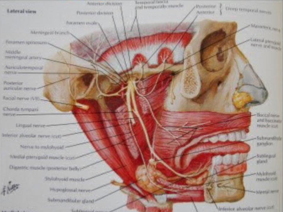
• TMJ dipersarafi oleh Nervus Trigeminus (N. V) divisi N. Mandibularis
• Persyarafan utama TMJ berasal dari N. Auriculotemporalis, yaitu divisi posterior dari N. Mandubularis.
• Persyarafan lain dapat berasal dari Masserericus nervus dan deep temporale.

1. N. Auriculotemporalis.
Syaraf ini berjalan pada bagian luar spina ossis spenoidalis, terletak dekat permukaan dalam capsula sendi.
Syaraf berjalan ke luar dan biasanya masuk ke lobus glenoidalis dari glandula parotidea dan disini syaraf akan mengeluarkan serabut Sekretomotoris ke glandula ( berasal dari n. glossopharyngeus melalui ganglion oticum ).
Syaraf kemudian berjalan ke atas di atas radix posterior processus Zygomaticus tepat dibelakang dan kebagian luar tuberculum Postglenoidalis.

• 2. N. Chorda TympaniKeluar dari ujung medial FissuraTympanosqumosa berjalan oblik ke bawah dan ke depan pada sisi medial spina ossis Spenoidalis dan bagian dalam capsula sendi untuk menghubungkan n. lingualis yang terletak jauh di dalam m. Pterygoideus
Lateralis.
• 3. A. Auricularis dan A. Tympanica cab. Pertama a. Maxillaris.
Masuk ke fissura Tympanosquamosa di belakang daerah perlekatan Capsula sendi dan mengeluarkan cabang-
cabangnya ke struktur sendi.

Auriculotemporalis


Internal structures of the TMJ
1. Articular disc -
-biconcave, thicker on outside edges
- no vascular or nervous supply except at edges
- made of fibrous tissue
2. 2 Joint cavities - TMJ is a compound joint
a. inferior - below the disc
b. superior - above the disc
3. Retrodiscal tissue
- superior retrodiscal lamina - attaches to glenoid fossa
and pulls disc back when mandible opens


TMJ Musculature
• Four muscles of mastication that move the mandible:– Masseter– Temporalis– Medial Pterygoid– Lateral Pterygoid

OTOT PENGUNYAHAN TMJ
– M. MASSETER• Suplai saraf : Cabang mandibularis dari n.
Cranialis V (trigeminus) melalui cabang yang berjalan melintasi incisura mandibula.
• Fungsi : M. Masseter mengangkat rahang bawah dan menariknya sedikit ke depan.bersama m. pterygoideus medialis dari sisi yang sama , otot ini berfungsi mengatur posisi angulus mandibula pada bidang vertikal.

• M. TEMPORALIS– Suplai syaraf : Cabang mandibularis n. cranialis
V ( Trigeminus )– Fungsi :
• Serabut anterior m.temporalis berfungsi mengangkat mandibula.
• Serabut posterior berfungsi untuk menarik processus Condylaris ke belakang masuk ke fossa mandibularis atau fossa glenoidalis dan membantu menghilangkan tekanan dari caput mandibula ketika gigi geligi ( Clenching )

• M. PTERYGOIDEUS
– Suplai Saraf:
N. Mandibularis Cabang N Cranialis V– Fungsi:
• M. Pterygoideus Lateralis : Caput superior m. pterygoideus lateralis berfungsi menarik discus articularis ke depan dan pada saat bersamaan caput condylaris akan ditarik kedepan oleh caput inferior.
• M. Pterygoideus Medialis : berfungsi mengangkat rahang bawah/ mandibula dan memajukannya. Dan juga mengungkit angulus mandibula ke medial.
•

• M. DIGASTRICUS– Suplai saraf :
• Venter anterior n. Mandibularis cabang n. trigeminus, venter posterior n. facialis.
• Fungsi :– Merupakan otot bantu pengunyahan ke dua
bagian m. digastricus bekerja bersama untuk mendepresi dagu dan membantu membuka mulut ketika os hyoideum dalam keadaan terfiksasi oleh mm.infra hyoidei. Otot ini juga berfungsi mengangkat os hyoideum pada saat mandibula pada posisi stabil.


TMJ Anatomy
• Meniscal Anatomy– Oval-shaped fibrocartilaginous articular disk (meniscus) between
the osseous components of the joint.– The central, intermediate portion of the disk is thin while the
anterior and posterior aspects, or bands, are thicker.– The bilaminar zone attaches to the posterior disc assists the
head of the condyle in moving forward.
• Ligaments– Temporomandibular ligament– Stylomandibular ligament– Sphenomandibular ligament

Biomechanics of the TMJ
Two distinct joint systems
1. Rotational movement
- inferior joint cavity
2. Translational movement
- superior joint cavity

Gerakan Mandibula
– A. Gerak Rotasi atau Sendi
Ketika Caput Processus Condylaris bergerak dalam kompartemen sendi bagian bawah dalam hubungannya dengan discus articularis
– B. Gerak meluncur atau Translasi
Caput mandibula dan discus articularis bergerak disepanjang permukaan bawah
os Temporale pada kompartemen sendi bagian atas. Kombinasi gerak sendi dan meluncur diperlukan agar cavum oris dibuka lebar-lebar.

• Gerak sendi pada individu dewasa yang normal mempunyai kisaran 20 – 25 mm antara gigi geligi anterior atas dan bawah.
• Bila dikombiasikan dengan gerak meluncur kisaran gerak membuka mulut yang normal akan meningkat menjadi 35 – 45 mm

TMJ Biomechanics
• Two motions:– First 20mm of motion is
rotation. The mandible and meniscus move anteriorly together beneath the articular eminence while opening or closing.
– Second motion is translation, which slides the jaw further forward or from side to side.

TMJ Classification
Functions as a unique joint.
Anatomically is a compound joint

Rotation occurs around the horizontal condylar axis

Translational movement begins after TM ligament becomes tight. Condylar head starts to move down the eminentia

Posterior border of the mandible moves back during rotation
Rotational movement stops when the oblique band of the Temporomandibular ligament becomes tight

TMJ Anatomy
• Osseous Anatomy– The articulation between the condyles of the mandible
and the temporal bone, which is part of the cranium.– The articular surface of the condyle is convex and the
articular eminence of the temporal bone is concave.

.
• .
Related Documents