RESEARCH ARTICLE Open Access Neuritogenic effect of standardized extract of Centella asiatica ECa233 on human neuroblastoma cells Oraphan Wanakhachornkrai 1 , Varisa Pongrakhananon 2,3 , Preedakorn Chunhacha 3 , Aree Wanasuntronwong 4 , Anusara Vattanajun 5 , Boonyong Tantisira 2,6 , Pithi Chanvorachote 2,3* and Mayuree H Tantisira 2,7* Abstract Background: In order to gain insight into neuroprotective effects of ECa 233, a standardized extract of Centella asiatica, previously demonstrated in animal models of memory impairment induced by transient global ischemia or intracerebroventricular injection of β-amyloid, the effect of ECa 233 on neurite outgrowth of human IMR-32 neuroblastoma cell line was investigated. Methods: Cells were seeded and incubated with various concentrations of ECa 233. Morphometric analysis was carried out by a measurement of the longest neurite growth of cells at 24 and 48 h. Contributing signaling pathways possibly involved were subsequently elucidated by western blot analysis. Results: While ECa 233 had only limited effects on cell viability, it significantly enhanced neurite outgrowth of IMR- 32 cells at the concentrations of 1–100 μg/ml. Western blot analysis revealed that ECa 233 significantly upregulated the level of activated ERK1/2 and Akt of the treated cells suggesting their involvement in the neuritogenic effect observed, which was subsequently verified by the finding that an addition of their respective inhibitors could reverse the effect of ECa 233 on these cells. Conclusions: The present study clearly demonstrated neurite outgrowth promoting activity of ECa 233. ERK1/2 and Akt signaling pathways seemed to account for the neurotrophic effect observed. In conjunction with in vivo neuroprotective effect of ECa 233 previously reported, the results obtained support further development of ECa 233 for clinical use in neuronal injury or neurodegenerative diseases. Keywords: Neurite outgrowth, Centella asiatica, ECa 233, ERK1/2, Akt, IMR-32 neuroblastoma Background Neurite outgrowth is an important initial step in the for- mation of neuronal network. Such neuronal behavior was also shown to implicate in neuronal regeneration and response to neural injury [1]. Mechanisms in regula- tion of neurite outgrowth have been intensively investi- gated and the most accepted concept involves MEK/ ERK and PI3K/Akt signaling pathways. Indeed, these molecular signals are triggered by tyrosine kinase recep- tor activation [2-6]. Several neurotrophic ligands for tyrosine kinase receptors such as nerve growth factor (NGF), brain derived neurotrophic factor (BDNF) and neurotrophin-3 (NT-3) have been identified [7,8]. The activation of MEK/ERK and PI3K/Akt results in an increase in cell survival as well as an enhancement of neuronal growth and differentiation [9,10]. In this con- text, molecules or compounds that exhibit potent neuro- trophic activity are of great interest and may be useful for the treatment of stroke, brain or spinal cord injury and neurodegenerative diseases [11-13]. In search for potential neurotrophic activity from herbs acting on the central nervous system, the present study focused on the neurotrophic activity of Centella asiatica which has, for a long time, been claimed to be beneficial for managing disorders of the brain, skin and * Correspondence: [email protected]; [email protected] 2 Department of Pharmacology and Physiology, Faculty of Pharmaceutical Sciences, Chulalongkorn University, Bangkok 10330, Thailand 3 Cell-based Drug and Health Product Development Research Unit, Chulalongkorn University, Bangkok 10330, Thailand Full list of author information is available at the end of the article © 2013 Wanakhachornkrai et al.; licensee BioMed Central Ltd. This is an Open Access article distributed under the terms of the Creative Commons Attribution License (http://creativecommons.org/licenses/by/2.0), which permits unrestricted use, distribution, and reproduction in any medium, provided the original work is properly cited. Wanakhachornkrai et al. BMC Complementary and Alternative Medicine 2013, 13:204 http://www.biomedcentral.com/1472-6882/13/204

Welcome message from author
This document is posted to help you gain knowledge. Please leave a comment to let me know what you think about it! Share it to your friends and learn new things together.
Transcript

Wanakhachornkrai et al. BMC Complementary and Alternative Medicine 2013, 13:204http://www.biomedcentral.com/1472-6882/13/204
RESEARCH ARTICLE Open Access
Neuritogenic effect of standardized extract ofCentella asiatica ECa233 on humanneuroblastoma cellsOraphan Wanakhachornkrai1, Varisa Pongrakhananon2,3, Preedakorn Chunhacha3, Aree Wanasuntronwong4,Anusara Vattanajun5, Boonyong Tantisira2,6, Pithi Chanvorachote2,3* and Mayuree H Tantisira2,7*
Abstract
Background: In order to gain insight into neuroprotective effects of ECa 233, a standardized extract of Centellaasiatica, previously demonstrated in animal models of memory impairment induced by transient global ischemia orintracerebroventricular injection of β-amyloid, the effect of ECa 233 on neurite outgrowth of human IMR-32neuroblastoma cell line was investigated.
Methods: Cells were seeded and incubated with various concentrations of ECa 233. Morphometric analysis wascarried out by a measurement of the longest neurite growth of cells at 24 and 48 h. Contributing signalingpathways possibly involved were subsequently elucidated by western blot analysis.
Results: While ECa 233 had only limited effects on cell viability, it significantly enhanced neurite outgrowth of IMR-32 cells at the concentrations of 1–100 μg/ml. Western blot analysis revealed that ECa 233 significantly upregulatedthe level of activated ERK1/2 and Akt of the treated cells suggesting their involvement in the neuritogenic effectobserved, which was subsequently verified by the finding that an addition of their respective inhibitors couldreverse the effect of ECa 233 on these cells.
Conclusions: The present study clearly demonstrated neurite outgrowth promoting activity of ECa 233. ERK1/2 andAkt signaling pathways seemed to account for the neurotrophic effect observed. In conjunction with in vivoneuroprotective effect of ECa 233 previously reported, the results obtained support further development of ECa 233for clinical use in neuronal injury or neurodegenerative diseases.
Keywords: Neurite outgrowth, Centella asiatica, ECa 233, ERK1/2, Akt, IMR-32 neuroblastoma
BackgroundNeurite outgrowth is an important initial step in the for-mation of neuronal network. Such neuronal behaviorwas also shown to implicate in neuronal regenerationand response to neural injury [1]. Mechanisms in regula-tion of neurite outgrowth have been intensively investi-gated and the most accepted concept involves MEK/ERK and PI3K/Akt signaling pathways. Indeed, thesemolecular signals are triggered by tyrosine kinase recep-tor activation [2-6]. Several neurotrophic ligands for
* Correspondence: [email protected]; [email protected] of Pharmacology and Physiology, Faculty of PharmaceuticalSciences, Chulalongkorn University, Bangkok 10330, Thailand3Cell-based Drug and Health Product Development Research Unit,Chulalongkorn University, Bangkok 10330, ThailandFull list of author information is available at the end of the article
© 2013 Wanakhachornkrai et al.; licensee BioMCreative Commons Attribution License (http:/distribution, and reproduction in any medium
tyrosine kinase receptors such as nerve growth factor(NGF), brain derived neurotrophic factor (BDNF) andneurotrophin-3 (NT-3) have been identified [7,8]. Theactivation of MEK/ERK and PI3K/Akt results in anincrease in cell survival as well as an enhancement ofneuronal growth and differentiation [9,10]. In this con-text, molecules or compounds that exhibit potent neuro-trophic activity are of great interest and may be usefulfor the treatment of stroke, brain or spinal cord injuryand neurodegenerative diseases [11-13].In search for potential neurotrophic activity from
herbs acting on the central nervous system, the presentstudy focused on the neurotrophic activity of Centellaasiatica which has, for a long time, been claimed to bebeneficial for managing disorders of the brain, skin and
ed Central Ltd. This is an Open Access article distributed under the terms of the/creativecommons.org/licenses/by/2.0), which permits unrestricted use,, provided the original work is properly cited.

Wanakhachornkrai et al. BMC Complementary and Alternative Medicine 2013, 13:204 Page 2 of 7http://www.biomedcentral.com/1472-6882/13/204
gastrointestinal tract [14]. Additionally, it was used as abrain booster for promoting brain growth and improvingmemory [12]. Like many other medicinal plants, C.asiatica contains several active compounds, includingasiatic acid, madecassic acid, asiaticoside, and madecas-soside [15]. To avoid a large fluctuation in biologicalresponses arising from variations of these bioactive con-stituents, we have established the standardized extract ofC. asiatica, namely ECa 233. It is defined as a white tooff-white extracted powder of C. asiatica containingtriterpenoids not less than 80% and the ratio betweenmadecassoside and asiaticoside was kept within 1.5 ± 0.5[16]. Restorative and neuroprotective effects of ECa 233have been demonstrated in animal models of learningand memory deficit induced by either a transient occlu-sion of common carotid arteries [17] or an intracerebro-ventricular injection of β-amyloid [18]. Protection ofoxidative stress was proposed to be one of the possibleunderlying mechanisms. However, effect of ECa 233 onneurite outgrowth which could possibly be involved inits neurotrophic/neuroprotective effects has not yet beenelucidated. Therefore, the present study aimed to inves-tigate the effect of ECa 233 on the neurite growth andits underlying mechanisms in IMR-32 human neuro-blastoma cells.
MethodsCell culture and reagentsIMR-32 neuroblastoma cells were obtained from theAmerican Type Culture Collection, ATCC (Manassas,VA, USA). Cells were cultured in DMEM/F12 supple-mented with 10% fetal bovine serum, 2 mmol/l L-glu-tamine and 100 units/ml penicillin/streptomycin in a 5%CO2 at 37°C. BDNF, PD 098059, LY 294002 were pur-chased from Sigma Chemical. Inc. (St. Louis, MO, USA).Resazurin was purchased from Invitrogen (Carlsbad,CA, USA). Specific antibody for phospho-Akt, Akt,phospho-ERK1/2, ERK1/2 and GAPDH were purchasedfrom Abcam (Cambridge, MA, USA), and peroxidaseconjugated anti-rabbit secondary antibody were pur-chased from Cell Signaling (Danvers, MA, USA).
Preparation of tested substancesECa 233 containing madecassoside 52% w/w andasiaticoside 32% w/w was kindly supplied by AssociateProfessor Ekarin Saifah, Ph.D and collaborates, Facultyof Pharmaceutical Sciences, Chulalongkorn University.Thailand. It was suspended in sterile PBS at 10 mg/mland served as stock solution. BDNF was dissolved in thesterile PBS to a stock solution at the concentration of50 μg/ml. PD098059 and LY294002 were dissolved byDMSO to concentration of 0.344 and 0.267 mg/ml,respectively.
Cell viability assayCells were seeded in 96-well plates and incubated for24 h. After incubation, the plating media were removedand replaced. The cell were subsequently incubated for30 minutes then subjected to treatments. After 24 h,cells were incubated with 1:50 resazurin at 37°C for30 minutes. The well-plate was transferred to a fluores-cence microplate reader with Softmax Pro software tomeasure fluorescence intensity of resorufin (resazurinproduct) at excitation wavelength of 530 nm and emis-sion wavelength of 590 nm. The percentage of cell via-bility was calculated and compared with non-treatedcontrol. All analyses were performed for at least threeindependent triplicate experiments.
Measurement of neurite outgrowthIMR-32 cells were cultured in a 96-well culture plate.After 24 h cells were subjected to various concentrationsof ECa 233 (0.1, 1, 10, 100 μg/ml) or BDNF (100 ng/ml).A neurite was identified as a process equal to or longerthan cell body diameter. The cells selected randomlyfrom 3–4 fields of each well were photographed (phasecontrast, Nikon, Inverted microscope ECLIPSE Ti-u) formorphometric analyses. The length of the longestneurite from each cell was measured using NIS-Elementimage software [19,20].To test the involvement of MEK and Akt pathway,
their respective inhibitors, PD098059 (5 μM) for MEKand LY294002 (7.5 μM) for PI3K/Akt, was added at30 min prior to the test substance.
Western blot analysisAfter specified treatments, cells were incubated in lysisbuffer containing 20 mM Tris–HCl (pH 7.5), 1%TritonX-100, 150 mM sodium chloride, 10% glycerol,1 mM sodium orthovanadate, 50 mM sodium fluoride,100 nM phenylmethylsulfonyl fluoride, and a commer-cial protease inhibitor cocktail (Roche Molecular Bio-chemicals, Indianapolis, IN, USA) for 30 minutes on ice.Cell lysates were collected and centrifuged 12,000 rpmat 4°C, the supernatant was collected and the proteincontent was determined using Bradford method (Bio-Rad, Hercules, CA, USA). Equal amount of proteinsfrom each sample were denatured by heating at 95°Cwith laemmli loading buffer for 5 min and were subse-quently loaded onto 10% SDS-PAGE. After separation,proteins were transferred onto 0.45 μm nitrocellulosemembranes (Bio-Rad). The transferred membranes wereblocked in 5% non-fat dry milk in TBST (25 mM Tris–HCl (pH 7.5), 125 mM NaCl, 0.1% tween20) for 1 h.Then washed with TBST and further incubated with theindicated primary antibodies at 4°C overnight. Mem-brane were washed twice with TBST for 10 min and in-cubated with secondary antibody for 1 h at room
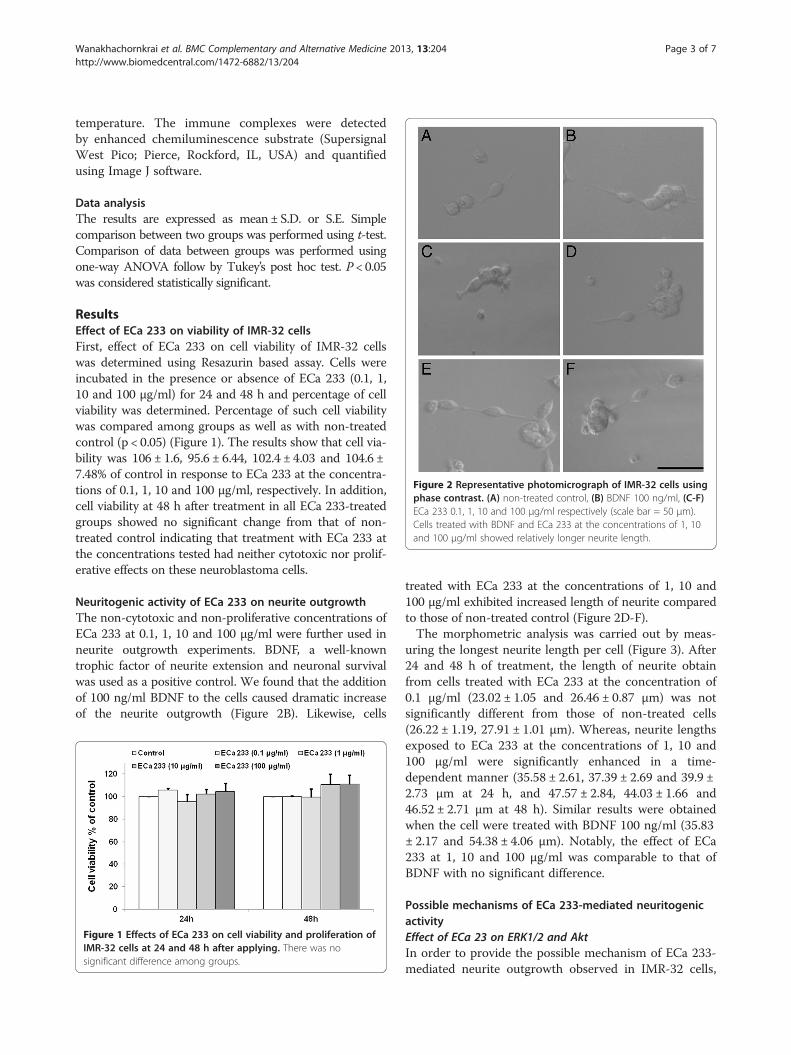
Figure 2 Representative photomicrograph of IMR-32 cells usingphase contrast. (A) non-treated control, (B) BDNF 100 ng/ml, (C-F)ECa 233 0.1, 1, 10 and 100 μg/ml respectively (scale bar = 50 μm).Cells treated with BDNF and ECa 233 at the concentrations of 1, 10and 100 μg/ml showed relatively longer neurite length.
Wanakhachornkrai et al. BMC Complementary and Alternative Medicine 2013, 13:204 Page 3 of 7http://www.biomedcentral.com/1472-6882/13/204
temperature. The immune complexes were detectedby enhanced chemiluminescence substrate (SupersignalWest Pico; Pierce, Rockford, IL, USA) and quantifiedusing Image J software.
Data analysisThe results are expressed as mean ± S.D. or S.E. Simplecomparison between two groups was performed using t-test.Comparison of data between groups was performed usingone-way ANOVA follow by Tukey’s post hoc test. P < 0.05was considered statistically significant.
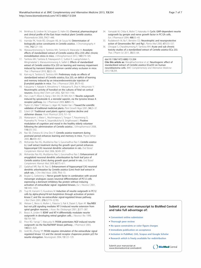
ResultsEffect of ECa 233 on viability of IMR-32 cellsFirst, effect of ECa 233 on cell viability of IMR-32 cellswas determined using Resazurin based assay. Cells wereincubated in the presence or absence of ECa 233 (0.1, 1,10 and 100 μg/ml) for 24 and 48 h and percentage of cellviability was determined. Percentage of such cell viabilitywas compared among groups as well as with non-treatedcontrol (p < 0.05) (Figure 1). The results show that cell via-bility was 106 ± 1.6, 95.6 ± 6.44, 102.4 ± 4.03 and 104.6 ±7.48% of control in response to ECa 233 at the concentra-tions of 0.1, 1, 10 and 100 μg/ml, respectively. In addition,cell viability at 48 h after treatment in all ECa 233-treatedgroups showed no significant change from that of non-treated control indicating that treatment with ECa 233 atthe concentrations tested had neither cytotoxic nor prolif-erative effects on these neuroblastoma cells.
Neuritogenic activity of ECa 233 on neurite outgrowthThe non-cytotoxic and non-proliferative concentrations ofECa 233 at 0.1, 1, 10 and 100 μg/ml were further used inneurite outgrowth experiments. BDNF, a well-knowntrophic factor of neurite extension and neuronal survivalwas used as a positive control. We found that the additionof 100 ng/ml BDNF to the cells caused dramatic increaseof the neurite outgrowth (Figure 2B). Likewise, cells
Figure 1 Effects of ECa 233 on cell viability and proliferation ofIMR-32 cells at 24 and 48 h after applying. There was nosignificant difference among groups.
treated with ECa 233 at the concentrations of 1, 10 and100 μg/ml exhibited increased length of neurite comparedto those of non-treated control (Figure 2D-F).The morphometric analysis was carried out by meas-
uring the longest neurite length per cell (Figure 3). After24 and 48 h of treatment, the length of neurite obtainfrom cells treated with ECa 233 at the concentration of0.1 μg/ml (23.02 ± 1.05 and 26.46 ± 0.87 μm) was notsignificantly different from those of non-treated cells(26.22 ± 1.19, 27.91 ± 1.01 μm). Whereas, neurite lengthsexposed to ECa 233 at the concentrations of 1, 10 and100 μg/ml were significantly enhanced in a time-dependent manner (35.58 ± 2.61, 37.39 ± 2.69 and 39.9 ±2.73 μm at 24 h, and 47.57 ± 2.84, 44.03 ± 1.66 and46.52 ± 2.71 μm at 48 h). Similar results were obtainedwhen the cell were treated with BDNF 100 ng/ml (35.83± 2.17 and 54.38 ± 4.06 μm). Notably, the effect of ECa233 at 1, 10 and 100 μg/ml was comparable to that ofBDNF with no significant difference.
Possible mechanisms of ECa 233-mediated neuritogenicactivityEffect of ECa 23 on ERK1/2 and AktIn order to provide the possible mechanism of ECa 233-mediated neurite outgrowth observed in IMR-32 cells,

Figure 3 Morphometric analysis of IMR-32 cells affected by ECa 233. After cells cultured for 24 and 48 h in the presence of either ECa 233(0.1, 1, 10 and 100 μg/ml) or BDNF (100 ng/ml). Morphometric analysis was carried out by measuring neurites from 60 living cells per treatedcondition, (n = 3). Average of the neurite length was significantly increased after being treated with ECa 233 (1, 10 and 100 μg/ml) or BDNF. Thedata presented as mean ± S.E. ** = p < 0.01, *** = p < 0.001 vs non-treated control.
Wanakhachornkrai et al. BMC Complementary and Alternative Medicine 2013, 13:204 Page 4 of 7http://www.biomedcentral.com/1472-6882/13/204
western blot analysis was carried out to determinewhether ERK and Akt signaling were involved. Cellswere treated with ECa 233 and the level of phosphory-lated ERK and phosphorylated Akt were determined.Figures 4A and B demonstrated that relative blot densityof pERK/ERK and pAkt/Akt in response to ECa 233 atthe concentration of 0.1 μg/ml was not different fromthat of non-treated group. However, ECa 233 at theconcentrations of 1, 10 and 100 μg/ml significantly in-creased the level of phosphorylated ERK and phosphory-lated Akt, suggesting that ECa 233 could possibly increasethe neurite outgrowth via these 2 major pathways.
Effect of ECa 233 on neurite outgrowth in the presence ofPD098059 and LY294002Subsequently, the involvement of ERK and Akt signalingpathways in the neurite outgrowth promoting activityof ECa 233 was confirmed by treatment of specific
Figure 4 Relative blot density of pERK/ERK and pAkt/Akt in responseAkt and Akt phosphorylation. The level of phospho-ERK1/2 and phospho-A6 h were significantly higher than that of control. Densitometric analysis ofwere plated and presented as mean ± S.D. of three independent experimen
inhibitor of MEK (PD098059) or PI3K (LY294002) priorto the exposure to ECa 233. In comparison to neuritelengths of non-treated group which was 23.39 ± 0.54 μm(Figure 5A), ECa 233 at the concentrations of 1, 10 and100 μg/ml significantly enhanced the neurite outgrowthinto 36.12 ± 1.53, 36.79 ± 1.60 and 36.69 ± 1.98 μm re-spectively. Pretreatment of the cell with PD098059significantly decreased the neurite outgrowth of the re-spective concentrations of ECa 233 into 22.45 ± 0.65,20.76 ± 0.532 and 21.83 ± 0.53 μm. Similar results weredemonstrated by LY294002 which significantly decreasedthe ECa 233-induced neurite outgrowth into 20.83 ±0.81, 20.03 ± 0.49 and 20.76 ± 0.53 μm, respectively(Figure 5B). As expected, PD098059 (5 μM) orLY294002 (7.5 μM) in the concentration that exhibitedno significant effect on neurite outgrowth (23.52 ± 0.84and 20.43 ± 0.54 μm, respectively) completely abolishedneurite stimulating effect of ECa 233.
to ECa 233 treatment. (A) ERK1/2 and ERK1/2 phosphorylation (B)kt in IMR-32 cells after treated with ECa 233 (1, 10 and 100 μg/ml) forthe indicated proteins normalized with the density of GAPDH bandts, * = p < 0.05, ** = p < 0.001 vs non-treated control.

Figure 5 Representative photomicrograph of IMR-32 cells. (A) after pre-treated with LY294002 or PD098059 in the presence or absence ofECa 233 (1, 10 and 100 μg/ml) (scale bar = 50 μm). (B) Morphometric analysis of the cell treated with LY294002 or PD098059 on the increment ofneurite outgrowth induced by ECa 233. Neurites from 60 living cells per treated condition were evaluated, (n = 3). Data are presented as mean± S.E., ** = p < 0.001 vs non-treated control. ## = p < 0.001 vs ECa 233 treated groups.
Wanakhachornkrai et al. BMC Complementary and Alternative Medicine 2013, 13:204 Page 5 of 7http://www.biomedcentral.com/1472-6882/13/204
DiscussionAlthough the plant originated extracts have garneredattentions as important sources of drugs and healthproducts, the inconsistency of main components foundin such extracts limits their use as well as further de-velopment to clinical use. Establishment of standardizedextract with consistency of its bioactive constituentscould enhance reproducible biological responses andsubsequently reliable use of herbal products [21]. Basedon well-defined constituents and numbers of scientificevidence, EGb 761, a standardized extract of Chineseancient plant, Ginko biloba, known as a brain tonic rem-edy, has been prescribed for memory deficit all over theworld [22]. By the same analogy, C. asiatica which haslong been used in Ayurveda to enhance memory was ex-tensively investigated [23]. In an attempt to make a reli-able herbal product, ECa 233, a standardized extract ofC. asiatica, was established and investigated for its effecton brain function. We have found that ECa 233 couldattenuate memory deficit and exerted anxiolytic effect in
animal models [16-18]. As administration of fresh leafjuice of C. asiatica was found to enhance dendriticaborization of hippocampal and amygdala neurons inneonatal and adult rats [24-27], we hypothesized thatECa 233 might enhance neurite outgrowth.As expected, the present study clearly demonstrated
neuritogenic effect of ECa 233 in the range of 1–100μg/ml. Further experiment was then conducted to gainan insight into signaling pathways possibly involved.Numerous signaling pathways involving in neurite out-growth have been proposed [2,5]. Among them, ERKand Akt activations have long been known to be import-ant for neurite elongation induced by certain neuro-trophic factors including NGF [28,29], neurotrophin-3(NT-3) [30], BDNF [31] as well as natural compoundsincluding FK506 [32,33], genipin [34], honokiol [4] andmilitarinone A [9]. In addition, ginsenoside, triterpe-noidglycoside from gingseng, has been shown to signifi-cantly increase the neurite outgrowth of neuroblastomacells [35].

Wanakhachornkrai et al. BMC Complementary and Alternative Medicine 2013, 13:204 Page 6 of 7http://www.biomedcentral.com/1472-6882/13/204
In line with the findings previously demonstrated bysome other natural products mentioned above, westernblot analysis demonstrated that administration of ECa233 significantly increased the level of phosphorylatedERK and Akt in IMR-32 cells indicating their activationin the presence of ECa 233. In addition, contribution ofERK and Akt signaling pathways was subsequently con-firmed by the results that the neurite outgrowth promot-ing effect of ECa 233 was abolished by specific inhibitorsof MEK (PD098059) or PI3K (LY294002) suggesting thatECa 233 promoted neurite outgrowth in human neuro-blastoma IMR-32 cells via MEK/ERK and PI3K/Akt-dependent mechanisms. Though, asiatic acid, one ofchemical constituents present in C. asiatica has beenproposed to elicit its effect via MAP kinase pathway[12], increases of phosphorylated ERK and Akt observedin the present study are likely to be attributable tomadecassoside and/or asiaticoside, the major constitu-ents of ECa 233.In agreement with the fact that C. asiatica is widely
consumed as food, ECa 233 in the concentrations thatpromoted neurite outgrowth in human neuroblastomaIMR-32 cells had no effect on cell viability under theexperimental condition indicating safety of ECa 233, astandardized extract derived from C. asiatica. Similarly,safety profiles of ECa 233 have been previously reportedin both acute and sub-chronic toxicity testing. Oral ad-ministration of ECa 233 in the dose up to 10 g/kg didnot cause any lethality and no significant adverse effectswas observed in experimental animals receiving dailyadministration of ECa 233 in the dose range of 10–1000 mg/kg/day [36]. In consideration to very favorablesafety profiles and significant in vivo neuroprotective ac-tivity of ECa 233 [18], the results of the present studysupports further development of ECa 233 for neuronalinjury as well as neurodegenerative diseases in human.
ConclusionsThe present study demonstrated the neurite outgrowthpromoting activity of ECa 233 in human IMR-32 neuro-blastoma cell line. The neuritogenic effect observedseemed to be mediated via ERK1/2 and Akt signalingpathways. The results obtained support potential benefitof ECa 233 for the management of neuronal injury andneurodegenerative diseases. Further pharmacodynamicand pharmacokinetic studies of ECa 233 are needed tobring ECa 233 into clinical use.
AbbreviationsC. asiatica: Centella asiatica; MEK: Mitogen activated protein kinase;ERK: Extracellular signaling regulated kinase; PI3K: Phosphoinositide-3-kinase;NGF: Nerve growth factor; BDNF: Brain derived neurotrophic factor;NT-3: Neurotrophin-3; PD: PD 098059; LY: LY 294002.
Competing interestsAll authors declare that they have no competing interests.
Authors’ contributionsOW participated in design and conducting the experiments, analysis of data,drafting the manuscript. VP, PrC and AW participated in technical supportsand interpretation of the data. AV and BT supervised, design of experiments,analyzed and interpretation of the data. PiC was a coordinator, designed thestudy, analysis and interpretation of data, and drafting the manuscript. MTsupervised, conceived and designed the experiment, interpretation of thedata and revision of the manuscript. All authors have read and approved thefinal manuscript.
AcknowledgementsThe work was supported by The 90th Anniversary of Chulalongkorn UniversityFund (Ratchadaphiseksomphot Endowment Fund) and Ratchadaphiseksom-phot Endowment Fund for Cell-based Drug and Health Product DevelopmentResearch Unit. The authors would like to thank Associate Professor Ekarin Saifahand collaborates for the provision of ECa 233.
Author details1Inter-disciplinary Program in Physiology, Graduate School, ChulalongkornUniversity, Bangkok 10330, Thailand. 2Department of Pharmacology andPhysiology, Faculty of Pharmaceutical Sciences, Chulalongkorn University,Bangkok 10330, Thailand. 3Cell-based Drug and Health Product DevelopmentResearch Unit, Chulalongkorn University, Bangkok 10330, Thailand. 4Faculty ofDentistry, Mahidol University, Bangkok 10400, Thailand. 5Department ofPhysiology, Phramongkutklao College of Medicine, Bangkok 10400, Thailand.6Faculty of Pharmacy, Silpakorn University, Nakonprathom 73000, Thailand.7Faculty of Pharmaceutical Scienccs, Burapha University, Chonburi 20131,Thailand.
Received: 1 March 2013 Accepted: 31 July 2013Published: 4 August 2013
References1. Plachez C, Richards LJ: Mechanisms of axon guidance in the developing
nervous system. Curr Top Dev Biol 2005, 69:267–346.2. Kaplan D, Miller F: Neurotrophin signaling transduction in the nervous
system. Curr Opin Neurobiol 2000, 10:381–391.3. Hur J, Lee P, Kim H, Kang I, Lee KR, Kim SY: (−)-3,5-Dicaffeoyl-muco-quinic
acid isolated from Aster scaber contributes to the differentiation of PC12cells: through tyrosine kinase cascade signaling. Biochem and Biophys ResCommun 2004, 313:948–953.
4. Zhai H, Nakade K, Oda M, Mitsumoto Y, Akagi M, Sakurai J, Fukuyama Y:Honokiol-induced neurite outgrowth promotion depends on activationof extracellular signal-regulated kinases (ERK1/2). Eur J Pharmacol 2005,516:112–117.
5. Read DE, Gorman AM: Involvement of Akt in neurite outgrowth. Cell MolLife Sci 2009, 66:2975–2984.
6. Takahashi K, Piao S, Yamatani H, Du B, Yin L, Ohta T, Kawagoe J, Takata K,Tsutsumi S, Kurachi H: Estrogen induces neurite outgrowth via Rho familyGTPases in neuroblastoma cells. Mol Cell Neurosci 2011, 48:217–224.
7. Labelle C, Leclerc N: Exogenouse BDNF, NT-3 and NT-4 differentiallyregulate neurite outgrowth in cultured hippocampal neurons. Brain ResDev Brain Res 2000, 123:1–11.
8. Chao MV, Rajagopal R, Lee FS: Neurotrophin signaling in health anddisease. Clin Sci (Lond) 2006, 110:167–173.
9. Küenzi P, Kiefer S, Koryalina A, Hamburger M: Promotion of cell death orneurite outgrowth in PC-12 and N2a cells by the fungal alkaloidmilitarinone A depends on basal expression of p53. Apoptosis 2008,13:364–376.
10. Wang X, Wang Z, Yao Y, Li J, Zhang X, Li C, Cheng Y, Ding G, Liu L, Ding Z:Essential role of ERK activation in neurite outgrowth induced by α-lipoicacid. Biochim Biophys Acta 1813, 2011:827–838.
11. Horner PJ, Gage FH: Regenerating the central nervous system. Nat 2000,407:963–970.
12. Soumyanath A, Zhong YP, Gold SA, Yu X, Koop DR, Bourdette D, Gold BG:Centella asiatica accelerates nerve regeneration upon oral administrationand contains multiple active fractions increasing neurite elongationin-vitro. J Pharm Pharmacol 2005, 57:1221–1229.
13. Maier IC, Schweb ME: Sprouting, regeneration and circuit formation inthe injured spinal cord: factors and activity. Philos Trans R Soc Lond B BiolSci 2006, 61:1611–1634.
Wanakhachornkrai et al. BMC Complementary and Alternative Medicine 2013, 13:204 Page 7 of 7http://www.biomedcentral.com/1472-6882/13/204
14. Brinkhaus B, Lindner M, Schuppan D, Hahn EG: Chemical, pharmacologicaland clinical profile of the East Asian medical plant Centella asiatica.Phytomedicine 2000, 7:427–448.
15. Inamdar PK, Yeole RD, Ghogare AB, de Souza NJ: Determination ofbiologically active constituents in Centella asiatica. J Chromatography A1996, 742:127–130.
16. Wanasuntronwong A, Tantisira MH, Tantisira B, Watanabe H: Anxiolyticeffects of standardized extract of Centella asiatica (ECa 233) after chronicimmobilization stress in mice. J Ethnopharmacol 2012, 143:579–585.
17. Tantisira MH, Tantisira B, Patarapanich C, Suttisri R, Luangcholatan S,Mingmalailak S, Wanasuntronwong A, Saifah E: Effects of standardizedextract of Centella asiatica ECa 233 on learning and memory impairmentinduced by transient bilateral common carotid artery occlusion in mice.Thai J Pharmacol 2010, 32:22–33.
18. Kam-eg A, Tantisira B, Tantisira MH: Preliminary study on effects ofstandardized extract of Centella asiatica, Eca 233, on deficit of learningand memory induced by an intracerebroventricular injection ofβ-amyloid peptide in mice. Thai J Pharmacol 2009, 31:79–82.
19. Fukuyama Y, Nakade K, Minoshima Y, Yokoyama R, Zhai H, Mitsumoto Y:Neurotrophic activity of honokiol on the cultures of fetal rat corticalneurons. Bioorg Med Chem Lett 2002, 12:1163–1166.
20. Hur J, Lee P, Moon E, Kang I, Kim SH, Oh MH, Kim Y: Neurite outgrowthinduced by spicatoside A, a steroidal saponin, via the tyrosine kinase Areceptor pathway. Eur J Pharmacol 2009, 620:9–15.
21. Taylor JLS, Rabe T, McGaw LJ, Jäger AK, Staden Van J: Toward the scientificvalidation of traditional medicinal plants. Plant Growth Regul 2001, 34:23–37.
22. Eckert GP: Traditional used plants against cognitive decline andalzheimer disease. Front Pharmcol 2010, 1:1–10.
23. Wattanatorn J, Mator L, Muchimapura S, Tongun T, Pasuriwong O,Piyawatkul N, Yimtae K, Sripanidkulchai B, Singkhoraard J: Positivemodulation of cognition and mood in the healthy elderly volunteerfollowing the administration of Centella asiatica. J Ethnopharmacol 2008,116:325–332.
24. Rao SB, Chetana M, Uma Devi P: Centella asiatica treatment duringpostnatal period enhances learning and memory in mice. Physiol Behav2005, 86:449–457.
25. Mohandas Rao KG, Muddanna Rao S, Gurumadhva Rao S: Centella asiatica(L.) Leaf extract treatment during the growth spurt period enhanceshippocampal CA3 neuronal dendritic arborization in rats. Evid BasedComplement Alternat Med 2006, 3:349–357.
26. Mohandas Rao KG, Muddanna Rao S, Gurumadhva Rao S: Enhancement ofamygdaloid neuronal dendritic arboritezation by fresh leaf juice ofCentella asiatica (Linn) during growth spurt period in rats. Evid BasedComplement Alternat Med 2009, 6:575–611.
27. Gadahad MR, Rao M, Rao G: Enhancement of hippocampal CA3 neuronaldendritic arboritezation by Centella asiatica (Linn) fresh leaf extract inadult rats. J Chin Med Assoc 2008, 71:6–13.
28. Boglari G, Szeberenyi J: Nerve growth factor in combination with secondmessenger analogues causes neuronal differentiation of PC12 cellsexpressing a dominant inhibitory Ras protein without inducingactivation of extracellular signal- regulated kinases. Eur J Neurosci 2001,14:1445–1454.
29. Tsuji M, Inanami O, Kuwabara M: Induction of neurite outgrowth in PC12cells by alpha-phenyl-N-tert-butylnitron through activation of proteinkinase C and the ras-extracellular signal-regulated kinase pathway.J Biol Chem 2001, 276:32779–32785.
30. Aletsee C, Beros A, Mullen L, Palacios S, Pak K, Dazert S, Ryan AF: Ras/MEKbut not p38 signaling mediates NT-3-induced neurite extension fromspiral ganglion neurons. J Assoc Res Otolaryngol 2001, 2:377–387.
31. Bosco A, Linden R: BDNF and NT-4 differentially modulate neuriteoutgrowth in developing retinal ganglion cells. J Neurosci Res 1999,15:759–769.
32. Price RD, Yamaji T, Matsuoka N: FK506 potentiates NGF-induced neuriteoutgrowth via the Ras/Raf/MAP kinase pathway. J Pharmacol 2003,140:825–829.
33. Gold BG, Zhong YP: FK506 requires stimulation of the extracellular signalregulated kinase 1/2 and the steroid receptor chaperone protein p23 forneurite elongation. Neurosignals 2004, 13:122–129.
34. Yamazaki M, Chiba K, Mohri T, Hatanaka H: Cyclic GMP-dependent neuriteoutgrowth by genipin and nerve growth factor in PC12h cells.Eur J Pharmacol 2004, 488:35–43.
35. Rudakewich M, Ba F, Benishin CG: Neurotrophic and neuroprotectiveaction of Ginsenosides Rb1 and Rg1. Planta Med 2000, 67:533–537.
36. Chivapat S, Chavalittumrong P, Tantisira MH: Acute and sub chronictoxicity studies of a standardized extract of Centella asiatica ECa 233.Thai J Pharm Sci 2011, 35:55–64.
doi:10.1186/1472-6882-13-204Cite this article as: Wanakhachornkrai et al.: Neuritogenic effect ofstandardized extract of Centella asiatica ECa233 on humanneuroblastoma cells. BMC Complementary and Alternative Medicine2013 13:204.
Submit your next manuscript to BioMed Centraland take full advantage of:
• Convenient online submission
• Thorough peer review
• No space constraints or color figure charges
• Immediate publication on acceptance
• Inclusion in PubMed, CAS, Scopus and Google Scholar
• Research which is freely available for redistribution
Submit your manuscript at www.biomedcentral.com/submit
Related Documents