Neural Responsesto Expression and Gaze in the Posterior Superior Temporal Sulcus Interact with Facial Identity Heidi A. Baseler, Richard J. Harris, Andrew W. Young and Timothy J. Andrews Department of Psychology, York Neuroimaging Centre, University of York, York, UK Address correspondence to Email: [email protected] Neural models of human face perception propose parallel pathways. One pathway (including posterior superior temporal sulcus, pSTS) is responsible for processing changeable aspects of faces such as gaze and expression, and the other pathway (including the fusiform face area, FFA) is responsible for relatively invariant aspects such as identity. However, to be socially meaningful, changes in expression and gaze must be tracked across an individual face. Our aim was to investigate how this is achieved. Using functional magnetic reson- ance imaging, we found a region in pSTS that responded more to sequences of faces varying in gaze and expression in which the identity was constant compared with sequences in which the iden- tity varied. To determine whether this preferential response to same identity faces was due to the processing of identity in the pSTS or was a result of interactions between pSTS and other regions thought to code face identity, we measured the functional connectivity between face-selective regions. We found increased functional connectivity between the pSTS and FFA when partici- pants viewed same identity faces compared with different identity faces. Together, these results suggest that distinct neuralpathways involved in expression and identity interact to process the change- able features of the face in a socially meaningful way. Keywords: expression, functional magnetic resonance imaging, face, functional connectivity, gaze direction Introduction When someone moves about or as their gaze direction or expression changes the size and shape of the image, their face subtends on your retina also changes. To be useful, the visual system must be able to ignore these sources of variation to facilitate the recognition of who the person is, but also use this information to detect changes that enable social com- munication. Models of human face perception suggest that human observers deal with this problem using separate func- tional pathways, with the pathways involved in the visual analysis of identity being partially or fully independent of the pathway involved in processing the changeable aspects of faces (Bruce and Young 1986, 2012; Haxby et al. 2000). Neuroimaging studies have identified a number of face- selective regions, which appear to provide support for the idea of separable visual pathways in face perception (Allison et al. 1994; Kanwisher et al. 1997): An occipital face area (OFA), an fusiform face area (FFA), and posterior superior temporal sulcus (pSTS) region. The OFA is thought to be involved in the early perception of facial features and has a feed-forward pro- jection to both the pSTS and the FFA. The connection between the OFA and pSTS is thought to be important in processing dynamic changes in the face, such as changes in expression and gaze, which are important for social interactions (Puce et al. 1998; Pelphrey et al. 2004; Calder et al. 2007; Engell and Haxby 2007). In contrast, the connection between the OFA and FFA is considered to be involved in the representation of invariant facial characteristics that are important for recognition (Grill-Spector et al. 2004; Rotshtein et al. 2005). Influenced by the models of face perception, studies over the past decade have concentrated on the functional roles of each of these face-selective pathways (Hoffman and Haxby 2000; Barton et al. 2002; Andrews and Ewbank 2004; Winston et al. 2004; Fox et al. 2009). Consequently, it has remained unclear whether there is any interaction between regions in- volved in the perception of facial identity and expression. Support for this possibility has come from recent studies that have reported functional connectivity between the pSTS and FFA (Zhang et al. 2009; Turk-Browne et al. 2010; Ethofer et al. 2011). Although the exact role of this interaction between the 2 more heavily investigated pathways is not known, one possibility is that, to process changeable aspects of faces in a socially meaningful way, it is important to track changes across the same identity. Support for this possibility has come from studies that have shown an increased response to sequences of images in with the same facial identity com- pared with sequences containing different facial identities (Andrews and Ewbank, 2004; Davies-Thompson et al. 2009). The aim of this study was to probe the interaction of the pathways involved in processing identity and changeable aspects of faces. First, we asked whether the neural response to sequences of faces that change in expression and gaze di- rection differs according to whether the face images were of the same person or from different people. Next, we used func- tional connectivity to determine whether the influence of facial identity resulted from neural processes within the pSTS face-selective region itself, or whether it was dependent on interactions with other face regions. To do this, we removed the stimulus-driven activity from the fMR signal and correlated the remaining or residual time-courses between face regions (Davies-Thompson and Andrews forthcoming). This can be an extension of resting-state connectivity in which correlations between regions, independent of a response to stimuli, are examined (Biswal et al. 1995; Margulies et al. 2010). Our aim was to determine how the correlations between regions change as a function of whether the same or different identity faces were viewed (cf. Norman-Haignere et al. 2012). Materials and Methods Participants Data were analyzed from functional localizer scans from 103 different participants (49 females; mean age 24) and run as a standard part of 6 © The Author 2012. Published by Oxford University Press. This is an Open Access article distributed under the terms of the Creative Commons Attribution License (http://creativecommons.org/licenses/by-nc/3.0/), which permits non-commercial reuse, distribution, and reproduction in any medium, provided the original work is properly cited. For commercial re-use, please contact journals. [email protected]. Cerebral Cortex doi:10.1093/cercor/bhs360 Cerebral Cortex Advance Access published November 21, 2012

Welcome message from author
This document is posted to help you gain knowledge. Please leave a comment to let me know what you think about it! Share it to your friends and learn new things together.
Transcript
Neural Responses to Expression and Gaze in the Posterior Superior Temporal SulcusInteract with Facial Identity
Heidi A. Baseler, Richard J. Harris, Andrew W. Young and Timothy J. Andrews
Department of Psychology, York Neuroimaging Centre, University of York, York, UK
Address correspondence to Email: [email protected]
Neural models of human face perception propose parallel pathways.One pathway (including posterior superior temporal sulcus, pSTS) isresponsible for processing changeable aspects of faces such asgaze and expression, and the other pathway (including the fusiformface area, FFA) is responsible for relatively invariant aspects suchas identity. However, to be socially meaningful, changes in expressionand gaze must be tracked across an individual face. Our aim was toinvestigate how this is achieved. Using functional magnetic reson-ance imaging, we found a region in pSTS that responded more tosequences of faces varying in gaze and expression in which theidentity was constant compared with sequences in which the iden-tity varied. To determine whether this preferential response tosame identity faces was due to the processing of identity in thepSTS or was a result of interactions between pSTS and otherregions thought to code face identity, we measured the functionalconnectivity between face-selective regions. We found increasedfunctional connectivity between the pSTS and FFA when partici-pants viewed same identity faces compared with different identityfaces. Together, these results suggest that distinct neural pathwaysinvolved in expression and identity interact to process the change-able features of the face in a socially meaningful way.
Keywords: expression, functional magnetic resonance imaging, face,functional connectivity, gaze direction
Introduction
When someone moves about or as their gaze direction orexpression changes the size and shape of the image, theirface subtends on your retina also changes. To be useful, thevisual system must be able to ignore these sources of variationto facilitate the recognition of who the person is, but also usethis information to detect changes that enable social com-munication. Models of human face perception suggest thathuman observers deal with this problem using separate func-tional pathways, with the pathways involved in the visualanalysis of identity being partially or fully independent of thepathway involved in processing the changeable aspects offaces (Bruce and Young 1986, 2012; Haxby et al. 2000).
Neuroimaging studies have identified a number of face-selective regions, which appear to provide support for the ideaof separable visual pathways in face perception (Allison et al.1994; Kanwisher et al. 1997): An occipital face area (OFA), anfusiform face area (FFA), and posterior superior temporalsulcus (pSTS) region. The OFA is thought to be involved in theearly perception of facial features and has a feed-forward pro-jection to both the pSTS and the FFA. The connection betweenthe OFA and pSTS is thought to be important in processingdynamic changes in the face, such as changes in expression andgaze, which are important for social interactions (Puce et al.
1998; Pelphrey et al. 2004; Calder et al. 2007; Engell and Haxby2007). In contrast, the connection between the OFA andFFA is considered to be involved in the representation ofinvariant facial characteristics that are important for recognition(Grill-Spector et al. 2004; Rotshtein et al. 2005).
Influenced by the models of face perception, studies overthe past decade have concentrated on the functional roles ofeach of these face-selective pathways (Hoffman and Haxby2000; Barton et al. 2002; Andrews and Ewbank 2004; Winstonet al. 2004; Fox et al. 2009). Consequently, it has remainedunclear whether there is any interaction between regions in-volved in the perception of facial identity and expression.Support for this possibility has come from recent studies thathave reported functional connectivity between the pSTS andFFA (Zhang et al. 2009; Turk-Browne et al. 2010; Ethoferet al. 2011). Although the exact role of this interactionbetween the 2 more heavily investigated pathways is notknown, one possibility is that, to process changeable aspectsof faces in a socially meaningful way, it is important to trackchanges across the same identity. Support for this possibilityhas come from studies that have shown an increased responseto sequences of images in with the same facial identity com-pared with sequences containing different facial identities(Andrews and Ewbank, 2004; Davies-Thompson et al. 2009).
The aim of this study was to probe the interaction of thepathways involved in processing identity and changeableaspects of faces. First, we asked whether the neural responseto sequences of faces that change in expression and gaze di-rection differs according to whether the face images were ofthe same person or from different people. Next, we used func-tional connectivity to determine whether the influence offacial identity resulted from neural processes within the pSTSface-selective region itself, or whether it was dependent oninteractions with other face regions. To do this, we removedthe stimulus-driven activity from the fMR signal and correlatedthe remaining or residual time-courses between face regions(Davies-Thompson and Andrews forthcoming). This can be anextension of resting-state connectivity in which correlationsbetween regions, independent of a response to stimuli, areexamined (Biswal et al. 1995; Margulies et al. 2010). Our aimwas to determine how the correlations between regionschange as a function of whether the same or different identityfaces were viewed (cf. Norman-Haignere et al. 2012).
Materials and Methods
ParticipantsData were analyzed from functional localizer scans from 103 differentparticipants (49 females; mean age 24) and run as a standard part of 6
© The Author 2012. Published by Oxford University Press.This is an Open Access article distributed under the terms of the Creative Commons Attribution License (http://creativecommons.org/licenses/by-nc/3.0/),which permits non-commercial reuse, distribution, and reproduction in any medium, provided the original work is properly cited. For commercial re-use, please contact [email protected].
Cerebral Cortexdoi:10.1093/cercor/bhs360
Cerebral Cortex Advance Access published November 21, 2012
different functional magnetic resonance imaging (fMRI) experimentsessions to identify face-selective regions. Taking advantage of thelarge number of participants run on the same paradigm allowed us tolook at general properties of identity in face processing and deliveredsubstantial statistical power to our functional connectivity analysis,which measured low-level interactions that might normally beswamped by stimulus-driven activity (main effects). All observers hadnormal or corrected-to-normal vision. Written consent was obtainedfor all participants and the study was approved by the York Neuroi-maging Centre Ethics Committee.
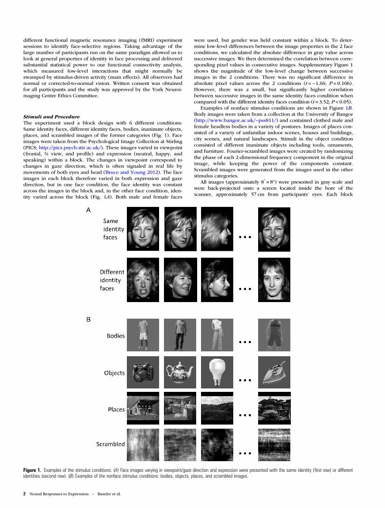
Stimuli and ProcedureThe experiment used a block design with 6 different conditions:Same identity faces, different identity faces, bodies, inanimate objects,places, and scrambled images of the former categories (Fig. 1). Faceimages were taken from the Psychological Image Collection at Stirling(PICS; http://pics.psych.stir.ac.uk/). These images varied in viewpoint(frontal, ¾ view, and profile) and expression (neutral, happy, andspeaking) within a block. The changes in viewpoint correspond tochanges in gaze direction, which is often signaled in real life bymovements of both eyes and head (Bruce and Young 2012). The faceimages in each block therefore varied in both expression and gazedirection, but in one face condition, the face identity was constantacross the images in the block and, in the other face condition, iden-tity varied across the block (Fig. 1A). Both male and female faces
were used, but gender was held constant within a block. To deter-mine low-level differences between the image properties in the 2 faceconditions, we calculated the absolute difference in gray value acrosssuccessive images. We then determined the correlation between corre-sponding pixel values in consecutive images. Supplementary Figure 1shows the magnitude of the low-level change between successiveimages in the 2 conditions. There was no significant difference inabsolute pixel values across the 2 conditions (t =−1.66; P = 0.106).However, there was a small, but significantly higher correlationbetween successive images in the same identity faces condition whencompared with the different identity faces condition (t = 3.52; P < 0.05).
Examples of nonface stimulus conditions are shown in Figure 1B.Body images were taken from a collection at the University of Bangor(http://www.bangor.ac.uk/~pss811/) and contained clothed male andfemale headless bodies in a variety of postures. Images of places con-sisted of a variety of unfamiliar indoor scenes, houses and buildings,city scenes, and natural landscapes. Stimuli in the object conditionconsisted of different inanimate objects including tools, ornaments,and furniture. Fourier-scrambled images were created by randomizingthe phase of each 2-dimensional frequency component in the originalimage, while keeping the power of the components constant.Scrambled images were generated from the images used in the otherstimulus categories.
All images (approximately 8° × 8°) were presented in gray scale andwere back-projected onto a screen located inside the bore of thescanner, approximately 57 cm from participants’ eyes. Each block
Figure 1. Examples of the stimulus conditions. (A) Face images varying in viewpoint/gaze direction and expression were presented with the same identity (first row) or differentidentities (second row). (B) Examples of the nonface stimulus conditions: bodies, objects, places, and scrambled images.
2 Neural Responses to Expression • Baseler et al.
consisted of 10 images from a single-stimulus condition; each image waspresented for 700 ms and followed by a 200-ms blank screen, resultingin a total block length of 9 s. Stimulus blocks were separated by a 9-sgray screen with a central fixation cross. Each condition was repeated 4times in a counterbalanced design, resulting in a total scan length of 7.2min. All participants viewed the same sequence of blocks and images.
Participants were required to monitor all images for the presence ofa red dot that was superimposed on 1 or 2 images in each block. Par-ticipants were required to respond, with a button press, as soon asthey saw the image containing the target. The target could appear inany location on the image and was counterbalanced across conditions.We found no significant differences in the accuracy or reaction timeduring any of the experimental conditions. Mean detection accuracywas 96.9% overall (same identity faces: 96.6%, different identity faces:97.5%, and non-face images: 96.9%). A 1-way analysis of variance(ANOVA) revealed no significant difference in accuracy across con-ditions (F2,200 = 1.01, P = 0.37). The mean reaction time was 441.4 msoverall (same identity faces: 438.7 ms, different identity faces: 441.6ms, and nonface images: 442.1 ms). A 1-way ANOVA found no signifi-cant effect of condition on reaction times (F2,200 = 0.51, P = 0.60).
Imaging ParametersThe experiment was carried out using a 3-T GE HD Excite MRIscanner at the York Neuroimaging Centre (YNiC) at the University ofYork. An 8-channel, phased-array head coil (GE, Milwaukee) tuned to127.4 MHz was used to acquire MRI data. A gradient-echo EPI (echoplanar imaging) sequence was used to collect the data from 38 contig-uous axial slices (time repition = 3 s, time echo = 25 ms, field of view28 × 28 cm, matrix size = 128 × 128, slice thickness 3 mm). These werecoregistered onto a T1-weighted anatomical image (1 × 1 × 1 mm)from each participant. To improve registrations, an additional T1--weighted image was taken in the same plane as the EPI slices.
Whole-Brain AnalysisStatistical analysis of the fMRI data was carried out using FEAT in theFSL toolbox (http://www.fmrib.ox.ac.uk/fsl). The first 3 volumes (9 s)of each scan were removed to minimize the effects of magnetic satur-ation, and slice-timing correction was applied. Motion correction wasfollowed by spatial smoothing (Gaussian, full width at half maximum6 mm) and temporal high-pass filtering (cut off, 0.01 Hz). Regressorsfor each condition in the general linear model (GLM) were convolvedwith a gamma hemodynamic response function. Individual partici-pant data were then entered into a higher level group analysis using amixed effects design (FLAME, http://www.fmrib.ox.ac.uk/fsl) whole-brain analysis. To define face-selective regions, “same identity faces”and “different identity faces” were compared with the responses fromeach of the nonface conditions (bodies, objects, places, andscrambled), and the average of these contrasts was taken. To deter-mine the effect of facial identity, we compared the response fromsame identity faces with the response from different identity faces.
Functional Connectivity AnalysisTo measure the functional connectivity between regions, face-selective regions of interest (ROIs) were identified for each participantusing the averaged contrasts of face > bodies, faces > objects, faces >places, and faces > scrambled, thresholded at P < 0.001 (uncorrected).Regions identified included the left and right occipital face area(OFA), the left and right fusiform face area (FFA), and the right pos-terior superior temporal sulcus (pSTS) face. Because the left pSTSwas found in only a small minority of participants (about 20%), thisregion was not included in the functional connectivity analysis. Acontrol region, which was visually responsive but not face selective,was also defined for each participant by transforming the anatomical“occipital pole, OccP” region from the Harvard-Oxford Cortical Struc-tural Atlas in the MNI standard brain into the participant’s functionaldata space (Desikan et al. 2006). This region included the OccP ofboth left and right hemispheres.
To assess functional connectivity between regions, we firstremoved any stimulus-driven activity, as 2 regions will appear highly
correlated if both are parallelly driven by the stimulus through acommon input. As such, this analysis with stimulus-driven activityremoved is orthogonal to the whole-brain GLM analysis. The stimulus-driven activity was removed through 2 steps (Supplementary Fig. 2):First, the stimulus-driven activation as modeled in the GLM analysiswas removed, resulting in a residual time series response for each par-ticipant. Secondly, to capture any remaining stimulus-driven responsethat might not be fully accounted for the hemodynamic model, thefirst residual time series response was averaged across all ROIs (leftand right OFA, left and right FFA, and right pSTS and OccP) andacross all 103 participants. The rationale for combining across regionsis that the average time-course of response was very similar acrossregions (Supplementary Fig. 4). The first-level analysis was then re-peated with the average first residual response as an additional regres-sor. This gave rise to a second residual for each participant. Thetime-points corresponding to the same identity and different identityface blocks were then extracted for the functional connectivity analy-sis. Correlations between different regions were calculated using theextracted time-points for each participant for the same identity anddifferent identity conditions. Mean correlations and standard error ofthe mean were calculated across participants for each condition andROI pair. To test statistically whether the functional connectivitybetween regions was influenced by facial identity, correlations fromeach participant were converted to a normal distribution using aFisher transformation, and then appropriate statistical tests (repeated-measures ANOVAs and post hoc t-tests) were performed.
Results
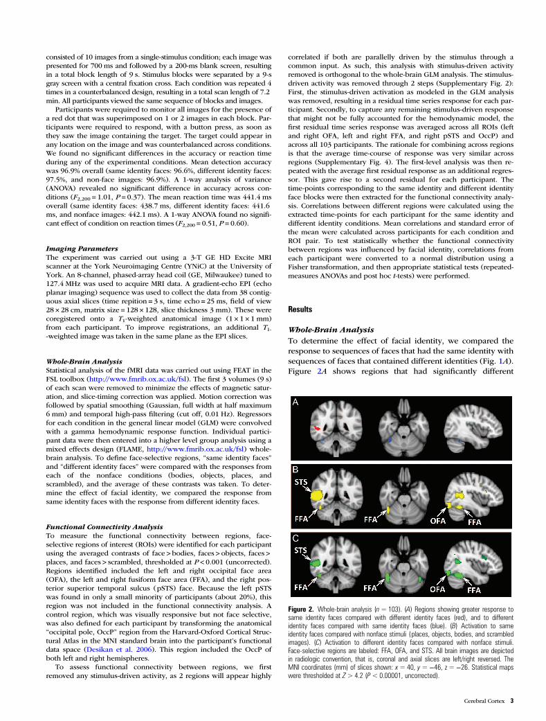
Whole-Brain AnalysisTo determine the effect of facial identity, we compared theresponse to sequences of faces that had the same identity withsequences of faces that contained different identities (Fig. 1A).Figure 2A shows regions that had significantly different
Figure 2. Whole-brain analysis (n=103). (A) Regions showing greater response tosame identity faces compared with different identity faces (red), and to differentidentity faces compared with same identity faces (blue). (B) Activation to sameidentity faces compared with nonface stimuli ( places, objects, bodies, and scrambledimages). (C) Activation to different identity faces compared with nonface stimuli.Face-selective regions are labeled: FFA, OFA, and STS. All brain images are depictedin radiologic convention, that is, coronal and axial slices are left/right reversed. TheMNI coordinates (mm) of slices shown: x= 40, y=−46, z =−26. Statistical mapswere thresholded at Z> 4.2 (P< 0.00001, uncorrected).
Cerebral Cortex 3
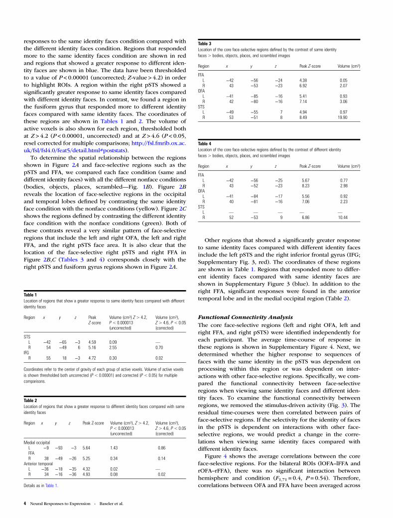
responses to the same identity faces condition compared withthe different identity faces condition. Regions that respondedmore to the same identity faces condition are shown in redand regions that showed a greater response to different iden-tity faces are shown in blue. The data have been thresholdedto a value of P < 0.00001 (uncorrected; Z-value > 4.2) in orderto highlight ROIs. A region within the right pSTS showed asignificantly greater response to same identity faces comparedwith different identity faces. In contrast, we found a region inthe fusiform gyrus that responded more to different identityfaces compared with same identity faces. The coordinates ofthese regions are shown in Tables 1 and 2. The volume ofactive voxels is also shown for each region, thresholded bothat Z > 4.2 (P < 0.00001, uncorrected) and at Z > 4.6 (P < 0.05,resel corrected for multiple comparisons; http://fsl.fmrib.ox.ac.uk/fsl/fsl4.0/feat5/detail.html#poststats).
To determine the spatial relationship between the regionsshown in Figure 2A and face-selective regions such as thepSTS and FFA, we compared each face condition (same anddifferent identity faces) with all the different nonface conditions(bodies, objects, places, scrambled—Fig. 1B). Figure 2Breveals the location of face-selective regions in the occipitaland temporal lobes defined by contrasting the same identityface condition with the nonface conditions (yellow). Figure 2Cshows the regions defined by contrasting the different identityface condition with the nonface conditions (green). Both ofthese contrasts reveal a very similar pattern of face-selectiveregions that include the left and right OFA, the left and rightFFA, and the right pSTS face area. It is also clear that thelocation of the face-selective right pSTS and right FFA inFigure 2B,C (Tables 3 and 4) corresponds closely with theright pSTS and fusiform gyrus regions shown in Figure 2A.
Other regions that showed a significantly greater responseto same identity faces compared with different identity facesinclude the left pSTS and the right inferior frontal gyrus (IFG;Supplementary Fig. 3, red). The coordinates of these regionsare shown in Table 1. Regions that responded more to differ-ent identity faces compared with same identity faces areshown in Supplementary Figure 3 (blue). In addition to theright FFA, significant responses were found in the anteriortemporal lobe and in the medial occipital region (Table 2).
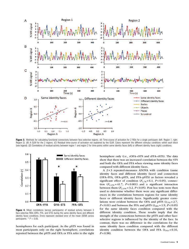
Functional Connectivity AnalysisThe core face-selective regions (left and right OFA, left andright FFA, and right pSTS) were identified independently foreach participant. The average time-course of response inthese regions is shown in Supplementary Figure 4. Next, wedetermined whether the higher response to sequences offaces with the same identity in the pSTS was dependent onprocessing within this region or was dependent on inter-actions with other face-selective regions. Specifically, we com-pared the functional connectivity between face-selectiveregions when viewing same identity faces and different iden-tity faces. To examine the functional connectivity betweenregions, we removed the stimulus-driven activity (Fig. 3). Theresidual time-courses were then correlated between pairs offace-selective regions. If the selectivity for the identity of facesin the pSTS is dependent on interactions with other face-selective regions, we would predict a change in the corre-lations when viewing same identity faces compared withdifferent identity faces.
Figure 4 shows the average correlations between the coreface-selective regions. For the bilateral ROIs (lOFA–lFFA andrOFA–rFFA), there was no significant interaction betweenhemisphere and condition (F1,71 = 0.4, P = 0.54). Therefore,correlations between OFA and FFA have been averaged across
Table 1Location of regions that show a greater response to same identity faces compared with differentidentity faces
Region x y z PeakZ-score
Volume (cm³) Z> 4.2,P< 0.000013(uncorrected)
Volume (cm³),Z> 4.6, P< 0.05(corrected)
STSL −42 −65 −3 4.59 0.09 —
R 54 −49 6 5.16 2.55 0.70IFGR 55 18 −3 4.72 0.30 0.02
Coordinates refer to the center of gravity of each group of active voxels. Volume of active voxelsis shown thresholded both uncorrected (P< 0.00001) and corrected (P< 0.05) for multiplecomparisons.
Table 2Location of regions that show a greater response to different identity faces compared with sameidentity faces
Region x y z Peak Z-score Volume (cm³), Z> 4.2,P< 0.000013(uncorrected)
Volume (cm³),Z> 4.6, P< 0.05(corrected)
Medial occipitalL −9 −93 −3 5.64 1.43 0.86FFAR 38 −49 −26 5.25 0.34 0.14
Anterior temporalL −36 −18 −35 4.32 0.02 —
R 34 −16 −36 4.93 0.08 0.02
Details as in Table 1.
Table 4Location of the core face-selective regions defined by the contrast of different identityfaces > bodies, objects, places, and scrambled images
Region x y z Peak Z-score Volume (cm³)
FFAL −42 −56 −25 5.67 0.77R 43 −52 −23 8.23 2.98
OFAL −41 −84 −17 5.56 0.92R 40 −81 −16 7.06 2.23
STSL — — — — —
R 52 −53 9 6.86 10.44
Table 3Location of the core face-selective regions defined by the contrast of same identityfaces > bodies, objects, places, and scrambled images
Region x y z Peak Z-score Volume (cm³)
FFAL −42 −56 −24 4.38 0.05R 43 −53 −23 6.92 2.07
OFAL −41 −85 −16 5.41 0.93R 42 −80 −16 7.14 3.06
STSL −49 −55 7 4.94 0.97R 53 −51 8 8.49 19.90
4 Neural Responses to Expression • Baseler et al.
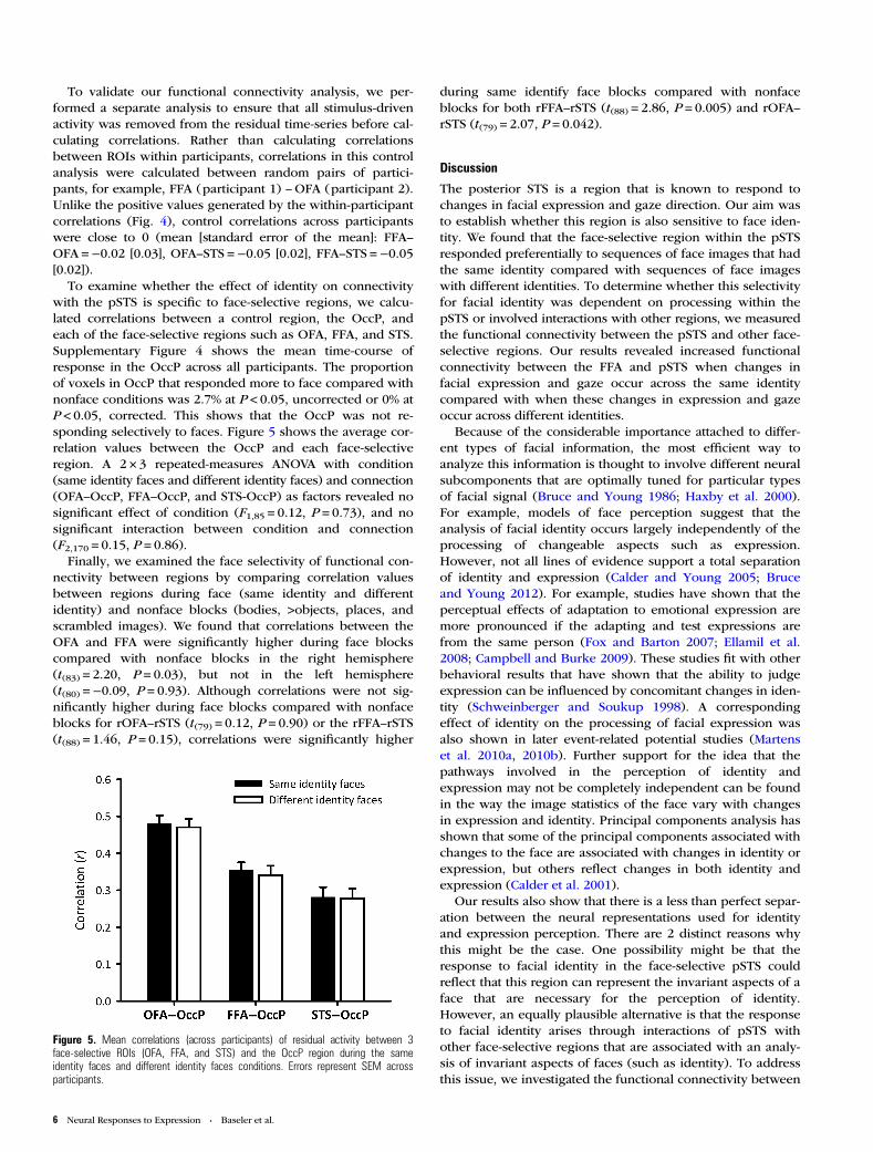
hemispheres for each participant. As the pSTS was found inmost participants only on the right hemisphere, correlationsreported between the pSTS and OFA or FFA refer to the right
hemisphere only (i.e., rOFA–rSTS and rFFA–rSTS). The datashow that there was an increased correlation between the STSand both the OFA and FFA when viewing same identity facescompared with different identity faces.
A 2 × 3 repeated-measures ANOVA with condition (sameidentity faces and different identity faces) and connection(OFA–FFA, OFA–pSTS, and FFA–pSTS) as factors revealed asignificant effect of condition (F1,78 = 6.1, P < 0.05), connec-tion (F2,156 = 41.7, P < 0.001) and a significant interactionbetween them (F2,156 = 3.2, P < 0.05). Post hoc tests were thenused to determine whether there were any significant differ-ences in the correlations between regions for same identityfaces or different identity faces. Significantly greater corre-lations were evident between the OFA and pSTS (t(79) = 2.7,P < 0.01) and between the FFA and pSTS (t(88) = 1.9, P < 0.05)for the same identity faces condition compared with thedifferent identity condition. These results imply that thestrength of the connections between the pSTS and other face-selective regions is influenced by the identity of the face. Incontrast, there was no difference in the correlations for thesame identity faces condition compared with the differentidentity condition between the OFA and FFA (t(92) = 0.16,P = 0.98).
Figure 3. Methods for calculating functional connectivity between face-selective regions. (A) Time-course of activation for 2 ROIs for a single participant (left: Region 1, right:Region 2). (B) A GLM for the 2 regions. (C) Residual time-course of activation not explained by the GLM. Colors represent the different stimulus conditions within each block(see legend). (D) Correlations of residual activity between region 1 and region 2 for time-points within same identity faces (left) or different identity faces (right) conditions.
Figure 4. Mean correlations (across participants) of residual activity between 3face-selective ROIs (OFA, FFA, and STS) during the same identity faces and differentidentity faces conditions. Errors represent standard error of the mean (SEM) acrossparticipants. * P< 0.05.
Cerebral Cortex 5
To validate our functional connectivity analysis, we per-formed a separate analysis to ensure that all stimulus-drivenactivity was removed from the residual time-series before cal-culating correlations. Rather than calculating correlationsbetween ROIs within participants, correlations in this controlanalysis were calculated between random pairs of partici-pants, for example, FFA (participant 1) –OFA (participant 2).Unlike the positive values generated by the within-participantcorrelations (Fig. 4), control correlations across participantswere close to 0 (mean [standard error of the mean]: FFA–OFA =−0.02 [0.03], OFA–STS =−0.05 [0.02], FFA–STS =−0.05[0.02]).
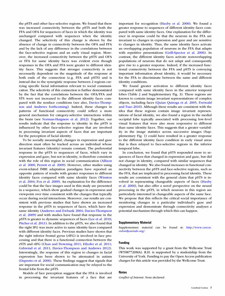
To examine whether the effect of identity on connectivitywith the pSTS is specific to face-selective regions, we calcu-lated correlations between a control region, the OccP, andeach of the face-selective regions such as OFA, FFA, and STS.Supplementary Figure 4 shows the mean time-course ofresponse in the OccP across all participants. The proportionof voxels in OccP that responded more to face compared withnonface conditions was 2.7% at P < 0.05, uncorrected or 0% atP < 0.05, corrected. This shows that the OccP was not re-sponding selectively to faces. Figure 5 shows the average cor-relation values between the OccP and each face-selectiveregion. A 2 × 3 repeated-measures ANOVA with condition(same identity faces and different identity faces) and connection(OFA–OccP, FFA–OccP, and STS-OccP) as factors revealed nosignificant effect of condition (F1,85 = 0.12, P = 0.73), and nosignificant interaction between condition and connection(F2,170 = 0.15, P = 0.86).
Finally, we examined the face selectivity of functional con-nectivity between regions by comparing correlation valuesbetween regions during face (same identity and differentidentity) and nonface blocks (bodies, >objects, places, andscrambled images). We found that correlations between theOFA and FFA were significantly higher during face blockscompared with nonface blocks in the right hemisphere(t(83) = 2.20, P = 0.03), but not in the left hemisphere(t(80) =−0.09, P = 0.93). Although correlations were not sig-nificantly higher during face blocks compared with nonfaceblocks for rOFA–rSTS (t(79) = 0.12, P = 0.90) or the rFFA–rSTS(t(88) = 1.46, P = 0.15), correlations were significantly higher
during same identify face blocks compared with nonfaceblocks for both rFFA–rSTS (t(88) = 2.86, P = 0.005) and rOFA–rSTS (t(79) = 2.07, P = 0.042).
Discussion
The posterior STS is a region that is known to respond tochanges in facial expression and gaze direction. Our aim wasto establish whether this region is also sensitive to face iden-tity. We found that the face-selective region within the pSTSresponded preferentially to sequences of face images that hadthe same identity compared with sequences of face imageswith different identities. To determine whether this selectivityfor facial identity was dependent on processing within thepSTS or involved interactions with other regions, we measuredthe functional connectivity between the pSTS and other face-selective regions. Our results revealed increased functionalconnectivity between the FFA and pSTS when changes infacial expression and gaze occur across the same identitycompared with when these changes in expression and gazeoccur across different identities.
Because of the considerable importance attached to differ-ent types of facial information, the most efficient way toanalyze this information is thought to involve different neuralsubcomponents that are optimally tuned for particular typesof facial signal (Bruce and Young 1986; Haxby et al. 2000).For example, models of face perception suggest that theanalysis of facial identity occurs largely independently of theprocessing of changeable aspects such as expression.However, not all lines of evidence support a total separationof identity and expression (Calder and Young 2005; Bruceand Young 2012). For example, studies have shown that theperceptual effects of adaptation to emotional expression aremore pronounced if the adapting and test expressions arefrom the same person (Fox and Barton 2007; Ellamil et al.2008; Campbell and Burke 2009). These studies fit with otherbehavioral results that have shown that the ability to judgeexpression can be influenced by concomitant changes in iden-tity (Schweinberger and Soukup 1998). A correspondingeffect of identity on the processing of facial expression wasalso shown in later event-related potential studies (Martenset al. 2010a, 2010b). Further support for the idea that thepathways involved in the perception of identity andexpression may not be completely independent can be foundin the way the image statistics of the face vary with changesin expression and identity. Principal components analysis hasshown that some of the principal components associated withchanges to the face are associated with changes in identity orexpression, but others reflect changes in both identity andexpression (Calder et al. 2001).
Our results also show that there is a less than perfect separ-ation between the neural representations used for identityand expression perception. There are 2 distinct reasons whythis might be the case. One possibility might be that theresponse to facial identity in the face-selective pSTS couldreflect that this region can represent the invariant aspects of aface that are necessary for the perception of identity.However, an equally plausible alternative is that the responseto facial identity arises through interactions of pSTS withother face-selective regions that are associated with an analy-sis of invariant aspects of faces (such as identity). To addressthis issue, we investigated the functional connectivity between
Figure 5. Mean correlations (across participants) of residual activity between 3face-selective ROIs (OFA, FFA, and STS) and the OccP region during the sameidentity faces and different identity faces conditions. Errors represent SEM acrossparticipants.
6 Neural Responses to Expression • Baseler et al.
the pSTS and other face-selective regions. We found that therewas increased connectivity between the pSTS and both theFFA and OFA for sequences of faces in which the identity wasunchanged compared with sequences when the identitychanged. The selectivity of this change is shown by theabsence of change in connectivity between the OFA and FFAand by the lack of any difference in the correlations betweenthe face-selective regions and an early visual region. More-over, the increased connectivity between the pSTS and OFAor FFA for same identity faces was evident even thoughresponses in the OFA and FFA were greater to different iden-tity faces. This suggests that stronger connectivity is notnecessarily dependent on the magnitude of the response atboth ends of the connection (e.g. FFA and pSTS) and isinstead due to the synergistic response between 2 regions car-rying specific facial information relevant to social communi-cation. The selectivity of this connection is further demonstratedby the fact that the correlations between the OFA/FFA andSTS were not increased to the different identity faces com-pared with the nonface conditions (see also, Davies-Thomp-son and Andrews forthcoming). Indeed, these changes inpatterns of functional connectivity could reflect a moregeneral mechanism for category-selective interactions withinthe brain (see Norman-Haignere et al. 2012). Together, ourresults indicate that the response to identity in the pSTS isinfluenced by other face-selective regions that are involvedin processing invariant aspects of faces that are importantfor the perception of facial identity.
To be socially meaningful, changes in expression and gazedirection must often be tracked across an individual whoseinvariant features (identity) remain constant. The preferentialresponse in the pSTS to sequences of faces, which vary inexpression and gaze, but not in identity, is therefore consistentwith the role of this region in social communication (Allisonet al. 2000; Perrett et al. 2009). However, other studies usingevent-related fMRI adaptation paradigms have reported anopposite pattern of results with greater responses to differentidentity faces compared with same identity faces (Winstonet al. 2004; Fox et al. 2009). An explanation for the differencecould be that the face images used in this study are presentedin a sequence, which show gradual changes in expression andviewpoint over time consistent with the changes that typicallyoccur during social interactions. Moreover, our results are con-sistent with previous studies that have shown an increasedresponse in the pSTS to sequences of faces, which have thesame identity (Andrews and Ewbank 2004; Davies-Thompsonet al. 2009) and with studies have found that response in thepSTS is greater to dynamic sequences of faces (Lee et al. 2010;Pitcher et al. 2011). In addition to the pSTS, we also found thatthe right IFG was more active to same identity faces comparedwith different identity faces. Previous studies have shown thatthe right inferior frontal gyrus (rIFG) is involved in face pro-cessing and that there is a functional connection between therSTS and rIFG (Chan and Downing 2011; Ethofer et al. 2011;Gshwind et al. 2011; Davies-Thompson and Andrews 2012).Interestingly, the response of this region to changes in facialexpression has been shown to be attenuated in autism(Dapretto et al. 2006). These findings suggest that signals thatare important for social communication may be relayed to thefrontal lobe from the pSTS.
Models of face perception suggest that the FFA is involvedin processing the invariant features of a face that are
important for recognition (Haxby et al. 2000). We found agreater response to sequences of different identity faces com-pared with same identity faces. One explanation for the differ-ence in response could be that the neurons in the FFA areinvariant to changes in expression and gaze and are sensitiveto changes in identity. Thus, the same identity faces activatean overlapping population of neurons in the FFA that adaptswith repetitive presentations (Grill-Spector et al. 2006). Incontrast, the different identity faces activate nonoverlappingpopulations of neurons that do not adapt and consequentlygive rise to a greater response. Indeed, if the increased func-tional connectivity between the FFA and pSTS is conveyingimportant information about identity, it would be necessaryfor the FFA to discriminate between the same and differentidentity conditions.
We found greater activation to different identity facescompared with same identity faces in the anterior temporallobes (Table 2 and Supplementary Fig. 3). These regions areknown to contain image invariant representations of complexobjects, including faces (Quian Quiroga et al. 2005, Freiwaldand Tsao 2010). Although these results are consistent with theidea that these regions contain image invariant represen-tations of facial identity, we also found a region in the medialoccipital lobe typically associated with processing low-levelvisual features that were also more responsive to differentthan same identity faces. This suggests that a greater variabil-ity in the image statistics across successive images (Sup-plementary Fig. 1) could have resulted in a greater responseto the different identity faces condition in early visual areasthat is then relayed to face-selective regions in the inferiortemporal lobe.
In conclusion, we found that pSTS responded more to se-quences of faces that changed in expression and gaze, but didnot change in identity, compared with similar sequences thatchanged in identity. We also found increases in functional con-nectivity between the pSTS and face-selective regions, such asthe FFA, that are implicated in processing facial identity. Theseresults are consistent with the general claim that pSTS is in-volved in representing changeable aspects of faces (Haxbyet al. 2000), but also offer a novel perspective on the neuralprocessing in the pSTS, in which neurons in this region areparticularly interested in changeable aspects of the same face.We propose that this reflects the critical social importance ofmonitoring changes in a particular individual’s gaze andexpression and demonstrate through connectivity analyses apotential mechanism through which this can happen.
Supplementary MaterialSupplementary material can be found at: http://www.cercor.oxfordjournals.org/.
Funding
This work was supported by a grant from the Wellcome Trust(WT087720MA). R.H. is supported by a studentship from theUniversity of York. Funding to pay the Open Access publicationcharges for this article was provided by the Wellcome Trust.
NotesConflict of Interest: None declared.
Cerebral Cortex 7
ReferencesAllison T, McCarthy G, Nobre A, Puce A, Belger A. 1994. Human ex-
trastriate visual cortex and the perception of faces, words,numbers, and colors. Cereb Cortex. 4:544–554.
Allison T, Puce A, McCarthy G. 2000. Social perception from visualcues: role of the pSTS region. Trends Cogn Sci. 4:267–278.
Andrews TJ, Ewbank MP. 2004. Distinct representations for facialidentity and changeable aspects of faces in the human temporallobe. Neuroimage. 23:905–913.
Barton JJS, Press DZ, Keenan JP, O’Connor M. 2002. Lesions of thefusiform face area impair perception of facial configuration in pro-sopagnosia. Neurology. 58:71–78.
Biswal B, Yetkin FZ, Haughton VM, Hyde JS. 1995. Functional con-nectivity in the motor cortex of resting human brain using echo-planar MRI. Magn Reson Med. 34:537–541.
Bruce V, Young A. 2012. Face perception. Hove, East Sussex: Psychol-ogy Press.
Bruce V, Young A. 1986. Understanding face recognition. Br JPsychol. 77:305–327.
Calder AJ, Beaver JD, Winston JS, Dolan RJ, Jenkins R, Eger E,Henson RN. 2007. Separate coding of different gaze directions inthe superior temporal sulcus and inferior parietal lobule. CurrBiol. 17:20–25.
Calder AJ, Burton AM, Miller P, Young AW, Akamatsu S. 2001. A prin-cipal component analysis of facial expressions. Vis Res. 41(9):1179–1208.
Calder AJ, Young AW. 2005. Understanding the recognition of facialidentity and facial expression. Nat Rev Neurosci. 6:641–651.
Campbell J, Burke D. 2009. Evidence that identity-dependent andidentity independent neural populations are recruited in the per-ception of five basic emotional facial expressions. Vis Res.49:1532–1540.
Chan AW, Downing PE. 2011. Faces and eyes in human lateral pre-frontal cortex. Front Hum Neurosci. 5:51.
Dapretto M, Davies MS, Pfeifer JH, Scott AA, Sigman M, BookheimerSY, Iacoboni M. 2006. Understanding emotions in others: mirrorneuron dysfunction in children with autism spectrum disorders.Nat Neurosci. 9:28–30.
Davies-Thompson J, Andrews TJ. Forthcoming. Intra- and inter-hemispheric connectivity between face-selective regions in thehuman brain. J Neurophysiol. doi: 10.1152/jn.01171.2011.
Davies-Thompson J, Gouws A, Andrews TJ. 2009. An image-dependent representation of familiar and unfamiliar faces in thehuman ventral stream. Neuropsychologia. 47(6):1627–1635.
Desikan RS, Ségonne F, Fischl B, Quinn BT, Dickerson BC, Blacker D,Buckner RL, Dale AM, Maguire RP, Hyman BT et al. 2006. Anautomated labeling system for subdividing the human cerebralcortex on MRI scans into gyral based regions of interest. Neuro-image. 31:968–980.
Ellamil M, Susskind JM, Anderson AK. 2008. Examinations of identityinvariance in facial expression adaptation. Cogn Affect BehavNeurosci. 8:273–281.
Engell AD, Haxby JV. 2007. Facial expression and gaze-direction in hu-man superior temporal sulcus. Neuropsychologia. 45:3234–3241.
Ethofer T, Gshwind M, Vuilleumier P. 2011. Processing social aspectsof human gaze: a combined fMRI-DTI study. Neuroimage. 55:411–419.
Fox CJ, Barton JJS. 2007. What is adapted in face adaptation? Theneural representations of expression in the human visual system.Brain Res. 1127:80–89.
Fox CJ, Moon S-Y, Iaria G, Barton JJS. 2009. The correlates of subjec-tive perception of identity and expression in the face network: anfMRI adaptation study. Neuroimage. 44:569–580.
Freiwald WA, Tsao DY. 2010. Functional compartmentalization andviewpoint generalization within the macaque face-processingsystem. Science. 330:845.
Grill-Spector K, Henson R, Martin A. 2006. Repetition and the brain:neural models of stimulus-specific effects. Trends Cogn Sci. 10:14–23.
Grill-Spector K, Knouf N, Kanwisher N. 2004. The fusiform face areasubserves face perception, not generic within-category identifi-cation. Nat Neurosci. 7:555–562.
Gshwind M, Pourtois G, Schwartz S, Van De Ville D, Vuilleumier P.2011. White matter connectivity between face-responsive regionsin the human brain. Cereb Cortex. doi: 10.1093/cercor/bhr226
Haxby JV, Hoffman EA, Gobbini MI. 2000. The distributed humanneural system for face perception. Trends Cogn Sci. 4:223–233.
Hoffman EA, Haxby JV. 2000. Distinct representation of eye gaze andidentity in the distributed human neural system for face percep-tion. Nat Neurosci. 3:80–84.
Kanwisher N, McDermott J, Chun MM. 1997. The fusiform face area: amodule in human extrastriate cortex specialized for face percep-tion. J Neurosci. 17:4302–4311.
Lee LC, Andrews TJ, Johnson SJ, Woods W, Gouws A, Green GGR,Young AW. 2010. Neural responses to rigidly moving faces dis-playing shifts in social attention investigated with fMRI and MEG.Neuropsychologia. 48:477–490.
Margulies DS, Bottger J, Long X, Yating L, Kelly C, Schafer A, GoldhahnD, Abbushi A, Milham MP, Lohmann G et al. 2010. Resting develop-ments: a review of fMRI post-processing methodologies for spon-taneous brain activity. Magn Reson Mater Phys Biol Med. 23:289–307.
Martens U, Leuthold H, Schweinberger SR. 2010a. Parallel processingin face perception. J Exp Psychol Hum Percept Perform.36:103–121.
Martens U, Leuthold H, Schweinberger SR. 2010b. On the temporalorganization of facial identity and expression analysis: Inferencesfrom event-related brain potentials. Cogn Affect Soc Neurosci.10:505–522.
Norman-Haignere SV, McCarthy G, Chun MM, Turk-Browne NB.2012. Category-selective background connectivity in ventral visualcortex. Cereb Cortex. 22:391–402.
Pelphrey KA, Viola RJ, McCarthy G. 2004. Processing of mutual andaverted social gaze in the superior temporal sulcus. Psychol Sci.15:598–603.
Perrett DI, Xiao D, Barraclough NE, Keysers C, Oram MW. 2009.Seeing the future: natural image sequences produce ‘anticipatory’neuronal activity and bias perceptual report. Q J Exp Psychol.62:2081–2104.
Pitcher D, Dilks DD, Saxe RR, Triantafyllou C, Kanwisher N. 2011.Differential selectivity for dynamic versus static information inface-selective cortical regions. Neuroimage. 56:2356–2363.
Puce A, Allison T, Bentin S, Gore JC, McCarthy G. 1998. Temporalcortex activation in humans viewing eye and mouth movements.J Neurosci. 18:2188–2199.
Quian Quiroga R, Reddy L, Kreiman G, Koch C, Fried I. 2005. Invar-iant visual representation by single-neurons in the human brain.Nature. 435:1102–1107.
Rotshtein P, Henson RN, Treves A, Driver J, Dolan RJ. 2005. MorphingMarilyn into Maggie dissociates physical and identity face rep-resentations in the brain. Nat Neurosci. 8:107–113.
Schweinberger SR, Soukup GR. 1998. Asymmetric relationshipsamong perceptions of facial identity, emotion, and facial speech.J Exp Psychol Hum Percep Perform. 24:1748–1765.
Turk-Browne NB, Norman-Haignere SV, McCarthy G. 2010. Face-specific resting functional connectivity between the fusiform gyrusand posterior superior temporal sulcus. Front Hum Neurosci. 4(176):1–15.
Winston JS, Henson RN, Fine-Goulden MR, Dolan RJ. 2004.fMRI-adaptation reveals dissociable neural representations of identityand expression in face perception. J Neurophysiol. 92:1830–1839.
Zhang H, Tian J, Liu J, Li J, Lee K. 2009. Intrinsically organizednetwork for face perception during the resting state. Neurosci Lett.454:1–5.
8 Neural Responses to Expression • Baseler et al.
Related Documents