Neural respiratory drive and breathlessness in COPD Caroline J. Jolley 1 , Yuanming M. Luo 2 , Joerg Steier 1,3 , Gerrard F. Rafferty 1 , Michael I. Polkey 4 and John Moxham 1 Affiliations: 1 King’s College London Division of Asthma, Allergy and Lung Biology, King’s College London School of Medicine, King’s Health Partners, London, UK. 2 State Key Laboratory of Respiratory Disease, Guangzhou Medical College, Guangzhou, China. 3 Lane Fox Respiratory Unit/Sleep Disorders Centre, Guy’s and St Thomas’ NHS Foundation Trust, London, UK. 4 NIHR Respiratory Biomedical Research Unit, Royal Brompton and Harefield NHS Foundation Trust and Imperial College, London, UK. Correspondence: Caroline Jolley, King’s College London Division of Asthma, Allergy and Lung Biology, King’s College London School of Medicine, King’s Health Partners, Bessemer Road, SE5 9PJ, London, UK. E-mail: [email protected] ABSTRACT The aim of this study was to test the hypothesis that neural respiratory drive, measured using diaphragm electromyogram (EMGdi) activity expressed as a percentage of maximum (EMGdi%max), is closely related to breathlessness in chronic obstructive pulmonary disease. We also investigated whether neuroventilatory uncoupling contributes significantly to breathlessness intensity over an awareness of levels of neural respiratory drive alone. EMGdi and ventilation were measured continuously during incremental cycle and treadmill exercise in 12 chronic obstructive pulmonary disease patients (forced expiratory volume in 1 s¡SD was 38.7¡14.5 % pred). EMGdi was expressed both as EMGdi%max and relative to tidal volume expressed as a percentage of predicted vital capacity to quantify neuroventilatory uncoupling. EMGdi%max was closely related to Borg breathlessness in both cycle (r50.98, p50.0001) and treadmill exercise (r50.94, p50.005), this relationship being similar to that between neuroventilatory uncoupling and breathlessness (cycling r50.94, p50.005; treadmill r5 0.91, p50.01). The relationship between breathlessness and ventilation was poor when expansion of tidal volume became limited. In chronic obstructive pulmonary disease the intensity of exertional breathlessness is closely related to EMGdi%max. These data suggest that breathlessness in chronic obstructive pulmonary disease can be largely explained by an awareness of levels of neural respiratory drive, rather than the degree of neuroventilatory uncoupling. EMGdi%max could provide a useful physiological biomarker for breathlessness in chronic obstructive pulmonary disease. @ERSpublications Exertional breathlessness in patients with severe COPD is closely related to levels of neural respiratory drive http://ow.ly/BO6MI Received: April 03 2014 | Accepted after revision: Aug 26 2014 Support statement: C. Jolley was funded by a MRC Clinical Research Training Fellowship. Both C. Jolley and Y.M. Luo were awarded a British Council Researcher Exchange Award to support the collaboration between the British (London) and Chinese (Guangzhou) research groups. The contribution by M.I. Polkey to this study was supported by the NIHR Respiratory Biomedical Research Unit at the Royal Brompton and Harefield NHS Foundation Trust and Imperial College, who part fund his salary. Conflict of interest: None declared. Copyright ßERS 2014 This article has supplementary material available from erj.ersjournals.com ORIGINAL ARTICLE IN PRESS | CORRECTED PROOF Eur Respir J 2014; in press | DOI: 10.1183/09031936.00063014 1 . Published on October 16, 2014 as doi: 10.1183/09031936.00063014 ERJ Express Copyright 2014 by the European Respiratory Society.

Welcome message from author
This document is posted to help you gain knowledge. Please leave a comment to let me know what you think about it! Share it to your friends and learn new things together.
Transcript
Neural respiratory drive andbreathlessness in COPD
Caroline J. Jolley1, Yuanming M. Luo2, Joerg Steier1,3, Gerrard F. Rafferty1,Michael I. Polkey4 and John Moxham1
Affiliations: 1King’s College London Division of Asthma, Allergy and Lung Biology, King’s College LondonSchool of Medicine, King’s Health Partners, London, UK. 2State Key Laboratory of Respiratory Disease,Guangzhou Medical College, Guangzhou, China. 3Lane Fox Respiratory Unit/Sleep Disorders Centre, Guy’s andSt Thomas’ NHS Foundation Trust, London, UK. 4NIHR Respiratory Biomedical Research Unit, Royal Bromptonand Harefield NHS Foundation Trust and Imperial College, London, UK.
Correspondence: Caroline Jolley, King’s College London Division of Asthma, Allergy and Lung Biology, King’sCollege London School of Medicine, King’s Health Partners, Bessemer Road, SE5 9PJ, London, UK.E-mail: [email protected]
ABSTRACT The aim of this study was to test the hypothesis that neural respiratory drive, measured using
diaphragm electromyogram (EMGdi) activity expressed as a percentage of maximum (EMGdi%max), is
closely related to breathlessness in chronic obstructive pulmonary disease. We also investigated whether
neuroventilatory uncoupling contributes significantly to breathlessness intensity over an awareness of levels
of neural respiratory drive alone.
EMGdi and ventilation were measured continuously during incremental cycle and treadmill exercise in 12
chronic obstructive pulmonary disease patients (forced expiratory volume in 1 s¡SD was 38.7¡14.5 % pred).
EMGdi was expressed both as EMGdi%max and relative to tidal volume expressed as a percentage of predicted
vital capacity to quantify neuroventilatory uncoupling.
EMGdi%max was closely related to Borg breathlessness in both cycle (r50.98, p50.0001) and treadmill
exercise (r50.94, p50.005), this relationship being similar to that between neuroventilatory uncoupling
and breathlessness (cycling r50.94, p50.005; treadmill r5 0.91, p50.01). The relationship between
breathlessness and ventilation was poor when expansion of tidal volume became limited.
In chronic obstructive pulmonary disease the intensity of exertional breathlessness is closely related to
EMGdi%max. These data suggest that breathlessness in chronic obstructive pulmonary disease can be largely
explained by an awareness of levels of neural respiratory drive, rather than the degree of neuroventilatory
uncoupling. EMGdi%max could provide a useful physiological biomarker for breathlessness in chronic
obstructive pulmonary disease.
@ERSpublications
Exertional breathlessness in patients with severe COPD is closely related to levels of neuralrespiratory drive http://ow.ly/BO6MI
Received: April 03 2014 | Accepted after revision: Aug 26 2014
Support statement: C. Jolley was funded by a MRC Clinical Research Training Fellowship. Both C. Jolley and Y.M. Luowere awarded a British Council Researcher Exchange Award to support the collaboration between the British (London)and Chinese (Guangzhou) research groups. The contribution by M.I. Polkey to this study was supported by the NIHRRespiratory Biomedical Research Unit at the Royal Brompton and Harefield NHS Foundation Trust and Imperial College,who part fund his salary.
Conflict of interest: None declared.
Copyright �ERS 2014
This article has supplementary material available from erj.ersjournals.com
ORIGINAL ARTICLEIN PRESS | CORRECTED PROOF
Eur Respir J 2014; in press | DOI: 10.1183/09031936.00063014 1
. Published on October 16, 2014 as doi: 10.1183/09031936.00063014ERJ Express
Copyright 2014 by the European Respiratory Society.
IntroductionBreathlessness is an important cause of exercise limitation and reduced quality of life for patients with
chronic obstructive pulmonary disease (COPD) [1, 2]. Our understanding of the psychophysiological
mechanisms underlying breathlessness remains incomplete [2, 3]. Although it is widely appreciated that an
awareness of levels of motor output to the respiratory muscles from the brainstem respiratory centre is
important to the sensation of breathlessness [4], the identification of reliable physiological measures of
neural respiratory drive (NRD) poses a significant challenge. Ventilation does not adequately reflect NRD in
COPD because changes in the mechanical properties of the respiratory system alter the relationship between
NRD and inspiratory flow [5]. Similarly, since respiratory muscle pressure generation is dependent on the
contractile function of the respiratory muscles and the length-tension properties of the muscle, independent
the NRD level, pressure-derived variables underestimate NRD when pulmonary hyperinflation is present [5, 6].
Breathlessness has also been shown to increase disproportionally if the ventilatory response is limited by
impaired pulmonary mechanics (neuroventilatory uncoupling (NVU)) [7]. To date, the relationship
between NVU and breathlessness in COPD has been most extensively studied using the ‘‘effort/
displacement ratio’’ (EDR). The EDR is the ratio of tidal swings of oesophageal pressure (Poes) relative to
maximum inspiratory pressure (PImax) i.e. ‘‘effort’’ and the tidal volume (VT) response expressed relative to
the predicted vital capacity (VC) i.e. ‘‘displacement’’, thus giving the ratio of Poes/PImax to VT/predicted VC
[8]. The EDR correlates well with Borg breathlessness in normal subjects and in patients with chronic
airflow limitation [8], increasing markedly with breathlessness after a critically low inspiratory reserve
volume (IRV) has been reached [9]. However because the oesophageal pressure swing is reduced by
hyperinflation [10], the reliability of indices such as the EDR, which rely on pressure-derived measures of
NRD, is also limited for COPD.
Recent advances in measuring the diaphragm electromyogram (EMGdi), using multi-pair oesophageal
recording electrodes by our group [11–14] and others [15–18], have enabled the accurate quantification of
NRD and NVU in terms of a measurement that is neurophysiologically more ‘‘upstream’’ of respiratory
pressure generation. The aim of the present study was to investigate the relationship between NRD and
breathlessness, using EMGdi as an index of NRD. The influence of NVU on increases in the intensity
of breathlessness was also assessed, using the ratio of EMGdi activity to ventilatory output as an index of
NVU. These relationships were examined during maximal cycle and treadmill exercise tests to assess the
consistency of relationships during different forms of exercise.
MethodsPatients12 COPD patients (mean¡SD) age 66.7¡7.0 years, forced expiratory volume in 1 s (FEV1) 38.7¡14.5 % pred,
n511 males) were studied. Participants were recruited in accordance with local research ethics committee
procedures, and written informed consent was given. Age, height, weight, body mass index and Medical
Research Council dyspnoea score were documented.
Participants made two visits at least 24 h apart. FEV1 and slow VC were measured on both days. Inspiratory
capacity (IC) and lung volumes (using body plethysmography) were measured prior to exercise on visit 2 only.
Exercise protocolsVisit 1Incremental cycle and incremental treadmill exercise tests were undertaken to familiarise participants with
the exercise and breathlessness assessment protocols. The patients were asked to score breathlessness
intensity (strength of sensation) and the intensity of leg fatigue using the modified Borg scale [19]. Each
subject was briefed by explaining that descriptors of the intensity of the sensations on the Borg scale are
anchored to numerical points on the scale, e.g. for breathlessness, varying between 0 (no breathlessness) and
10 (maximum breathlessness the patient had ever experienced). Further details of the equipment and
exercise protocols are provided in the online data supplement. Modified Borg breathlessness and leg fatigue
scores were assessed at baseline, at the end of each minute of exercise and at the time of exercise limitation
by symptoms (‘‘symptom limitation’’) in both protocols.
Visit 2EMGdi was recorded using a multi-pair oesophageal catheter [20] and ventilation recorded using a
pneumotachograph connected to the patient via the full face mask with a noseclip in place. Following
measurements made seated at rest, subjects performed the incremental cycle and treadmill exercise tests to
symptom limitation, with Borg breathlessness and leg fatigue assessments as in visit 1. At recruitment,
patients were alternately assigned to undertake either the cycle or treadmill exercise first. Both exercise tests
COPD | C.J. JOLLEY ET AL.
DOI: 10.1183/09031936.000630142
were performed on the same day with a minimum rest of 1 h between. Oxygen uptake, carbon dioxide
production and minute ventilation (V9E) were measured throughout exercise (AD Instruments, Castle Hill,
Australia), using a full face mask. Data were acquired using a desktop computer (Apple Computer Inc,
Cupertino, CA, USA) running Chart software (version 5.4.2, AD Instruments). Metabolic data were
analysed online and stored for further offline analysis. Haemoglobin oxygen saturation was assessed by
pulse oximetry in all patients. IC was assessed immediately before each exercise test and as close to the
exercise termination point as possible.
Data analysisEMGdi, metabolic and other cardiorespiratory data were averaged over 30-s time periods. Group data were
reported at each 20% epoch of exercise time (i.e. from the end of the rest period onwards) and the patterns
of increase in each variable during each exercise modality examined.
EMGdi activity, expressed as a percentage of maximum (EMGdi%max), was calculated per breath as the
mean peak root mean square (RMS) EMGdi per breath expressed as a percentage of the peak RMS EMGdi
activity, which were recorded during inspiratory manoeuvres performed before exercise (inspiration to total
lung capacity from functional residual capacity (FRC), maximum sniff from FRC, PImax manoeuvre against
a closed shutter from FRC and sprint maximum voluntary ventilation .15 s). In addition EMGdi%max was
also calculated during performance of IC manoeuvres at end exercise, and during breaths taken throughout
the exercise protocols. The NVU index was calculated as the ratio of EMGdi%max to VT expressed as a
percentage of predicted vital capacity i.e. EMGdi%max/VT%VCpred). Further details, including EMGdi signal
processing and analysis of composite indices derived from EMGdi%max, and respiratory rate and inspiratory
time, are provided in the online supplementary material.
Analysis of slopes of the relationships between Borg breathlessness, EMGdi%max and ventilationafter the ‘‘breathlessness threshold’’Previous studies in both healthy subjects and patients have shown that patterns of increases in
breathlessness usually follow an alinear ‘‘tick’’ shape, with little change in breathlessness over the first few
minutes of exercise, followed by a phase of rapid increases in breathlessness after the breathlessness
threshold [9, 14]. The breathlessness threshold was determined by plotting individual Borg breathlessness
time-curves throughout exercise, identifying, by eye, a point of inflection for each subject as the point of
intersection of two linear relationships. In the present study, for each patient, each physiological variable
was normalised to the value of that variable at the timepoint at which the slope of the breathlessness–time
relationship became more steeply positive at the breathlessness threshold (figure S1). For example, if V9E at
baseline, 20% and 40% epochs of exercise time were 10, 20 and 25 L?min-1, respectively, and breathlessness
increased sharply at 20% exercise time, the normalised values of V9E were 10/20 (0.5), 20/20 (1) and 25/20
(1.25) at baseline, 20% and 40% exercise time, respectively, a normalised value of 2 indicating doubling of
that variable relative to the take-off point. This allowed the slopes of the increases in each variable to be
compared regardless of the absolute numerical value. The purpose of this process was to evaluate the
relationships between incremental changes in physiological variables with increases in breathlessness, so as
to evaluate the discriminatory powers of each variable. More positive (‘‘steeper’’) slopes would indicate that
increases in breathlessness were associated with little incremental numerical increase in the physiological
variable, indicating that changes in such variables discriminated poorly between successive step increases in
the intensity of breathlessness.
Statistical analysisStatistical analysis was carried out using SPSS software version 15.0.1 for Windows (SPSS Inc, Chicago, IL,
USA) and Graphpad Prism 5 for Windows v5.00 (Graphpad Software Inc, La Jolla, CA, USA). EMGdi%max was
non-normally distributed during exercise and, therefore, described as median (interquartile range (IQR)) and
analysed using nonparametric statistics. Inter-subject comparisons were made using Wilcoxon signed-rank
tests (nonparametric) unless otherwise stated. A p-value ,0.05 was considered statistically significant.
ResultsThe results obtained during cycle and treadmill exercise testing, were similar. Consequently only the results
from the cycle exercise are reported here to highlight the principal findings. The treadmill data and cycle/
treadmill comparisons are reported in the online supplementary data. Results of analysis for composite
indices derived from EMGdi%max, respiratory rate and inspiratory time are also reported in the online
supplementary data.
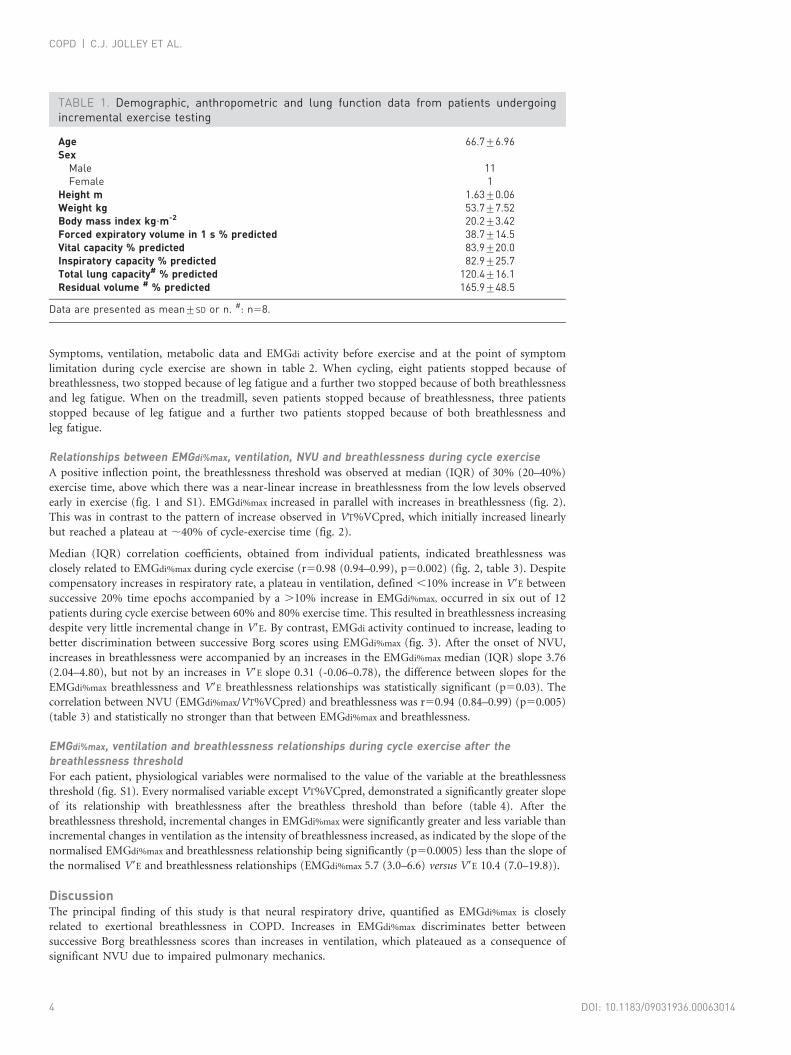
Demographic and anthropometric data, spirometry, and lung volume measurements are shown in table 1.
Lung volume measurements were obtained from only eight patients.
COPD | C.J. JOLLEY ET AL.
DOI: 10.1183/09031936.00063014 3
Symptoms, ventilation, metabolic data and EMGdi activity before exercise and at the point of symptom
limitation during cycle exercise are shown in table 2. When cycling, eight patients stopped because of
breathlessness, two stopped because of leg fatigue and a further two stopped because of both breathlessness
and leg fatigue. When on the treadmill, seven patients stopped because of breathlessness, three patients
stopped because of leg fatigue and a further two patients stopped because of both breathlessness and
leg fatigue.
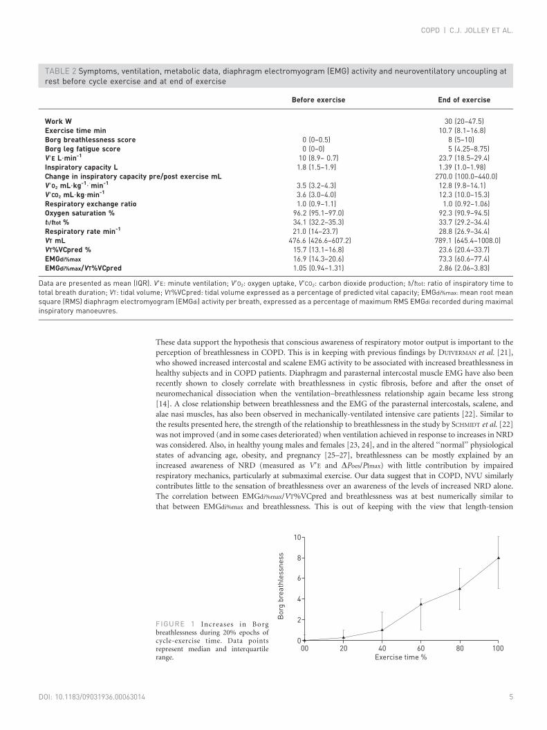
Relationships between EMGdi%max, ventilation, NVU and breathlessness during cycle exerciseA positive inflection point, the breathlessness threshold was observed at median (IQR) of 30% (20–40%)
exercise time, above which there was a near-linear increase in breathlessness from the low levels observed
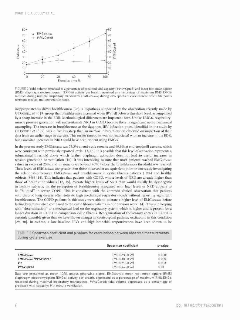
early in exercise (fig. 1 and S1). EMGdi%max increased in parallel with increases in breathlessness (fig. 2).
This was in contrast to the pattern of increase observed in VT%VCpred, which initially increased linearly
but reached a plateau at ,40% of cycle-exercise time (fig. 2).
Median (IQR) correlation coefficients, obtained from individual patients, indicated breathlessness was
closely related to EMGdi%max during cycle exercise (r50.98 (0.94–0.99), p50.002) (fig. 2, table 3). Despite
compensatory increases in respiratory rate, a plateau in ventilation, defined ,10% increase in V9E between
successive 20% time epochs accompanied by a .10% increase in EMGdi%max, occurred in six out of 12
patients during cycle exercise between 60% and 80% exercise time. This resulted in breathlessness increasing
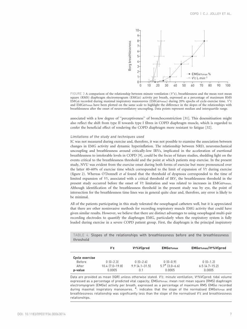
despite very little incremental change in V9E. By contrast, EMGdi activity continued to increase, leading to
better discrimination between successive Borg scores using EMGdi%max (fig. 3). After the onset of NVU,
increases in breathlessness were accompanied by an increases in the EMGdi%max median (IQR) slope 3.76
(2.04–4.80), but not by an increases in V9E slope 0.31 (-0.06–0.78), the difference between slopes for the
EMGdi%max breathlessness and V9E breathlessness relationships was statistically significant (p50.03). The
correlation between NVU (EMGdi%max/VT%VCpred) and breathlessness was r50.94 (0.84–0.99) (p50.005)
(table 3) and statistically no stronger than that between EMGdi%max and breathlessness.
EMGdi%max, ventilation and breathlessness relationships during cycle exercise after thebreathlessness thresholdFor each patient, physiological variables were normalised to the value of the variable at the breathlessness
threshold (fig. S1). Every normalised variable except VT%VCpred, demonstrated a significantly greater slope
of its relationship with breathlessness after the breathless threshold than before (table 4). After the
breathlessness threshold, incremental changes in EMGdi%max were significantly greater and less variable than
incremental changes in ventilation as the intensity of breathlessness increased, as indicated by the slope of the
normalised EMGdi%max and breathlessness relationship being significantly (p50.0005) less than the slope of
the normalised V9E and breathlessness relationships (EMGdi%max 5.7 (3.0–6.6) versus V9E 10.4 (7.0–19.8)).
DiscussionThe principal finding of this study is that neural respiratory drive, quantified as EMGdi%max is closely
related to exertional breathlessness in COPD. Increases in EMGdi%max discriminates better between
successive Borg breathlessness scores than increases in ventilation, which plateaued as a consequence of
significant NVU due to impaired pulmonary mechanics.
TABLE 1. Demographic, anthropometric and lung function data from patients undergoingincremental exercise testing
Age 66.7¡6.96Sex
Male 11Female 1
Height m 1.63¡0.06Weight kg 53.7¡7.52Body mass index kg?m-2 20.2¡3.42Forced expiratory volume in 1 s % predicted 38.7¡14.5Vital capacity % predicted 83.9¡20.0Inspiratory capacity % predicted 82.9¡25.7Total lung capacity# % predicted 120.4¡16.1Residual volume # % predicted 165.9¡48.5
Data are presented as mean¡SD or n. #: n58.
COPD | C.J. JOLLEY ET AL.
DOI: 10.1183/09031936.000630144
These data support the hypothesis that conscious awareness of respiratory motor output is important to the
perception of breathlessness in COPD. This is in keeping with previous findings by DUIVERMAN et al. [21],
who showed increased intercostal and scalene EMG activity to be associated with increased breathlessness in
healthy subjects and in COPD patients. Diaphragm and parasternal intercostal muscle EMG have also been
recently shown to closely correlate with breathlessness in cystic fibrosis, before and after the onset of
neuromechanical dissociation when the ventilation–breathlessness relationship again became less strong
[14]. A close relationship between breathlessness and the EMG of the parasternal intercostals, scalene, and
alae nasi muscles, has also been observed in mechanically-ventilated intensive care patients [22]. Similar to
the results presented here, the strength of the relationship to breathlessness in the study by SCHMIDT et al. [22]
was not improved (and in some cases deteriorated) when ventilation achieved in response to increases in NRD
was considered. Also, in healthy young males and females [23, 24], and in the altered ‘‘normal’’ physiological
states of advancing age, obesity, and pregnancy [25–27], breathlessness can be mostly explained by an
increased awareness of NRD (measured as V9E and DPoes/PImax) with little contribution by impaired
respiratory mechanics, particularly at submaximal exercise. Our data suggest that in COPD, NVU similarly
contributes little to the sensation of breathlessness over an awareness of the levels of increased NRD alone.
The correlation between EMGdi%max/VT%VCpred and breathlessness was at best numerically similar to
that between EMGdi%max and breathlessness. This is out of keeping with the view that length-tension
TABLE 2 Symptoms, ventilation, metabolic data, diaphragm electromyogram (EMG) activity and neuroventilatory uncoupling atrest before cycle exercise and at end of exercise
Before exercise End of exercise
Work W 30 (20–47.5)Exercise time min 10.7 (8.1–16.8)Borg breathlessness score 0 (0–0.5) 8 (5–10)Borg leg fatigue score 0 (0–0) 5 (4.25–8.75)V9E L?min-1 10 (8.9– 0.7) 23.7 (18.5–29.4)Inspiratory capacity L 1.8 (1.5–1.9) 1.39 (1.0–1.98)Change in inspiratory capacity pre/post exercise mL 270.0 (100.0–440.0)V9O2 mL?kg-1??min-1 3.5 (3.2–4.3) 12.8 (9.8–14.1)V9CO2 mL?kg?min-1 3.6 (3.0–4.0) 12.3 (10.0–15.3)Respiratory exchange ratio 1.0 (0.9–1.1) 1.0 (0.92–1.06)Oxygen saturation % 96.2 (95.1–97.0) 92.3 (90.9–94.5)tI/ttot % 34.1 (32.2–35.3) 33.7 (29.2–34.4)Respiratory rate min-1 21.0 (14–23.7) 28.8 (26.9–34.4)VT mL 476.6 (426.6–607.2) 789.1 (645.4–1008.0)VT%VCpred % 15.7 (13.1–16.8) 23.6 (20.4–33.7)EMGdi%max 16.9 (14.3–20.6) 73.3 (60.6–77.4)EMGdi%max/VT%VCpred 1.05 (0.94–1.31) 2.86 (2.06–3.83)
Data are presented as mean (IQR). V9E: minute ventilation; V9O2: oxygen uptake, V9CO2: carbon dioxide production; tI/ttot: ratio of inspiratory time tototal breath duration; VT: tidal volume; VT%VCpred: tidal volume expressed as a percentage of predicted vital capacity; EMGdi%max: mean root meansquare (RMS) diaphragm electromyogram (EMGdi) activity per breath, expressed as a percentage of maximum RMS EMGdi recorded during maximalinspiratory manoeuvres.
10
8
6
4
2
0
Bor
g br
eath
less
ness
Exercise time %2000 40 60 80 100
▲
▲
▲
▲
▲▲
FIGURE 1 Increases in Borgbreathlessness during 20% epochs ofcycle-exercise time. Data pointsrepresent median and interquartilerange.
COPD | C.J. JOLLEY ET AL.
DOI: 10.1183/09031936.00063014 5
inappropriateness drives breathlessness [28], a hypothesis supported by the observation recently made by
O’DONNELL et al. [9] group that breathlessness increased when IRV fell below a threshold level, accompanied
by a sharp increase in the EDR. Methodological differences are important here. Unlike EMGdi, respiratory-
muscle pressure generation will underestimate NRD in COPD because there is significant neuromechanical
uncoupling. The increase in breathlessness at the dyspnoea-IRV inflection point, identified in the study by
O’DONNELL et al. [9], was in fact less steep than an increase in breathlessness observed on inspection of their
data from an earlier stage in exercise. This earlier timepoint was not associated with an increase in the EDR,
but associated increases in NRD could have been evident using EMGdi.
In the present study EMGdi%max was 73.3% at end-cycle exercise and 69.9% at end-treadmill exercise, which
were consistent with previously reported levels [13, 16]. It is possible that this level of activation represents a
submaximal threshold above which further diaphragm activation does not lead to useful increases in
tension generation or ventilation [16]. It was interesting to note that most patients reached EMGdi%max
values in excess of 25%, and in some cases beyond 40%, before the breathlessness threshold was reached.
These levels of EMGdi%max are greater than those observed at an equivalent point in our study investigating
the relationship between EMGdi%max and breathlessness in cystic fibrosis patients (19%) and healthy
subjects (9%) [14]. This indicates that patients with COPD, whose levels of NRD are already higher than
those of healthy individuals [12, 15], tolerate higher levels of NRD than would usually be dyspnegenic
in healthy subjects, i.e. the perception of breathlessness associated with high levels of NRD appears to
be ‘‘blunted’’ in severe COPD. This is consistent with the common clinical observation that patients
with chronic lung disease often tolerate high mechanical respiratory loads without reporting significant
breathlessness. The COPD patients in this study were able to tolerate a higher level of EMGdi%max before
feeling breathless when compared to the cystic fibrosis patients in our previous work [14]. This is in keeping
with ‘‘desensitisation’’ to a mechanical load on the respiratory system, which is higher and is present for a
longer duration in COPD in comparison cystic fibrosis. Reorganisation of the sensory cortex in COPD is
certainly plausible given that we have shown changes in corticospinal pathway excitability in this condition
[29, 30]. In asthma, a low baseline FEV1 and high bronchial responsiveness have been shown to be
TABLE 3 Spearman coefficient and p-values for correlations between observed measurementsduring cycle exercise
Spearman coefficient p-value
EMGdi%max 0.98 (0.94–0.99) 0.0001EMGdi%max/VT%VCpred 0.94 (0.84–0.99) 0.005V9E 0.96 (0.93–0.99) 0.003VT%VCpred 0.90 (0.67–0.96) 0.01
Date are presented as mean (IQR), unless otherwise stated. EMGdi%max: mean root mean square (RMS)diaphragm electromyogram (EMGdi) activity per breath, expressed as a percentage of maximum RMS EMGdi
recorded during maximal inspiratory manoeuvres; VT%VCpred: tidal volume expressed as a percentage ofpredicted vital capacity; V9E: minute ventilation.
80
70
60
50
40
30
10
20
0
EMG
di%
max
80
70
60
50
40
30
10
20
0
VT%
VCpr
ed
Exercise time %0 20 40 60 80 100
▲
▲
▲
▲
▲
▲
▲
EMGdi%max
VT%VCpred
FIGURE 2 Tidal volume expressed as a percentage of predicted vital capacity (VT%VCpred) and mean root mean square(RMS) diaphragm electromyogram (EMGdi) activity per breath, expressed as a percentage of maximum RMS EMGdi
recorded during maximal inspiratory manoeuvres (EMGdi%max) during 20% epochs of cycle-exercise time. Data pointsrepresent median and interquartile range.
COPD | C.J. JOLLEY ET AL.
DOI: 10.1183/09031936.000630146
associated with a low degree of ‘‘perceptiveness’’ of bronchoconstriction [31]. This desensitisation might
also reflect the shift from type II towards type I fibres in COPD diaphragm muscle, which is regarded to
confer the beneficial effect of rendering the COPD diaphragm more resistant to fatigue [32].
Limitations of the study and techniques usedIC was not measured during exercise and, therefore, it was not possible to examine the association between
changes in EMG activity and dynamic hyperinflation. The relationship between NRD, neuromechanical
uncoupling and breathlessness around critically-low IRVs, implicated in the acceleration of exertional
breathlessness to intolerable levels in COPD [9], could be the focus of future studies, shedding light on the
events critical to the breathlessness threshold and the point at which patients stop exercise. In the present
study, NVU was evident from the exercise onset during both forms of exercise but more pronounced over
the latter 40–60% of exercise time which corresponded to the limit of expansion of VT during exercise
(figure 2). Whereas O’Donnell et al found that the threshold of dyspnoea corresponded to the time of
limited expansion of VT, associated with a critical threshold of IRV, the breathlessness threshold in the
present study occurred before the onset of VT limitation and was related to increases in EMGdi%max.
Although identification of the breathlessness threshold in the present study was by eye, the point of
intersection for the breathlessness time lines was in general quite clear and, therefore, any error is likely to
be minimal.
All of the patients participating in this study tolerated the oesophageal catheters well, but it is appreciated
that there are other noninvasive methods for recording respiratory muscle EMG activity that could have
given similar results. However, we believe that there are distinct advantages to using oesophageal multi-pair
recording electrodes to quantify the diaphragm EMG, particularly when the respiratory system is fully
loaded during exercise in a severe COPD patient group. First, the diaphragm is the principal inspiratory
10
98
67
45
3210
Bor
g br
eath
less
ness
100 20 30 40
EMGdi%max %V'E L·min-1
6050 70 80 90 100
FIGURE 3 A comparison of the relationship between minute ventilation (V9E), breathlessness and the mean root meansquare (RMS) diaphragm electromyogram (EMGdi) activity per breath, expressed as a percentage of maximum RMSEMGdi recorded during maximal inspiratory manoeuvres (EMGdi%max) during 20% epochs of cycle-exercise time. V9E
and EMGdi%max have been plotted on the same scale to highlight the difference in the slopes of the relationships withbreathlessness after the onset of neuroventilatory uncoupling. Data points represent median and interquartile range.
TABLE 4. Slopes of the relationships with breathlessness before and the breathlessnessthreshold
V9E VT%VCpred EMGdi%max EMGdi%max/VT%VCpred
Cycle exerciseBefore 0 (0–2.3) 0 (0–2.6) 0 (0–0.9) 0 (0–1.2)After 10.4 (7.0–19.8) 9.9 (4.1–31.5) 5.7# (3.0–6.6) 6.5 (4.7–15.2)
p-value 0.0005 0.1 0.0005 0.0005
Data are provided as mean (IQR) unless otherwise stated. V9E: minute ventilation; VT%VCpred: tidal volumeexpressed as a percentage of predicted vital capacity; EMGdi%max: mean root mean square (RMS) diaphragmelectromyogram (EMGdi) activity per breath, expressed as a percentage of maximum RMS EMGdi recordedduring maximal inspiratory manoeuvres. #: indicates that the slope of the normalised EMGdi%max andbreathlessness relationship was significantly less than the slope of the normalised V9E and breathlessnessrelationships.
COPD | C.J. JOLLEY ET AL.
DOI: 10.1183/09031936.00063014 7
muscle, accounting for ,70% of V9E in normal humans [33] and the majority of inspiratory work [34]. It is
acknowledged that despite this, earlier work in healthy subjects has suggested that the sensation of respiratory
effort in nonexercise tasks is related to an awareness of increased NRD directed to extradiaphragmatic obligate
inspiratory muscles and accessory muscles of inspiration, rather than NRD to the diaphragm itself [35, 36].
Quantification of NRD using surface recordings of another obligate inspiratory muscle, parasternal intercostal
muscle EMG activity (EMGpara), have the advantage over EMGdi recorded using oesophageal catheters of
being noninvasive. Surface recordings of EMGpara have also been shown to be closely related to breathlessness
in healthy subjects [14, 21] and in respiratory disease [14, 21, 22, 37]. However, work by our group [13, 14, 20]
and others [16, 24] has shown that EMGdi activity in patients with chronic airflow limitation and healthy
individuals increases continuously throughout the duration of incremental exercise protocols, and that
breathlessness is closely related to EMGdi activity in healthy subjects [14, 24] and in cystic fibrosis [14]. In
COPD, improvements in breathlessness following lung volume reduction surgery are closely related to
reductions in NRD as assessed by EMGdi [38]. These results are in contrast to earlier findings, and are in
keeping with the hypothesis that breathlessness in COPD patients is closely related to an awareness of motor
drive to the diaphragm, rather than an awareness of NRD to extradiaphragmatic muscles alone. One
explanation for the difference in the findings is likely to be the significant advances in EMGdi recording
technology and analysis software that have been made over recent decades. This includes the development of
oesophageal catheters wired as multiple, overlapping, pairs of bipolar electrodes and analysed using digital
analysis algorithms [12–16, 18, 20, 24, 39, 40], which have greatly improved the reliability and signal-to-noise
ratio of the technique over earlier methods [35, 36, 41, 42].
Secondly, although the parasternal intercostal muscles are activated synchronously with the diaphragm at
rest, the pattern of recruitment of the diaphragm and parasternal intercostal muscles, as NRD increases, is
similar but not identical. As shown by GANDEVIA et al. [43], the parasternal intercostal muscles and
diaphragm adopt different ‘‘strategies’’ to increase motor unit output when NRD increases; the diaphragm
showing a predominance for frequency modulation, and the parasternal intercostal muscles a predominance
for motor unit recruitment. Furthermore, diaphragm activation approaching the upper limit of ventilatory
capacity appears to be closer to maximum than that of the parasternal intercostal muscles under the same
condition. Whereas EMGdi%max reaches near-maximal levels at the end of exercise, surface EMGpara%max
levels at an equivalent level of NRD are significantly lower [14]. EMGdi%max levels are also consistently
higher than surface EMGpara%max when NRD is increased under hypercapnic conditions or when breathing
against an inspiratory threshold load [44]. This difference is particularly marked at high levels of diaphragm
activation, such that the slope of the increase in surface EMGpara activity with increasing respiratory effort is
less steep than that of EMGdi [14]. Hyperinflation-associated muscle shortening is also considerably less in
the parasternal intercostal muscles [45] compared to the diaphragm [46, 47], and so the extent of NVU can
be expected to be greater in the diaphragm than in the parasternal intercostal muscles. Surface recordings of
respiratory muscles also have the additional general disadvantage of susceptibility to crosstalk from nearby
respiratory and nonrespiratory muscles [48], which can be pronounced during vigorous exercise. Thus
although surface EMGpara activity is a useful objective measure of NRD that does relate closely to
breathlessness, surface EMGpara may be a less reliable index of NRD than EMGdi at levels of NRD that are
close to maximum.
Breathlessness is a multidimensional symptom that can be described quantitatively, in terms of the
‘‘intensity’’ of breathlessness, or qualitatively, through selection of descriptors describing the experience of
breathing discomfort and associated unpleasantness or distress [49]. In this study, patients were asked only
to rate the intensity of breathlessness, without reference to breathlessness descriptors or affective
components. It is generally accepted that distinct sensations of dyspnoea, most importantly ‘‘work/effort’’,
‘‘air hunger’’ (‘‘unsatisfied inspiration’’/’’urge to breathe’’) and ‘‘chest tightness’’, are likely to originate
from cortical processing of differing sources of afferent information [4, 49]. Although in our studies
breathlessness is usually used in the context of descriptors in the air hunger cluster, it is recognised that this
study could have been improved by ensuring that patients were explicitly aware of the sensation under
investigation to reduce potential inter-individual differences in the scoring of breathless intensity.
Calibrating the top of the Borg scale to each participant’s experience as the ‘‘maximum breathlessness ever
experienced’’ could amplify intersubject variability, which could potentially obscure correlations of
objective physiological measurements with breathlessness mechanisms. This is unlikely to be a significant
issue in the current group of patients who, having severe COPD, are likely to have experienced similar very
high levels of breathlessness. However, it is an important consideration to make in studies comparing the
responses of groups of individuals who do not share a common experience of breathlessness, particularly
given that much of the variation in dyspnoea reporting on a population level cannot be predicted by
consideration of standard demographic, clinical and lung function variables [50].
COPD | C.J. JOLLEY ET AL.
DOI: 10.1183/09031936.000630148
In summary, NRD to the diaphragm, expressed by quantifying the diaphragm EMG as EMGdi%max, is
closely related to breathlessness in COPD. Although V9E can perform well as a surrogate for NRD on an
individual level if NVU is minimal, EMGdi%max is a more reliable index of NRD for a population of subjects
in whom significant NVU is expected to be highly prevalent. EMGdi%max could, therefore, provide a useful
physiological marker of breathlessness in COPD and other diseases in which NVU limits the use of
ventilatory or respiratory pressure measurements as indices of NRD. EMGdi%max can be measured
continuously, unlike periodic maximum volitional inspiratory efforts to assess IRV [9]. Although COPD
has been used as a model of NVU in this study, the finding that breathlessness is better related to levels of
NRD than ventilatory output can, potentially, be extrapolated to any physiological condition in which the
mechanical output of the respiratory muscle pump becomes uncoupled from increases in neural respiratory
drive, in respiratory, cardiac and neuromuscular disease.
AcknowledgementsThe authors thank the Masters students working in the State Key Laboratory of Respiratory Disease of GuangzhouMedical College (Guangzhou, China), for their practical assistance in carrying out this project.
References1 Global Initiative for Chronic Obstructive Lung Disease. Global Strategy for the Diagnosis, Management and
Prevention of Chronic Obstructive Lung Disease, Global Initiative for Chronic Obstructive Lung Disease (GOLD):Updated 2014. Global Initiative for Chronic Obstructive Lung Disease, 2014.
2 O’Donnell DE, Banzett RB, Carrieri-Kohlman V, et al. Pathophysiology of dyspnea in chronic obstructivepulmonary disease: a roundtable. Proc Am Thorac Soc 2007; 4: 145–168.
3 Jolley CJ, Moxham J. A physiological model of patient-reported breathlessness during daily activities in COPD. EurRespir Rev 2009; 18: 66–79.
4 Parshall MB, Schwartzstein RM, Adams L, et al. An Official American Thoracic Society statement: update on themechanisms, assessment, and management of dyspnea. Am J Respir Crit Care Med 2012; 185: 435–452.
5 Cherniack RM, Snidal DP. The effect of obstruction to breathing on the ventilatory response to CO2. J Clin Invest1956; 35: 1286–1290.
6 Evanich MJ, Franco MJ, Lourenco RV. Force output of the diaphragm as a function of phrenic nerve firing rate andlung volume. J Appl Physiol 1973; 35: 208–212.
7 Scano G, Innocenti-Bruni G, Stendardi L. Do obstructive and restrictive lung diseases share common underlyingmechanisms of breathlessness? Respir Med 2010; 104: 925–933.
8 O’Donnell DE, Bertley JC, Chau LK, et al. Qualitative aspects of exertional breathlessness in chronic airflowlimitation: pathophysiologic mechanisms. Am J Respir Crit Care Med 1997; 155: 109–115.
9 O’Donnell DE, Hamilton AL, Webb KA. Sensory-mechanical relationships during high-intensity, constant-work-rate exercise in COPD. J Appl Physiol 2006; 101: 1025–1035.
10 Polkey MI, Hamnegard CH, Hughes PD, et al. Influence of acute lung volume change on contractile properties ofhuman diaphragm. J Appl Physiol 1998; 85: 1322–1328.
11 Luo YM, Lyall RA, Harris ML, et al. Quantification of the esophageal diaphragm electromyogram with magneticphrenic nerve stimulation. Am J Respir Crit Care Med 1999; 160: 1629–1634.
12 Jolley CJ, Luo YM, Steier J, et al. Neural respiratory drive in healthy subjects and in COPD. Eur Respir J 2009; 33:289–297.
13 Luo YM, Li RF, Jolley C, et al. Neural respiratory drive in patients with COPD during exercise tests. Respiration2010; 81: 294–301.
14 Reilly CC, Ward K, Jolley CJ, et al. Neural respiratory drive, pulmonary mechanics and breathlessness in patientswith cystic fibrosis. Thorax 2011; 66: 240–246.
15 Sinderby C, Beck J, Spahija J, et al. Voluntary activation of the human diaphragm in health and disease. J ApplPhysiol 1998; 85: 2146–2158.
16 Sinderby C, Spahija J, Beck J, et al. Diaphragm activation during exercise in chronic obstructive pulmonary disease.Am J Respir Crit Care Med 2001; 163: 1637–1641.
17 Spahija J, de Marchie M, Albert M, et al. Patient-ventilator interaction during pressure support ventilation andneurally adjusted ventilatory assist. Crit Care Med 2010; 38: 518–526.
18 Tuchscherer D, Z’Graggen WJ, Passath C, et al. Neurally adjusted ventilatory assist in patients with critical illness-associated polyneuromyopathy. Intensive Care Med 2011; 37: 1951–1961.
19 Borg GA. Psychophysical bases of perceived exertion. Med Sci Sports Exerc 1982; 14: 377–381.20 Luo YM, Moxham J. Measurement of neural respiratory drive in patients with COPD. Respir Physiol Neurobiol
2005; 146: 165–174.21 Duiverman ML, de Boer EWJ, van Eykern LA, et al. Respiratory muscle activity and dyspnea during exercise in
chronic obstructive pulmonary disease. Respir Physiol Neurobiol 2009; 167: 195–200.22 Schmidt M, Kindler F, Gottfried SB, et al. Dyspnea and surface inspiratory electromyograms in mechanically
ventilated patients. Intensive Care Med 2013; 39: 1368–1376.23 Mendonca CT, Schaeffer MR, Riley P, et al. Physiological mechanisms of dyspnea during exercise with external
thoracic restriction: role of increased neural respiratory drive. J Appl Physiol 2014; 116: 570–581.24 Schaeffer MR, Mendonca CT, Levangie MC, et al. Physiological mechanisms of sex differences in exertional
dyspnea: role of neural respiratory motor drive. Exp Physiol 2014; 99: 427–441.25 Ofir D, Laveneziana P, Webb KA, et al. Ventilatory and perceptual responses to cycle exercise in obese women.
J Appl Physiol 2007; 102: 2217–2226.26 Ofir D, Laveneziana P, Webb KA, et al. Sex differences in the perceived intensity of breathlessness during exercise
with advancing age. J Appl Physiol 2008; 104: 1583–1593.
COPD | C.J. JOLLEY ET AL.
DOI: 10.1183/09031936.00063014 9
27 Jensen D, Webb KA, Davies GAL, et al. Mechanical ventilatory constraints during incremental cycle exercise inhuman pregnancy: implications for respiratory sensation. J Physiol 2008; 586: 4735–4750.
28 Campbell EJ, Howell JB. The sensation of breathlessness. Br Med Bull 1963; 19: 36–40.29 Hopkinson NS, Sharshar T, Dayer MJ, et al. The effect of acute non-invasive ventilation on corticospinal pathways
to the respiratory muscles in chronic obstructive pulmonary disease. Respir Physiol Neurobiol 2012; 183: 41–47.30 Hopkinson NS, Sharshar T, Ross ET, et al. Corticospinal control of respiratory muscles in chronic obstructive
pulmonary disease. Respir Physiol Neurobiol 2004; 141: 1–12.31 Burdon JG, Juniper EF, Killian KJ, et al. The perception of breathlessness in asthma. Am Rev Respir Dis 1982; 126:
825–828.32 Levine S, Kaiser L, Leferovich J, et al. Cellular adaptations in the diaphragm in chronic obstructive pulmonary
disease. N Engl J Med 1997; 337: 1799–1806.33 Wade OL. Movements of the thoracic cage and diaphragm in respiration. J Physiol 1954; 124: 193–212.34 Mead J, Loring SH. Analysis of volume displacement and length changes of the diaphragm during breathing. J Appl
Physiol Respir Environ Exerc Physiol 1982; 53: 750–755.35 Ward ME, Eidelman D, Stubbing DG, et al. Respiratory sensation and pattern of respiratory muscle activation
during diaphragm fatigue. J Appl Physiol 1988; 65: 2181–2189.36 Breslin EH, Garoutte BC, Kohlman-Carrieri V, et al. Correlations between dyspnea, diaphragm and sternomastoid
recruitment during inspiratory resistance breathing in normal subjects. Chest 1990; 98: 298–302.37 Murphy PB, Kumar A, Reilly C, et al. Neural respiratory drive as a physiological biomarker to monitor change
during acute exacerbations of COPD. Thorax 2011; 66: 602–608.38 Lahrmann H, Wild M, Wanke T, et al. Neural drive to the diaphragm after lung volume reduction surgery. Chest
1999; 116: 1593–1600.39 Sinderby CA, Beck JC, Lindstrom LH, et al. Enhancement of signal quality in esophageal recordings of diaphragm
EMG. J Appl Physiol 1997; 82: 1370–1377.40 Laghi F, Shaikh HS, Morales D, et al. Diaphragmatic neuromechanical coupling and mechanisms of hypercapnia
during inspiratory loading. Respir Physiol Neurobiol 2014; 198: 32–41.41 Agostoni E, Sant’Ambrogio G, Del Portillo Carrasco H. Electromyography of the diaphragm in man and
transdiaphragmatic pressure. J Appl Physiol 1960; 15: 1093–1097.42 Petit JM, Milic-Emili G, Delhez L. Role of the diaphragm in breathing in conscious normal man: an
electromyographic study. J Appl Physiol 1960; 15: 1101–1106.43 Gandevia SC, Gorman RB, McKenzie DK, et al. Effects of increased ventilatory drive on motor unit firing rates in
human inspiratory muscles. Am J Respir Crit Care Med 1999; 160: 1598–1603.44 Reilly CC, Jolley CJ, Ward K, et al. Neural respiratory drive measured during inspiratory threshold loading and
acute hypercapnia in healthy individuals. Exp Physiol 2013; 98: 1190–1198.45 Decramer M, De Troyer A. Respiratory changes in parasternal intercostal length. J Appl Physiol Respir Environ Exerc
Physiol 1984; 57: 1254–1260.46 Decramer M, Xi JT, Reid MB, et al. Relationship between diaphragm length and abdominal dimensions. J Appl
Physiol 1986; 61: 1815–1820.47 Gauthier AP, Verbanck S, Estenne M, et al. Three-dimensional reconstruction of the in vivo human diaphragm
shape at different lung volumes. J App Physiol 1994; 76: 495–506.48 De Troyer A, Peche R, Yernault JC, et al. Neck muscle activity in patients with severe chronic obstructive
pulmonary disease. Am J Respir Crit Care Med 1994; 150: 41–47.49 Laviolette L, Laveneziana P. Dyspnoea: a multidimensional and multidisciplinary approach. Eur Respir J 2014; 43:
1750–1762.50 Grønseth R, Vollmer WM, Hardie JA, et al. Predictors of dyspnoea prevalence: results from the BOLD study. Eur
Respir J 2014; 43: 1610–1620.
COPD | C.J. JOLLEY ET AL.
DOI: 10.1183/09031936.0006301410
Related Documents