REVIEWS NATURE REVIEWS | NEUROSCIENCE VOLUME 4 | MARCH 2003 | 193 Imagine that you are in a classroom listening to a lecture. As you pay attention to the speaker, you might fail to notice other ongoing events, such as the students taking notes next to you or the flickering of a fluorescent light. Then, suddenly, your attention is diverted when a naked man enters the room. This anecdote was drawn from the experiences of one of the (fully-clothed) authors while he was an undergraduate at the University of California, Berkeley. Suffice to say, the entrance of the ‘naked guy’ was a novel event in that it was unexpected and out of context. The story illustrates two points — novel events attract atten- tion and they are more effectively encoded in memory than are predictable events. In nature, the ability to respond rapidly to novel events is fundamental to survival, but little theoretical work has been carried out to establish how the brain processes novelty. Recent research has shown that the occurrence of a novel event triggers a cascade of neural events that are relevant to perception, attention, learn- ing and memory. Although several models have been used to study the effects of novelty, the results from these different approaches might reveal common underlying mechanisms by which the brain responds to novel events. First, we discuss research into how the brain responds to two types of novelty — stimulus nov- elty and contextual novelty. Next, we review research revealing the neural mechanisms by which novel events are encoded into memory. Finally, we consider the role of neurotransmitter systems in coordinating a wide range of neural responses to novel stimuli. Stimulus novelty One type of novelty that has been studied extensively in humans, non-human primates and rodents is stimulus novelty. The effects of stimulus novelty can be seen as changes in behavioural and neural responses to a stimu- lus as it is repeated. Behaviourally, repetition often results in priming — that is, repeated items are often processed more fluently and efficiently 1 . In addition, studies of per- ceptual learning that involve extensive repetition of stim- uli during training have documented improvements in identification or classification of learned items 2 . Stimulus repetition is often (but not always) accompanied by reductions in associated neural activity in cortical and subcortical brain regions 3,4 . Note that a reduction of activity for repeated items is equivalent to increased activation when these items are novel 5 . The systematic repetition-related differences in neural and behavioural responses can therefore be thought of as effects of repe- tition, as has usually been done in previous studies, or as effects of stimulus novelty, as we do here. Single-unit recording studies have shown that repeti- tion suppression — the reduction of neural activity with repetition of a stimulus across a brief interval — is a common feature of neurons in inferior temporal, medial NEURAL MECHANISMS FOR DETECTING AND REMEMBERING NOVEL EVENTS Charan Ranganath* and Gregor Rainer ‡ The ability to detect and respond to novel events is crucial for survival in a rapidly changing environment. Four decades of neuroscientific research has begun to delineate the neural mechanisms by which the brain detects and responds to novelty. Here, we review this research and suggest how changes in neural processing at the cellular, synaptic and network levels allow us to detect, attend to and subsequently remember the occurrence of a novel event. *Center for Neuroscience and Department of Psychology, University of California, Davis, 1544 Newton Ct., Davis, California 95616, USA. ‡ Max-Planck-Institute for Biological Cybernetics, Spemannstrasse 38, D-72076 Tübingen, Germany e-mails: cranganath@ ucdavis.edu; gregor.rainer@ tuebingen.mpg.de doi:10.1038/nrn1052 COGNITIVE NEUROSCIENCE

Welcome message from author
This document is posted to help you gain knowledge. Please leave a comment to let me know what you think about it! Share it to your friends and learn new things together.
Transcript
R E V I E W S
NATURE REVIEWS | NEUROSCIENCE VOLUME 4 | MARCH 2003 | 193
Imagine that you are in a classroom listening to a lecture.As you pay attention to the speaker, you might fail tonotice other ongoing events, such as the students takingnotes next to you or the flickering of a fluorescent light.Then, suddenly, your attention is diverted when a nakedman enters the room.
This anecdote was drawn from the experiences ofone of the (fully-clothed) authors while he was anundergraduate at the University of California, Berkeley.Suffice to say, the entrance of the ‘naked guy’ was a novelevent in that it was unexpected and out of context. Thestory illustrates two points — novel events attract atten-tion and they are more effectively encoded in memorythan are predictable events.
In nature, the ability to respond rapidly to novelevents is fundamental to survival, but little theoreticalwork has been carried out to establish how the brainprocesses novelty. Recent research has shown that theoccurrence of a novel event triggers a cascade of neuralevents that are relevant to perception, attention, learn-ing and memory. Although several models have beenused to study the effects of novelty, the results fromthese different approaches might reveal commonunderlying mechanisms by which the brain responds tonovel events. First, we discuss research into how thebrain responds to two types of novelty — stimulus nov-elty and contextual novelty. Next, we review researchrevealing the neural mechanisms by which novel events
are encoded into memory. Finally, we consider the roleof neurotransmitter systems in coordinating a widerange of neural responses to novel stimuli.
Stimulus noveltyOne type of novelty that has been studied extensively inhumans, non-human primates and rodents is stimulusnovelty. The effects of stimulus novelty can be seen aschanges in behavioural and neural responses to a stimu-lus as it is repeated. Behaviourally, repetition often resultsin priming — that is, repeated items are often processedmore fluently and efficiently1. In addition, studies of per-ceptual learning that involve extensive repetition of stim-uli during training have documented improvements inidentification or classification of learned items2. Stimulusrepetition is often (but not always) accompanied byreductions in associated neural activity in cortical andsubcortical brain regions3,4. Note that a reduction ofactivity for repeated items is equivalent to increasedactivation when these items are novel5. The systematicrepetition-related differences in neural and behaviouralresponses can therefore be thought of as effects of repe-tition, as has usually been done in previous studies, or aseffects of stimulus novelty, as we do here.
Single-unit recording studies have shown that repeti-tion suppression — the reduction of neural activity withrepetition of a stimulus across a brief interval — is acommon feature of neurons in inferior temporal, medial
NEURAL MECHANISMS FORDETECTING AND REMEMBERINGNOVEL EVENTSCharan Ranganath* and Gregor Rainer‡
The ability to detect and respond to novel events is crucial for survival in a rapidly changingenvironment. Four decades of neuroscientific research has begun to delineate the neuralmechanisms by which the brain detects and responds to novelty. Here, we review this researchand suggest how changes in neural processing at the cellular, synaptic and network levels allowus to detect, attend to and subsequently remember the occurrence of a novel event.
*Center for Neuroscienceand Department ofPsychology, University ofCalifornia, Davis,1544 Newton Ct., Davis,California 95616, USA.‡Max-Planck-Institute forBiological Cybernetics,Spemannstrasse 38,D-72076 Tübingen,Germany e-mails: [email protected]; [email protected]:10.1038/nrn1052
C O G N I T I V E N E U R O S C I E N C E
194 | MARCH 2003 | VOLUME 4 www.nature.com/reviews/neuro
R E V I E W S
temporal and prefrontal cortices. This effect occurs in a wide variety of tasks, including DELAYED MATCHING-
TO-SAMPLE6–9 and classification10, as well as during passiveviewing and even under general anaesthesia11. Theeffects of repetition suppression are stimulus specific, inthat a particular neuron will show reduced responses torepeated stimuli whereas the responses of the same neu-ron to novel stimuli will be largely unaffected. Theseeffects occur over short timescales — it is estimated thatneurons recover their responsiveness after about six sec-onds in awake monkeys11. Finally, because the effects ofrepetition suppression carry information about recentlyseen stimuli, it has been proposed that they might be apassive mechanism for short-term memory9,12, but onlyin simple tasks without intervening distractors.
A largely separate body of work deals with a propertyof neurons in sensory visual areas known as adaptation.Adaptation is the reduced response of cortical neurons toa particular stimulus after previous exposure to that stim-ulus13,14.Although adaptation has generally been studiedwith long-adapting stimulus durations of several secondsin the anaesthetized cat, it also affects neural responses inthe visual cortex of awake monkeys15. Adaptation seemsto share many of the features of repetition suppression, asdescribed above. For example, it operates over shorttimescales (of the order of seconds), it is at least partlystimulus specific16 and it seems to be automatic.
Recent work has identified a cellular mechanism thatcontributes to both adaptation and repetition suppres-sion. An intracellular recording study identified a cal-cium-dependent potassium current as an importantcontributor to adaptation in the primary visual cortex17.The same current was identified as a plausible mecha-nism for repetition suppression in a study that used aneural network model of the inferior temporal cortex18.So, adaptation and repetition suppression might be twoinstantiations of the same cellular mechanism. Theeffects of this mechanism on neural activity might becompounded at higher levels of the visual processinghierarchy. This could help to explain the relatively highlevel of repetition suppression that is seen in someperirhinal cortex neurons19, in which a single repetitionof a stimulus can sometimes lead to marked attenuationof neural responses. Interestingly, whereas repetitionsuppression is thought to contribute to short-termrecognition memory12, adaptation has been linked tovarious perceptual illusions, such as the TILT AFTER-EFFECT,and to optimization of information transmissionthrough TEMPORAL DECORRELATION20. So, the same biophysi-cal mechanism seems to contribute to two differentbehavioural effects depending on where in the brain itoperates. This mechanism might therefore reflect aninherent property of cortical neurons rather than beinga specialized feature designed to solve a particular task.
Reductions in neural activity with repetition have alsobeen observed over longer timescales, as documented bytrends observed in a session lasting several hours or assystematic changes across training days. In the inferiortemporal cortex, neural responses decline systematicallyover several hours in a delayed matching-to-sampletask21, and similar effects are seen in neurons in the
a
b
c
Novel
Familiar
0.3
0.2
0.1
0.0
–0.1
% B
OLD
sig
nal c
hang
e
0 2 4 6 8 10 12 14 16 18 20 22
Sample Delay Probe
FamiliarNovel
Obj
ect s
elec
tivity
% Stimulus Time (ms)
0.50
0.75
1.00
10095
65 55 45 0 0650
1650
Obj
ect s
elec
tivity
% Stimulus Time (ms)
0.50
0.75
1.00
10095 85 75 65 55 45 0 0650
165085 75
Novel
Familiar
0
10
20
30
Firin
g ra
te (H
z)
0 650 1650
Sample Delay
Time (s)
Time (ms)
0% 50% 100%
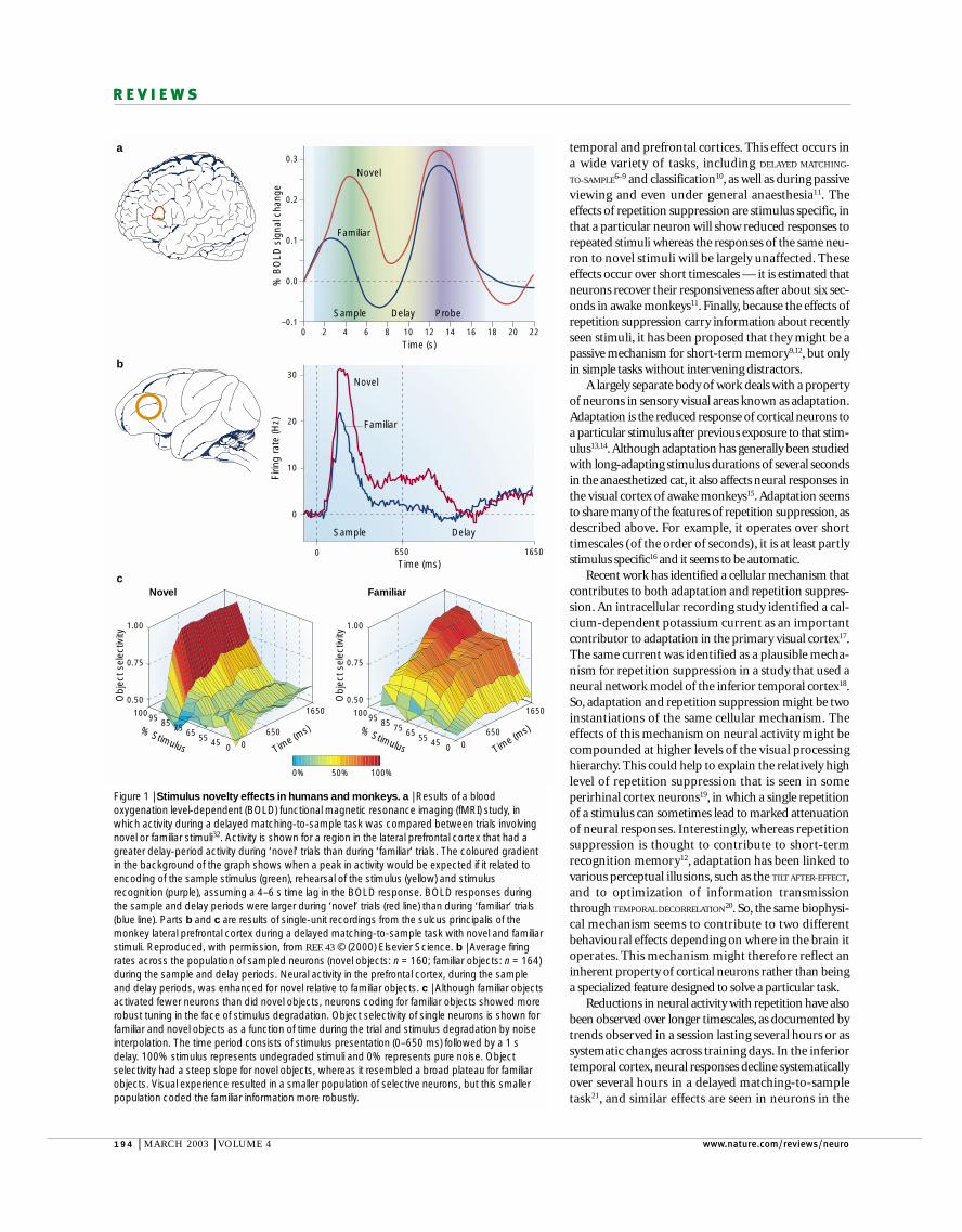
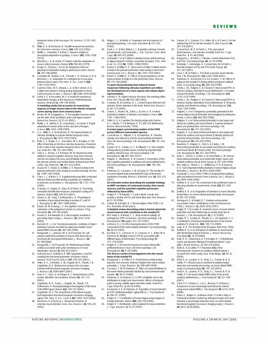
Figure 1 | Stimulus novelty effects in humans and monkeys. a | Results of a bloodoxygenation level-dependent (BOLD) functional magnetic resonance imaging (fMRI) study, inwhich activity during a delayed matching-to-sample task was compared between trials involvingnovel or familiar stimuli32. Activity is shown for a region in the lateral prefrontal cortex that had agreater delay-period activity during ‘novel’ trials than during ‘familiar’ trials. The coloured gradientin the background of the graph shows when a peak in activity would be expected if it related toencoding of the sample stimulus (green), rehearsal of the stimulus (yellow) and stimulusrecognition (purple), assuming a 4–6 s time lag in the BOLD response. BOLD responses duringthe sample and delay periods were larger during ‘novel’ trials (red line) than during ‘familiar’ trials(blue line). Parts b and c are results of single-unit recordings from the sulcus principalis of themonkey lateral prefrontal cortex during a delayed matching-to-sample task with novel and familiarstimuli. Reproduced, with permission, from REF. 43 © (2000) Elsevier Science. b | Average firingrates across the population of sampled neurons (novel objects: n = 160; familiar objects: n = 164)during the sample and delay periods. Neural activity in the prefrontal cortex, during the sampleand delay periods, was enhanced for novel relative to familiar objects. c | Although familiar objectsactivated fewer neurons than did novel objects, neurons coding for familiar objects showed morerobust tuning in the face of stimulus degradation. Object selectivity of single neurons is shown forfamiliar and novel objects as a function of time during the trial and stimulus degradation by noiseinterpolation. The time period consists of stimulus presentation (0–650 ms) followed by a 1 sdelay. 100% stimulus represents undegraded stimuli and 0% represents pure noise. Objectselectivity had a steep slope for novel objects, whereas it resembled a broad plateau for familiarobjects. Visual experience resulted in a smaller population of selective neurons, but this smallerpopulation coded the familiar information more robustly.
NATURE REVIEWS | NEUROSCIENCE VOLUME 4 | MARCH 2003 | 195
R E V I E W S
largely reflect the synaptic inputs to a given region) thanwith spiking activity (which reflects the region’soutput)36, such modulatory influences might be rela-tively amplified in BOLD signal levels compared withspiking activity37.
To link the generally observed reductions in neuralactivity in response to repetition with improved pro-cessing of these repeated stimuli, several authors havesuggested that neurons showing repetition effects are‘dropping out’ of the object representation38,39. One wayin which this might occur is through synaptic plastic-ity40. Consistent with this hypothesis, one study foundthat blocking NMDA (N-methyl-D-aspartate)-receptor-dependent synaptic plasticity eliminated medial tem-poral lobe field potentials that were correlated withstimulus repetition41 (BOX 1).
The models described earlier indicate not only thatstimulus repetition produces less overall activity, butalso that the net effect of this reduction is a more finelytuned object representation. Direct evidence for thisidea comes from a study of the prefrontal cortex, inwhich familiarity resulted in a reduction in the popu-lation-level activity in a delayed matching-to-sampletask (FIG. 1b). As shown in FIG. 1c, the smaller populationof neurons that responded to familiar objects codedthese objects more robustly with respect to stimulusdegradation42,43.
However, this account cannot explain activityreductions in early visual areas. It would predict thatneurons that represent non-optimal orientations orlocations might become less responsive with training,which would cause a reduction in their activity.However, activity reductions with perceptual learningare seen specifically for neurons at the trained locationand orientation26,27. What might be the source of thesereductions? We suggest that the slight reductions inearly sensory areas result from a network effect. That is,reductions in early sensory areas might reflect reducedfeedback coming from higher areas as a result of asparser representation of the learned stimuli in thosehigher areas.
The results reviewed above show that repetitive pre-sentation of a stimulus facilitates the processing of thatstimulus and leads to reductions in the average neuralactivity in many cortical regions. Reductions are seenfor timescales ranging from seconds to many months oftraining, and they reflect distinct mechanisms operatingat the cellular, synaptic and network levels. These find-ings indicate that, in one sense, novel stimuli might beless efficiently processed than familiar or repeated stim-uli. As described later, however, the increased activityelicited by novel stimuli can confer other processingadvantages.
Contextual noveltyAnother type of novelty that has been researched exten-sively is contextual novelty. A stimulus or event can bethought of as contextually novel if it arises in an unex-pected context (for example, the ‘naked guy’ enteringthe classroom). When an event is particularly novel orsurprising, it will elicit an orienting response44, with
superior temporal sulcus during passive viewing22 andin the medial temporal cortex during a serial recogni-tion task23. Robust reductions in activity have also beenseen in the prefrontal cortex24 and in the frontal andsupplementaryEYE FIELDS25 during CONDITIONAL OCULOMOTOR
LEARNING, reflecting many weeks of training. Familiarity-dependent reductions in neural activity have also beenrevealed in early visual areas by two perceptual learn-ing studies26,27. Monkeys were trained on a task thatinvolved repeated presentation (and discrimination) of gratings of a particular orientation in one part ofthe visual field. After many months of training, themonkeys’ perceptual ability to discriminate gratingsaround the trained location and orientation was greatlyimproved26. In addition to other effects not discussedhere, both studies found a slight reduction of V1 neuralpopulation activity for the trained orientation at thetrained location. Unlike repetition suppression, these‘familiarity effects’ are long-lasting. Although familiarityeffects can be detected after only minutes of experience,they probably continue to develop over longer periodsof time (hours to days) and are thought to be mediatedby synaptic plasticity28.
Effects that are similar to the repetition-related phe-nomena observed in neural activity can also be seen inactivity-dependent correlates, such as the blood oxy-genation level-dependent (BOLD) signal measured infunctional magnetic resonance imaging (fMRI) studiesof human subjects4. Reductions in BOLD signal levelswith stimulus repetition have been observed in severalstudies in prefrontal, medial temporal and inferior tem-poral regions29–33, as well as in sensory areas34,35 (FIG. 1).It is probable that adaptation of the BOLD signal is inpart due to the same mechanisms that give rise to thestimulus novelty effects described above. However, it isimportant to keep in mind that, in addition to localactivity, other mechanisms such as intracortical feed-back contribute to the BOLD signal. Because BOLD sig-nals correlate better with local field potentials (which
DELAYED MATCHING-TO-
SAMPLE TASKS
Recognition memory tasks inwhich presentation of a stimulusis followed by a delay, afterwhich a choice is offered. Inmatching tasks, the originallypresented stimulus must bechosen; in non-matching tasks, anew stimulus must be selected.With small stimulus sets, thestimuli are frequently repeatedand therefore become highlyfamiliar. So, typically, such tasksare most readily solved by short-term or working memory ratherthan by long-term memorymechanisms.
TILT AFTER-EFFECT
If you stare at a set of lines thatare tilted in one direction fromupright, upright lines willsubsequently look as thoughthey are tilted in the oppositedirection.
TEMPORAL DECORRELATION
Small eye movements madeduring free viewing of naturalscenes tend to expose neurons tosimilar but not identicalstructure. Adaptation can reduceresponses to structure that issimilar across fixations,removing correlations.
EYE FIELD
An area that receives visualinputs and produces movementsof the eye.
Box 1 | Stimulus novelty, synaptic plasticity and memory formation
Most investigators studying the effects of stimulus repetition frame their results in termsof the effects of repetition, rather than the effects of novelty. However, investigations offield potentials recorded from the human medial temporal lobes during performance ofverbal memory tasks indicate that some neural mechanisms might specifically affect theprocessing of relatively novel information. In these studies, field potentials were recordedin patients with severe epilepsy who had electrodes placed in their medial temporal lobesfor presurgical evaluation. Several previous intracranial event-related-potential studiesobserved a potential generated in the anterior medial temporal cortex, known as theAMTL-N400, the amplitude of which is sensitive to the novelty of a word41,133–138.One study found that epilepsy patients with hippocampal sclerosis had a reduced N400response to novel words, relative to epilepsy patients without hippocampal sclerosis134.By contrast, the N400 response to familiar words was not affected by hippocampalsclerosis, indicating that integrity of the hippocampus was crucial specifically for theenhanced N400 response to novel stimuli.A follow-up study showed that the magnitudeof the N400 response to novel words was directly correlated with neuronal density in theCA1 subfield of the hippocampus41. In addition, administration of the NMDA (N-methyl-D-aspartate) receptor antagonist ketamine selectively attenuated the N400 response tonovel words41. Collectively, these results indicate that there is a link between hippocampalNMDA receptor function and the encoding of novel stimuli.
196 | MARCH 2003 | VOLUME 4 www.nature.com/reviews/neuro
R E V I E W S
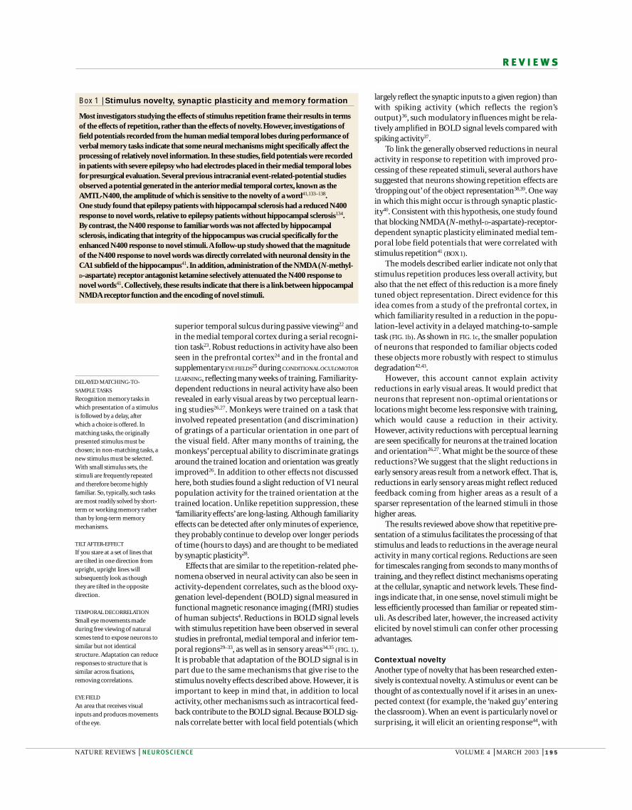
The functional characteristics of the novelty P3, andthe cognitive processes it might index, have been a topicof active investigation51. Results from these studies haveshown four important characteristics of the P3a. First,novelty P3 responses habituate across successive presen-tations of novel items, indicating that as these stimulibecome more predictable, the magnitude of theresponse wanes57–61. Second, novelty P3 responses arenot tied to any particular modality — similar novelty P3responses have been observed for novel visual, auditoryand somatosensory events57,62,63. Third, although thenovelty P3 is typically elicited experimentally by complex sounds, similar potentials can be derived withsimple stimuli, provided that they are contextuallydeviant49,64. Fourth, although task manipulations canaffect the magnitude of the novelty P3 (REFS 65–68), astimulus can elicit a robust novelty P3 even if it is task-irrelevant48 or if it is ignored49,58,69. The early latency ofthe novelty P3, together with the functional characteris-tics described above, indicate that the novelty P3 reflectsthe activity of a general network for rapidly orienting tonovel stimuli or events51,52.
Several sources of evidence implicate regions in theprefrontal cortex and the medial temporal lobes asimportant components of the network that generates thenovelty P3. For example, evidence has come from studiesof patients with epilepsy in whom intracranial electrodeshave been implanted for presurgical evaluation. Intra-cranial ERP studies in these patients have reported cortical-field potentials that have properties analogous tothose of the scalp-recorded P3 (REFS 54,70–78; BOX 2).These field potentials have been most commonlyobserved in the dorsolateral, ventrolateral and orbitalprefrontal cortex, cingulate cortex, lateral temporo-parietal cortex, hippocampus and parahippocampalcortical regions. Using an experimental design to elicitresponses specific to novel stimuli73–75, Halgren and col-leagues observed field potentials generated in orbital,ventrolateral and dorsolateral prefrontal regions thatwere temporally and functionally similar to the noveltyP3 (REF. 75). Outside the prefrontal cortex, similar fieldpotentials were recorded from sites in the medial tem-poral (perirhinal and posterior parahippocampal) cortex,subicular complex (in the hippocampal formation),temporoparietal cortex and cingulate gyrus73,74.
As shown in FIG. 3, studies of patients with focalbrain lesions also indicate that prefrontal, temporo-parietal and medial temporal cortical regions areimportant for responding to contextual novelty. Forexample, patients with lateral prefrontal57,63,79–82, lateraltemporoparietal63,81,83 or posterior medial temporallobe lesions62,82 resulting from strokes have attenuatednovelty P3 responses to novel auditory, visual orsomatosensory stimuli. Further studies have shownthat patients with prefrontal or medial temporallesions also do not show peripheral indices of orient-ing to contextually novel events, as indexed by skinconductance responses62,84. In addition, some findingsindicate that patients with lateral prefrontal lesionsdivert less attention towards novel stimuli, and thisreduction is directly correlated with the attenuation of
attentional resources being automatically divertedtowards the stimulus45.
Substantial insights into the nature of the orientingresponse have come from studies of scalp-recordedEVENT-RELATED POTENTIALS (ERPs) in humans. An ERP asso-ciated with contextual novelty — the P300 or P3 — wasfirst described by Sutton and colleagues in 1965 (REF. 46).Although the P3 has been studied most extensively inhumans, ‘P3-like’ potentials have been recorded frommacaque and squirrel monkeys, cats, rabbits, rats, dogsand dolphins, indicating that the P3 might representprocesses that are conserved across mammalian species47.Four decades of research have shown that the P3 proba-bly reflects a family of potentials that are related to differ-ent types of attentional processes48–56. The potential thatis most directly linked with novelty is the P3a (REF. 49) or‘novelty P3’ (REF. 48). In a typical novelty P3 experiment, asubject will perform an auditory target detection taskwith simple pure tone stimuli, and will occasionally heara contextually novel sound (such as a dog bark) amidstthese tones. Unlike the standard and target tones, thesenovel sounds elicit a scalp-recorded potential that peaksabout 200–300 ms after the stimulus and that is largestover the central and frontal scalp electrodes (FIG. 2). Theearly latency of this potential indicates that novel stimulirapidly modulate cognitive processing.
CONDITIONAL OCULOMOTOR
LEARNING
An association between a set ofstimuli and a set of eyemovements has to be learned bytrial and error, where eachstimulus is associated with aparticular eye movement.
EVENT-RELATED POTENTIALS
Electrical potentials that aregenerated in the brain as aconsequence of thesynchronized activation ofneuronal networks by externalstimuli. These evoked potentialsare recorded at the scalp andconsist of precisely timedsequences of waves or‘components’.
Novelty P3(P3a)
Target P3(P3b)
Volta
ge (µ
V)
0
0
5
–5
400Time (ms)
Volta
ge (µ
V)
0
0
5
–5
400Time (ms)
Standard toneTarget toneNovel sound
Figure 2 | The novelty P3. In typical experiments used to investigate contextual novelty, event-related potentials are recorded during an auditory target-detection task. For example, subjectsmight be instructed to respond to an infrequent target tone amidst a series of frequent ‘standard’sounds and infrequent novel distracter sounds (such as a dog’s bark). Scalp-recorded brainpotentials elicited during one such study are shown in the plots on the left. Infrequent novel sounds(purple line) elicited a novelty P3 potential at anterior scalp sites that peaked ~240 ms after thesound. Reproduced, with permission, from REF. 58 © (1999) MIT Press. A topographic ERPamplitude map (right) illustrates the relatively anterior topography of the novelty P3 potential on thescalp (as viewed from above). This potential was not elicited by the standard sounds, which werealso task-irrelevant, or the target sounds, which were also infrequent. Target sounds did, however,elicit a positive potential that peaked ~400 ms after the sound over posterior scalp sites. The map(right) illustrates the relatively posterior topography of the target P3 (also called the P3b).
NATURE REVIEWS | NEUROSCIENCE VOLUME 4 | MARCH 2003 | 197
R E V I E W S
correlate well with local field potentials37, indicating thatthey might also correlate well with scalp-recorded fieldpotentials. Nonetheless, because of the different time-scales of ERP and fMRI methods, these two measuresmight not always be in close correspondence.
With these caveats in mind, results from fMRI studiesof contextual novelty have generally corresponded wellwith results from intracranial ERP and lesion studies. Inevent-related fMRI experiments similar to ERP studiesused to evoke a novelty P3, novel stimuli elicit BOLDresponses in the inferior frontal gyrus, insula, temporo-parietal junction and anterior cingulate87–92. One limita-tion of these studies, however, is that it was unclearwhether activations in response to novel stimuli weredue to the contextual novelty of the items or whetherthey were due to other factors related to the lower-levelfeatures of the novel stimuli. Nonetheless, in accordancewith previous ERP results64, the results of two fMRIstudies indicate that this network responds robustly to violations of stimulus context, even when stimulusfactors are well controlled93,94.
In each of these fMRI studies93,94, a train of simplestimuli were presented, and brain responses were exam-ined in response to events that violated a pattern of pre-ceding events. In one study94, subjects were presentedwith a random string of two shapes, each requiring adifferent response. Response latencies to an item thatviolated a pattern in the previous trials (for example, asquare presented after three circles) increased linearlywith the length of the preceding pattern. Activation inthe middle and inferior frontal gyri, anterior insula anddorsal anterior cingulate regions showed a similar pat-tern, indicating that activity in these regions was sensi-tive to contextual novelty. In another study93, subjectspassively experienced simultaneous trains of auditory,visual and somatosensory stimuli. These stimuli wererepeated except for at certain times, when a stimulus inone modality would change. Such changes were associ-ated with enhanced activity in the inferior frontal gyrus,anterior insula and dorsal anterior cingulate, regardlessof the modality in which the deviation occurred. Theseresults strongly implicate regions in the ventrolateralprefrontal cortex, cingulate gyrus and anterior insulain orienting to contextually novel events.
Few functional imaging studies have found evidencefor medial temporal responses to contextual novelty,despite the evidence from ERP and lesion studies.However, most fMRI studies that have investigatedresponses to contextually novel events did not examinethe dynamics of activity over the course of stimulation.One study that did find evidence of medial temporallobe activation95 specifically investigated whetherresponses to novel events habituated over time.Consistent with the previous ERP results57–61, theseinvestigators found evidence for initial activation of thehippocampus upon presentation of contextually novelstimuli, and this activation habituated with repeatedpresentations. This finding converges with resultsfrom three other studies using different models thatalso found evidence for initial activation and rapidhabituation of hippocampal activation96–98.
the novelty P3 (REFS 79,80,85). In contrast to the effects oflateral prefrontal lesions, which reduce or eliminate thenovelty P3 response, the results of one study indicatethat damage to the orbital prefrontal cortex enhancesthe novelty P3 (REF. 86). These investigators concludedthat the orbital prefrontal cortex might suppress or modulate the novelty response, although furtherevidence is required to confirm this finding.
Functional neuroimaging — particularly EVENT-
RELATED fMRI — has been another valuable tool in identi-fying the neural sources of the scalp-recorded noveltyP3. However, correspondence between fMRI and ERPstudies must be interpreted with caution. Whereas ERPsdirectly index electrophysiological activity with a highdegree of temporal resolution, the BOLD responsereflects changes in blood oxygenation that occur sev-eral seconds after the corresponding neural events. Aswe noted earlier, BOLD responses detected by fMRI
EVENT-RELATED fMRI
A variant of functional magneticresonance imaging (fMRI)methods that allows neuralcorrelates of individual trials orclasses of trials to be isolated andcompared.
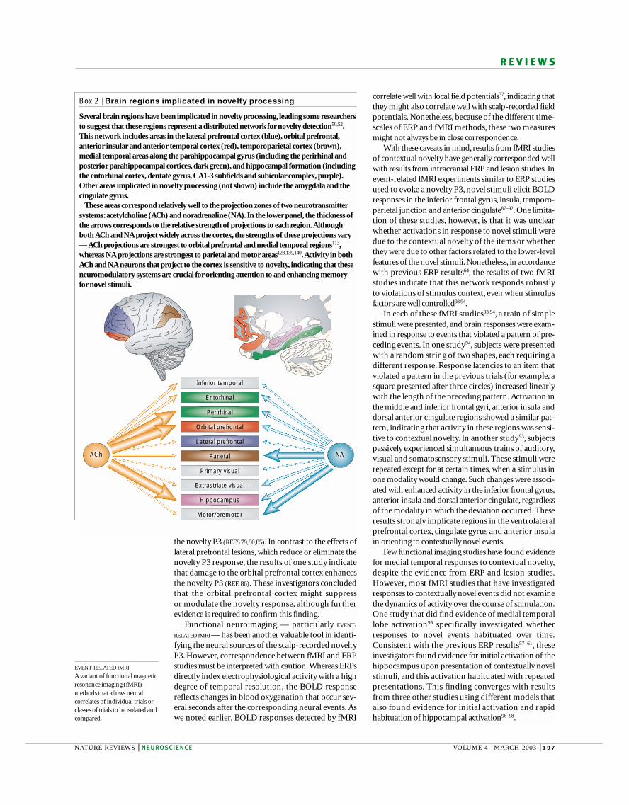
Box 2 | Brain regions implicated in novelty processing
Several brain regions have been implicated in novelty processing, leading some researchersto suggest that these regions represent a distributed network for novelty detection50,52.This network includes areas in the lateral prefrontal cortex (blue), orbital prefrontal,anterior insular and anterior temporal cortex (red), temporoparietal cortex (brown),medial temporal areas along the parahippocampal gyrus (including the perirhinal andposterior parahippocampal cortices, dark green), and hippocampal formation (includingthe entorhinal cortex, dentate gyrus, CA1-3 subfields and subicular complex, purple).Other areas implicated in novelty processing (not shown) include the amygdala and thecingulate gyrus.
These areas correspond relatively well to the projection zones of two neurotransmittersystems: acetylcholine (ACh) and noradrenaline (NA). In the lower panel, the thickness ofthe arrows corresponds to the relative strength of projections to each region.Althoughboth ACh and NA project widely across the cortex, the strengths of these projections vary— ACh projections are strongest to orbital prefrontal and medial temporal regions113,whereas NA projections are strongest to parietal and motor areas128,139,140.Activity in bothACh and NA neurons that project to the cortex is sensitive to novelty, indicating that theseneuromodulatory systems are crucial for orienting attention to and enhancing memoryfor novel stimuli.
Orbital prefrontal
Extrastriate visual
Primary visual
Lateral prefrontal
Parietal
Perirhinal
Entorhinal
Inferior temporal
Hippocampus
Motor/premotor
NAACh
198 | MARCH 2003 | VOLUME 4 www.nature.com/reviews/neuro
R E V I E W S
Novelty and memoryAs well as diverting attentional resources, contextuallynovel events tend to be encoded in memory more effec-tively than events that are less distinctive. Contextualnovelty might contribute to the ‘primacy effect’described in behavioural studies of memory — in whichitems that are presented first in a list are rememberedbetter than subsequently presented items.
Enhanced memory for contextually novel items hasbeen studied most directly in experiments using variantsof a model developed by von Restorff 99. In these studies,a few contextually novel items are studied amidst agroup of relatively homogeneous items (for example,a word printed in red among a series of words printed inblue). Several such studies have shown that memory isenhanced for contextually novel items relative to the lessdistinctive items99–102, regardless of in what way the eventis novel. Collectively, these findings indicate that contex-tually novel events recruit a neural network that divertsattention towards the salient event45 and modulates theencoding of this event in memory103,104.
As we noted earlier, the novelty P3 can be thought ofas an index of the diversion of attentional resourcestowards contextually novel stimuli. If the corticolimbicnetwork that generates the novelty P3 modulates mem-ory encoding, it might be predicted that the magnitudeof the P3 response to a contextually novel event wouldcorrelate with the degree to which that information issubsequently remembered. ERP studies indicate thatthis is the case — contextually novel stimuli elicit a largeP3 response relative to homogeneous items, and themagnitude of this response correlates with subsequentmemory for these items105–107.
Consistent with the results of studies on the neuralsubstrates of orienting responses, results from one lesionstudy indicate that interactions between prefrontal andmedial temporal lobe regions might be crucial for thegeneration of enhanced memory for contextually novelitems100. In this study, the authors developed an ana-logue of the von Restorff model and showed that bothhumans and intact monkeys had enhanced memory forcontextually novel stimuli. They also found that lesionsthat disconnected the frontal and perirhinal cortices inthe same hemisphere (that is, lesioning the perirhinalcortex in one hemisphere, and lesioning the prefrontalcortex in the other hemisphere) eliminated the memoryenhancement. Recent findings from another group indi-cate that amnesic patients with extensive medial temporallesions and patients with lesions limited to the hippo-campus do not show a von Restorff effect (Kishiyama, M.,Yonelinas, A. and Lazzara, M. submitted). These resultsunderscore the importance of prefrontal and medialtemporal regions for the encoding of contextuallynovel stimuli.
Results from a human neuroimaging study providefurther support for this point108. Volunteers werescanned while studying a series of scenes and words, andBOLD responses were compared between words and scenes that were relatively novel — not encounteredbefore in the session — and words and scenes that werehighly familiar — encountered several times just before
So, converging evidence indicates that a network ofbrain regions responds to stimuli that are contextuallynovel. This network includes areas in the prefrontal cor-tex, anterior insula, cingulate gyrus, temporoparietalcortex, medial temporal (entorhinal, perirhinal andparahippocampal) cortex, and the hippocampal for-mation (BOX 2). Although the results reviewed earlierindicate that these regions are recruited during the pro-cessing of novel stimuli, a subset of these regions, as wereview below, might also be crucial for encoding novelevents into memory.
Control
Lateralprefrontal
Medialtemporal
Auditory Visual Somatic
+ 100%
50%
0%
Auditory Visual Somatic
Novelty P3
Time (ms)
Lateralprefrontal
Medialtemporal
ControlLesion
0 400 800
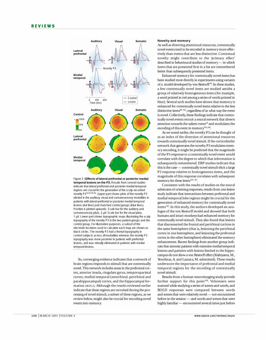
Figure 3 | Effects of lateral prefrontal or posterior medialtemporal lesions on the P3. Results from several studiesindicate that lateral prefrontal and posterior medial temporalregions are crucial for the generation of the scalp-recordednovelty P362,63,81,82. Upper part shows plots of the novelty P3elicited in the auditory, visual and somatosensory modalities inpatients with lateral prefrontal or posterior medial temporallesions (red lines) and matched control groups (blue lines).Positive is plotted upwards. Scale bar for the auditory andsomatosensory plots, 2 µV. Scale bar for the visual plots, 1 µV. Lower part shows topographic maps illustrating the scalptopography of the novelty P3 in the two patient groups and thecontrol group. For illustration purposes, a subset of theelectrode locations used to calculate each map are shown asblack circles. The novelty P3 had a frontal topography incontrol subjects across all modalities whereas the novelty P3topography was more posterior in patients with prefrontallesions, and was virtually eliminated in patients with medialtemporal lesions.
NATURE REVIEWS | NEUROSCIENCE VOLUME 4 | MARCH 2003 | 199
R E V I E W S
ACh comes from an in vivo microdialysis study in freelybehaving rats, which found robust increases in corticallevels of ACh while rats explored novel environments120.The increases were not correlated with motor activity andthere was no change in average glutamate release.
In humans, two studies found that administration ofscopolamine, an anticholinergic agent, attenuatedbehavioural indices of repetition priming and BOLDcorrelates of stimulus repetition (leading to a reduceddifference in the BOLD signal between novel andrepeated items)121,122. Administration of scopolaminewas associated with impaired learning of novelface–name associations and reduced the BOLD signal inventrolateral prefrontal, inferior temporal and hippo-campal regions123. Although one study reported behav-ioural impairments in a short-term memory task afterscopolamine administration in monkeys124, the studyfailed to find a robust effect of scopolamine on theresponses of inferotemporal neurons. Finally, adminis-tration of scopolamine in humans reduced frontal P300responses to infrequently occurring target stimuli125,126.These findings support a direct link between cholinergicneuromodulation and neural responses to novel or contextually deviant stimuli.
Other neuromodulators are probably also involved ingenerating novelty responses. For example, nor-adrenaline has similar biophysical properties to ACh, inthat it tends to enhance stimulus-evoked activity andpromote NMDA-receptor-dependent plasticity111. Thestrong reciprocal interactions between the ACh andnoradrenaline systems127 indicate that these systems arehighly interconnected and are often activated together.Noradrenaline projections originate in the locuscoeruleus , and unlike ACh projections, these projectionsparticularly target structures that are involved in atten-tion, such as the parietal cortex128 (BOX 2). The noradrena-line system might therefore also contribute to the noveltyP3 (REFS 129,130). Single-unit evidence from freely movingrats confirmed that, similar to neurons in the perirhinalcortex23, locus coeruleus neurons fire in response to novelobjects and that this firing habituates rapidly, leading tothe release of noradrenaline on just the first few trials131.
The projection patterns of noradrenaline (focusingon the parietal cortex) and ACh (focusing on frontaland anterior medial temporal cortices) could accountfor the topography of cortical generators of the noveltyP3. Consistent with this idea, an ERP study has found adouble dissociation between two components of thenovelty P3 centred on frontal and parietal corticalareas59. Furthermore, administration of a noradrenalineantagonist in monkeys selectively abolishes the parietalP3, but leaves the P3 in other regions unaffected132. Bycontrast, administration of an ACh antagonist in humansattenuated frontal, but not parietal, P3 responses125,126.
An integrated account of novelty responsesThe evidence cited in this review highlights the seeminglycontradictory effects of novelty on behaviour. The stim-ulus novelty effects show that novel stimuli can be at adisadvantage compared with familiar ones. Behaviouralevidence indicates that processing of familiar stimuli is
the scanning session. Regions in the ventrolateral prefrontal, parahippocampal and fusiform cortices andthe posterior hippocampus showed greater activity inresponse to novel items than familiar items. Each of these regions showed greater activity for novel itemsthat were successfully recognized than for novel items that were later forgotten. These two findings indi-cate a potential link between the sensitivity of theseregions to stimulus novelty and their role in encodinginformation in memory. These findings, together withthose in REF. 100, strongly implicate prefrontal andmedial temporal lobe regions in a circuit that mediatesthe enhanced encoding of novel stimuli and events109.
Neuromodulatory influencesThe idea that prefrontal and medial temporal corticalregions modulate encoding of novel events indicatesthat there must be a mechanism by which these regionsinfluence the rapid encoding of events that are processedacross various cortical regions. One mechanism bywhich these regions might influence the processing andencoding of novel information is through regulation ofneurotransmitter modulation of cortical processing47,110
(BOX 2). Several neurotransmitter systems have beenimplicated in the generation of responses to novelty, butvarious lines of evidence indicate that acetylcholine(ACh) has a particular role in modulating the encodingof novel events.
ACh tends to elevate the baseline firing rate whileenhancing stimulus-evoked activity in certain brainregions, as well as facilitating NMDA-receptor-dependent synaptic plasticity111. The main source ofcortical ACh is the nucleus basalis of Meynert (NBM),which receives inputs from only a few cortical regions,including the orbital prefrontal, medial temporal(including perirhinal and entorhinal) and anteriorinsular cortex112. However, the NBM provides AChinput to almost the entire neocortical mantle, with theorbital prefrontal, perirhinal, entorhinal and insularcortex receiving the strongest projections as assessed byACh-related enzymatic activity113. By modulating AChrelease from the NBM, prefrontal and medial temporalregions can modulate memory encoding across manybrain regions.
Several studies have implicated this pathway in learn-ing and memory. For example, disconnection of eitherthe frontal or the temporal cortex from ACh afferentsleads to deficits in visual recognition memory andobject–reward association learning114,115. In addition,immunotoxic lesions of the NBM lead to reductions in the level of ACh in frontal and temporal cortices,the extent of which are correlated with concurrentbehavioural impairments in a learning task116.
These links between ACh and learning are consistentwith a view in which ACh systems are preferentially acti-vated by novel stimuli and are instrumental in consolidat-ing memories about such novel stimuli117. Consistentwith this view, single-unit recordings from monkeysshow that neurons in the NBM have enhanced firing ratesto novel stimuli that decline with repetition118,119. Directevidence for novelty-associated enhancement of cortical
200 | MARCH 2003 | VOLUME 4 www.nature.com/reviews/neuro
R E V I E W S
activity for familiar inputs, so that when novel stimuliare encountered these can cause the release of modula-tory neurotransmitters. Familiar stimuli are processedmore efficiently because synaptic plasticity produces asmaller population of neurons that represents thesefamiliar stimuli more robustly. By contrast, novel stim-uli cause activation in a larger network, leading torelease of the neuromodulators ACh and noradrena-line. This results in attention being directed towardsprocessing these novel stimuli, and in more successfulmemory formation.
ConclusionsThe evidence we have reviewed indicates that theoccurrence of a novel stimulus elicits a cascade of neuralresponses that results in enhanced attention to andmemory for that stimulus. Recent research has begun toreveal the complex mechanisms by which these changesoccur, and we have suggested a tentative model toaccount for these findings. Nonetheless, substantialwork remains to be carried out. Research on stimuluseffects and contextual novelty effects has proceededlargely independently of each other. For example, thereare very few single-unit studies of contextual noveltyeffects in monkeys, compared with the wealth of infor-mation that has been gathered in ERP studies of humansubjects. Filling this gap, and assembling a more unifiedpicture of the various phenomena observed at differenttimescales and using different techniques, might lead toimportant breakthroughs in our understanding of basicneural mechanisms for memory and attention. We haveadvanced the hypothesis that the neuromodulatorytransmitters ACh and noradrenaline are closely involvedin novelty processing. Although this hypothesis canaccount for a wide range of findings, further experi-ments are required to directly test and refine it. In par-ticular, although the anatomical connections betweenthe orbital prefrontal cortex, perirhinal cortex and NBMhave been documented, these connections have yet to befunctionally characterized.
Finally, we note that the tentative account we haveoffered to explain novelty processing is a relatively mech-anistic model of neural processing. To characterize nov-elty processing more fully, it will be necessary to clarifythese mechanisms at the cognitive level. Associating theeffects of novelty at these levels with subjective experi-ence will be a daunting task and might require thedevelopment of new psychological constructs. Such anoccurrence might turn out to be the most significantbenefit to emerge from the development of a cognitiveneuroscience of novelty processing.
facilitated, and this is usually accompanied by reduc-tions in neural activity across several cortical areas —particularly in prefrontal and medial temporal corticalregions. The available evidence indicates that at least twomechanisms contribute to these effects, one operatingover short timescales (seconds) and the other over longertimescales (minutes to days).
Conversely, the contextual novelty effects reviewedearlier indicate that novel stimuli also have advantagesover repeated items. For example, novel items tend toattract attention and are encoded into memory more effi-ciently than are familiar items. These contextual noveltyeffects have been associated with increases in neural activ-ity across a distributed cortical network that includesfrontal, parietal and medial temporal areas. We havehypothesized that neuromodulators — in particular ACh— might provide the means by which this occurs38.
Prefrontal and medial temporal areas receive highlyprocessed information from many modalities.Assumingthat activity in these regions reflects summated inputsfrom lower-level processing areas, we would expectactivity in these regions to be most sensitive to the rela-tive familiarity of a stimulus19. So, when a novel stimu-lus is encountered, an increase in activity would beexpected in these areas. The unique connectivity of pre-frontal and medial temporal areas with cholinergicneurons in the NBM might provide a mechanism bywhich these regions can cause ACh release to targetregions of the NBM, thereby directing attentional andmnemonic resources towards novel events. The relativeincrease in activity in response to novel stimuli in pre-frontal and medial temporal regions would thereforelead to the release of greater amounts of ACh from theNBM when compared with amounts that are releasedin response to familiar stimuli.
By this account, during a typical task used to studycontextual novelty, repeated stimulation of neurons rep-resenting the standard or target items will — throughstimulus repetition effects — lead to reductions inneural activity that will be most robust in prefrontal andmedial temporal regions. The occurrence of the infre-quent novel item will elicit relatively higher amounts ofactivity — leading, for example, to release of AChthrough interactions of the prefrontal and medial tem-poral cortices with the basal forebrain. The resultantcascade of activity prompted by cholinergic as well asnoncholinergic modulation results in the diversion ofattentional resources (as indexed by the novelty P3) andenhanced memory encoding elicited by novel events.
So, stimulus novelty effects are constantly at work atall levels of the processing hierarchy to reduce mean
1. Tulving, E. & Schacter, D. L. Priming and human memorysystems. Science 247, 301–306 (1990).
2. Gilbert, C. D., Sigman, M. & Crist, R. E. The neural basis ofperceptual learning. Neuron 31, 681–697 (2001).
3. Ringo, J. L. Stimulus specific adaptation in inferior temporaland medial temporal cortex of the monkey. Behav. BrainRes. 76, 191–197 (1996).
4. Henson, R. N. & Rugg, M. D. Neural responsesuppression, haemodynamic repetition effects, andbehavioural priming. Neuropsychologia 41, 263–270(2003).
5. Habib, R. On the relation between conceptual priming,neural priming, and novelty assessment. Scand. J. Psychol.42, 187–195 (2001).
6. Miller, E. K., Erickson, C. A. & Desimone, R. Neuralmechanisms of visual working memory in prefrontal cortexof the macaque. J. Neurosci. 16, 5154–5167 (1996).
7. Rainer, G., Rao, S. C. & Miller, E. K. Prospective coding forobjects in primate prefrontal cortex. J. Neurosci. 19,5493–5505 (1999).
8. Riches, I. P., Wilson, F. A. & Brown, M. W. The effects ofvisual stimulation and memory on neurons of the
hippocampal formation and the neighboringparahippocampal gyrus and inferior temporal cortex of theprimate. J. Neurosci. 11, 1763–1779 (1991).
9. Baylis, G. C. & Rolls, E. T. Responses of neurons in theinferior temporal cortex in short term and serial recognitionmemory tasks. Exp. Brain Res. 65, 614–622 (1987).
10. Sobotka, S. & Ringo, J. L. Stimulus specific adaptation inexcited but not in inhibited cells in inferotemporal cortex ofmacaque. Brain Res. 646, 95–99 (1994).
11. Miller, E. K., Gochin, P. M. & Gross, C. G. Habituation-like decrease in the responses of neurons in inferior
NATURE REVIEWS | NEUROSCIENCE VOLUME 4 | MARCH 2003 | 201
R E V I E W S
temporal cortex of the macaque. Vis. Neurosci. 7, 357–362(1991).
12. Miller, E. K. & Desimone, R. Parallel neuronal mechanismsfor short-term memory. Science 263, 520–522 (1994).
13. Maffei, L., Fiorentini, A. & Bisti, S. Neural correlate ofperceptual adaptation to gratings. Science 182, 1036–1038(1973).
14. Movshon, J. A. & Lennie, P. Pattern-selective adaptation invisual cortical neurones. Nature 278, 850–852 (1979).
15. Dragoi, V., Sharma, J. & Sur, M. Adaptation-inducedplasticity of orientation tuning in adult visual cortex. Neuron28, 287–298 (2000).
16. Carandini, M., Barlow, H. B., O’Keefe, L. P., Poirson, A. B. &Movshon, J. A. Adaptation to contingencies in macaqueprimary visual cortex. Phil. Trans. R. Soc. Lond. B 352,1149–1154 (1997).
17. Sanchez-Vives, M. V., Nowak, L. G. & McCormick, D. A.Cellular mechanisms of long-lasting adaptation in visualcortical neurons in vitro. J. Neurosci. 20, 4286–4299 (2000).
18. Sohal, V. S. & Hasselmo, M. E. A model for experience-dependent changes in the responses of inferotemporalneurons. Network 11, 169–190 (2000).A modelling study that accounts for several keyaspects of single-neuron data in the inferior temporalcortex during short-term memory tasks.
19. Brown, M. W. & Aggleton, J. P. Recognition memory: whatare the roles of the perirhinal cortex and hippocampus?Nature Rev. Neurosci. 2, 51–61 (2001).
20. Muller, J. R., Metha, A. B., Krauskopf, J. & Lennie, P. Rapidadaptation in visual cortex to the structure of images.Science 285, 1405–1408 (1999).
21. Li, L., Miller, E. K. & Desimone, R. The representation ofstimulus familiarity in anterior inferior temporal cortex. J. Neurophysiol. 69, 1918–1929 (1993).
22. Rolls, E. T., Baylis, G. C., Hasselmo, M. E. & Nalwa, V. Theeffect of learning on the face selective responses of neuronsin the cortex in the superior temporal sulcus of the monkey.Exp. Brain Res. 76, 153–164 (1989).
23. Fahy, F. L., Riches, I. P. & Brown, M. W. Neuronal activityrelated to visual recognition memory: long-term memoryand the encoding of recency and familiarity information inthe primate anterior and medial inferior temporal and rhinalcortex. Exp. Brain Res. 96, 457–472 (1993).
24. Asaad, W. F., Rainer, G. & Miller, E. K. Neural activity in theprimate prefrontal cortex during associative learning. Neuron21, 1399–1407 (1998).
25. Chen, L. L. & Wise, S. P. Supplementary eye field contrastedwith the frontal eye field during acquisition of conditionaloculomotor associations. J. Neurophysiol. 73, 1122–1134(1995).
26. Schoups, A., Vogels, R., Qian, N. & Orban, G. Practisingorientation identification improves orientation coding in V1neurons. Nature 412, 549–553 (2001).
27. Ghose, G. M., Yang, T. & Maunsell, J. H. Physiologicalcorrelates of perceptual learning in monkey V1 and V2. J. Neurophysiol. 87, 1867–1888 (2002).
28. Brown, M. W. & Xiang, J. Z. Recognition memory: neuronalsubstrates of the judgement of prior occurrence. Prog.Neurobiol. 55, 149–189 (1998).
29. Kourtzi, Z. & Kanwisher, N. Cortical regions involved inperceiving object shape. J. Neurosci. 20, 3310–3318.(2000).
30. Buckner, R. L. et al. Functional-anatomic correlates of objectpriming in humans revealed by rapid presentation event-related fMRI. Neuron 20, 285–296 (1998).
31. Ranganath, C., Johnson, M. K. & D’Esposito, M. Leftanterior prefrontal activation increases with demands torecall specific perceptual information. J. Neurosci. 20,RC108 (2000).
32. Ranganath, C. & D’Esposito, M. Medial temporal lobeactivity associated with active maintenance of novelinformation. Neuron 31, 865–873 (2001).
33. Grill-Spector, K. & Malach, R. fMR-adaptation: a tool forstudying the functional properties of human corticalneurons. Acta Psychol. (Amst.) 107, 293–321 (2001).
34. Tolias, A. S., Smirnakis, S. M., Augath, M. A., Trinath, T. &Logothetis, N. K. Motion processing in the macaque:revisited with functional magnetic resonance imaging. J. Neurosci. 21, 8594–8601 (2001).
35. Huk, A. C., Ress, D. & Heeger, D. J. Neuronal basis of themotion aftereffect reconsidered. Neuron 32, 161–172(2001).
36. Logothetis, N. K., Pauls, J., Augath, M., Trinath, T. &Oeltermann, A. Neurophysiological investigation of the basisof the fMRI signal. Nature 412, 150–157 (2001).
37. Logothetis, N. K. The neural basis of the blood-oxygen-level-dependent functional magnetic resonance imagingsignal. Phil. Trans. R. Soc. Lond. B 357, 1003–1037 (2002).
38. Desimone, R. & Duncan, J. Neural mechanisms of selective visual attention. Annu. Rev. Neurosci. 18, 193–222(1995).
39. Wiggs, C. L. & Martin, A. Properties and mechanisms ofperceptual priming. Curr. Opin. Neurobiol. 8, 227–233(1998).
40. Stark, C. E. & McClelland, J. L. Repetition priming of words,pseudowords, and nonwords. J. Exp. Psychol. Learn. Mem.Cogn. 26, 945–972 (2000).
41. Grunwald, T. et al. Evidence relating human verbal memoryto hippocampal N-methyl-D-aspartate receptors. Proc. NatlAcad. Sci. USA 96, 12085–12089 (1999).
42. Rainer, G. & Miller, E. K. Timecourse of object-related neuralactivity in the primate prefrontal cortex during a short-termmemory task. Eur. J. Neurosci. 15, 1244–1254 (2002).
43. Rainer, G. & Miller, E. K. Effects of visual experience on therepresentation of objects in the prefrontal cortex. Neuron27, 179–189 (2000).A report demonstrating that reduced neuralresponses following stimulus repetition can reflectthe development of a more sparse and robust objectrepresentation.
44. Sokolov, E. N. Higher nervous functions: the orienting reflex.Annu. Rev. Physiol. 25, 545–580 (1963).
45. Corbetta, M. & Shulman, G. L. Control of goal-directed andstimulus-driven attention in the brain. Nature Rev. Neurosci.3, 201–215 (2002).
46. Sutton, S., Braren, M., Zubin, J. & John, E. R. Evoked-potential correlates of stimulus uncertainty. Science 150,1187–1188 (1965).
47. Paller, K. A. in Cognitive Electrophysiology (eds Heinze, H. J., Munte, T. F. & Mangun, G. R.) 300–333 (Birkhauser,Boston, Massachusetts, 1994).A review paper summarizing studies of the P300across different mammalian species.
48. Courchesne, E., Hillyard, S. A. & Galambos, R. Stimulusnovelty, task relevance and the visual evoked potential inman. Electroencephalogr. Clin. Neurophysiol. 39, 131–143(1975).
49. Squires, N. K., Squires, K. C. & Hillyard, S. A. Two varietiesof long-latency positive waves evoked by unpredictableauditory stimuli in man. Electroencephalogr. Clin.Neurophysiol. 38, 387–401 (1975).
50. Halgren, E., Marinkovic, K. & Chauvel, P. Generators of thelate cognitive potentials in auditory and visual oddball tasks.Electroencephalogr. Clin. Neurophysiol. 106, 156–164(1998).
51. Friedman, D., Cycowicz, Y. M. & Gaeta, H. The novelty P3:an event-related brain potential (ERP) sign of the brain’sevaluation of novelty. Neurosci. Biobehav. Rev. 25, 355–373(2001).An extensive review describing the available findingson ERP correlates of contextual novelty, their neuralsources, and the potential cognitive processesreflected by these ERPs.
52. Knight, R. T. & Nakada, T. Cortico-limbic circuits andnovelty: a review of EEG and blood flow data. Rev. Neurosci.9, 57–70 (1998).
53. Soltani, M. & Knight, R. T. Neural origins of the P300. Crit.Rev. Neurobiol. (in the press).A detailed review describing what is known about theneural sources that contribute to the novelty P3.
54. McCarthy, G. & Wood, C. C. Intracranial recordings ofendogenous ERPs in humans. Electroencephalogr. Clin.Neurophysiol. 39 (suppl.), 331–337 (1987).
55. Johnson, R. Jr. On the neural generators of the P300component of the event-related potential. Psychophysiology30, 90–97 (1993).
56. Ruchkin, D. S., Johnson, R. Jr, Canoune, H. L., Ritter, W. &Hammer, M. Multiple sources of P3b associated withdifferent types of information. Psychophysiology 27,157–176 (1990).
57. Knight, R. T. Decreased response to novel stimuli afterprefrontal lesions in man. Electroencephalogr. Clin.Neurophysiol. 59, 9–20 (1984).The first in a series of investigations into the neuralbasis of the novelty P3.
58. Ranganath, C. & Paller, K. A. Frontal brain activity duringepisodic and semantic retrieval: insights from event-relatedpotentials. J. Cogn. Neurosci. 11, 598–609 (1999).
59. Cycowicz, Y. M. & Friedman, D. Effect of sound familiarity onthe event-related potentials elicited by novel environmentalsounds. 36, 30–51 (1998).
60. Friedman, D. & Simpson, G. V. ERP amplitude and scalpdistribution to target and novel events: effects of temporalorder in young, middle-aged and older adults. Brain Res.Cogn. Brain Res. 2, 49–63 (1994).
61. Kazmerski, V. A. & Friedman, D. Repetition of novel stimuli inan ERP oddball paradigm: aging effects. J. Psychophysiol.9, 298–311 (1995).
62. Knight, R. T. Contribution of human hippocampal region tonovelty detection. Nature 383, 256–259 (1996).
63. Knight, R. T. Distributed cortical network for visual attention.J. Cogn. Neurosci. 9, 75–91 (1997).
64. Simons, R. F., Graham, F. K., Miles, M. A. & Chen, X. On therelationship of P3a and the novelty-P3. Biol. Psychol. 56,207–218 (2001).
65. Comerchero, M. D. & Polich, J. P3a, perceptualdistinctiveness, and stimulus modality. Brain Res. Cogn.Brain Res. 7, 41–48 (1998).
66. Katayama, J. & Polich, J. Stimulus context determines P3aand P3b. Psychophysiology 35, 23–33 (1998).
67. Demiralp, T., Ademoglu, A., Comerchero, M. & Polich, J.Wavelet analysis of P3a and P3b. Brain Topogr. 13,251–267 (2001).
68. Jeon, Y. W. & Polich, J. P3a from a passive visual stimulustask. Clin. Neurophysiol. 112, 2202–2208 (2001).
69. Friedman, D., Kazmerski, V. A. & Cycowicz, Y. M. Effects ofaging on the novelty P3 during attend and ignore oddballtasks. Psychophysiology 35, 508–520 (1998).
70. Clarke, J. M., Halgren, E. & Chauvel, P. Intracranial ERPs inhumans during a lateralized visual oddball task: I. Occipitaland peri-Rolandic recordings. Clin. Neurophysiol. 110,1210–1225 (1999).
71. Clarke, J. M., Halgren, E. & Chauvel, P. Intracranial ERPs inhumans during a lateralized visual oddball task: II. Temporal,parietal, and frontal recordings. Clin. Neurophysiol. 110,1226–1244 (1999).
72. Halgren, E. et al. Endogenous potentials generated in thehuman hippocampal formation and amygdala by infrequentevents. Science 210, 803–805 (1980).
73. Halgren, E. et al. Intracerebral potentials to rare target anddistractor auditory and visual stimuli. I. Superior temporalplane and parietal lobe. Electroencephalogr. Clin.Neurophysiol. 94, 191–220 (1995).
74. Halgren, E. et al. Intracerebral potentials to rare target anddistractor auditory and visual stimuli. II. Medial, lateral andposterior temporal lobe. Electroencephalogr. Clin.Neurophysiol. 94, 229–250 (1995).
75. Baudena, P., Halgren, E., Heit, G. & Clarke, J. M.Intracerebral potentials to rare target and distractor auditoryand visual stimuli. III. Frontal cortex. Electroencephalogr.Clin. Neurophysiol. 94, 251–264 (1995).
76. Alain, C., Richer, F., Achim, A. & Saint Hilaire, J. M. Humanintracerebral potentials associated with target, novel, andomitted auditory stimuli. Brain Topogr. 1, 237–245 (1989).
77. McCarthy, G., Wood, C. C., Williamson, P. D. & Spencer, D. D. Task-dependent field potentials in human hippocampalformation. J. Neurosci. 9, 4253–4268 (1989).
78. Grunwald, T. et al. Limbic P300s in temporal lobe epilepsywith and without Ammon’s horn sclerosis. Eur. J. Neurosci.11, 1899–1906 (1999).
79. Daffner, K. R. et al. The central role of the prefrontal cortex indirecting attention to novel events. Brain 123, 927–939(2000).
80. Daffner, K. R. et al. Regulation of attention to novel stimuli byfrontal lobes: an event-related potential study. Neuroreport9, 787–791 (1998).
81. Yamaguchi, S. & Knight, R. T. Anterior and posteriorassociation cortex contributions to the somatosensoryP300. J. Neurosci. 11, 2039–2054 (1991).
82. Knight, R. T. & Scabini, D. Anatomic bases of event-relatedpotentials and their relationship to novelty detection inhumans. J. Clin. Neurophysiol. 15, 3–13 (1998).
83. Knight, R. T., Scabini, D., Woods, D. L. & Clayworth, C. C.Contributions of temporal-parietal junction to the humanauditory P3. Brain Res. 502, 109–116 (1989).
84. Luria, A. R. The Working Brain (Penguin, New York, 1966).85. Daffner, K. R. et al. Disruption of attention to novel events
after frontal lobe injury in humans. J. Neurol. Neurosurg.Psychiatry 68, 18–24 (2000).
86. Rule, R. R., Shimamura, A. P. & Knight, R. T. Orbitofrontalcortex and dynamic filtering of emotional stimuli. Cogn.Affect. Behav. Neurosci. 2, 264–270 (2002).
87. Kiehl, K. A. & Liddle, P. F. Reproducibility of thehemodynamic response to auditory oddball stimuli: a six-week test-retest study. Hum. Brain Mapp. 18, 42–52(2003).
88. Kiehl, K. A., Laurens, K. R., Duty, T. L., Forster, B. B. &Liddle, P. F. Neural sources involved in auditory targetdetection and novelty processing: an event-related fMRIstudy. Psychophysiology 38, 133–142 (2001).
89. Kiehl, K. A., Laurens, K. R., Duty, T. L., Forster, B. B. &Liddle, P. F. An event-related fMRI study of visual andauditory oddball tasks. J. Psychophysiol. 15, 221–240(2001).
90. Clark, V. P., Fannon, S., Lai, S., Benson, R. & Bauer, L.Responses to rare visual target and distractor stimuli using event-related fMRI. J. Neurophysiol. 83, 3133–3139(2000).
91. Kirino, E., Belger, A., Goldman-Rakic, P. & McCarthy, G.Prefrontal activation evoked by infrequent target and novelstimuli in a visual target detection task: an event-relatedfunctional magnetic resonance imaging study. J. Neurosci.20, 6612–6618 (2000).
202 | MARCH 2003 | VOLUME 4 www.nature.com/reviews/neuro
R E V I E W S
92. Opitz, B., Mecklinger, A., Friederici, A. D. & von Cramon, D. Y. The functional neuroanatomy of novelty processing:integrating ERP and fMRI results. Cereb. Cortex 9, 379–391(1999).
93. Downar, J., Crawley, A. P., Mikulis, D. J. & Davis, K. D. A multimodal cortical network for the detection of changesin the sensory environment. Nature Neurosci. 3, 277–283(2000).
94. Huettel, S. A., Mack, P. B. & McCarthy, G. Perceivingpatterns in random series: dynamic processing of sequence in prefrontal cortex. Nature Neurosci. 5, 485–490(2002).
95. Strange, B. A. & Dolan, R. J. Adaptive anterior hippocampalresponses to oddball stimuli. Hippocampus 11, 690–698(2001).
96. Strange, B. A., Fletcher, P. C., Henson, R. N., Friston, K. J. &Dolan, R. J. Segregating the functions of humanhippocampus. Proc. Natl Acad. Sci. USA 96, 4034–4039(1999).
97. Poldrack, R. A. et al. Interactive memory systems in thehuman brain. Nature 414, 546–550 (2001).
98. Buchel, C., Dolan, R. J., Armony, J. L. & Friston, K. J.Amygdala-hippocampal involvement in human aversivetrace conditioning revealed through event-related functionalmagnetic resonance imaging. J. Neurosci. 19,10869–10876 (1999).
99. von Restorff, H. Uber die Wirkung von Bereichsbildungen imSpurenfeld. Psychol. Forsch. 18, 299–342 (1933).
100. Parker, A., Wilding, E. L. & Akerman, C. The von Restorffeffect in visual object recognition memory in humans andmonkeys: the role of frontal/perirhinal interaction. J. Cogn.Neurosci. 10, 691–703 (1998).An experiment demonstrating that the processingadvantage for contextually novel items, in anotherwise homogeneous list, is dependent on intactperirhinal–frontal interactions.
101. Hunt, R. R. The subtlety of distinctiveness: what von Restorffreally did. Psychon. Bull. Rev. 2, 105–112 (1995).
102. Wallace, W. P. Review of the historical, empirical, andtheoretical status of the von Restorff phenomenon. Psychol.Bull. 63, 410–424 (1965).
103. Habib, R., McIntosh, A. R., Wheeler, M. A. & Tulving, E.Memory encoding and hippocampally-basednovelty/familiarity discrimination networks.Neuropsychologia 41, 271–279 (2003).
104. Tulving, E., Markowitsch, H. J., Craik, F. E., Habib, R. &Houle, S. Novelty and familiarity activations in PET studies ofmemory encoding and retrieval. Cereb. Cortex 6, 71–79(1996).
105. Fabiani, M. & Donchin, E. Encoding processes and memoryorganization: a model of the von Restorff effect. J. Exp.Psychol. Learn. Mem. Cogn. 21, 224–240 (1995).
106. Fabiani, M., Karis, D. & Donchin, E. Effects of mnemonicstrategy manipulation in a von Restorff paradigm.Electroencephalogr. Clin. Neurophysiol. 75, 22–35 (1990).
107. Fabiani, M., Karis, D. & Donchin, E. P300 and recall in anincidental memory paradigm. Psychophysiology 23,298–308 (1986).
108. Kirchhoff, B. A., Wagner, A. D., Maril, A. & Stern, C. E.Prefrontal–temporal circuitry for episodic encoding and subsequent memory. J. Neurosci. 20, 6173–6180(2000).
109. Fernandez, G. & Tendolkar, I. Integrated brain activity inmedial temporal and prefrontal areas predicts subsequent memory performance: human declarativememory formation at the system level. Brain Res. Bull. 55,1–9 (2001).
110. Frodl-Bauch, T., Bottlender, R. & Hegerl, U. Neurochemicalsubstrates and neuroanatomical generators of the event-related P300. Neuropsychobiology 40, 86–94 (1999).
111. Gu, Q. Neuromodulatory transmitter systems in the cortexand their role in cortical plasticity. Neuroscience 111,815–835 (2002).
112. Mesulam, M. M. & Mufson, E. J. Neural inputs into thenucleus basalis of the substantia innominata (Ch4) in therhesus monkey. Brain 107, 253–274 (1984).A careful characterization of the inputs to the NBM,revealing that, although this area modulates activityacross several cortical regions, it receives input fromonly a few.
113. Mesulam, M. M., Volicer, L., Marquis, J. K., Mufson, E. J. &Green, R. C. Systematic regional differences in thecholinergic innervation of the primate cerebral cortex:distribution of enzyme activities and some behavioralimplications. Ann. Neurol. 19, 144–151 (1986).
114. Easton, A. & Gaffan, D. Crossed unilateral lesions of themedial forebrain bundle and either inferior temporal or frontalcortex impair object-reward association learning in Rhesusmonkeys. Neuropsychologia 39, 71–82 (2001).One of a series of recent lesion studies in monkeys bythese authors that underscore the importance of thecholinergic system in learning and memory.
115. Easton, A., Parker, A. & Gaffan, D. Crossed unilateral lesionsof medial forebrain bundle and either inferior temporal orfrontal cortex impair object recognition memory in Rhesusmonkeys. Behav. Brain Res. 121, 1–10 (2001).
116. Fine, A. et al. Learning impairments following injection of aselective cholinergic immunotoxin, ME20.4 IgG-saporin, intothe basal nucleus of Meynert in monkeys. Neuroscience 81,331–343 (1997).A study demonstrating that immunotoxic lesionsspecific to cholinergic neurons in the basal forebrainresult in cell loss in inferior temporal and frontalcortex of monkeys, the severity of which is correlatedwith behavioural impairments.
117. Hasselmo, M. E. Neuromodulation: acetylcholine andmemory consolidation. Trends Cogn. Sci. 3, 351–359 (1999).
118. Wilson, F. A. & Rolls, E. T. Neuronal responses related to thenovelty and familiarity of visual stimuli in the substantiainnominata, diagonal band of Broca and periventricularregion of the primate basal forebrain. Exp. Brain Res. 80,104–120 (1990).
119. Wilson, F. A. & Rolls, E. T. Learning and memory is reflectedin the responses of reinforcement-related neurons in theprimate basal forebrain. J. Neurosci. 10, 1254–1267 (1990).
120. Giovannini, M. G. et al. Effects of novelty and habituation onacetylcholine, GABA, and glutamate release from the frontalcortex and hippocampus of freely moving rats.Neuroscience 106, 43–53 (2001).A microdialysis study in behaving rats showing robustnovelty-related changes in levels of ACh, but notglutamate, in rat frontal cortex.
121. Thiel, C. M., Henson, R. N. & Dolan, R. J. Scopolamine butnot lorazepam modulates face repetition priming: apsychopharmacological fMRI study.Neuropsychopharmacology 27, 282–292 (2002).
122. Thiel, C. M., Henson, R. N., Morris, J. S., Friston, K. J. &Dolan, R. J. Pharmacological modulation of behavioral andneuronal correlates of repetition priming. J. Neurosci. 21,6846–6852 (2001).
123. Sperling, R. et al. Functional MRI detection ofpharmacologically induced memory impairment. Proc. Natl Acad. Sci. USA 99, 455–460 (2002).
124. Miller, E. K. & Desimone, R. Scopolamine affects short-termmemory but not inferior temporal neurons. Neuroreport 4,81–84 (1993).
125. Potter, D. D., Pickles, C. D., Roberts, R. C. & Rugg, M. D.The effect of cholinergic receptor blockade by scopolamineon memory performance and the auditory P3. J. Psychophysiol. 14, 11–23 (2000).
126. Potter, D. D., Pickles, C. D., Roberts, R. C. & Rugg, M. D.Scopolamine impairs memory performance and reducesfrontal but not parietal visual P3 amplitude. Biol. Psychol. 52,37–52 (2000).
127. Zaborszky, L., Cullinan, W. E. & Luine, V. N.Catecholaminergic–cholinergic interaction in the basalforebrain. Prog. Brain Res. 98, 31–49 (1993).
128. Morrison, J. H. & Foote, S. L. Noradrenergic andserotoninergic innervation of cortical, thalamic, and tectalvisual structures in Old and New World monkeys. J. Comp.Neurol. 243, 117–138 (1986).
129. Aston-Jones, G., Chiang, C. & Alexinsky, T. Discharge ofnoradrenergic locus coeruleus neurons in behaving rats andmonkeys suggests a role in vigilance. Prog. Brain Res. 88,501–520 (1991).
130. Foote, S. L., Berridge, C. W., Adams, L. M. & Pineda, J. A.Electrophysiological evidence for the involvement of thelocus coeruleus in alerting, orienting, and attending. Prog.Brain Res. 88, 521–532 (1991).
131. Vankov, A., Herve-Minvielle, A. & Sara, S. J. Response tonovelty and its rapid habituation in locus coeruleus neuronsof the freely exploring rat. Eur. J. Neurosci. 7, 1180–1187(1995).A direct demonstration of novelty-related responsesin single neurons of the locus coeruleus of freelybehaving rats.
132. Pineda, J. A. & Westerfield, M. Monkey P3 in an ‘oddball’paradigm: pharmacological support for multiple neuralsources. Brain Res. Bull. 31, 689–696 (1993).
133. Guillem, F., Rougier, A. & Claverie, B. Short- and long-delayintracranial ERP repetition effects dissociate memorysystems in the human brain. J. Cogn. Neurosci. 11,437–458 (1999).
134. Grunwald, T., Lehnertz, K., Heinze, H. J., Helmstaedter, C. &Elger, C. E. Verbal novelty detection within the humanhippocampus proper. Proc. Natl Acad. Sci. USA 95,3193–3197 (1998).
135. Grunwald, T., Elger, C. E., Lehnertz, K., Van Roost, D. &Heinze, H. J. Alterations of intrahippocampal cognitivepotentials in temporal lobe epilepsy. Electroencephalogr.Clin. Neurophysiol. 95, 53–62 (1995).
136. Elger, C. E. et al. Human temporal lobe potentials in verballearning and memory processes. Neuropsychologia 35,657–667 (1997).
137. McCarthy, G., Nobre, A. C., Bentin, S. & Spencer, D. D.Language-related field potentials in the anterior-medialtemporal lobe: I. Intracranial distribution and neuralgenerators. J. Neurosci. 15, 1080–1089 (1995).
138. Nobre, A. C. & McCarthy, G. Language-related fieldpotentials in the anterior-medial temporal lobe: II. Effects ofword type and semantic priming. J. Neurosci. 15,1090–1098 (1995).
139. Loy, R., Koziell, D. A., Lindsey, J. D. & Moore, R. Y.Noradrenergic innervation of the adult rat hippocampal formation. J. Comp. Neurol. 189, 699–710(1980).
140. Lewis, D. A. & Morrison, J. H. Noradrenergic innervation ofmonkey prefrontal cortex: a dopamine-β-hydroxylaseimmunohistochemical study. J. Comp. Neurol. 282,317–330 (1989).
AcknowledgementsWe thank C. Clayworth, R. Knight, K. Lamberty, K. Paller and M.Soltani for their generous assistance in figure preparation, and C.Brozinsky, N. Kroll and M. Kishiyama for helpful comments on ear-lier versions of this article. G.R. was supported by the AustrianAcademy of Sciences and the Max Planck Society.
ONLINE LINKS
FURTHER INFORMATIONDynamic Memory Lab: http://dynamicmemorylab.org/Encyclopedia of Life Sciences: http:www.els.net/acetylcholine | brain imaging: localization of brain functions | brainimaging: observing ongoing neural activity | learning and memory |NMDA receptorsGregor Rainer’s homepage:http://www.kyb.tuebingen.mpg.de/~gregor/Access to this interactive links box is free online.
Related Documents


![Practical deep neural nets for detecting marine mammals ...danielnouri.org/docs/dclde2013-neural-nets.pdfConvolutional Neural Networks [Krizhevsky 2012] Improving neural networks by](https://static.cupdf.com/doc/110x72/5fef2f54b164744e7046f536/practical-deep-neural-nets-for-detecting-marine-mammals-convolutional-neural.jpg)