Neural Correlates of Subliminal Language Processing The Harvard community has made this article openly available. Please share how this access benefits you. Your story matters Citation Axelrod, Vadim, Moshe Bar, Geraint Rees, and Galit Yovel. 2014. “Neural Correlates of Subliminal Language Processing.” Cerebral Cortex (New York, NY) 25 (8): 2160-2169. doi:10.1093/cercor/bhu022. http://dx.doi.org/10.1093/cercor/bhu022. Published Version doi:10.1093/cercor/bhu022 Citable link http://nrs.harvard.edu/urn-3:HUL.InstRepos:17820882 Terms of Use This article was downloaded from Harvard University’s DASH repository, and is made available under the terms and conditions applicable to Other Posted Material, as set forth at http:// nrs.harvard.edu/urn-3:HUL.InstRepos:dash.current.terms-of- use#LAA

Welcome message from author
This document is posted to help you gain knowledge. Please leave a comment to let me know what you think about it! Share it to your friends and learn new things together.
Transcript
Neural Correlates of SubliminalLanguage Processing
The Harvard community has made thisarticle openly available. Please share howthis access benefits you. Your story matters
Citation Axelrod, Vadim, Moshe Bar, Geraint Rees, and Galit Yovel. 2014.“Neural Correlates of Subliminal Language Processing.” CerebralCortex (New York, NY) 25 (8): 2160-2169. doi:10.1093/cercor/bhu022.http://dx.doi.org/10.1093/cercor/bhu022.
Published Version doi:10.1093/cercor/bhu022
Citable link http://nrs.harvard.edu/urn-3:HUL.InstRepos:17820882
Terms of Use This article was downloaded from Harvard University’s DASHrepository, and is made available under the terms and conditionsapplicable to Other Posted Material, as set forth at http://nrs.harvard.edu/urn-3:HUL.InstRepos:dash.current.terms-of-use#LAA
Neural Correlates of Subliminal Language Processing
Vadim Axelrod1,2, Moshe Bar1,4, Geraint Rees2,3 and Galit Yovel5,6
1The Gonda Multidisciplinary Brain Research Center, Bar Ilan University, Ramat Gan, Israel, 2UCL Institute of CognitiveNeuroscience, 3Wellcome Trust Centre for Neuroimaging, University College London, London, UK, 4Athinoula A. Martinos Centerfor Biomedical Imaging, Massachusetts General Hospital and Harvard Medical School, Charlestown, MA, USA, 5School ofPsychological Sciences and 6Sagol School of Neuroscience, Tel Aviv University, Tel Aviv, Israel
Address correspondence to Vadim Axelrod. Email: [email protected]
Language is a high-level cognitive function, so exploring the neuralcorrelates of unconscious language processing is essential for un-derstanding the limits of unconscious processing in general. Theresults of several functional magnetic resonance imaging studieshave suggested that unconscious lexical and semantic processing isconfined to the posterior temporal lobe, without involvement of thefrontal lobe—the regions that are indispensable for consciouslanguage processing. However, previous studies employed a similarlydesigned masked priming paradigm with briefly presented single andcontextually unrelated words. It is thus possible, that the stimulationlevel was insufficiently strong to be detected in the high-level frontalregions. Here, in a high-resolution fMRI and multivariate patternanalysis study we explored the neural correlates of subliminallanguage processing using a novel paradigm, where written mean-ingful sentences were suppressed from awareness for extended dur-ation using continuous flash suppression. We found that subjectivelyand objectively invisible meaningful sentences and unpronounceablenonwords could be discriminated not only in the left posteriorsuperior temporal sulcus (STS), but critically, also in the left middlefrontal gyrus. We conclude that frontal lobes play a role in uncon-scious language processing and that activation of the frontal lobesper se might not be sufficient for achieving conscious awareness.
Keywords: continuous flash suppression (CFS), decoding subliminalcontent, fMRI imaging of unconscious processing, multivoxel patternclassification analysis (MVPA), subliminal language processing
Introduction
What are the limits of unconscious language processing? Thisquestion has been intensively researched during last 50 years(for reviews see Kouider and Dehaene 2007; Lin and He 2009;Van den Bussche et al. 2009). Though no consensus has beenreached, many behavioral experiments show that subliminallypresented text can be processed not only at a relatively loworthographic level (e.g., Dehaene et al. 2001; Devlin et al.2004), but also at a higher semantic level (e.g., Marcel 1983;Jiang et al. 2007; Costello et al. 2009; Sklar et al. 2012; but seeHolender 1986; Abrams and Greenwald 2000). Neuroimagingstudies show that the visual word form area (VWFA) (Cohenet al. 2000) is involved in unconscious orthographic word pro-cessing (e.g., Dehaene et al. 2001; Kouider et al. 2007),whereas unconscious semantic language processing is mostconsistently observed along the left posterior STS (Devlin et al.2004; Nakamura et al. 2007; see also Nakamura et al. 2005).Whereas the central role of frontal lobes in various aspects ofconscious language processing is unquestionable (for reviews
see Vigneau et al. 2006; Price 2012), the role of the frontallobes in unconscious language processing remains elusive. Inparticular, only one study reports activations in the inferiorfrontal gyrus for subliminally presented words (Diaz andMcCarthy 2007). However, this study did not implement sub-jective/objective awareness reports after each trial (Seth et al.2008) and it is therefore difficult to confidently determinewhether the activations indeed reflect unconscious languageprocessing. It is noteworthy that while such limited empiricalevidence for unconscious activations in frontal lobes (e.g., Lauand Passingham 2007; van Gaal et al. 2010) is in line withsome prominent theoretical models, such as Global Workspacemodel (Dehaene et al. 1998), it is also possible that subliminalsensory stimulation in the previous studies was too weak to ac-tivate the frontal lobes (Haynes 2009). Specifically, the earlierimaging studies used subliminal priming masking paradigmwith a brief stimulus exposure, which could have resulted ininsufficient brain stimulation. In addition, a more general limit-ation of previous studies was that none of them measuredawareness on each individual trial during the neuroimagingexperiment (Seth et al. 2008). Consequently, if on some trialsthe primes were visible or at least partially visible (Kouideret al. 2010) they would still be considered unaware and there-fore their neural correlates may not reflect only unconsciouslyprocessed material.
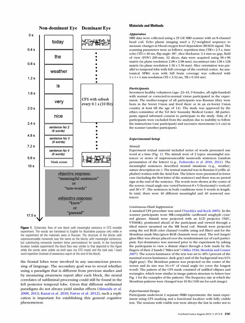
In the current functional magnetic resonance imaging(MRI) study we explored the neural correlates of subliminallanguage processing, with a novel design that addresses theconcerns reviewed above. Observers were presented withseries of consecutively presented textual stimuli: meaningfulsentences or unpronounceable nonwords (Fedorenko et al.2010), which were rendered invisible using continuous flashsuppression (CFS) (Tsuchiya and Koch 2005) for extendedperiod of time (10 s) (Fig. 1). Critically, as we sought for evi-dence of unconscious language processing of any type, wedecided to use meaningful sentences that required not onlysemantic, but also syntactic and structural processing—thedesign which permitted to increase potential differencesbetween meaningful (sentences) and meaningless (non-words) conditions. After each block of either sentences ornonwords participants reported whether they had beenaware of even a single word—a procedure which ensured thatdata analyses were conducted only on blocks judged invisibleby participants. To discriminate between neural activity eli-cited by the 2 conditions we used multivoxel pattern classifi-cation analyses (MVPA) focusing on the language network(Fedorenko et al. 2010), which was localized on a per-participant basis using the same stimuli while they were fullyvisible. The principal goal of our research was to test whether
© The Author 2014. Published by Oxford University Press.This is an Open Access article distributed under the terms of the Creative Commons Attribution License (http://creativecommons.org/licenses/by/3.0/), which permits unrestrictedreuse, distribution, and reproduction in any medium, provided the original work is properly cited.
Cerebral Cortex August 2015;25:2160–2169doi:10.1093/cercor/bhu022Advance Access publication February 20, 2014
the frontal lobes were involved in any unconscious proces-sing of language. The secondary goal was to reveal whetherusing a paradigm that is different from previous studies andby measuring awareness report after each block, the neuralcorrelates of subliminal processing could still be found in theleft posterior temporal lobe. Given that different subliminalparadigms do not always yield similar effects (Almeida et al.2008, 2013; Kanai et al. 2010; Faivre et al. 2012), such a repli-cation is important for establishing this general cognitivephenomenon.
Materials and Methods
ApparatusMRI data were collected using a 3T GE MRI scanner with an 8-channelhead coil. Echo planar imaging used a T2*-weighted sequence tomeasure changes in blood oxygen level dependent (BOLD) signal. Thescanning parameters were as follows: repetition time (TR) = 2.5 s, timeecho (TE) = 30 ms, flip angle: 90°, slice thickness: 3.4 mm no gap, fieldof view (FOV) 200 mm, 32 slices; data were acquired using 96 × 96matrix (in plane resolution 2.08 × 2.08 mm), reconstruct into 128 × 128matrix (in plane resolution 1.56 × 1.56 mm). Slice orientation was par-allel to temporal lobe with full coverage of the cerebral cortex. An ana-tomical SPRG scan with full brain coverage was collected with1 × 1 × 1 mm resolution (TE = 3.52 ms, TR = 9.104 ms).
ParticipantsSeventeen healthy volunteers (age: 23–43, 9 females, all right-handed)with normal or corrected-to-normal vision participated in the exper-iment. The mother-tongue of all participants was Russian (they wereborn in the Soviet Union and lived there or in an ex-Soviet Unioncountry at least till the age of 14). The study was approved by theethics committee of the Tel Aviv Sourasky Medical Center. All partici-pants signed informed consent to participate in the study. Data of 2participants were excluded from the analysis due to inability to followthe instructions (one participant) and excessive movements (>1 cm) inthe scanner (another participant).
Experimental Setup
StimuliExperiment textual material included series of words presented oneword at a time (Fig. 1). The stimuli were of 2 types: meaningful sen-tences or series of unpronounceable nonwords sentences (randompermutation of the letters) (e.g., Fedorenko et al. 2010, 2011). Themeaningful sentences described neutral situations (e.g., weather,nature description etc.). The textual material was in Russian (Cyrillic al-phabet) written with the Arial font. The letters were presented in lowercase (including the first letter of the sentence) and there was no periodsign at the end of the sentence. The words were shown at the center ofthe screen; visual angle size varied between 8 × 5 (horizontal × vertical)and 30 × 5°. The sentences in both conditions were 6 words in length.In total, there were 40 different meaningful and 40 nonword sen-tences.
Continuous Flash SuppressionA standard CFS procedure was used (Tsuchiya and Koch 2005). In thescanner participants wore MR-compatible cardboard anaglyph cyan/red glasses. Stimuli were projected with an LCD projector (NEC,VT660K), positioned ahead of the participant and viewed through atilted mirror mounted on the MR head coil. Stimuli were projectedusing the red RGB color channel (visible using red filter) and for theMondrian mask blue/green RGB channels were used. The red (target)glass filter was always placed over the nondominant eye of each partici-pant. Eye-dominance was assessed prior to the experiment by askingthe participants to view a distant object through a hole made by thefingers of their 2 hands (“Miles test”) (Miles 1930; Mendola and Conner2007). The screen luminance of the text was set to 40% (percent of themaximal screen luminance; dark gray) and of the background was 61%(light gray). The Mondrian pattern was projected on the center of thescreen and its size was 34 × 6° of visual angle (to cover the longestword). The pattern of the CFS mask consisted of unfilled ellipses andrectangles, which were similar in image pattern structure to letters (seeFig. 1 for the examples of the pattern). The frequency rate at which theMondrian patterns were changed was 10 Hz (100 ms for each image).
Experimental DesignParticipants underwent 2 separate fMRI experiments: the main exper-iment using CFS masking and a functional localizer with fully visibletext. The sessions with visible text were always the last in order not to
Figure 1. Schematic flow of one block with meaningful sentence in CFS invisibleexperiment. The words are translated to English for illustrative purpose only while inthe experiment all the materials were in Russian. The structure of the blocks withunpronounceable nonwords was the same as the blocks with meaningful sentences,but substituting nonwords (random letter permutations) for words. In the functionallocalizer (visible experiment) the block flow was similar to that depicted in this figurewhile the words were visible via both eyes (no CFS mask) and the task was 1-backword repetition (instead of awareness report at the end of the block).
Cerebral Cortex August 2015, V 25 N 8 2161

provide additional cues about the appearance of invisible stimuli. Thedesign and flow of both experiments were identical except for the visi-bility level of the stimuli and the behavioral task (described below). Inthe functional localizer experiment, no Mondrian mask was used andthe text was projected to both eyes using all 3 RGB channels (to elimin-ate potential head movements in the scanner between 2 experiments,participants still wore anaglyph glasses during this experiment aswell). Screen luminance of the text in the functional localizer exper-iment was 0% (black) and the background was 61% (light gray).
The experiment used a block-design with each experimental blocklasting 10 s and interleaved with a fixation block of 7.5-s duration. Fix-ation block was an empty screen with “+” sign at the middle (0.2 × 0.2°of visual angle; background luminosity: 61% [light gray color], fore-ground luminosity: 0% [black color]). Each session consisted of 12experimental blocks (6 blocks of meaningful sentences and unpro-nounceable nonwords, respectively). Each session started with a 10 sfixation cross. Total session duration was 3 min and 40 s. Number ofsessions per participant varied between participants: for the mainexperiment (invisibility experiment) it was between 7 and 11 and forthe functional localizer experiment it was between 3 and 5. The use ofa larger number of short experimental sessions compared with asmaller number of long experimental sessions improves classificationperformance (Coutanche and Thompson-Schill 2012).
The schematic flow of one block (invisible CFS experiment) consist-ing of meaningful sentences is shown in Figure 1. The flow of the con-dition with unpronounceable nonwords was similar. For illustrativepurposes only, in the figure the words have been translated to English,while all the experiment materials were in Russian. Duration of asingle word was 0.4 s; duration of a single sentence (6 words) was 2.4 s(words appeared one after another without interstimulus interval).Each experimental block consisted of 4 sentences (or 4 lists of non-words). The sentences appeared back to back without delay betweenthe last word of the previous sentence and the first word of a next sen-tence. For some sentences of the block the last word of the previoussentence and the first word of the consecutive sentence was identical(e.g., “the queue was extremely long today”, “today the weather is verynice”). The number of repetitions of the last and first word varied ran-domly between blocks (minimum 0, maximum 2). The first word ofeach sentence appeared with a random horizontal position jitter (oneor 2 letters from centered position). This ensured that when the firstand last word of the sentence repeated, there was no effect of word“freezing” on the screen. The blocks of unpronounceable nonwordswere similar to blocks of meaningful sentences (Fig. 1) but substitutingnonwords (random letter permutations) for words. We decided to usethe random letter permutation and not the letter permutation of thereal words, since the later can be still occasionally recognized as realwords (e.g., Wentura et al. 2005). The 6-word sentence-like structureand the repetition of the last and first word were preserved for non-words as well.
The behavioral tasks in the functional localizer and in the CFS sub-liminal experiment were different. In the functional localizer partici-pants were asked to press any button on the response box when theydetected a consecutive repetition of a word (1-back task). The repeatedwords could only be the last and first words of a sentence (see above).This task ensured that participants were attentive to the stimuli.
In the CFS main experiment, participants were required to make 2separate responses after every block (either 4 sentences or 4 nonwordlists, see Fig. 1). These responses were made during the fixation block(no instructions were presented). The participants were first required(“objective response”) whether they thought that a block consisted ofsentences (comprising meaningful words) or of nonwords. Partici-pants were then required to make a second response (“subjectiveresponse”) indicating whether their first response was based on seeingthe stimuli or on a “guess”. Below are the instructions, which weregiven to participants: “You will be presented with blocks of either sen-tences composed of meaningful words or series of nonwords. Thewords or nonwords will be presented sequentially. The stimuli are pre-sented in a way that makes it very hard and probably impossible to seethem. At the end of each block you need to make 2 responses. The firstresponse asks you to indicate whether it was a block of sentences(words) or nonwords. Because in each block we present only words or
nonwords, detecting one of the stimuli (single word or nonword)during the block would allow you to indicate the correct response.Even if you did not see anything, we ask you to guess. The secondresponse asks you to indicate whether your first response was basedon seeing the text or guessing”. It should be noted that as the conditionof sentences is comprised of various types of language processing(sentence syntax and structure, words semantics etc.), it was crucial toensure that any part of language processing remained unconscious.This was the reason that we made it clear to participants, that detectinga single word in the block is sufficient for a correct answer. Participantsunderwent a short training session outside the scanner as well as ashort training session inside the scanner at the beginning of the exper-iment to ensure that they understood the instructions. Prior to startingthe experimental sessions all participants confirmed that the instruc-tions are clear for them. The instructions were also repeated during theexperiment, between the sessions. At the end of the experiment,during the informal debriefing, none of the participants indicated anydifficulty with performing the task according to the instructions.
Data Analysis
PreprocessingData analysis used SPM5 (Wellcome Trust Centre for Neuroimaging,London, UK; http://www.fil.ion.ucl.ac.uk). The first 4 volumes (4 TRs,10 s) of each session were discarded to allow for T1 equilibrationeffects. Preprocessing steps applied for functional (EPI) scans in-cluded: realignment, slice-time correction, motion correction, normali-zation to 2 × 2 × 3 voxel resolution using Montreal NeurologicalInstitute (MNI) template and spatial smoothing with a full-width athalf-maximum = 6 mm kernel. For the normalization we used a unifiedsegmentation procedure (Ashburner and Friston 2005).
Region of Interest LocalizationFor the language functional localizer (visible text) we estimated a GLMmodel (HRF boxcar function) with 2 regressors: meaningful sentencesand nonwords. We used the contrast “meaningful sentences >nonwords” to identify a network of language processing regions foreach participant (Fedorenko et al. 2010, 2011, 2012). To constraintindividual GLM-defined functional activations we used probabilisticgroup-level functional masks (Fedorenko et al. 2010; http://web.mit.edu/evelina9/www/funcloc/funcloc_parcels.html). Thus, for eachmask region/participant based on individual “meaningful sentences >nonwords” GLM contrast we selected a contiguous cluster of most se-lective voxels (number of voxels is specified below). The regionsdefined by the masks are shown in Figure 2. There were 11 regions intotal: 5 regions in the left parieto-temporal lobe (angular gyrus, supra-marginal gyrus, posterior STS, middle anterior temporal gyrus, andanterior temporal gyrus), 2 regions in the right hemisphere of the tem-poral lobe (posterior STS, middle anterior temporal gyrus) and 4regions in the left hemisphere of the frontal lobe (orbital inferiorfrontal gyrus, inferior frontal gyrus, middle frontal gyrus, superiorfrontal gyrus). Critically, as multivariate prediction is influenced byregion of interest (ROI) size (e.g., Eger et al. 2008; Walther et al. 2009;Said et al. 2010) we ensured the ROIs of different regions were of anequal size of 100 voxels (1200 mm3). In additional analyses we also ex-plored a range of different ROI sizes of 50 and 150 voxels. The ROIsize could not be increased further since the size of probabilisticgroup-level functional masks (Fedorenko et al. 2010) of some of theregions (e.g., left superior frontal gyrus) was <200 voxels. DefiningROIs of equal size was undertaken using custom MATLAB code, wherefor each region/participant the code selected the contiguous cluster ofvoxels with the highest z-score values relating to the “meaningfulsentences > nonwords” contrast in the independent localizer withvisible stimuli (similar procedure had been previously applied for face-selective voxels here [Axelrod and Yovel 2012]). The list of the ROIs(100 voxels size) with their coordinates and average z-score values canbe found in Table 3.
2162 Neural Correlates of Subliminal Language Processing • Axelrod et al.

Main Experiment: Multivariate Pattern AnalysisA multivariate pattern classification analysis was performed on rawBOLD signal values. After a preprocessing stage (the step which wasidentical for all EPI data), the data of the main (CFS) experiment weredetrended and normalized using the z-score MATLAB function. Thisprocedure was applied to the full scan voxel time course. The timecourse was also shifted 3 TRs to account for hemodynamic lag. In themultivariate analysis the global signal average for each of the 2 con-ditions was subtracted (e.g., Serences et al. 2009; Misaki et al. 2010),while this procedure was performed separately for each session inorder to prevent information leakage in course of cross-validationprocedure. Global signal average subtraction, which was applied aspart of per-condition normalization procedure, increases classificationperformance (Raizada et al. 2010; see also Aksoy and Haralick 2001)and may be beneficial, as it prevents voxels with higher values andrange to dominate the classifier’’s weights (Coutanche 2013). Inaddition, subtraction of the global signal average from each conditionpermits a more straightforward interpretation of the results, as evenminimal differences in global average between conditions might be aresult of different level of arousal or attention (Coutanche 2013). Weobtained qualitatively similar results when the analyses were con-ducted without subtracting global signal average. The TRs within eachblock were averaged, resulting in a single average data point value perblock that was used as the input to pattern classification analysis. TheLibSVM MATLAB implementation of a linear support vector machinewas used for classification (http://www.csie.ntu.edu.tw/~cjlin/libsvm/) using a leave-one-session out cross-validation procedure, which wasrepeated according to number of scans available for each participant.Significance of prediction rate was established using a one-tailed t-testabove the chance for the group classification rates (Eger et al. 2008;Meyer et al. 2010; Nestor et al. 2011). Multiple comparison Bonferronicorrection was made based on the number of ROIs in posterior tem-poral lobes (7 ROIs, P-value significance threshold = 0.0071) andfrontal lobe: 4 ROIs, P-value significance threshold = 0.0125). Of note,the significance of the results persists when the correction is madebased on total number of ROIs (11 ROIs). Supplementary analysestested whether successful prediction could also be achieved based onsignal global level and this analysis differed from the main analysis in 2ways: 1) the average signal was not subtracted; 2) the timecourses of allvoxels in a ROI were averaged resulting in one timecourse (i.e., classifi-cation using one dimension). In both main and supplementary ana-lyses only the blocks where the second (“subjective”) report of the
participants was “guessed” were used. To ensure that for each partici-pant the equal number of data points (blocks) per condition (otherwisethe use of imbalanced data set might bias classification performance[Japkowicz and Stephen 2002]) we randomly discarded the data pointsfrom the condition with largest amount of data.
Results
Behavioral ResultsTo establish the level of stimulus awareness (during fMRI scan-ning) after each block (duration of 10 s) participants wereasked to make 2 separate judgments: an objective response(“meaningful sentences”/“nonwords”) and a subjective (confi-dence) response (“knew”/“guessed”). Most blocks of both con-ditions were judged to be invisible: the percentage of blockson which participants responded with “guessed” for meaning-ful sentences was 80.3% (standard error of mean [SEM] = 4.5%)and for nonwords was 82.6% (SEM = 3.4%). Critically, objectiveresponses for these invisible (“guessed”) blocks were atchance level: meaningful sentences = 51.7% (SEM = 2.1%;P-value = 0.22, t < 1, one-tailed t-test vs. 50%) and nonwords =51.7% (SEM = 2.4%; P-value = 0.24, t < 1, one-tailed t-test vs.50%). The distribution of all subjective and objective responsesis shown in Tables 1 and 2. In addition, during the informal de-briefing after the experiment, we asked the participants whatexactly they saw when they choose to answer “guessed”. Criti-cally, all the participants indicated that when they responded“guessed” then they could not see even a single letter within apresented stimulus. Thus, taken together, we conclude thatinvisibility manipulation was effective and that the trials,which were reported as “guessed”, were genuinely invisible.To explore unconscious processing, we restricted all sub-sequent analyses to blocks ranked by participants as “guessed”in their subjective response. The average number of blocks perparticipant/condition was 38.53 (mean squared error [MSE] =2.49). It was not feasible to investigate neural correlates of
Figure 2. Probabilistic group-level masks of language network (Fedorenko et al. 2010; http://web.mit.edu/evelina9/www/funcloc/funcloc_parcels.html) projected on a SPMtemplate T1 image. The names of the regions are: 1—left angular gyrus, 2—left supramarginal gyrus, 3—left posterior STS, 4—left middle anterior temporal, 5—left anteriortemporal, 6—left orbital inferior frontal gyrus, 7—left inferior frontal gyrus, 8—left middle frontal gyrus, 9—left superior frontal gyrus, 10—right posterior STS, 11—right middleanterior temporal. Regions’ IDs correspond to the IDs in Table 3.
Cerebral Cortex August 2015, V 25 N 8 2163

conscious processing (“knew” subjective, second response)since there was not sufficient data: 45% of the participants had<5 blocks per condition of this type and the average number ofblocks per participant/condition was 7.93 (MSE = 1.94).
Imaging ResultsThe goal of our analyses was to identify brain regions wherepattern signals were sufficient to discriminate subliminalmeaningful sentences from nonwords. Our multivoxel patternclassification analysis (MVPA) (Norman et al. 2006) approachfocused on nodes in the language network (Fedorenko et al.2010) identified by an independent localizer using visiblestimuli. Summary statistics (average selectivity z-score and co-ordinates) of the ROIs is presented in the Table 3. It can beseen that all the regions except for the left superior frontalgyrus showed higher activation for meaningful sentences com-pared with nonwords. Notably, the left superior frontal gyruswas also among the less selective regions in the study of Fedor-enko et al. (2010). It is noteworthy, that statistical contrast ofvisible meaningful sentences versus nonwords identifies onlythe high-level language processing network and does notinclude inferior temporal cortex (e.g., VWFA [Cohen et al.2000]), which is implicated in more low-level orthographicalprocessing (Dehaene and Cohen 2011).
The performance of the support vector machine in dis-tinguishing subliminal meaningful sentences from nonwordsin the parieto-temporal ROIs is shown in Figure 3A. Group-level statistical significance was assessed using one-tailedt-tests against chance level of 50% (Bonferroni multiple com-parison correction, see Materials and Methods). The onlyparieto-temporal region, which showed prediction signifi-cantly above chance was left posterior STS: 56.2% (MSE: 2%,t(14) = 3.01, P = 0.004). Prediction rate in the right posterior STSwas greater than chance (53.1% [MSE: 1.6%]), but it did notreach statistical significance after multiple comparison correc-tion (t(14) = 1.93, P = 0.036)]. Performance in the other ROIsdid not differ from chance: left supramarginal gyrus: 53.1%(MSE: 2.8%, t(14) = 1.1, P = 0.22), left angular gyrus: 51.6%(MSE: 2.5%, t(14) < 1), left middle anterior temporal: 49.9%(MSE: 2%, t(14) < 1), left anterior temporal: 50.7% (MSE: 2.7%,t(14) < 1) and right middle anterior temporal: 49.1% (MSE:2.8%, t(14) < 1). To compare the prediction rates between hemi-spheres, for 2 regions which were localized in both hemi-spheres (the posterior STS and the middle anterior temporalregion) we ran a 2-way repeated measures ANOVA with aregion and a hemisphere as factors. The results showed signifi-cant main effect of region [F1,14 = 7.781, P = 0.014], but no sig-nificant effect of hemisphere [F1,14 < 1] and no significantinteraction [F1,14 < 1] suggesting that higher prediction rate inthe posterior STS comparing to the middle anterior temporalwas a property of both hemispheres.
Decoding performance comparing subliminal meaningfulsentences and nonwords in frontal regions is shown in Figure 3B.The only region which showed prediction rate significantlyabove chance was left middle frontal gyrus: 54.7% (MSE: 1.5%,
Table 1Distribution of “subjective” responses (confidence rating, second response)
“Knew” “Guessed”
Meaningful sentences 19.7% (SEM: 4.5%) 80.3% (SEM: 4.5%)Nonwords 17.4% (SEM: 3.4%) 82.6% (SEM: 3.4%)
Table 2Percent of correct “objective” (first answer) responses per each category (numbers in the tablecells) binned for corresponding “subjective” (second answer) response (table columns)
“Knew” “Guessed”
Meaningful sentences 62.4% (SEM: 9.9%) 51.7% (SEM: 2.1%)]Nonwords 86.5% (SEM: 4.9%) 51.7% (SEM: 2.4%)
Table 3Average z-scores and average MNI coordinates (center of mass) of ROI used in the decodinganalysis of invisible stimuli
ID Region of interest Average z-score MNI coordinates
X Y Z
1 Left angular gyrus 1.46 (SEM: 0.47) −43 −73 302 Left supramarginal gyrus 3.35 (SEM: 0.64) −54 −59 153 Left posterior superior temporal sulcus (STS) 5.85 (SEM: 0.47) −57 −42 44 Left middle anterior temporal 4.67 (SEM: 0.55) −57 −18 −95 Left anterior temporal 3.9 (SEM: 0.44) −54 2 −176 Left orbital inferior frontal gyrus 3.5 (SEM: 0.51) −48 28 −37 Left inferior frontal gyrus 4.94 (SEM: 0. 64) −50 17 238 Left middle frontal gyrus 3.72 (SEM: 0.62) −43 1 529 Left superior frontal gyrus 0.13 (SEM: 0.47) −7 54 3810 Right posterior superior temporal sulcus (STS) 3.24 (SEM: 0.31) 59 −45 811 Right middle anterior temporal 3.08 (SEM: 0.47) 54 −14 −13
ID numbers in the first column correspond to the numbers of anatomical masks in Figure 2. TheZ-score values are based on meaningful sentence > nonwords contrast in visible stimuli localizer.Volume of all ROIs was 100 voxels (1200 mm3). Details of how the ROIs were generated aredescribed in the Materials and Methods section.
Figure 3. Prediction rate of discrimination between subliminal meaningful sentencesand nonwords in language network regions in the temporal lobe (A) and the frontallobe (B). Black line is a chance level = 50%; error bars denote standard error of themean.
2164 Neural Correlates of Subliminal Language Processing • Axelrod et al.
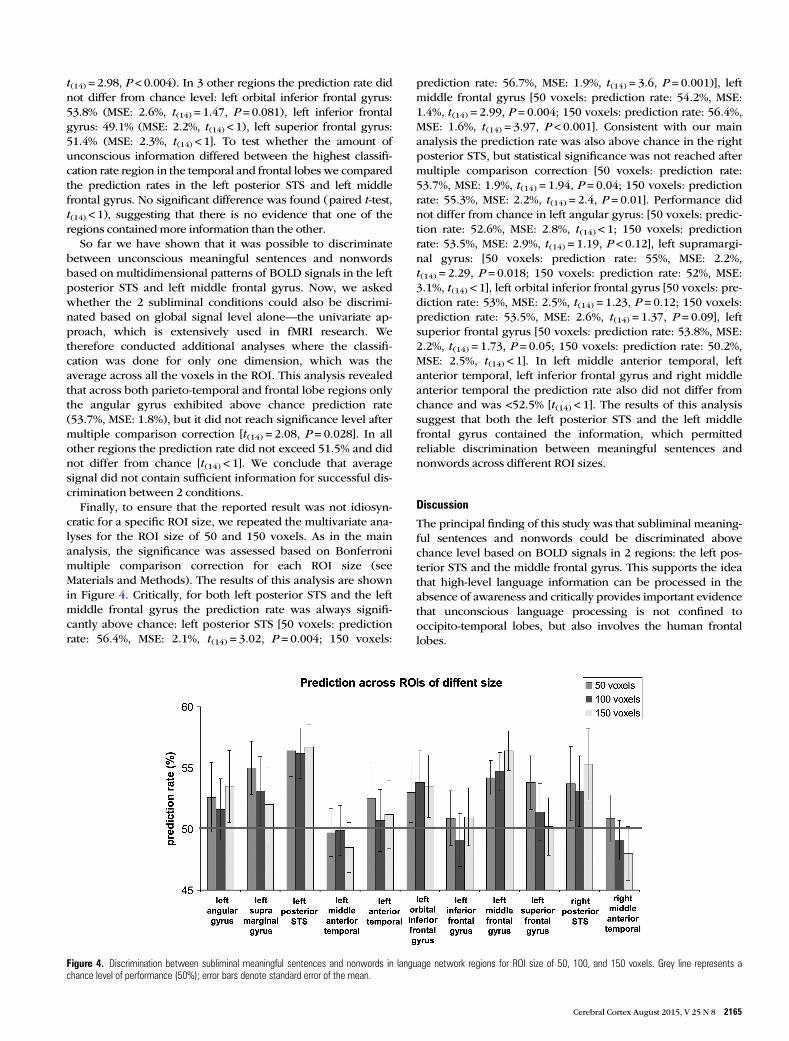
t(14) = 2.98, P < 0.004). In 3 other regions the prediction rate didnot differ from chance level: left orbital inferior frontal gyrus:53.8% (MSE: 2.6%, t(14) = 1.47, P = 0.081), left inferior frontalgyrus: 49.1% (MSE: 2.2%, t(14) < 1), left superior frontal gyrus:51.4% (MSE: 2.3%, t(14) < 1]. To test whether the amount ofunconscious information differed between the highest classifi-cation rate region in the temporal and frontal lobes we comparedthe prediction rates in the left posterior STS and left middlefrontal gyrus. No significant difference was found (paired t-test,t(14) < 1), suggesting that there is no evidence that one of theregions contained more information than the other.
So far we have shown that it was possible to discriminatebetween unconscious meaningful sentences and nonwordsbased on multidimensional patterns of BOLD signals in the leftposterior STS and left middle frontal gyrus. Now, we askedwhether the 2 subliminal conditions could also be discrimi-nated based on global signal level alone—the univariate ap-proach, which is extensively used in fMRI research. Wetherefore conducted additional analyses where the classifi-cation was done for only one dimension, which was theaverage across all the voxels in the ROI. This analysis revealedthat across both parieto-temporal and frontal lobe regions onlythe angular gyrus exhibited above chance prediction rate(53.7%, MSE: 1.8%), but it did not reach significance level aftermultiple comparison correction [t(14) = 2.08, P = 0.028]. In allother regions the prediction rate did not exceed 51.5% and didnot differ from chance [t(14) < 1]. We conclude that averagesignal did not contain sufficient information for successful dis-crimination between 2 conditions.
Finally, to ensure that the reported result was not idiosyn-cratic for a specific ROI size, we repeated the multivariate ana-lyses for the ROI size of 50 and 150 voxels. As in the mainanalysis, the significance was assessed based on Bonferronimultiple comparison correction for each ROI size (seeMaterials and Methods). The results of this analysis are shownin Figure 4. Critically, for both left posterior STS and the leftmiddle frontal gyrus the prediction rate was always signifi-cantly above chance: left posterior STS [50 voxels: predictionrate: 56.4%, MSE: 2.1%, t(14) = 3.02, P = 0.004; 150 voxels:
prediction rate: 56.7%, MSE: 1.9%, t(14) = 3.6, P = 0.001)], leftmiddle frontal gyrus [50 voxels: prediction rate: 54.2%, MSE:1.4%, t(14) = 2.99, P = 0.004; 150 voxels: prediction rate: 56.4%,MSE: 1.6%, t(14) = 3.97, P < 0.001]. Consistent with our mainanalysis the prediction rate was also above chance in the rightposterior STS, but statistical significance was not reached aftermultiple comparison correction [50 voxels: prediction rate:53.7%, MSE: 1.9%, t(14) = 1.94, P = 0.04; 150 voxels: predictionrate: 55.3%, MSE: 2.2%, t(14) = 2.4, P = 0.01]. Performance didnot differ from chance in left angular gyrus: [50 voxels: predic-tion rate: 52.6%, MSE: 2.8%, t(14) < 1; 150 voxels: predictionrate: 53.5%, MSE: 2.9%, t(14) = 1.19, P < 0.12], left supramargi-nal gyrus: [50 voxels: prediction rate: 55%, MSE: 2.2%,t(14) = 2.29, P = 0.018; 150 voxels: prediction rate: 52%, MSE:3.1%, t(14) < 1], left orbital inferior frontal gyrus [50 voxels: pre-diction rate: 53%, MSE: 2.5%, t(14) = 1.23, P = 0.12; 150 voxels:prediction rate: 53.5%, MSE: 2.6%, t(14) = 1.37, P = 0.09], leftsuperior frontal gyrus [50 voxels: prediction rate: 53.8%, MSE:2.2%, t(14) = 1.73, P = 0.05; 150 voxels: prediction rate: 50.2%,MSE: 2.5%, t(14) < 1]. In left middle anterior temporal, leftanterior temporal, left inferior frontal gyrus and right middleanterior temporal the prediction rate also did not differ fromchance and was <52.5% [t(14) < 1]. The results of this analysissuggest that both the left posterior STS and the left middlefrontal gyrus contained the information, which permittedreliable discrimination between meaningful sentences andnonwords across different ROI sizes.
Discussion
The principal finding of this study was that subliminal meaning-ful sentences and nonwords could be discriminated abovechance level based on BOLD signals in 2 regions: the left pos-terior STS and the middle frontal gyrus. This supports the ideathat high-level language information can be processed in theabsence of awareness and critically provides important evidencethat unconscious language processing is not confined tooccipito-temporal lobes, but also involves the human frontallobes.
Figure 4. Discrimination between subliminal meaningful sentences and nonwords in language network regions for ROI size of 50, 100, and 150 voxels. Grey line represents achance level of performance (50%); error bars denote standard error of the mean.
Cerebral Cortex August 2015, V 25 N 8 2165

The language processing system is a widely distributedbrain network, which spans large regions of the left temporaland frontal lobes (for reviews see Vigneau et al. 2006; Price2012) as well as regions in the right temporal lobe (for reviewssee Jung-Beeman 2005; Vigneau et al. 2011). Strong activationof the language network was observed in our functional locali-zer experiment with visible stimuli, where all the regionsexcept for the superior frontal gyrus showed higher activationto meaningful sentences compared with unpronounceablenonwords (Table 3). Notably, the level of activations and theamount of information is drastically attenuated with uncon-scious stimulation. Even more relevant for the current discus-sion, is that some regions like frontal lobes are usually notactivated at all during unconscious processing (for review seeDehaene and Changeux 2011)—evidence that is taken tosupport theoretical models, such as Global Workspace Theory(Dehaene et al. 1998). Yet, alternatively, it can be suggestedthat weak sensory stimulation, which is an inevitable conse-quence of rendering stimuli invisible, is simply not sufficientlystrong to lead to activation in areas processing high-level infor-mation such as the frontal lobes (Haynes 2009). In the currentstudy, to increase the probability of detecting the signals in thefrontal lobes we: 1) increased sensory stimulation by projectinginvisible stimuli for an extended period of time (10 s); 2) en-hanced linguistic processing by showing meaningful sen-tences, which in addition to semantics also contained syntaxand structure; 3) used multivariate ROI analysis approachesthat focused on language-selective regions, which were loca-lized using independent experiment with visible stimuli. Criti-cally, by implementing the awareness report procedure aftereach block we ensured that the only blocks used in our analysiswere subjectively and objectively invisible on a per-participantand per-block basis. We found that invisible meaningful sen-tences and unpronounceable nonwords could be discriminatedbeyond chance level in the left middle frontal gyrus (Fig. 3B).The classification result was stable and did not depend on theexact size of the ROI used (Fig. 4).
It is noteworthy, that because none of the previous studiesthat have examined unconscious language processing couldreliably demonstrate unconscious language activity in thefrontal lobes, we deliberately designed the study to maximizethe difference between 2 conditions (meaningful sentences vs.nonwords). Accordingly, the current design was not intendedto answer the question what type of language information (se-mantic, syntactic, structural, and semantic context etc.) contrib-uted to successful unconscious discrimination. Yet, the factthat in the conscious language experiments the left middlefrontal gyrus has been shown to be most active in studies withsentences (Bottini et al. 1994; Baumgaertner et al. 2002), textprocessing (Vingerhoets et al. 2003), and complex languagematerial in general (for review see Vigneau et al. 2006) makesit plausible that subliminal syntactic/structural informationmight have contributed to successful discrimination betweenmeaningful sentences and nonwords. This interpretation isalso in line with a recent event-related potentials (ERP) study(Batterink and Neville 2013), where using auditory–visual at-tentional blink paradigm (Raymond et al. 1992) the authorsshowed that consciously undetected violations in written sen-tence syntax processing elicited early frontal negativity ∼100–400 ms. Interestingly, we found no successful decoding in theorbital inferior frontal gyrus and inferior frontal gyrus regions(including Broca’s area)—the key regions of language
processing in general (Broca 1861). This result is apparently atodds with the results of Diaz and McCarthy (2007) study, whodid report extended activations in the left inferior frontal gyrusduring a subliminal semantic task. Yet, as this study did notassess awareness after each trial, it is possible that invisibilitywas not complete and the participants were aware of the sub-liminal stimuli during some of the trials.
From a broader conscious awareness theoretical perspec-tive, the fact that unconscious information was successfullydecoded from a region in the frontal lobes suggests that infor-mation can be processed by frontal lobes without automati-cally triggering conscious awareness. This result is consistentwith first order theories (Block 2005) and recurrent processingview (Lamme 2006; van Gaal and Lamme 2011) which do notattribute special role to the frontal lobes in achieving consciousawareness. The present result does not support the originalformulation of Global Workspace Theory (Dehaene et al.1998), which does not expect the frontal lobes to be activatedby unconscious stimulation. Yet, based on the recent elabor-ation of this theory (Dehaene and Changeux 2011), our resultmight not contradict it either since the activity we report waslocalized in a specific region and did not span large portions ofthe frontal lobe, as is the case for many conscious experiences(Dehaene and Changeux 2011). Indeed, localized fMRI acti-vations of the frontal lobes were previously shown in uncon-scious cognitive control tasks (Lau and Passingham 2007; vanGaal et al. 2008; see also EEG findings: van Gaal et al. 2010,2011). Yet, the neural systems responsible for cognitive controland language processing are very different. Thus, the presentfindings complement current knowledge by showing thatunconscious language processing can also elicit localizedactivity in the frontal lobes.
Interestingly, many previous studies using various stimuli,such as faces (e.g., Sterzer et al. 2008; Schurger et al. 2010; Fah-renfort et al. 2012) or words (Devlin et al. 2004; Nakamura et al.2005, 2007) failed to find unconscious information in the frontallobes. In the present study, we used a combination of exper-imental design (long stimulation duration using CFS and mean-ingful sentences) and data analysis (MVPA) procedures, whichhas not been previously applied together. There was no way toestimate a contribution of each one of these procedures, as ourexperiment did not include direct, within experiment compari-son, between different parameters (e.g., long stimulation dur-ation using CFS vs. short backward masking stimulation).Having said that, we were able to establish that the use of multi-variate analysis (MVPA) played an important role in decodingunconscious activity in the frontal lobes, as the successful de-coding could not be achieved using univariate analysis. Similarobservation was made by another study, where invisible face/scene stimuli could be decoded in the temporal lobe only byusing multivariate, but not using univariate approach (Sterzeret al. 2008). Thus, future studies are needed to examinewhetherthe use of MVPAwill permit to find unconscious information inthe frontal lobes, also for the backward masking paradigms(Devlin et al. 2004; Nakamura et al. 2005, 2007).
Additional finding of the current study was that subliminalmeaningful sentences and nonwords could be discriminatedbeyond chance level from signals in the left posterior STS. Thisresult is consistent with previous studies that also reportedsignals associated with unconscious processing of words inthis area (Devlin et al. 2004; Nakamura et al. 2005, 2007). Criti-cally, only the present study implemented awareness report
2166 Neural Correlates of Subliminal Language Processing • Axelrod et al.
after each block allowing characterization of subjective and ob-jective invisibility. Compared with previous studies we used adifferent methodological approach: experimental paradigm(CFS vs. backward/forward mask priming), language stimuli(sentences vs. words) and data analysis approach (MVPA)(Norman et al. 2006) vs. fMR-adaptation (Grill-Spector et al.1999)]. Importantly, despite the differences between thestudies the result in the left posterior STS was successfully re-plicated, providing converging evidence and making a strongcase for the involvement of the left posterior STS in subliminaltext processing. It should be also noted, that discriminationrate from signals in the right posterior STS was also relativelyhigh (Fig. 3B) and stable across ROIs of different sizes (Fig. 4),thought it did not reach statistical significance after correctionfor multiple comparisons. An important role of bilateral pos-terior STS in semantic processing with visible stimuli has beenshown using various tasks and paradigms (for reviews seeJung-Beeman 2005; Vigneau et al. 2006, 2011). As this regionwas also shown to be involved in syntactic processing (e.g.,Ben-Shachar et al. 2004) it remains to be established what typeof information contributed the most to successful discrimi-nation of the invisible stimuli. Interestingly, in our functionallocalizer task with visible stimuli, while the activation of theleft posterior STS was the strongest among all the regions(Table 3, average z-score column), the activation of the rightposterior STS was weaker than most other regions. Thus, suc-cessful discrimination of invisible stimuli was not just a directconsequence of a strong level of activation for visible stimuli(Smith et al. 2011; Tong et al. 2012), but might also reflectregional specialization in the absence of aware processing.
Interestingly, while fMRI studies including the current(Devlin et al. 2004; Nakamura et al. 2005, 2007; Diaz andMcCarthy 2007) consistently find neural correlates of sublim-inal meaningful text processing in the left posterior temporallobe, one recent ERP study, which also used CFS (Tsuchiyaand Koch 2005) failed to find any modulation of the N400 se-mantic component to invisible text content (Kang et al. 2011;see also: Vogel et al. 1998; Heyman and Moors 2012). Thoughdifferences in the signal measured by the 2 neuroimagingmethods can potentially explain these different results, basedon our current findings we propose an additional interpret-ation. The N400 is thought to originate from multiple sourcesin the left temporal lobe (Kutas and Federmeier 2011). In thepresent study we identified 5 regions in the left temporal lobe(Fig. 2) that were all selective to meaningful text processingwhen stimuli were visible (Table 3). Yet, when the text waspresented subliminally out of those 5 temporal regions onlyone of them (left posterior STS) afforded successful discrimi-nation of meaningful sentences from nonwords, while in theother temporal ROIs performance did not differ from chancelevel. Given that the N400 ERP component reflects these mul-tiple sources (Kutas and Federmeier 2011), it is possible thatthe neural activity that originated in discriminative left pos-terior STS was intermixed with the activity which originated inneighboring, nondiscriminative regions (e.g., left middleanterior temporal, left anterior temporal). As a result, the N400component recorded on the scalp was not sensitive enough toreflect the subliminal semantic processing.
Finally, a novel methodological aspect of our work was thepresentation of long invisible sentences. While the CFS para-digm was previously used for presenting subliminal words(Costello et al. 2009; Kang et al. 2011; Yang and Yeh 2011) or 3
words sentence on one screen (Sklar et al. 2012) here wepropose a method of presenting invisible sentences of the un-limited length. The presentation of invisible sentences for pro-longed duration (e.g., 10 s in our case) gives an opportunity topresent not only subliminal text with much richer semanticinformation, but also permits exploration of unconsciousneural correlates of complex language processing (e.g., syntaxprocessing). We suggest that this paradigm can be useful forfuture studies in the field.
In conclusion, in the current study we demonstrated thatbased on the activity in the human left posterior STS and leftmiddle frontal gyrus it was possible to discriminate betweensubliminally presented meaningful sentences and nonwords.This result supports the notion that high-level language func-tions might be processed subliminally and provides importantevidence that frontal regions might be involved in unconsciouslanguage processing.
Funding
This study was supported by the Israeli Center of Research Ex-cellence in Cognitive Sciences (V.A., M.B., and G.Y.), DanielTurnberg Travel Fellowship (V.A.) and the Wellcome Trust(G.R.). Funding to pay the Open Access publication chargesfor this article was provided by Wellcome Trust grant 100227/Z/12/Z and Wellcome Trust strategic award 091593/Z/10/Z.
NotesWe thank Ev Fedorenko for assistance with establishing experimentalparadigm and for the permission to use anatomical masks. We wouldalso like to thank 2 anonymous reviewers for constructive comments.Conflict of Interest: None declared
ReferencesAbrams RL, Greenwald AG. 2000. Parts outweigh the whole (word) in
unconscious analysis of meaning. Psychol Sci. 11:118–124.Aksoy S, Haralick RM. 2001. Feature normalization and likelihood-
based similarity measures for image retrieval. Pattern Recognit Lett.22:563–582.
Almeida J, Mahon BZ, Nakayama K, Caramazza A. 2008. Unconsciousprocessing dissociates along categorical lines. Proc Natl Acad SciUSA. 105:15214–15218.
Almeida J, Pajtas P, Mahon B, Nakayama K, Caramazza A. 2013. Affectof the unconscious: Visually suppressed angry faces modulate ourdecisions. Cogn Affect Behav Neurosci. 13:94–101.
Ashburner J, Friston KJ. 2005. Unified segmentation. Neuroimage.26:839–851.
Axelrod V, Yovel G. 2012. Hierarchical processing of face viewpoint inhuman visual cortex. J Neurosci. 32:2442–2452.
Batterink L, Neville HJ. 2013. The human brain processes syntax in theabsence of conscious awareness. J Neurosci. 33:8528–8533.
Baumgaertner A, Weiller C, Büchel C. 2002. Event-related fMRI revealscortical sites involved in contextual sentence integration. Neuro-image. 16:736–745.
Ben-Shachar M, Palti D, Grodzinsky Y. 2004. Neural correlates of syn-tactic movement: converging evidence from two fMRI experiments.Neuroimage. 21:1320–1336.
Block N. 2005. Two neural correlates of consciousness. Trends CognSci. 9:46–52.
Bottini G, Corcoran R, Sterzi R, Paulesu E, Schenone P, Scarpa P, Frack-owiak RS, Frith D. 1994. The role of the right hemisphere in theinterpretation of figurative aspects of language A positron emissiontomography activation study. Brain. 117:1241–1253.
Cerebral Cortex August 2015, V 25 N 8 2167
Broca P. 1861. Remarques sur le siège de la faculté du langage articulé,suivies d’une observation d’aphémie (perte de la parole). Bull SocAnat. 6:330–357.
Cohen L, Dehaene S, Naccache L, Lehéricy S, Dehaene-Lambertz G,Hénaff M-A, Michel F. 2000. The visual word form area. Brain.123:291–307.
Costello P, Jiang Y, Baartman B, McGlennen K, He S. 2009. Semanticand subword priming during binocular suppression. ConsciousCogn. 18:375–382.
Coutanche MN. 2013. Distinguishing multi-voxel patterns and meanactivation: why, how, and what does it tell us? Cogn Affect BehavNeurosci. 13:667–673.
Coutanche MN, Thompson-Schill SL. 2012. The advantage of brieffMRI acquisition runs for multi-voxel pattern detection across runs.Neuroimage. 61:1113–1119.
Dehaene S, Changeux J-P. 2011. Experimental and theoretical ap-proaches to conscious processing. Neuron. 70:200–227.
Dehaene S, Cohen L. 2011. The unique role of the visual word formarea in reading. Trends Cogn Sci. 15:254–262.
Dehaene S, Kerszberg M, Changeux J-P. 1998. A neuronal model of aglobal workspace in effortful cognitive tasks. Proc Natl Acad SciUSA. 95:14529–14534.
Dehaene S, Naccache L, Cohen L, Bihan DL, Mangin J-F, Poline J-B,Riviere D. 2001. Cerebral mechanisms of word masking and uncon-scious repetition priming. Nat Neurosci. 4:752–758.
Devlin JT, Jamison HL, Matthews PM, Gonnerman LM. 2004. Mor-phology and the internal structure of words. Proc Natl Acad SciUSA. 101:14984–14988.
Diaz MT, McCarthy G. 2007. Unconscious word processing engages adistributed network of brain regions. J Cogn Neurosci. 19:1768–1775.
Eger E, Ashburner J, Haynes JD, Dolan RJ, Rees G. 2008. fMRI activitypatterns in human LOC carry information about object exemplarswithin category. J Cogn Neurosci. 20:356–370.
Fahrenfort JJ, Snijders TM, Heinen K, van Gaal S, Scholte HS, LammeVAF. 2012. Neuronal integration in visual cortex elevates face cat-egory tuning to conscious face perception. Proc Natl Acad Sci USA.109:21504–21509.
Faivre N, Berthet V, Kouider S. 2012. Nonconscious influences fromemotional faces: a comparison of visual crowding, masking, andcontinuous flash suppression. Front Psychol. 3:129.
Fedorenko E, Behr MK, Kanwisher N. 2011. Functional specificity forhigh-level linguistic processing in the human brain. Proc Natl AcadSci USA. 108:16428–16433.
Fedorenko E, Hsieh P-J, Nieto-Castañón A, Whitfield-Gabrieli S, Kanw-isher N. 2010. New method for fMRI investigations of language: de-fining ROIs functionally in individual subjects. J Neurophysiol.104:1177–1194.
Fedorenko E, Nieto-Castañon A, Kanwisher N. 2012. Lexical and syn-tactic representations in the brain: an fMRI investigation with multi-voxel pattern analyses. Neuropsychologia. 50:499–513.
Grill-Spector K, Kushnir T, Edelman S, Avidan G, Itzchak Y, Malach R.1999. Differential processing of objects under various viewing con-ditions in the human lateral occipital complex. Neuron. 24:187–203.
Haynes J-D. 2009. Decoding visual consciousness from human brainsignals. Trends Cogn Sci. 13:194–202.
Heyman T, Moors P. 2012. Using interocular suppression and EEG tostudy semantic processing. J Neurosci. 32:1515–1516.
Holender D. 1986. Semantic activation without conscious identificationin dichotic listening, parafoveal vision, and visual masking: asurvey and appraisal. Behav Brain Sci. 9:1–23.
Japkowicz N, Stephen S. 2002. The class imbalance problem: a sys-tematic study. Intell Data Anal. 6:429–449.
Jiang Y, Costello P, He S. 2007. Processing of invisible stimuli: advan-tage of upright faces and recognizable words in overcoming intero-cular suppression. Psychol Sci. 18:349–355.
Jung-Beeman M. 2005. Bilateral brain processes for comprehendingnatural language. Trends Cogn Sci. 9:512–518.
Kanai R, Walsh V, Tseng C-h. 2010. Subjective discriminability of invisi-bility: a framework for distinguishing perceptual and attentionalfailures of awareness. Conscious Cogn. 19:1045–1057.
Kang M-S, Blake R, Woodman GF. 2011. Semantic analysis does notoccur in the absence of awareness induced by interocular suppres-sion. J Neurosci. 31:13535–13545.
Kouider S, de Gardelle V, Sackur J, Dupoux E. 2010. How rich is con-sciousness? The partial awareness hypothesis. Trends Cogn Sci.14:301–307.
Kouider S, Dehaene S. 2007. Levels of processing during non-conscious perception: a critical review of visual masking. PhilosTrans R Soc B Biol Sci. 362:857–875.
Kouider S, Dehaene S, Jobert A, Le Bihan D. 2007. Cerebral bases ofsubliminal and supraliminal priming during reading. Cereb Cortex.17:2019–2029.
Kutas M, Federmeier KD. 2011. Thirty years and counting: findingmeaning in the N400 component of the event-related brain poten-tial (ERP). Annu Rev Psychol. 62:621–647.
Lamme VAF. 2006. Towards a true neural stance on consciousness.Trends Cogn Sci. 10:494–501.
Lau HC, Passingham RE. 2007. Unconscious activation of the cognitivecontrol system in the human prefrontal cortex. J Neurosci.27:5805–5811.
Lin Z, He S. 2009. Seeing the invisible: the scope and limits of uncon-scious processing in binocular rivalry. Prog Neurobiol. 87:195–211.
Marcel AJ. 1983. Conscious and unconscious perception: experimentson visual masking and word recognition. Cogn Psychol. 15:197–237.
Mendola JD, Conner IP. 2007. Eye dominance predicts fMRI signals inhuman retinotopic cortex. Neurosci Lett. 414:30–34.
Meyer K, Kaplan JT, Essex R, Webber C, Damasio H, Damasio A. 2010.Predicting visual stimuli on the basis of activity in auditory cortices.Nat Neurosci. 13:667–668.
Miles WR. 1930. Ocular dominance in human adults. J Gen Psychol.3:412–430.
Misaki M, Kim Y, Bandettini PA, Kriegeskorte N. 2010. Comparison ofmultivariate classifiers and response normalizations for pattern-information fMRI. Neuroimage. 53:103–118.
Nakamura K, Dehaene S, Jobert A, Bihan DL, Kouider S. 2005. Sublim-inal convergence of Kanji and Kana words: further evidence forfunctional parcellation of the posterior temporal cortex in visualword perception. J Cogn Neurosci. 17:954–968.
Nakamura K, Dehaene S, Jobert A, Le Bihan D, Kouider S. 2007. Task-specific change of unconscious neural priming in the cerebrallanguage network. Proc Natl Acad Sci USA. 104:19643–19648.
Nestor A, Plaut DC, Behrmann M. 2011. Unraveling the distributedneural code of facial identity through spatiotemporal pattern analy-sis. Proc Natl Acad Sci USA. 108:9998–10003.
Norman KA, Polyn SM, Detre GJ, Haxby JV. 2006. Beyond mind-reading: multi-voxel pattern analysis of fMRI data. Trends Cogn Sci.10:424–430.
Price CJ. 2012. A review and synthesis of the first 20 years of PET andfMRI studies of heard speech, spoken language and reading. Neu-roimage. 62:816–847.
Raizada RD, Tsao F-M, Liu H-M, Holloway ID, Ansari D, Kuhl PK.2010. Linking brain-wide multivoxel activation patterns to behav-iour: examples from language and math. Neuroimage. 51:462–471.
Raymond JE, Shapiro KL, Arnell KM. 1992. Temporary suppression ofvisual processing in an RSVP task: an attentional blink. J ExpPsychol Hum Percept Perform. 18:849–860.
Said CP, Moore CD, Engell AD, Todorov A, Haxby JV. 2010. Distributedrepresentations of dynamic facial expressions in the superior tem-poral sulcus. J Vis. 10:1–12.
Schurger A, Pereira F, Treisman A, Cohen JD. 2010. Reproducibility dis-tinguishes conscious from nonconscious neural representations.Science. 327:97–99.
Serences JT, Saproo S, Scolari M, Ho T, Muftuler LT. 2009. Estimating theinfluence of attention on population codes in human visual cortexusing voxel-based tuning functions. Neuroimage. 44:223–231.
Seth AK, Dienes Z, Cleeremans A, Overgaard M, Pessoa L. 2008.Measuring consciousness: relating behavioural and neurophysiolo-gical approaches. Trends Cogn Sci. 12:314–321.
Sklar AY, Levy N, Goldstein A, Mandel R, Maril A, Hassin RR. 2012.Reading and doing arithmetic nonconsciously. Proc Natl Acad SciUSA. 109:19614–19619.
2168 Neural Correlates of Subliminal Language Processing • Axelrod et al.
Smith AT, Kosillo P, Williams AL. 2011. The confounding effect ofresponse amplitude on MVPA performance measures. Neuroimage.56:525–530.
Sterzer P, Haynes J-D, Rees G. 2008. Fine-scale activity patterns in high-level visual areas encode the category of invisible objects. J Vis. 8:1–12.
Tong F, Harrison SA, Dewey JA, Kamitani Y. 2012. Relationship betweenBOLD amplitude and pattern classification of orientation-selectiveactivity in the human visual cortex. Neuroimage. 63:1212–1222.
Tsuchiya N, Koch C. 2005. Continuous flash suppression reduces nega-tive afterimages. Nat Neurosci. 8:1096–1101.
Van den Bussche E, Van den Noortgate W, Reynvoet B. 2009. Mechan-isms of masked priming: a meta-analysis. Psychol Bull. 135:452–477.
van Gaal S, Lamme VA, Fahrenfort JJ, Ridderinkhof KR. 2011. Dissoci-able brain mechanisms underlying the conscious and unconsciouscontrol of behavior. J Cogn Neurosci. 23:91–105.
van Gaal S, Lamme VAF. 2011. Unconscious high-level information pro-cessing: implication for neurobiological theories of consciousness.The Neuroscientist. 18:287–301.
van Gaal S, Ridderinkhof KR, Fahrenfort JJ, Scholte HS, Lamme VA.2008. Frontal cortex mediates unconsciously triggered inhibitorycontrol. J Neurosci. 28:8053–8062.
van Gaal S, Ridderinkhof KR, Scholte HS, Lamme VA. 2010. Uncon-scious activation of the prefrontal no-go network. J Neurosci.30:4143–4150.
Vigneau M, Beaucousin V, Hervé PY, Duffau H, Crivello F, Houdé O,Mazoyer B, Tzourio-Mazoyer N. 2006. Meta-analyzing left hemi-sphere language areas: Phonology, semantics, and sentence proces-sing. Neuroimage. 30:1414–1432.
Vigneau M, Beaucousin V, Hervé P-Y, Jobard G, Petit L, Crivello F,Mellet E, Zago L, Mazoyer B, Tzourio-Mazoyer N. 2011. What isright-hemisphere contribution to phonological, lexico-semantic,and sentence processing? Insights from a meta-analysis. Neuro-image. 54:577–593.
Vingerhoets G, Borsel JV, Tesink C, van den Noort M, Deblaere K, Seur-inck R, Vandemaele P, Achten E. 2003. Multilingualism: an fMRIstudy. Neuroimage. 20:2181–2196.
Vogel EK, Luck SJ, Shapiro KL. 1998. Electrophysiological evi-dence for a postperceptual locus of suppression during the at-tentional blink. J Exp Psychol Hum Percept Perform.24:1656–1674.
Walther DB, Caddigan E, Fei-Fei L, Beck DM. 2009. Natural scene cat-egories revealed in distributed patterns of activity in the humanbrain. J Neurosci. 29:10573–10581.
Wentura D, Kulfanek M, Greve W. 2005. Masked affective priming byname letters: evidence for a correspondence of explicit and implicitself-esteem. J Exp Soc Psychol. 41:654–663.
Yang Y-H, Yeh S-L. 2011. Accessing the meaning of invisible words.Conscious Cogn. 20:223–233.
Cerebral Cortex August 2015, V 25 N 8 2169
Related Documents


![[SfN 2013] Neural correlates of flow](https://static.cupdf.com/doc/110x72/5564e56bd8b42afb268b4978/sfn-2013-neural-correlates-of-flow.jpg)