Neural correlates of perceptual priming of visual motion Yang Jiang 1,2,* , Yue J. Luo 3 , and Raja Parasuraman 1 1 Cognitive Science Laboratory, The Catholic University of America, Washington DC, USA; 2 Laboratory of Brain and Cognition, National Institute of Mental Health, Bethesda, MD, USA; and 3 Institute of Psychology, The Chinese Academy of Sciences, Beijing, China Abstract In two experiments, the temporal dynamics of neural activity underlying perceptual priming of visual motion was examined using event-related potentials (ERPs) during directional judgments of the apparent motion of two-dimensional sine-wave gratings. Compared to perceptually ambiguous motion, unambiguous left- or rightward motion was associated with enhanced ERP activity about 300 ms after the onset of apparent motion. In the second experiment, ERPs were recorded to two successive motion jumps in which an unambiguous motion jump served as a prime for a subsequent target motion that was ambiguous. The prime-target time interval was varied between 200, 400, and 1000 ms. In a control (motion reversal) condition, the two motion jumps were both unambiguous but in opposite directions. Compared to the motion reversal condition, motion priming was associated with an enhancement of ERP amplitudes at 100 ms and 350 ms following target stimulus onset. ERP enhancement was greatest at a short prime-target interval of 200 ms, which was also associated behaviorally with the strongest priming. The ERP enhancement and behavioral priming were both eliminated at the long 1000 ms prime-target interval. Functional magnetic resonance imaging (fMRI) data from a subset of subjects supported the view that motion priming involves modulation of neural responses both in early visual cortex and in later stages of visual processing. Keywords Perceptual priming; Visual motion; Brain imaging; ERP; fMRI INTRODUCTION Perceptual priming is an important form of automatic learning (or implicit memory) that provides for efficient perception and action in a changing visual environment. The perception of a visual event can be biased, or primed, by prior exposure to the same or related visual information. For instance, people generally respond faster to an object they have seen previously than to a new object, even though they may not consciously remember having seen the object before [25]. Perceptual priming occurs for many features of objects, including object motion. Motion perception can be biased in favor of a particular direction by previous exposure to a preceding stimulus in that direction [2,6,15,20,21,23]. Although perceptual priming reflects automatic or implicit learning, it differs from conscious recollection. The neural basis of perceptual priming of stationary objects has been studied extensively using electrophysiological and neuroimaging techniques (e.g., [4,5,8,14,17]). For example, repetition priming of visual objects is typically associated with reduced neural activation (see [28] for a review). Other forms of priming, such as word-stem completion, have * Address for correspondence: Dr. Yang Jiang, E-mail:[email protected]; or Dr. Yue J. Luo, E-mail:[email protected]. NIH Public Access Author Manuscript Brain Res Bull. Author manuscript; available in PMC 2006 January 25. Published in final edited form as: Brain Res Bull. 2002 January 15; 57(2): 211–219. NIH-PA Author Manuscript NIH-PA Author Manuscript NIH-PA Author Manuscript

Welcome message from author
This document is posted to help you gain knowledge. Please leave a comment to let me know what you think about it! Share it to your friends and learn new things together.
Transcript
Neural correlates of perceptual priming of visual motion
Yang Jiang1,2,*, Yue J. Luo3, and Raja Parasuraman11 Cognitive Science Laboratory, The Catholic University of America, Washington DC, USA;
2 Laboratory of Brain and Cognition, National Institute of Mental Health, Bethesda, MD, USA; and
3 Institute of Psychology, The Chinese Academy of Sciences, Beijing, China
AbstractIn two experiments, the temporal dynamics of neural activity underlying perceptual priming of visualmotion was examined using event-related potentials (ERPs) during directional judgments of theapparent motion of two-dimensional sine-wave gratings. Compared to perceptually ambiguousmotion, unambiguous left- or rightward motion was associated with enhanced ERP activity about300 ms after the onset of apparent motion. In the second experiment, ERPs were recorded to twosuccessive motion jumps in which an unambiguous motion jump served as a prime for a subsequenttarget motion that was ambiguous. The prime-target time interval was varied between 200, 400, and1000 ms. In a control (motion reversal) condition, the two motion jumps were both unambiguous butin opposite directions. Compared to the motion reversal condition, motion priming was associatedwith an enhancement of ERP amplitudes at 100 ms and 350 ms following target stimulus onset. ERPenhancement was greatest at a short prime-target interval of 200 ms, which was also associatedbehaviorally with the strongest priming. The ERP enhancement and behavioral priming were botheliminated at the long 1000 ms prime-target interval. Functional magnetic resonance imaging (fMRI)data from a subset of subjects supported the view that motion priming involves modulation of neuralresponses both in early visual cortex and in later stages of visual processing.
KeywordsPerceptual priming; Visual motion; Brain imaging; ERP; fMRI
INTRODUCTIONPerceptual priming is an important form of automatic learning (or implicit memory) thatprovides for efficient perception and action in a changing visual environment. The perceptionof a visual event can be biased, or primed, by prior exposure to the same or related visualinformation. For instance, people generally respond faster to an object they have seenpreviously than to a new object, even though they may not consciously remember having seenthe object before [25]. Perceptual priming occurs for many features of objects, including objectmotion. Motion perception can be biased in favor of a particular direction by previous exposureto a preceding stimulus in that direction [2,6,15,20,21,23].
Although perceptual priming reflects automatic or implicit learning, it differs from consciousrecollection. The neural basis of perceptual priming of stationary objects has been studiedextensively using electrophysiological and neuroimaging techniques (e.g., [4,5,8,14,17]). Forexample, repetition priming of visual objects is typically associated with reduced neuralactivation (see [28] for a review). Other forms of priming, such as word-stem completion, have
* Address for correspondence: Dr. Yang Jiang, E-mail:[email protected]; or Dr. Yue J. Luo, E-mail:[email protected].
NIH Public AccessAuthor ManuscriptBrain Res Bull. Author manuscript; available in PMC 2006 January 25.
Published in final edited form as:Brain Res Bull. 2002 January 15; 57(2): 211–219.
NIH
-PA Author Manuscript
NIH
-PA Author Manuscript
NIH
-PA Author Manuscript

also been extensively studied using electrophysiological techniques. The typical finding fromthese studies is a greater electrical positivity at about 300–500 ms from occipitoparietal scalpsites for primed visual items (see [19] for a review). However, little is known of the neuralmechanisms underlying perceptual priming of dynamic visual motion stimuli.
Many forms of perceptual priming are quite long lasting, from several seconds to minutes todays (e.g., [9]). Neuroimaging techniques that assess neural activity over several seconds orminutes are therefore appropriate for investigating the neural correlates of these types ofpriming. Motion priming, on the other hand, may last only hundreds of milliseconds.Examining the neural correlates of this form of priming thus requires a technique with hightemporal resolution, such as event-related brain potentials (ERPs).
The millisecond resolution of ERPs allows for investigation of the temporal dynamics of neuralactivity underlying fast-acting motion priming. In the present study, we used ERPs to examinetwo-dimensional visual motion priming. We used a priming paradigm developed by Pinkusand Pantle [21] in which observers were shown sine-wave gratings that moved ambiguouslyto either the left or the right. When such a target stimulus is preceded by a prime that movesunambiguously in one direction, motion direction is disambiguated, and most observers reportseeing the target move in the same direction as the prime. Such motion priming typically decaysover a prime-target interval of about 800 ms [21].
In the present studies we used ERPs to examine the temporal characteristics of neural activityassociated with motion priming of 2-D sine-wave gratings. The objective was to determinewhether motion priming occurs at a relatively early stage of processing in visual cortex bymodulating early-latency ERP components, at a later visual processing stage, or both. We alsotook advantage of the high spatial resolution of functional magnetic resonance imaging (fMRI)to localize the early- and late-stage visual cortical areas involved in this motion priming task.
EXPERIMENT 1. SINGLE MOTION JUMPS: AMBIGUOUS VS. UNAMBIGUOUSMOVING GRATINGS
Before examining motion priming, i.e., the neural signals associated with the influence ofunambiguous motion jumps on the subsequent perception of an ambiguous motion jump, wefirst examined the ERPs associated with perceiving single motion jumps. The aim ofExperiment 1 was to compare the ERP components associated with perception of unambiguous(90° phase shift) and ambiguous (180° counter-phase shift) motion. This would then allowsubsequent examination of ERPs associated with the effects of unambiguous motion onambiguous motion.
Materials and MethodsSubjects.—Seventeen young adults (mean age 22) participated in two experiments in whichERPs were recorded. They were college students from the Catholic University of America. Allparticipants had corrected vision of at least 20/40 in Snellen and Rosenbaum eye examinationtests.
Visual stimuli and display.—The apparent motion stimuli used in the current study wereimage sequences of vertical sine-wave gratings. They were constructed as in the 2-D motionpriming experiments reported previously [21]. The motion jumps were viewed through acircular hole of a cardboard of 16 cm in diameter, at viewing distance of 104 cm (8.8° of visualangle). The average luminance of the display was 14 cd/m2. The circular viewing aperture wasstationary while the gratings moved horizontally (Fig. 1A).
Jiang et al. Page 2
Brain Res Bull. Author manuscript; available in PMC 2006 January 25.
NIH
-PA Author Manuscript
NIH
-PA Author Manuscript
NIH
-PA Author Manuscript
In Experiment 1, there were three types of one-step single motion sequences. An abrupt 90°or −90° phase shift of a sine-wave grating is associated with perception of an unambiguous,single motion step to the right or to the left. A 0°–180° counter-phase shift between frames,on the other hand, results in perception of movement to either the left or right (ambiguous orbistable), even though the physical stimuli remain the same. When shown rapidly one afteranother, the sequence produced a vivid impression of leftward or rightward motion. Theschematic diagram of the sinusoidal gratings of apparent motion sequences for leftward,rightward, and ambiguous motion jumps is shown in Fig. 1B. Thus, the three types of singlemovement steps were: (1) unambiguous left; (2) unambiguous right; and (3) ambiguous (leftor right). The order of the presentation of the three types of single motion jumps wascounterbalanced.
Task and data analysis.—For each trial, an observer viewed an apparent motion sequence.The participants were instructed to look globally towards the center of the computer screenand not to track a particular grating. After viewing each trial, the observer was required to pressthe left or right button on the response box to indicate their perceived direction of each motionjump (right or left). There were 70 trials for each of the three conditions in Experiment 1.Participants could not predict on any given trial whether an ambiguous or an unambiguousjump would be presented.
The electroencephalogram (EEG) was recorded from 14 scalp electrodes at Fz, Cz, Pz, Oz, C3,C4, P3, P4, T5, T6, O1, O2, OL, and OR. The EEG was amplified with a bandpass of 0.1–100Hz and continuously sampled (250 Hz/channel) and digitized for off-line analysis. ERPs wereselectively averaged to the last apparent motion frame (target). Each ERP component wasmeasured relative to a 200 ms baseline preceding the onset of the stimuli. Peak amplitudes andlatencies were computed for the P1 (50–150 ms), N1 (100–200 ms), and P3 (250–550 ms)components.
Results and DiscussionBehavioral results for ambiguous and unambiguous single motion jumps.—Single leftward motion was consistently perceived 99% of the time within and across subjectsin the 90° (leftward) phase shift condition. The corresponding proportion in the −90°(rightward) condition was also 99%. Thus in these conditions, motion direction wasperceptually unambiguous. In the 180° counter-phase shift condition, however, leftwardmotion was reported 56% of the time within and across subjects. This counter-phase apparentmotion was therefore perceptually ambiguous. These psychophysical results are consistentwith Pinkus and Pantle’s definition of ambiguous and unambiguous motion stimuli [21].
ERPs to ambiguous and unambiguous single motion jumps.—Figure 2 shows theERPs associated with the different single motion jump conditions. There were no significantdifferences in ERP component amplitudes or latencies between the unambiguous leftward (Fig.2 in light grey) and rightward (Fig. 2 in dark grey) motion directions. ANOVA (3 conditions× 14 sites) revealed no significant difference among leftward, rightward, and ambiguousconditions for the P1 component [F(2,32) = 1.509, p > 0.2]. There was also no difference amongleftward, rightward, and ambiguous conditions for the N1 component [F(2,32) = 1.294, p >0.2]. However, the amplitude of the P3 component elicited by ambiguous motion wassignificantly smaller than that evoked by unambiguous motion, [F(2,32) = 8.248, p < 0.005](Fig. 2 in black).
Previous ERP studies of motion direction judgments have found that an electrophysiologicalcorrelate of visual motion processing can be found in the N2, a transient negativity triggeredby the onset of a coherent motion [3,7,16,18]. The visual ERPs in the present experiment also
Jiang et al. Page 3
Brain Res Bull. Author manuscript; available in PMC 2006 January 25.
NIH
-PA Author Manuscript
NIH
-PA Author Manuscript
NIH
-PA Author Manuscript

generated negativity 160–200 ms following the onset of coherent movement (Fig. 2). However,this early component associated with motion perception was identical for ambiguous andunambiguous motion.
The main difference between ambiguous vs. unambiguous motion was in the late P3component. Previous perceptual studies [1,22,24,27] suggest that visual motion perception ismediated by neural mechanisms that correspond to mutual suppressive populations of neuronsthat are able to detect motion in opposite directions. Using fMRI, Heeger et al. [12] reportedthat the suppressive motion mechanism was found at a late visual motion processing stage—known as the human MT complex, but not in the early visual processing stage of primary visualcortex (V1). Our results suggest that the decision concerning motion direction is made at a laterstage of visual information processing at about 300 ms. This finding is consistent with Heegeret al.’s results [12].
EXPERIMENT 2: VISUAL MOTION PRIMING AND MOTION REVERSALIn the second experiment, observers were asked to judge two successive motion jumps. In themotion priming condition, an unambiguous jump (left or right) was followed by an ambiguousjump. If priming occurred, observers would report both jumps in the same direction (both leftor both right). A motion reversal condition was used as a control condition. In this condition,two unambiguous jumps were presented in succession, but in opposite directions (e.g., left-right or right-left). In both the motion priming and motion reversal conditions, therefore, thefirst motion jump was always unambiguous. However, the second target motion wasambiguous in the priming condition and unambiguously in the opposite direction to the firstin the motion reversal condition. We included this control condition to test whether observersare sensitive to motion reversals. If so, any motion priming results could not be attributedsimply to a bias or tendency for perceiving two motion jumps in the same direction.
The time interval between jumps in the priming condition was varied and ERPs were recordedto examine the temporal dynamics of the neural mechanisms underlying visual motion priming.
Materials and MethodsExperimental design.—The stimuli for double motion steps were presented on a computerscreen as in Experiment 1. The corresponding perception was two moving steps. As shown inFig. 3A, the first motion step was always an unambiguous motion step as used in Experiment1. The second motion step was either ambiguous motion in the case of the motion primingcondition, or unambiguous motion in the motion reversal condition. The phase shifts betweensuccessive sine-wave gratings in the motion reversal trials were in the sequence 0° → 90° →0°, which was perceived consistently as two opposite motion directions (right then left; or leftthen right). This is because, as indicated before, 90° phase shifts, whether negative (0° → −90°→ 0°) or positive (0° → 90° → 0°) are unambiguous. The phase shifts between successiveframes in the priming condition were in the sequence of 0° → 90° → 270° or 0° → −90 →−270°. This resulted in an unambiguous movement to the right or left (90° shift), followed byambiguous movement (180° shift). There were three time delays or SOA (200, 400, and 1000ms) between the first and the second motion steps. There were 80 trials in each of the sixconditions in Experiment 2. The reversal and priming conditions were presented in a randomorder. The six conditions in Experiment 2 were counterbalanced to avoid response bias.Observers were asked to press the left button if two successive motion jumps were perceivedto be in the same direction, and the right button if motion jumps were in opposite directions.The instructions to press the left and right buttons were switched for half the subjects.
Jiang et al. Page 4
Brain Res Bull. Author manuscript; available in PMC 2006 January 25.
NIH
-PA Author Manuscript
NIH
-PA Author Manuscript
NIH
-PA Author Manuscript
Results and DiscussionThe effect of time delay and early and late components.—The three-way ANOVA(3 Time delays × 2 conditions × 14 sites) revealed a very significant main effect of time delay(p < 0.0001). The degree of priming was reduced as the prime-target intervals increased from200 to 1000 ms. For the 200 ms interval, relative to the reversal condition, motion priming wasassociated with significantly more positive-going ERP activity at both early (P1: 100 ms, F(1,16) = 5.898, p < .05) and late (P3: 350 ms, F(1,16) = 11.373, p < .005) stages of processingfollowing the onset of the target motion. When the delay interval was increased to 400 ms, thelate P3 positivity in the priming condition was significantly greater than that in the reversalcondition, [F(1,16) = 14.408, p < .001], the earlier P1 components of the two conditionsapproaching significant difference at the p = 0.5 level [F(1,16) = 3.698]. At a prime-targetinterval of 1000 ms, there were no significant differences between the priming and reversalconditions for any ERP component. As illustrated in Fig. 3B, ERP measures at O2 for the twotypes of motion perception at time interval of 200, 400, and 1000 ms. The P1 and P3components of the ERP were significantly different between the motion priming and reversalconditions with a delay of 200 ms, less different with SOA of 400 ms, and not statisticallydifferent at prime-target delay of 1000 ms.
Behavioral results and corresponding ERPs in motion priming and motionreversal conditions.—Motion priming was measured by computing the percentage ofsame-direction motion judgments. These showed that priming decayed over the prime-targetinterval: target motion was perceived to move in the same direction as the prime on 96%, 80%,and 54% of trials at the 200, 400, and 1000 ms prime-target intervals, respectively (Fig. 4A).A 100% response proportion of same-direction responses represents perfect priming, whereas50% corresponds to a chance level of same-direction responses, or lack of priming. Theseresults therefore justify labeling of the three prime-target intervals as leading to “strongpriming” (200 ms interval), “weak priming” (400 ms), and “no priming” (1000 ms),respectively. In general these findings agree with the original psychophysical findings ofPinkus and Pantle [21].
Because the priming and reversal conditions had identical unambiguous first jumps, we wereable to compare ERPs to the motion priming condition to those in the reversal motion conditionso that neural activity specific to motion priming would be revealed. The ERP differencewaveforms between the motion priming and motion reversal conditions at the three prime-target intervals. The difference waveform represents neural activity evoked by the targetstimulus specifically associated with motion priming. Both the early 80–130 ms (p < 0.05) andlate 250–500 ms (p < 0.001) phases of ERP activity were positively enhanced in the strongpriming condition (200 ms prime-target interval) compared to the weak (400 ms) and nopriming (1000 ms) conditions (Fig. 4B).
We also tested for possible motion priming effects for the N1, P2, and N2 components, whichare associated with motion perception, However, no significant effects (p > 0.5) of primingcondition were found for N1 [F(1,16) = 0.002], P2 [F(1,16) = 0.404], and N2 [F(1,16) = 0.002].The interaction between time delay and priming condition was also not significant for anycomponent (p > 0.3).
The topographic maps of the difference ERP waveforms (priming – reversal), partitioned bydifferent time epochs of ERP activity are shown in Fig. 5A. At 98 ms following the targetmotion (in the P1 component latency range), there was significant priming-relatedenhancement in activity at posterior scalp electrode sites. At 386 ms following target motion(in the P3 component latency range), the difference appeared more extensively at posteriorsites, but included some anterior areas as well. Because of the limited spatial resolution of scalpERPs, no strong claims can be made for the intracortical localization of these neural effects.
Jiang et al. Page 5
Brain Res Bull. Author manuscript; available in PMC 2006 January 25.
NIH
-PA Author Manuscript
NIH
-PA Author Manuscript
NIH
-PA Author Manuscript
However, the P1 component of the visual ERP has been localized to extrastriate cortex [13].Thus the observed regional scalp distribution of priming-related ERP activity suggests thatmotion priming has its first effect in extrastriate cortical areas associated with relatively earlystages of visual processing.
fMRI Localization of Motion Priming-Related Cortical AreasWe used fMRI to localize the cortical areas involved in visual perception of moving sine-wavegratings. Inside of the MR scanner, subjects repeated the same motion perception tasks as theyperformed in both ERP experiments, i.e., judging single motion jumps or successive jumpsinvolving motion priming. The priming condition in the fMRI experiment only included doublejumps with a 200 ms prime-target interval. In between these conditions, subjects viewedstationary sine-wave gratings as a baseline condition.
MR data acquisition and analysis.—Two of the 17 subjects who participated in the ERPtesting were scanned. There were eight functional imaging runs (each run lasted about 5 min)for each subject. A GE 1.5 Tesla magnet was used to obtain T2*-weighted gradient echo-planarimages with blood oxygen level dependent (BOLD) signals. Twenty-two 5-mm whole brainvolumes of axial slices were acquired for each subject (repetition time 3 s, echo time 40 ms,flip angle 90°) and analyzed using multiple regression [10,11]. Brain regions showingsignificant signal enhancement to motion perception were defined as brain regions thatactivated significantly during perceiving both single motion jump and motion priming (Z >3.09; corrected).
fMRI results.—We compared MR signals associated with perceptual judgments of motiondirection with the perception of stationary sine-wave gratings. Significant (p < .001) voxels ofMR responses associated with the sine-wave motion directions were found constrained to smallcortical brain regions in the human brain. Significant activation was found in ventral occipitalarea, inferior temporal cortex known as motion processing area MT, superior temporal cortex,and intraparietal cortices. Figure 5B shows the resulting activation patterns of a single subjectoverlaid on a high-resolution structural MRI of the subject’s brain.
GENERAL DISCUSSION AND CONCLUSIONSThe results of the two experiments reveal the temporal dynamics of neural activity related tovisual motion priming. This is a form of perceptual priming in which the ambiguous motiondirection (left or right) of a 2-D sine wave grating (target) can be primed by previouspresentation of motion that is perceived unambiguously in one direction only. The primingeffect is fast acting, lasting about 800 ms. The results of the first experiment showed that forsingle motion jumps, neural activity for ambiguous motion differs from unambiguous motionat about 300 ms after motion onset. The second experiment showed that when the SOA, or theinterval between prime and target motion was as short as 200 ms, priming of ambiguous motiondirection was associated with enhanced neural responses at posterior scalp sites at both early(100 ms) and late (350 ms) stages of processing, following the onset of target motion. Thisenhancement was greatest at a short prime-target interval of 200 ms, which was also associatedbehaviorally with the strongest priming. The positive enhancement associated with motionpriming was maintained significantly for late ERP activity (P3), and approached significancefor the P1 component at the intermediate prime-target interval (400 ms). Both the early andlate ERP priming effects were eliminated at a longer SOA of 1000 ms.
The use of the motion reversal condition confirmed that these ERP amplitude changes wereassociated specifically with motion priming and were not simply due to an observer tendencyto report any two motion jumps as occurring in the same direction. In contrast, when two
Jiang et al. Page 6
Brain Res Bull. Author manuscript; available in PMC 2006 January 25.
NIH
-PA Author Manuscript
NIH
-PA Author Manuscript
NIH
-PA Author Manuscript

unambiguous jumps in opposite directions were presented, observers consistently reportedthem to move in a different, not same direction, and no ERP enhancement was obtained. TheERP signature of motion priming was quite well correlated with the behavioral reports, showinggreatest enhancement at a short SOA, reduced enhancement at the medium SOA at which weakbehavioral priming was observed, and no enhancement at the long SOA at which priming waseliminated.
The similar time course (over SOA) of neural activity and behavioral reports suggests thatperceptual decisions of motion direction result from the summation of direction-selectiveneural responses. Previous psychophysical studies have proposed such a temporal interactionview of motion priming based on motion energy models [21,26]. In this view, temporalinteractions between the neural responses of successive stimuli produce motion priming effectsthat decay as the interval between stimuli increases. In the priming condition, the neuralresponse to the priming motion favoring one particular motion direction is combined with aweaker neural response associated with ambiguous motion. Such a summation of neural signalsis reduced as the time interval between the two motion stimuli increases, consistent with ourfinding of reduced ERP amplitudes with increasing time delay between prime and target motionsteps (Fig. 4B). In contrast, in the reversal condition opposite direction motion signals resultin a net subtraction of neural responses, as reflected in smaller ERP amplitudes.
In contrast to conscious recollection, perceptual priming is an automatic process that reflectsprior exposure to a related stimulus. Electrophysiological correlates of visual word-formpriming typically are found for latencies between 300 and 500 ms, whereas consciousrecollection is usually associated with modulated of neural responses in the 600–800 ms latencyrange [19]. Perceptual priming tasks, such as word-form or repetition priming of a stationaryvisual stimulus, are longer lasting than motion priming (minutes or hours vs. 1 s).Neuroimaging evidence using PET or fMRI have associated perceptual priming of static stimuliwith occipitotemporal and other brain regions in late visual processing stages. The presentresults suggest that the neural mechanisms underlying visual motion priming differ from otherforms of perceptual priming. Visual motion priming involves both early and late stages ofvisual processing. Combined with our fMRI data, the results of the present study indicate thatmotion priming involves modulation of neural responses beginning as early as 100 ms in earlyvisual occipital cortical areas, as well as later processing areas in inferior temporal (MT),superior temporal and intraparietal cortices.
Acknowledgements
Authors Y. J. and Y.J.L. made equal contributions to the paper. We thank two anonymous reviewers, L. Chao, P.Grossenberg, A. Meyer-Lindenberg, and X.H. Peng, for their helpful comments, and the NMR center of the NationalInstitutes of Health for assistance in MR imaging. Supported by NIH grant AG07569 to R.P., AG00986 to Y.J., andNSF (China) grant 30070262 and Chinese Academy of Science grant KJCX1-07, both to Y.J.L.
References1. Adelson EH, Bergen JR. Spatial temporal energy models for the perception of motion. J Opt Soc Am
A 1985;2:284–299. [PubMed: 3973762]2. Anstis S, Ramachandran V. Visual inertia in apparent motion. Vision Res 1987;27:755–764. [PubMed:
3660637]3. Bach M, Ullrich D. Motion adaption governs the shape of motion-evoked cortical potentials. Vision
Res 1994;34:1541–1547. [PubMed: 7941362]4. Begleiter H, Projesz B, Wang W. Event-related brain potentials differentiate priming and recognition
to familiar and unfamiliar faces. Electroencephalogr Clin Neurophysiol 1995;94:41–49. [PubMed:7530638]
5. Bentin S, McCarthy G. The effects of immediate stimulus repetition on reaction time and event-relatedpotentials in tasks of different complexity. J Exp Psychol Learn Mem Cogn 1994;20:130–149.
Jiang et al. Page 7
Brain Res Bull. Author manuscript; available in PMC 2006 January 25.
NIH
-PA Author Manuscript
NIH
-PA Author Manuscript
NIH
-PA Author Manuscript
6. Blake R. What can be “perceived” in the absence of visual awareness. Curr Dir Psychol Sci 1998;6:157–162.
7. Buchner H, Gobbele R, Wager M, Fuchs M, Waberski TD, Bechmann R. Fast visual evoked potentialinput into human area V5. Neuroreport 1997;8:2419–2422. [PubMed: 9261801]
8. Buckner RL, Goodman J, Burock M, Rotte M, Koustaal W, Schacter D, Rosen B, Dale A. Functionalanatomic correlates of object priming in humans revealed by rapid presentation event related fMRI.Neuron 1998;20:285–296. [PubMed: 9491989]
9. Cave CB. Very long-lasting priming in picture naming. Psychol Sci 1997;8:322–325.10. Friston KJ, Holmes AP, Poline JB, Grasby PJ, Williams SC, Frackowiak RS, Turner R. Analysis of
fMRI time-series revisited. Neuroimage 1995;2:45–53. [PubMed: 9343589]11. Haxby, J. V.; Maisog, J. M.; Courtney, S. M. Multiple regression analysis of effects of interest in
fMRI time series. In: Lancaster, J.; Fox, P.; Friston, K., eds. Mapping and modeling the human brain.New York: Wiley; in press.
12. Heeger D, Boynton GM, Demb JB, Seidemann E, Newsome WT. Motion opponency in visual cortex.J Neurosci 1999;19:7182–7174. [PubMed: 10436071]
13. Hillyard SA. Electrical and magnetic brain recordings: Contributions to cognitive neuroscience. CurrOpin Neurobiol 1993;3:217–224. [PubMed: 8513235]
14. Jiang Y, Haxby JV, Martin A, Ungerleider LG, Parasuraman R. Complementary neural mechanismsfor tracking familiar items in human working memory. Science 2000;287:643–646. [PubMed:10649996]
15. Jiang Y, Pantle AJ, Mark LS. Visual inertia of rotating 3-D objects. Percept Psychophys 1998;60:275–286. [PubMed: 9529911]
16. Kuba M, Tonyonaga N, Kubova Z. Motion-reversal visual evoked responses. Physiol Res1992;41:369–373. [PubMed: 1286108]
17. Miller EK, Desimone R. Parallel neuronal mechanisms for short-term memory. Science1994;263:520. [PubMed: 8290960]
18. Niedeggen M, Wist ER. Characteristics of visual evoked potentials generated by motion coherenceonset. Brain Res Cogn Brain Res 1999;8:95–105. [PubMed: 10407199]
19. Paller KA. Neural measures of conscious and unconscious memory. Behav Neurosci 2000;12:127–141.
20. Pantle AJ, Gallogly DP, Piehler OC. Direction biasing by brief apparent motion stimuli. Vision Res2000;40:1979–1991. [PubMed: 10828466]
21. Pinkus A, Pantle A. Probing visual motion signals with a priming paradigm. Vision Res 1997;37:541–552. [PubMed: 9156198]
22. Qian N, Andersen RA, Adelsen EH. Transparent motion perception as detection of unbalanced motionsignals: III. Modeling. J Neurosci 1994;14:7381–7392. [PubMed: 7996183]
23. Ramachandran V, Anstis S. Extrapolation of motion path in human visual perception. Vision Res1983;23:83– 85. [PubMed: 6868383]
24. Raymond J, Braddick O. Responses to opposed directions of motion: Continuum or independentmechanisms? Vision Res 1996;36:1931–1937. [PubMed: 8759432]
25. Schacter DL, Buckner RL. Priming and the brain. Neuron 1998;20:185–195. [PubMed: 9491981]26. Strout JJ, Pantle A, Mills SL. An energy model of interframe interval effects in single-step apparent
motion. Vision Res 1994;33:3223–3240. [PubMed: 7975353]27. Van Santen J, Sperling G. Temporal covariance model of human motion perception. J Opt Soc Am
A 1984;1:451–473. [PubMed: 6726493]28. Wiggs CL, Martin A. Properties and mechanisms for perceptual priming. Curr Opin Neurobiol
1998;8:227–233. [PubMed: 9635206]
Jiang et al. Page 8
Brain Res Bull. Author manuscript; available in PMC 2006 January 25.
NIH
-PA Author Manuscript
NIH
-PA Author Manuscript
NIH
-PA Author Manuscript

FIG. 1.Visual stimuli for single apparent motion steps, corresponding perception of motion direction,from Experiment 1. (A) In the actual experiment, the stimuli were presented through a circularwindow on a computer screen. (B) Three types of apparent motion stimuli. The figure showsthe whole frame to illustrate the phase shift between frames. The abrupt 90° phase shift isassociated with perception of an unambigous single motion step to the right or to the left. A0–180° counterphase shift between frames results in perception of movement to either the leftor right (ambiguous or bistable), even though the physical stimuli remain the same.
Jiang et al. Page 9
Brain Res Bull. Author manuscript; available in PMC 2006 January 25.
NIH
-PA Author Manuscript
NIH
-PA Author Manuscript
NIH
-PA Author Manuscript
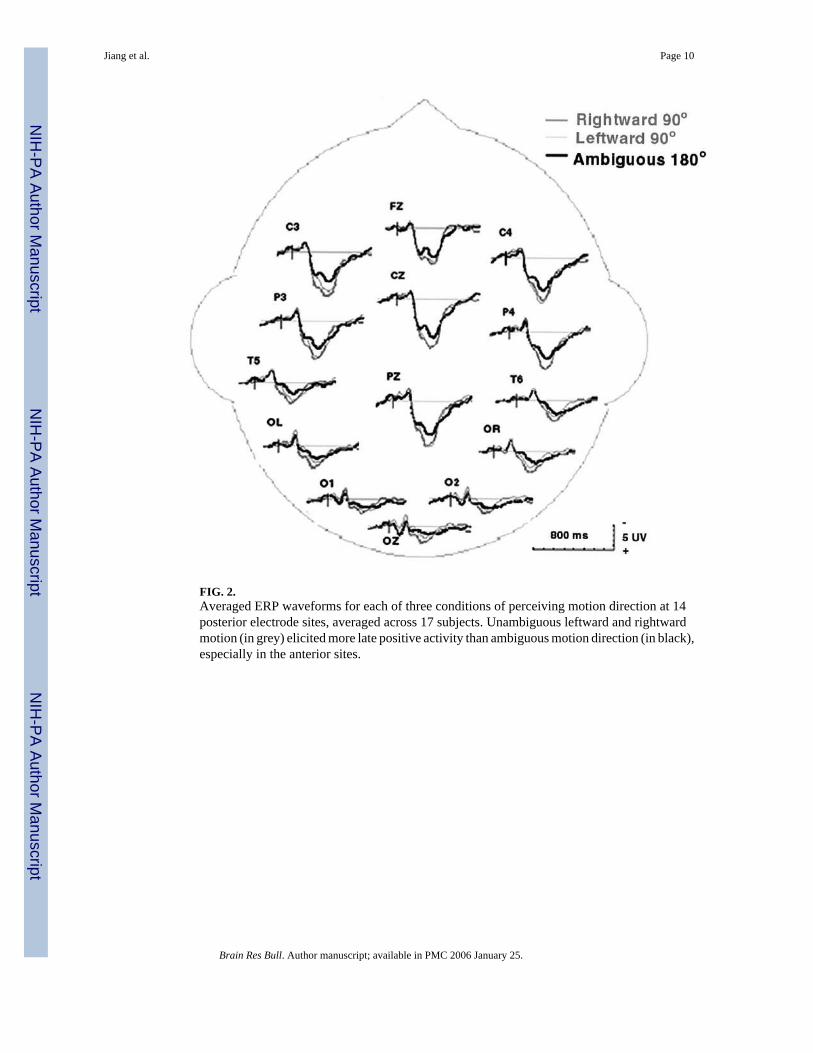
FIG. 2.Averaged ERP waveforms for each of three conditions of perceiving motion direction at 14posterior electrode sites, averaged across 17 subjects. Unambiguous leftward and rightwardmotion (in grey) elicited more late positive activity than ambiguous motion direction (in black),especially in the anterior sites.
Jiang et al. Page 10
Brain Res Bull. Author manuscript; available in PMC 2006 January 25.
NIH
-PA Author Manuscript
NIH
-PA Author Manuscript
NIH
-PA Author Manuscript
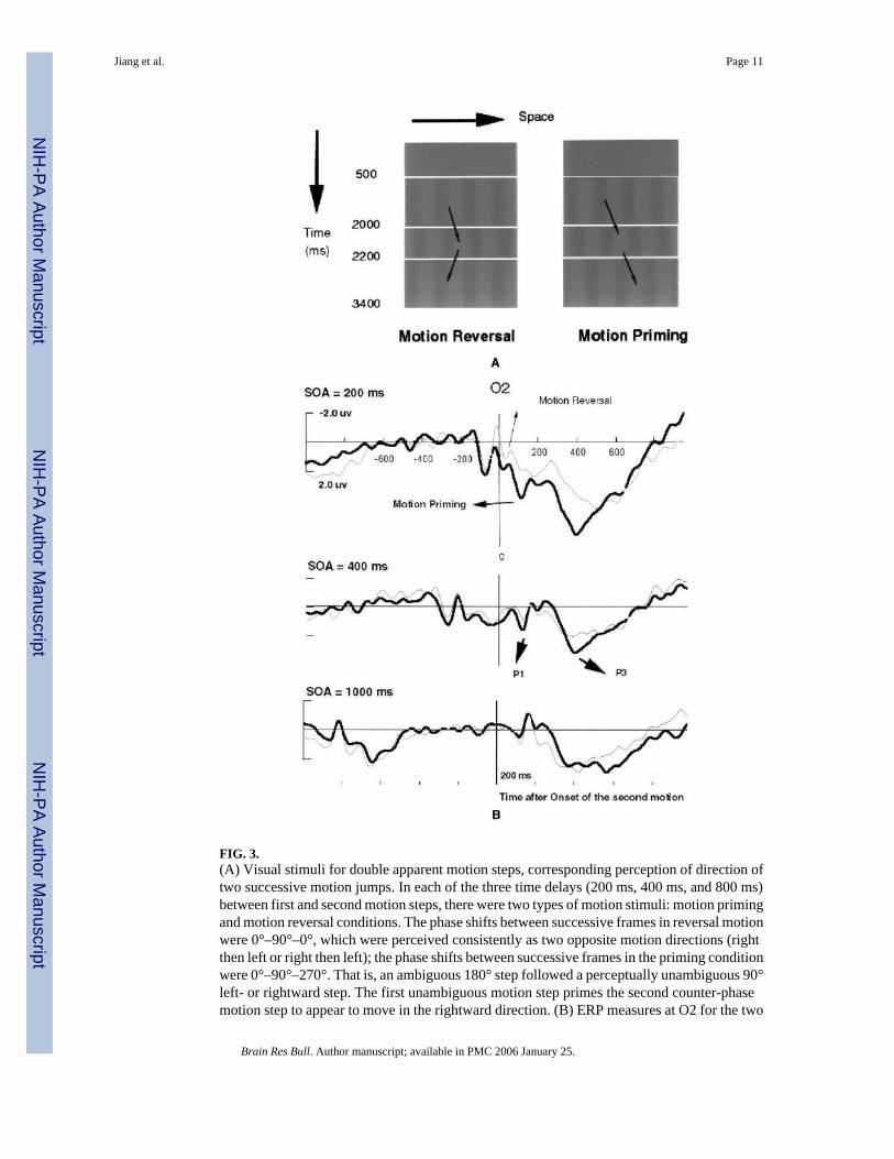
FIG. 3.(A) Visual stimuli for double apparent motion steps, corresponding perception of direction oftwo successive motion jumps. In each of the three time delays (200 ms, 400 ms, and 800 ms)between first and second motion steps, there were two types of motion stimuli: motion primingand motion reversal conditions. The phase shifts between successive frames in reversal motionwere 0°–90°–0°, which were perceived consistently as two opposite motion directions (rightthen left or right then left); the phase shifts between successive frames in the priming conditionwere 0°–90°–270°. That is, an ambiguous 180° step followed a perceptually unambiguous 90°left- or rightward step. The first unambiguous motion step primes the second counter-phasemotion step to appear to move in the rightward direction. (B) ERP measures at O2 for the two
Jiang et al. Page 11
Brain Res Bull. Author manuscript; available in PMC 2006 January 25.
NIH
-PA Author Manuscript
NIH
-PA Author Manuscript
NIH
-PA Author Manuscript

types of motion perception at time delay (SOA) of 200 ms, 400 ms, and 1000 ms. The verticalline (time 0) indicates the onset of the fourth frame as in Fig. 3A, i.e., the time of onset of thesecond motion stimulus. The P1 and P3 components of the ERP were significantly differentbetween the motion priming (in black) and reversal conditions (in grey) with a delay of 200ms, less different with time delay of 400 ms, and not statistically different at prime-target delayof 1000 ms.
Jiang et al. Page 12
Brain Res Bull. Author manuscript; available in PMC 2006 January 25.
NIH
-PA Author Manuscript
NIH
-PA Author Manuscript
NIH
-PA Author Manuscript
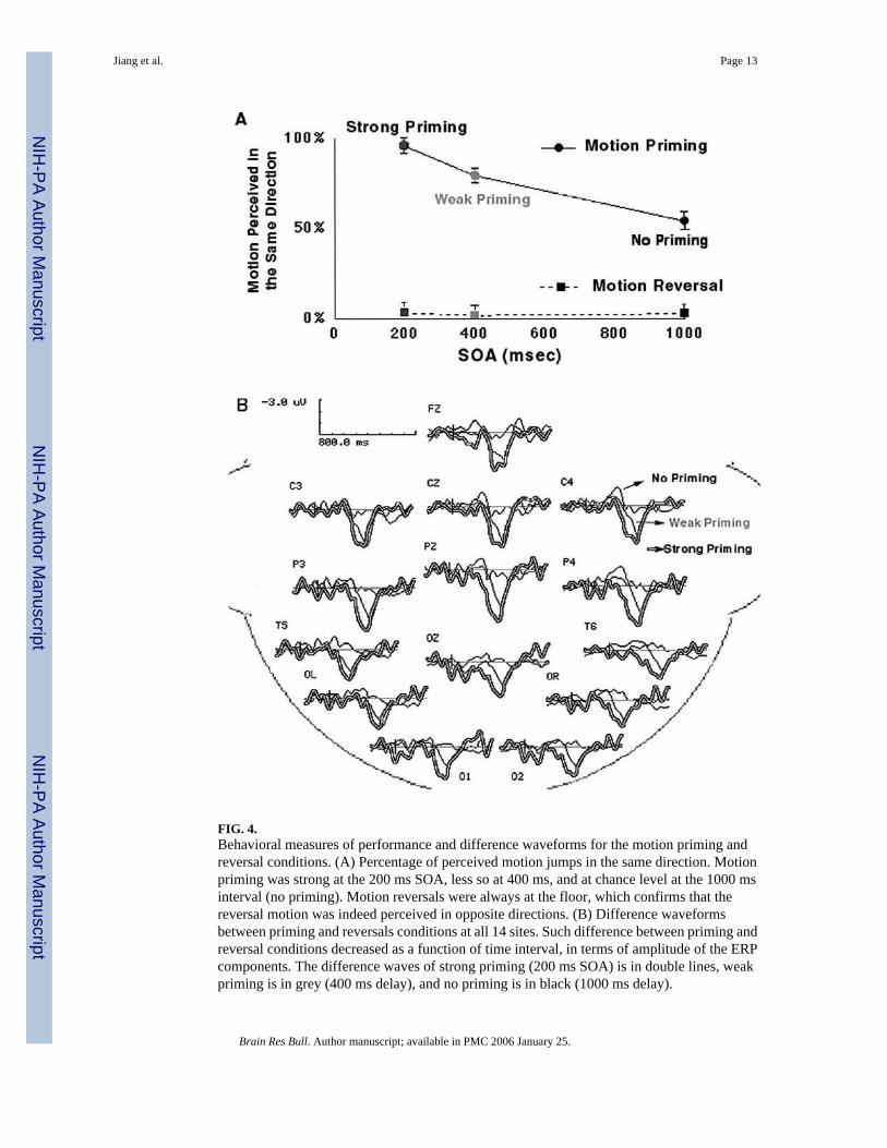
FIG. 4.Behavioral measures of performance and difference waveforms for the motion priming andreversal conditions. (A) Percentage of perceived motion jumps in the same direction. Motionpriming was strong at the 200 ms SOA, less so at 400 ms, and at chance level at the 1000 msinterval (no priming). Motion reversals were always at the floor, which confirms that thereversal motion was indeed perceived in opposite directions. (B) Difference waveformsbetween priming and reversals conditions at all 14 sites. Such difference between priming andreversal conditions decreased as a function of time interval, in terms of amplitude of the ERPcomponents. The difference waves of strong priming (200 ms SOA) is in double lines, weakpriming is in grey (400 ms delay), and no priming is in black (1000 ms delay).
Jiang et al. Page 13
Brain Res Bull. Author manuscript; available in PMC 2006 January 25.
NIH
-PA Author Manuscript
NIH
-PA Author Manuscript
NIH
-PA Author Manuscript

FIG. 5.Two dimensional plots of the ERP difference waveforms and fMRI measure of perception ofthe moving sine-wave gratings. (A) Plots of difference in ERP activity between the motionpriming and reversal conditions at three levels of priming. (B) Cortical regions involved in theperception of apparent motion jumps for one subject. High-resolution fMRI revealed thespecific cortical regions that activated by perceptual judgment of the motion direction,compared with perception of the stationary sine-wave gratings. These areas, in ventraloccipital, human motion processing area MT/V5, superior temporal, and intraparietal cortices,could have contributed to the source of the ERP responses associated with perceiving motionjumps.
Jiang et al. Page 14
Brain Res Bull. Author manuscript; available in PMC 2006 January 25.
NIH
-PA Author Manuscript
NIH
-PA Author Manuscript
NIH
-PA Author Manuscript
Related Documents