Neural activity changes to emotional stimuli in healthy individuals under chronic use of clomipramine Jorge Renner Cardoso de Almeida, MD A,B , Mary L Phillips, MD B , Carlos Toledo Cerqueira, MD A , Monica Zilberman, MD A , Daniela Lobo, MD A , Elaine Henna, MD A , Hermano Tavares, MD A , Edson Amaro Junior, MD D , Clarice Gorenstein, PhD A,C , Valentim Gentil, MD A , and Geraldo F. Busatto, MD A A Departamento de Psiquiatria, Faculdade de Medicina, Universidade de São Paulo, Brasil B Department of Psychiatry, University of Pittsburgh School of Medicine, Pittsburgh, PA, USA C Departamento de Farmacologia, Instituto de Ciências Biomédicas, Universidade de São Paulo, São Paulo, SP, Brasil D Departamento de Radiologia, Faculdade de Medicina, Universidade de São Paulo, Brasil Abstract Objective—Previous functional magnetic resonance imaging(fMRI) studies examined neural activity responses to emotive stimuli in healthy individuals after acute/subacute administration of antidepressants. We now report the effects of repeated use of the antidepressant clomipramine on fMRI data acquired during presentation of emotion-provoking and neutral stimuli on healthy volunteers. Methods—Twelve volunteers were evaluated with fMRI after receiving low doses of clomipramine for four weeks, and again after four weeks of washout. Fear-, happiness-, anger- provoking and neutral pictures from the IAPS were used. Data analysis was conducted with statistical parametric mapping(p<0.05). Results—Paired t-test comparisons for each condition between medicated and unmedicated states showed, to negative valence paradigms, decrease brain activity in the amygdala when participants were medicated. We also demonstrated, across both positive and negative valence paradigms, consistent brain activity decreases in the medicated state in the anterior cingulate gyrus and insula. Discussion—This is the first report of modulatory effects of repeated antidepressant use on the central representation of somatic states in response to emotions of both negative and positive valence in healthy individuals. Also, our results corroborate findings of antidepressant-induced temporolimbic activity changes to emotion-provoking stimuli obtained in studies of subjects treated acutely with such agents. Keywords MRI; Functional; Antidepressive Agents; Tricyclic; Clomipramine; Emotions; healthy subjects Correspondent Author: Name: Geraldo Busatto Filho, Address: Centro de Medicina Nuclear, Rua Ovídio Pires Campos, s/n, CEP 05403-903 - São Paulo - SP -Brazil, Phone/FAX: -55-11-3082 1015, [email protected]. NIH Public Access Author Manuscript J Psychopharmacol. Author manuscript; available in PMC 2010 September 1. Published in final edited form as: J Psychopharmacol. 2010 August ; 24(8): 1165–1174. doi:10.1177/0269881109105786. NIH-PA Author Manuscript NIH-PA Author Manuscript NIH-PA Author Manuscript

Welcome message from author
This document is posted to help you gain knowledge. Please leave a comment to let me know what you think about it! Share it to your friends and learn new things together.
Transcript
Neural activity changes to emotional stimuli in healthyindividuals under chronic use of clomipramine
Jorge Renner Cardoso de Almeida, MDA,B, Mary L Phillips, MDB, Carlos Toledo Cerqueira,MDA, Monica Zilberman, MDA, Daniela Lobo, MDA, Elaine Henna, MDA, Hermano Tavares,MDA, Edson Amaro Junior, MDD, Clarice Gorenstein, PhDA,C, Valentim Gentil, MDA, andGeraldo F. Busatto, MDAA Departamento de Psiquiatria, Faculdade de Medicina, Universidade de São Paulo, BrasilB Department of Psychiatry, University of Pittsburgh School of Medicine, Pittsburgh, PA, USAC Departamento de Farmacologia, Instituto de Ciências Biomédicas, Universidade de São Paulo,São Paulo, SP, BrasilD Departamento de Radiologia, Faculdade de Medicina, Universidade de São Paulo, Brasil
AbstractObjective—Previous functional magnetic resonance imaging(fMRI) studies examined neuralactivity responses to emotive stimuli in healthy individuals after acute/subacute administration ofantidepressants. We now report the effects of repeated use of the antidepressant clomipramine onfMRI data acquired during presentation of emotion-provoking and neutral stimuli on healthyvolunteers.
Methods—Twelve volunteers were evaluated with fMRI after receiving low doses ofclomipramine for four weeks, and again after four weeks of washout. Fear-, happiness-, anger-provoking and neutral pictures from the IAPS were used. Data analysis was conducted withstatistical parametric mapping(p<0.05).
Results—Paired t-test comparisons for each condition between medicated and unmedicatedstates showed, to negative valence paradigms, decrease brain activity in the amygdala whenparticipants were medicated. We also demonstrated, across both positive and negative valenceparadigms, consistent brain activity decreases in the medicated state in the anterior cingulate gyrusand insula.
Discussion—This is the first report of modulatory effects of repeated antidepressant use on thecentral representation of somatic states in response to emotions of both negative and positivevalence in healthy individuals. Also, our results corroborate findings of antidepressant-inducedtemporolimbic activity changes to emotion-provoking stimuli obtained in studies of subjectstreated acutely with such agents.
KeywordsMRI; Functional; Antidepressive Agents; Tricyclic; Clomipramine; Emotions; healthy subjects
Correspondent Author: Name: Geraldo Busatto Filho, Address: Centro de Medicina Nuclear, Rua Ovídio Pires Campos, s/n, CEP05403-903 - São Paulo - SP -Brazil, Phone/FAX: -55-11-3082 1015, [email protected].
NIH Public AccessAuthor ManuscriptJ Psychopharmacol. Author manuscript; available in PMC 2010 September 1.
Published in final edited form as:J Psychopharmacol. 2010 August ; 24(8): 1165–1174. doi:10.1177/0269881109105786.
NIH
-PA Author Manuscript
NIH
-PA Author Manuscript
NIH
-PA Author Manuscript
INTRODUCTIONThe serotonin (5-HT) and noradrenaline (NA) transmitter systems are critical for thedevelopment of normal social behavior. Their efferent neurons project to several brainregions involved in emotional processing in healthy individuals: the amygdala, the ventralstriatum, the orbitofrontal and visual cortical areas (implicated in the evaluation ofemotionally-salient stimuli and generation of emotional states), the ventral anterior cingulatecortex and the insula (involved in the central mapping of autonomic and visceral reactionsassociated with emotions), the hippocampus, the dorsal anterior cingulate, and thedorsolateral prefrontal cortices (regulating emotional behaviors)(Phillips et al. 2003;Critchley 2005; Phillips et al. 2008).
To date, many studies have shown functional abnormalities in the brain regions involved inemotional processing of negative and positive emotionally-salient stimuli in unipolardepression (Siegle 1999; Sheline et al. 2001; Fu et al. 2004; Keedwell et al. 2005; Keedwellet al. 2005; Surguladze et al. 2005; Schaefer et al. 2006; Siegle et al. 2006). Subjects withunipolar depression have shown normalization of neural activity after response toantidepressant drugs acting as 5-HT reuptake inhibitors (SRIs) and/or NA reuptakeinhibitors (NRIs)(Mayberg et al. 2000; Kennedy et al. 2001; Fu et al. 2004; Canli et al.2005; Schaefer et al. 2006). In such studies, however, the detection of the direct neuralmechanism by which SRIs/NRIs mediate their antidepressant effects was confounded by thesignificant changes in mood which might influence activity of brain regions involved inemotional processing.
Understanding the effects of 5-HT/NA augmentation upon activity in the above neuralsystem can be facilitated by examining the effect of such medication in healthy individualswithout family history of psychiatric disorders. Acute, intravenous administration of a SRI(citalopram) to healthy individuals has been associated with increased recognition of fearand happy facial expressions(Harmer et al. 2002), whereas sub-acute (seven-day) oraladministration of citalopram or a NRI (reboxetine) has been associated with reduced fearand anger recognition and enhanced positive stimuli recall(Harmer et al. 2004). Regardingdirect effects upon neural activity, acute administration of a SRI (citalopram) has beenassociated with increased amplitude of visual evoked potentials in parieto-occipital areas topleasant scenes and suppressed responses in frontal and occipital regions to unpleasantscenes(Kemp et al. 2004). In addition, in recent functional magnetic resonance imaging(fMRI) studies acute administration of a SRI (citalopram) has been associated withincreased resting activity in amygdala, striatum, thalamus and hippocampus(McKie et al.2005); with attenuated ventrolateral prefrontal cortical and amygdalar responses to aversivefaces(Del-Ben et al. 2005), and, in one study, with increased amygdala activity whenprocessing faces (Bigos et al. 2008). Acute administration of a SRI (fluvoxamine) has beenassociated with decreased amygdalar response to aversive compared with neutralstimuli(Takahashi et al. 2005). Also, seven-day administration of citalopram has beenassociated with decreased amygdalar, hippocampal and medial frontal activity to fearfulfaces(Harmer et al. 2006). Similarly, seven-day administration of reboxetine has been shownto provoke reduced amygdalar responses to fear-provoking faces(Norbury et al. 2007).These findings suggest that 5-HT/NA reuptake inhibiting antidepressants may attenuate theactivity in subcortical limbic neural regions to a higher degree when in presence of negativeemotional stimuli compared to positive emotional stimuli.
The above findings need to be interpreted in the light of some limitations. First, the studieshave not examined neural activity responses to emotive stimuli in healthy individuals afterseveral weeks of sustained 5-HT and/or NA re-uptake inhibition, which more closelyparallels the clinical uses of antidepressants in unipolar depression and anxiety disorders.
de Almeida et al. Page 2
J Psychopharmacol. Author manuscript; available in PMC 2010 September 1.
NIH
-PA Author Manuscript
NIH
-PA Author Manuscript
NIH
-PA Author Manuscript
Second, previous fMRI studies used paradigms requiring passive viewing or identificationof emotion in emotional displays, rather than paradigms that more closely resemble theemotion and cognitive processes involved in social interactions, namely, the appraisal of,and empathic response to, more complex emotionally-salient stimuli. The latter paradigmsmay be more appropriate to examine the putative neural mechanisms mediatingantidepressant response.
The present fMRI study sought to compare fMRI data acquired during presentation ofemotion-provoking (happiness, fear and anger) and neutral stimuli among healthy volunteersduring 4-week administration of the 5HT/NE re-uptake inhibitor clomipramine (medicatedstatus) and after 4-week of clomipramine washout (unmedicated status). We hypothesizedthat:
1. There would be decrease amygdala activity after repeated clomipramine use inresponse to negative stimuli;
2. There would be no change or increased neural response in the amygdala afterrepeated clomipramine use in response to positive stimuli;
3. Repeated clomipramine use would engage other brain regions involved in emotionprocessing in response to positive and negative stimuli.
METHODSSubjects
Twelve healthy individuals [3 males and 9 females, mean age = 33.5 years (SD=6.9), meaneducation = 11.1 years (SD=0.7)] were selected from a larger sample included in acontrolled drug trial conducted at the Institute of Psychiatry of São Paulo, Brazil. This studyaimed at investigating whether low doses of clomipramine had an effect on moodimprovement in healthy subjects (Gentil et al. 2007). Healthy subjects were recruitedthrough newspaper and radio advertisements, and were screened by a team of experiencedpsychiatrists using the Structured Clinical Interview for Diagnostic and Statistical Manual ofMental Disorders (First et al. 1995). Subjects did not have either first-degree family historyof psychiatric disorders including psychosis, recurrent mood disorders and substancedependence, as assessed by the Family History Screen (Weissman et al. 2000). As part ofthe drug trial, clomipramine was gradually increased according to subject tolerance to up40Mg/day over 2 weeks and maintained thereafter for an additional period of 2 weeks (meanclomipramine dose 37.5Mg/day – SD=4.5). Subjects were followed weekly for subjectivechanges in emotional response to every day stimuli. Side-effects (mainly constipation, drymouth and sexual dysfunction) were present in few subjects and considered mild. Novolunteer dropped-out due to any of these complains. Only subjects showing no subjectivemood change to clomipramine (as assessed every week blindly by two psychiatrists) (Gentilet al. 2007), and who were right-handed according to the Edinburgh Handedness Inventorywere included in this study. Exclusion criteria were: (a) current age greater than 50 or lowerthan 21 years; (b) current or previous history of neurological and/or general medicalconditions, as assessed by non-structured clinical interviewing, physical examination,electrocardiogram and blood/urinary work-up; (c) current use of other drugs psychoactiveeffects; (d) and for female subjects, history of pregnancy or lactation during the past sixmonths.
Local ethical committees approved the study and all participants signed a written informedconsent forms.
de Almeida et al. Page 3
J Psychopharmacol. Author manuscript; available in PMC 2010 September 1.
NIH
-PA Author Manuscript
NIH
-PA Author Manuscript
NIH
-PA Author Manuscript
Experimental ParadigmWe used the following stimuli: 45 pictures from the International Affective Picture System(IAPS)(Lang et al. 1997) added by 3 pictures obtained from internet versions of localnewspapers, divided in three sets with different emotional content (fear, happiness or anger);and 27 pictures of neutral scenes from the IAPS. The selection of happiness-inducingpictures was restricted to the excited (rather than relaxed) kind, in order to minimize thepossibility of major decrements in the level of arousal elicited by happiness-provokingstimuli compared to the fear- and anger-inducing pictures. All pictures were presented via amirror mounted on the head coil of the fMRI scanner.
Three, 5-minute experiments (order counterbalanced across subjects) were carried outduring fMRI acquisition, each consisting of a block design paradigm, with five blocks ofone type of emotion (fear, anger or happiness) alternated with three blocks of neutralpictures. Before the experiments, subjects were instructed to imagine themselves in thesituations while looking at the pictures. Each block comprised three pictures (each presentedfor 7.5s, with no inter-stimuli interval), all with the same emotional content (fear, anger,happiness or neutral). The themes for each picture set were the same, involving respectively:accidents, natural phenomena, dangerous animals and dentist for the fear blocks; adventurescenes and soccer for the happiness-aroused blocks; observational view of threateningsituations or revolting situations for the anger blocks; and personal objects and domesticutilities for the neutral blocks. Stimuli presentation was immediately preceded by astatement shown visually on a black background (for 5s); this strategy was aimed atdirecting the subject toward experiencing one of the three specific emotions, thus enhancingthe desired emotional response. For example, for a block of three fear stimuli including agun, a thief and a robbery, the instruction was “you are being robbed”.
After each 3-picture set, subjective self-ratings (for fear, anger, happiness and arousal) wereobtained over a period of 20s using discretized scales presented visually, in pseudo-randomized order. For each scale, individuals quantified the intensity of the emotionalexperience as follows: 1. almost nothing, 2. a little, 3. moderate, 4.intense.
In order to confirm that each set of pictures and its associated cue sentence would elicit thedesired emotion in a specific fashion, an off-scanner validation study was carried outpreviously with 156 undergraduate students (91 men and 65 women) at the same universitywhere the current imaging study was conducted (Tavares et al, submitted). A larger pool ofpictures were rated using the same four scales cited above. The final sets of three picturesselected for the fMRI study were matched across emotional conditions (happiness, fear oranger) with regard to the mean arousal scores and mean intensity of the key emotionobtained in the off-scanner study. Also, the emotion-provoking sets of pictures werematched to the neutral pictures in regard to their degree of visual complexity.
Occasions of Data AcquisitionEach subject participated in the above imaging experiment on two occasions: (1 -medicated) after a 4-week repeated use of low doses of clomipramine (mean dose 37.5Mg/day) and (2 - unmedicated) after a 4-week period of washout. A simulated session wasperformed in the week prior to the medicated state scan using a mock scanner that replicatedthe MRI environment, in order to habituate each subject to the fMRI procedure. In this shamsession, each subject was trained to use the response glove, and became used to theparadigm projection, gradient noise and room temperature. During the fMRI session, nosubject presented side effects impeditive of successful realization of the scan.
de Almeida et al. Page 4
J Psychopharmacol. Author manuscript; available in PMC 2010 September 1.
NIH
-PA Author Manuscript
NIH
-PA Author Manuscript
NIH
-PA Author Manuscript
Image AcquisitionGradient-echo imaging was used to acquire T2*-weighted image volumes using a 1.5 TeslaGE (Milkwalkee, USA) signa LX v8.3 gradient of 23mT/m, in the Institute of Radiology atthe FMUSP. For each of the 152 brain volumes, twenty-five axial planes parallel to the AC-PC plane were collected with the following parameters: TR 2500 ms, TE 40ms, slicethickness: 5 mm, 0.7 mm gap, FOV 240 mm and matrix 64×64. In addition, a high-resolution three-dimensional structural axial SPGR sequence was acquired (TE: 4.2ms, TI:400ms, TR: 10.5ms, flip angle 15°, FOV: 240mm, slice thickness: 1.6mm - zero filled to0.8mm, matrix: 256×256, 232 slices). Image acquisition, stimulus presentation and subjects’response were synchronized via an optical-electrical trigger based on the RF signal used toexcite each image slice.
Behavioral Data AnalysisPlanned comparison using paired t-tests were calculated contrasting scale scores between themedicated and unmedicated states for each condition (emotional or neutral pictures) in eachof the three paradigms.
Image AnalysisData were pre-processed and analyzed using statistical parametric mapping software (SPM5;www.fil.ion.ucl.ac.uk/spm). Data for each participant were initially realigned using the firstbrain volume acquired as a reference, and unwarped to correct for static inhomogeneity ofthe magnetic field and movement by inhomogeneity interactions. They were then co-registered with the subjects structural brain dataset, segmented/normalized to the standardMontreal Neurological Institute template (Ashburner and Friston 2005), resampled to 2×2×2mm3 voxels, and spatially smoothed with a Gaussian kernel of 6-mm full-width at half-maximum. The time-series in each voxel were high pass-filtered to 1/128 Hz. Images fromone subject during the Fear paradigm were not available due to technical problems duringdata acquisition. The structural dataset was always co-registered with the functional imagesacquired during the same session.
A single-subject, first-level fixed effect model was constructed with emotion and neutralpictures as separate conditions in a block design, using the period of visual scalepresentation and responses as baseline in the design matrix. Movement parameters from therealignment stage were entered as a covariate of no interest to further control for subjectmovement. Trials were modeled using the Canonical Haemodynamic Response Function(Friston 2007). The two conditions were then entered into the second-level analyses usingthe relevant t-contrast images (emotional pictures > baseline and neutral pictures > baseline).
A group analysis was conducted on the t-contrasts generated in the previous single-subjectanalyses using planned comparisons (paired t-test) to compare each condition (eitheremotional pictures or neutral pictures) at time 1 (medicated state) with the analogouscondition in time 2 (unmedicated state). Paired t-testing has been used in previousneuroimaging studies investigating the effect of psychotropic medications on brainfunctioning (Hariri et al. 2002; Aizenstein et al. 2008).
Region of Interest (ROI) AnalysesA priori regions of interest were targeted using anatomical masks created with the WakeForest University (WFU) Pick Atlas (Maldjian et al. 2003). To control for multiple statisticaltesting we maintained conservative threshold with a cluster-level false positive detection rateat p < 0.05 by using a voxel threshold of p < 0.05 with a cluster (k) extent empiricallydetermined by Monte Carlo simulations implemented in AlphaSim, which accounted forspatial correlations between BOLD signal changes in neighboring voxels (Forman et al.
de Almeida et al. Page 5
J Psychopharmacol. Author manuscript; available in PMC 2010 September 1.
NIH
-PA Author Manuscript
NIH
-PA Author Manuscript
NIH
-PA Author Manuscript
1995; Gianaros et al. 2008). We first accessed neural response in the amygdala.Sequentially, we explored regions involved in emotion processing as follows: ventrolateralprefrontal cortex (vlPFC - BA47), dorsolateral prefrontal cortex (DLPFC – BA9 and 46),ventromedial prefrontal cortex (vmPFC - BA11), dorsomedial prefrontal cortex (mPFC -medial region of BA10), anterior cingulate gyrus (ACG - BA24, 25 and 32), hippocampus,parahippocampal gyrus, insula, caudate, thalamus and putamen.
RESULTSBehavioral Data – Subjective State Ratings
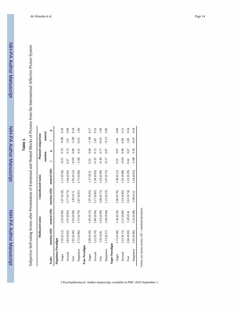
There was no significant difference in the mean scores for each scale between medicatedand unmedicated states, with the exception of trends toward greater arousal scores in themedicated state during the presentation of neutral pictures in the Happiness and Fearparadigms (p=0.081 and p=0.095 respectively), and greater anger scale scores in themedicated state during the presentation of anger-provoking pictures (p=0.065) (Table 1).
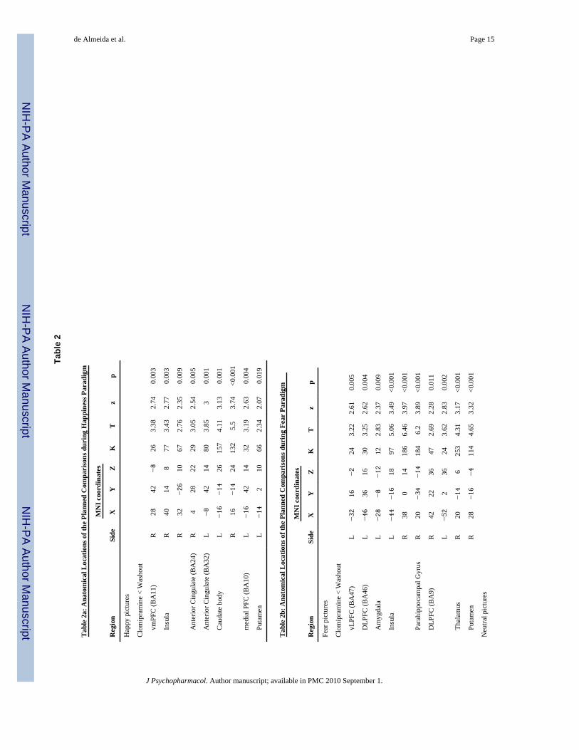
Functional Neuroimaging ResultsDuring emotion induction in the Happiness paradigm, ROI paired t-tests did not detectsignificant activity changes in the right or left amygdala. Exploratory analyses revealedsignificant reductions in brain activity in the right insula, right and left caudate body, rightand left ACG (BA24 and 32), right vmPFC (BA11), left mPFC (BA10) and left putamenwhen subjects were medicated relative to not medicated (Table 2a; Figure 1). There were nostatistical increases in the BOLD effect during the Happiness paradigm when we comparedthe medicated state (clomipramine) with the unmedicated state (washout) using ROIapproach. There were no differences in brain activity (at p<0.05) between the medicatedrelative to not medicated state during presentation of the neutral pictures.
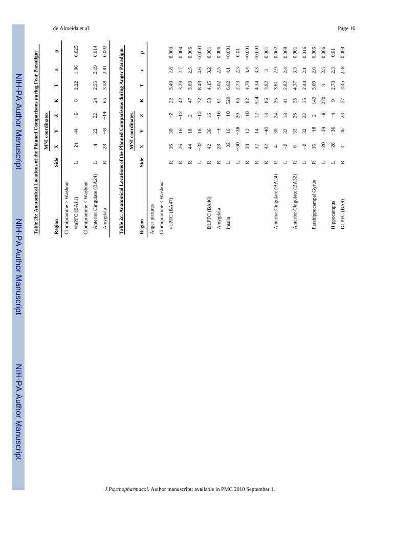
Emotion induction in the Fear Paradigm, using paired t-test comparisons, revealed decreasedactivity in the left amygdala when subjects were medicated. We also observed decreasedactivity in the right amygdala during visualization of neutral pictures when subjects weremedicated. Exploratory analyses revealed significant reduction in brain activity in the rightand left insula, right putamen, right parahippocampal gyrus, right thalamus, right and leftDLPFC (BA9/46), left vlPFC (BA47), when subjects were medicated relative to notmedicated. To neutral pictures, decreased activity was found in the left ACG (BA24)(Table2b, Figure 1); however an increase in activity was found in the vmPFC (BA11) whensubjects were medicated relative to not medicated.
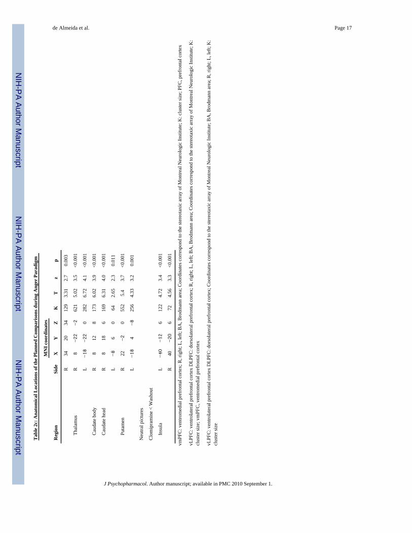
Finally, during emotion induction in the Anger paradigm, paired t-tests comparisons showedreduced activity in the right amygdala. Exploratory analyses revealed significant activityreduction when subjects were medicated relative to not medicated in the left and right insula,right and left thalamus, left and right caudate, right and left ACG (BA24/BA32), right andleft vlPFC (BA47), right and left DLPFC (BA09/46), left hippocampus, right and leftparahippocampal gyrus, right and left putamen (Table 2c, Figure 1). To neutral pictures,BOLD signal was reduced in the bilateral insula in the medicated relative to theunmedicated state (Table 2c).
DISCUSSIONThis fMRI study evaluated the neural responses to visually presented emotional stimuli inhealthy subjects during 4-week of repeated use of low doses of clomipramine (mean 37.5Mg/day) and after a 4-week washout period. Healthy subjects had no personal or familyhistory of psychiatric disorders, and showed no detectable subjective changes in mood oremotional reactivity to everyday stimuli under repeated use of clomipramine. We were
de Almeida et al. Page 6
J Psychopharmacol. Author manuscript; available in PMC 2010 September 1.
NIH
-PA Author Manuscript
NIH
-PA Author Manuscript
NIH
-PA Author Manuscript
able to replicate previous findings of reduction of amygdala activity after antidepressant usein response to negative stimuli. However, our analysis did not find change in amygdalaactivity in response to positive stimuli. Although there were some degree of variability in thespecific locations of emotion-related neural activity changes across the three emotion-provoking paradigms employed (happiness, fear and anger), we identified a consistentpattern of regionally decreased activity after repeated clomipramine use. The decreasedactivity occurred in neural regions implicated in the processing of emotional stimuli inhealthy individuals, such as the parahippocampal gyrus, anterior cingulate gyrus, insula,subcortical nuclei (striatum and thalamus), and prefrontal cortex (Phillips et al. 2003;Phillips et al. 2008). The healthy subjects in the study showed no detectable subjectivechanges in mood or emotional reactivity to everyday stimuli under repeated use ofclomipramine. Thus, the reduced BOLD signal during the medicated state may reflectintrinsic pharmacological effects of clomipramine upon neural activity, rather thanimprovements in sub-clinical psychopathology or differences in emotional engagement ofthe subjects during the processing of visual emotion stimuli between two consecutivescanning sessions.
Our findings of decreased activity in amygdala after repeated clomipramine use during theFear and Anger paradigm parallels previous studies in healthy volunteers. Studies of acuteantidepressant use of citalopram revealed decrease amygdala response to aversive faces(Del-Ben et al. 2005). Moreover, decreased amygdala response was also demonstrated afteracute fluvoxamine use in response to aversive faces (Takahashi et al. 2005). Finally, areduction of amygdala activity after seven days use of citalopram and reboxetine wasdemonstrated after fearful faces (Harmer et al. 2006; Norbury et al. 2007). The consistentobservation of reduction in the amygdala activity in acute, subacute and repeatedantidepressant use is consistent with the idea of antidepressant effects in emotion appraisalrather than regulatory emotion processing mechanisms (Norbury et al. 2007). However, onestudy found increased activation in the amygdala after acute use of citalopram (Bigos et al.2008). The contrasting findings are interesting and need replication. Moreover, studies inunipolar depression groups using sophisticated functional connectivity analysis methods,reported increased connectivity between amygdala and subgenual ACC, a regulatory area(Anand et al. 2007).
Interestingly, we did not find any significant change in the amygdala activity in response topositive stimuli. We might speculate a specific effect of antidepressant in the neuralresponse only after negative stimuli. However, more studies using positive stimuli arenecessary to replicate this finding.
Across the three emotion-provoking paradigms, the brain regions with the most significantactivity differences between the medicated and the unmedicated state were the anteriorcingulate gyrus, putamen and insula. These three regions have been implicated in the brainnetwork critical for the mediation of emotional responses to emotional stimuli (Phillips et al.2008; Critchley 2005). In particular, the anterior cingulate gyrus and insula areas are thoughtto be relevant to the cortical mapping of information pertaining to bodily responses thataccompany emotional reactions (Drevets et al. 1997; Craig 2002; Critchley 2005), while theputamen is involved with reward behavior (Forbes et al. 2008). Furthermore, the anteriorcingulate gyrus has been implicated in the generation of changes in autonomic arousal, andthe insula is thought to play a key role in the central representation of internal visceralresponses during emotional processing (Bechara et al. 1997). In addition, the putamen hasbeen implicated in emotional appraisal and identification (Phillips et al. 2008).Contemporary theories propose that the central mapping of such emotion-based somaticmarkers is crucial to the guiding of decisions and behaviors in complex situations,consciously or unconsciously (Del-Ben et al. 2005; Harmer et al. 2006; Norbury et al. 2007).
de Almeida et al. Page 7
J Psychopharmacol. Author manuscript; available in PMC 2010 September 1.
NIH
-PA Author Manuscript
NIH
-PA Author Manuscript
NIH
-PA Author Manuscript
Curiously, we found reductions in brain activity in response to negative stimuli (Fear andAnger paradigm) but not to positive stimuli in dorso- and ventro-lateral prefrontal corticalregions. These regions are known to be involved with regulation of behavior and emotion.The use of sophisticated analysis determining the function integration between these regionsmight help to elucidate the effect of antidepressants in the regulatory process and couplingbetween the subcortical regions and the prefrontal cortex.
To our knowledge, this is the first study reporting significant activity decrements in theinsula and anterior cingulate gyrus in healthy subjects after repeated clomipramine use (4weeks). Previous fMRI studies on healthy subjects during the presentation of emotion-inducing stimuli have examined the changes in neural activity following only acute (singledose) or subacute (seven days) administration of 5-HT/NA re-uptake inhibiting drugs (Del-Ben et al. 2005; Harmer et al. 2006; Norbury et al. 2007; Bigos et al. 2008; Takahashi et al.2005). Thus, our findings suggest that a sustained 5-HT/NA re-uptake inhibition (4 weeks)may induce changes in regions involved in the central representation of emotion-relatedautonomic and visceral responses. Since the subjects in the study were selected withexclusion criteria of vulnerability to depressive, anxiety or other mental disorders (they hadneither personal nor familiar history of any psychiatric disorders), our findings suggest anintrinsic capacity of clomipramine to modify brain activity patterns involved in the normalprocessing of emotional responses, irrespectively of pathological mood features.
Although the duration of clomipramine treatment in this study is close to the latency forantidepressants effects of this drug, the low doses and the absence of anxiety or depressivepsychopathology limit the extrapolation of the current findings to clinical states.Nevertheless, one could tentatively take our findings as suggestive that treatment-inducedmodifications in the processing of emotion-related bodily information are relevant to themediation of the clinical efficacy of antidepressant drugs. This is consistent with theproposed role of the anterior cingulate gyrus as a surrogate biomarker of antidepressanttreatment response, as indicated in previous neuroimaging studies of major depressedpatients evaluated before and after antidepressant treatment (Fraguas et al. 2007). Moreover,there is evidence that the intensity of autonomic responses during the processing of emotion-provoking stimuli in unmedicated major depressed patients predicts depressive symptomimprovement after chronic antidepressant treatment (Fu et al. 2004).
It is noteworthy that as other tricyclic antidepressants with SNRI properties, clomipraminedisplays high affinity to histaminergic H1, α1-adrenergic, 5-HT2A and muscarinicpostsynaptic receptors (Marcourakis et al. 1993). Therefore, the differences observed inemotion-related neural activity between medicated and non-medicated states may be due tothe effect of clomipramine on one or more of those postsynaptic receptor sites.
The choice of clomipramine for this experiment was based on our group’s previousexperiments showing that it is clinically effective in doses lower than those usuallyrecommended for major depression, and therefore is likely to decrease unwanted side-effectsthat could mask the more subtle effects on normal mood regulation (Lotufo-Neto et al.2001). Indeed, clomipramine is highly efficacious in relatively low doses in panic disorder(Wijkstra et al. 2006) Also, its antidepressant efficacy may be higher than that of non-tricyclic antidepressants in severe depression (Gillman 2007). Moreover, low doses ofclomipramine, such as 10Mg, has been demonstrated to occupy 80% of 5-HT transporter,similar with the occupation pattern of more selective inhibitors in clinical doses, (20–40Mgof fluoxetine occupy 80% of the 5-HT transporter)(Wong et al. 2008). Finally,pharmacological studies using animal tissue or human cloned receptors have shown thatclomipramine displays strong, dual action both as 5-HT and NA reuptake inhibitor, fulfillingthe criteria of a serotonin/noradreline reuptake inhibiting (SNRI) antidepressant even in
de Almeida et al. Page 8
J Psychopharmacol. Author manuscript; available in PMC 2010 September 1.
NIH
-PA Author Manuscript
NIH
-PA Author Manuscript
NIH
-PA Author Manuscript
greater conformity than the more recently marketed agents venlafaxine and duloxetine(Gillman 2007).
As we emphasized the detection of any patterns of changes in subjective mood underclomipramine use (and the exclusion from the present report of responders to this drug), wechose to use a fixed scanning order with the first session under the medicated state.Therefore, it is possible that the findings were influenced by non-specific practice effectsfrom the first, chronically medicated, to the second, not medicated, fMRI session. However,such practice effects are likely to have been minimized since all subjects underwent asimulated scan session one week prior to the medicated assessment. Moreover, previousstudies have shown decreased activity in neural regions involved in emotional processingafter repeated exposure to emotion-provoking stimuli, rather than the increase in neuralactivity seen in the post-treatment scan in our subjects. Therefore, the differences in activityin regions implicated in the representation of autonomic states may be related to the directaction of clomipramine, decreasing activity in those neural regions, rather than resultingfrom practice effects.
Our sample was not balanced in gender distribution and we did not control for possibleinfluence of menstrual cycle phase in our female subsample. Other studies should replicateour finding assessing the menstrual cycle and/or study a larger sample with only females.
In conclusion, the findings of this study indicate that clomipramine, possibly by sustained 5-HT/NA re-uptake inhibition, reduces the central representation of somatic states in responseto positive and negative emotional stimuli in healthy subjects. Our finding will inform futureresearch on patients with major depression or panic/agoraphobia in order to clarify thetherapeutic effect of antidepressants on neural activity changes in regions involved in theprocessing of the somatic markers that accompany the generation of emotional states.
AcknowledgmentsWe thank Antonio Cesário Cruz for technical assistance on the preparation of the devices for stimuli presentationand scale response recording, as well as for his continuing support during neuroimaging data acquisition.
Disclosure/Conflict of Interest
This study was supported by a grant from the ‘Fundação de Amparo à Pesquisa do Estado de São Paulo’ (FAPESP-Brazil; 01/00189-9). JRCA was supported by “Coordenação de Aperfeiçoamento de Pessoal de Nível Superior”(CAPES-Brazil; 190105-2)
ReferencesAizenstein HJ, Butters MA, Wu M, Mazurkewicz LM, Stenger VA, Gianaros PJ, Becker JT, Reynolds
CF 3rd, Carter CS. Altered Functioning of the Executive Control Circuit in Late-Life Depression:Episodic and Persistent Phenomena. Am J Geriatr Psychiatry. 2008
Anand A, Li Y, Wang Y, Gardner K, Lowe MJ. Reciprocal effects of antidepressant treatment onactivity and connectivity of the mood regulating circuit: an FMRI study. J Neuropsychiatry ClinNeurosci 2007;19(3):274–82. [PubMed: 17827412]
Ashburner J, Friston KJ. Unified segmentation. Neuroimage 2005;26(3):839–51. [PubMed: 15955494]Bechara A, Damasio H, Tranel D, Damasio AR. Deciding advantageously before knowing the
advantageous strategy. Science 1997;275(5304):1293–5. [PubMed: 9036851]Bigos KL, Pollock BG, Aizenstein HJ, Fisher PM, Bies RR, Hariri AR. Acute 5-HT Reuptake
Blockade Potentiates Human Amygdala Reactivity. Neuropsychopharmacology 2008;33(13):3221.[PubMed: 18463627]
de Almeida et al. Page 9
J Psychopharmacol. Author manuscript; available in PMC 2010 September 1.
NIH
-PA Author Manuscript
NIH
-PA Author Manuscript
NIH
-PA Author Manuscript
Canli T, Cooney RE, Goldin P, Shah M, Sivers H, Thomason ME, Whitfield-Gabrieli S, Gabrieli JD,Gotlib IH. Amygdala reactivity to emotional faces predicts improvement in major depression.Neuroreport 2005;16(12):1267–70. [PubMed: 16056122]
Craig AD. How do you feel? Interoception: the sense of the physiological condition of the body. NatRev Neurosci 2002;3(8):655–66. [PubMed: 12154366]
Critchley HD. Neural mechanisms of autonomic, affective, and cognitive integration. J Comp Neurol2005;493(1):154–66. [PubMed: 16254997]
Del-Ben CM, Deakin JF, McKie S, Delvai NA, Williams SR, Elliott R, Dolan M, Anderson IM. Theeffect of citalopram pretreatment on neuronal responses to neuropsychological tasks in normalvolunteers: an FMRI study. Neuropsychopharmacology 2005;30(9):1724–34. [PubMed: 15827569]
Drevets WC, Price JL, Simpson JR Jr, Todd RD, Reich T, Vannier M, Raichle ME. Subgenualprefrontal cortex abnormalities in mood disorders. Nature 1997;386(6627):824–7. [PubMed:9126739]
First, MB.; Spitzer, RL.; Gibbon, M.; Willians, JBW. Structured Clinical Interview for DSM-IV Axis IDisorders _ (SCID-, version 2.0). New York: Biometric research Department, New York StatePsychiatric Institute; 1995.
Forbes EE, Hariri AR, Martin SL, Silk JS, Moyles DL, Fisher PM, Brown SM, Ryan ND, Birmaher B,Axelson DA, Dahl RE. Altered Striatal Activation Predicting Real-World Positive Affect inAdolescent Major Depressive Disorder. Am J Psychiatry. 2008
Forman SD, Cohen JD, Fitzgerald M, Eddy WF, Mintun MA, Noll DC. Improved assessment ofsignificant activation in functional magnetic resonance imaging (fMRI): use of a cluster-sizethreshold. Magn Reson Med 1995;33(5):636–47. [PubMed: 7596267]
Fraguas R Jr, Marci C, Fava M, Iosifescu DV, Bankier B, Loh R, Dougherty DD. Autonomicreactivity to induced emotion as potential predictor of response to antidepressant treatment.Psychiatry Res 2007;151(1–2):169–72. [PubMed: 17360044]
Friston, KJ. Statistical parametric mapping: the analysis of funtional brain images. Amsterdam;Boston: Elsevier/Academic Press; 2007.
Fu CHY, Williams SCR, Cleare AJ, Brammer MJ, Walsh ND, Kim J, Andrew CM, Pich EM,Williams PM, Reed LJ, Mitterschiffthaler MT, Suckling J, Bullmore ET. Attenuation of the NeuralResponse to Sad Faces in Major Depression by Antidepressant Treatment: A Prospective, Event-Related Functional Magnetic Resonance Imaging Study. Arch Gen Psychiatry 2004;61(9):877–889. [PubMed: 15351766]
Gentil V, Zilberman ML, Lobo D, Henna E, Moreno RA, Gorenstein C. Clomipramine-induced moodand perceived performance changes in selected healthy individuals. J Clin Psychopharmacol2007;27(3):314–5. [PubMed: 17502789]
Gianaros PJ, Sheu LK, Matthews KA, Jennings JR, Manuck SB, Hariri AR. Individual differences instressor-evoked blood pressure reactivity vary with activation, volume, and functional connectivityof the amygdala. J Neurosci 2008;28(4):990–9. [PubMed: 18216206]
Gillman PK. Tricyclic antidepressant pharmacology and therapeutic drug interactions updated. Br JPharmacol 2007;151(6):737–48. [PubMed: 17471183]
Hariri AR V, Mattay S, Tessitore A, Fera F, Smith WG, Weinberger DR. Dextroamphetaminemodulates the response of the human amygdala. Neuropsychopharmacology 2002;27(6):1036–40.[PubMed: 12464460]
Harmer CJ, Bhagwagar Z, Perrett DI, Vollm BA, Cowen PJ, Goodwin GM. Acute SSRIAdministration Affects the Processing of Social Cues in Healthy Volunteers.Neuropsychopharmacology 2002;28(1):148. [PubMed: 12496951]
Harmer CJ, Shelley NC, Cowen PJ, Goodwin GM. Increased positive versus negative affectiveperception and memory in healthy volunteers following selective serotonin and norepinephrinereuptake inhibition. Am J Psychiatry 2004;161(7):1256–63. [PubMed: 15229059]
Harmer CJ, Mackay CE, Reid CB, Cowen PJ, Goodwin GM. Antidepressant drug treatment modifiesthe neural processing of nonconscious threat cues. Biol Psychiatry 2006;59(9):816–20. [PubMed:16460693]
de Almeida et al. Page 10
J Psychopharmacol. Author manuscript; available in PMC 2010 September 1.
NIH
-PA Author Manuscript
NIH
-PA Author Manuscript
NIH
-PA Author Manuscript
Keedwell PA, Andrew C, Williams SC, Brammer MJ, Phillips ML. A double dissociation ofventromedial prefrontal cortical responses to sad and happy stimuli in depressed and healthyindividuals. Biol Psychiatry 2005;58(6):495–503. [PubMed: 15993859]
Keedwell PA, Andrew C, Williams SC, Brammer MJ, Phillips ML. The neural correlates of anhedoniain major depressive disorder. Biol Psychiatry 2005;58(11):843–53. [PubMed: 16043128]
Kemp AH, Gray MA, Silberstein RB, Armstrong SM, Nathan PJ. Augmentation of serotonin enhancespleasant and suppresses unpleasant cortical electrophysiological responses to visual emotionalstimuli in humans. Neuroimage 2004;22(3):1084–1096. [PubMed: 15219580]
Kennedy SH, Evans KR, Kruger S, Mayberg HS, Meyer JH, McCann S, Arifuzzman AI, Houle S,Vaccarino FJ. Changes in Regional Brain Glucose Metabolism Measured With Positron EmissionTomography After Paroxetine Treatment of Major Depression. Am J Psychiatry 2001;158(6):899–905. [PubMed: 11384897]
Lang, PJ.; Bradley, MM.; Cuthbert, BN. International Affective Picture System (IAPS):TechnicalManual and Affective Ratings. Gainesville, FL: University of Florida, Center for Research inPsychophysiology; 1997.
Lotufo-Neto F, Bernik MA, Ramos RT, Andrade L, Gorenstein C, Cordas T, Melo M, Gentil V. Adose-finding and discontinuation study of clomipramine in panic disorder. J Psychopharmacol2001;15(1):13–17. [PubMed: 11277602]
Maldjian JA, Laurienti PJ, Kraft RA, Burdette JH. An automated method for neuroanatomic andcytoarchitectonic atlas-based interrogation of fMRI data sets. Neuroimage 2003;19(3):1233–9.[PubMed: 12880848]
Marcourakis T, Gorenstein C, Gentil V. Clomipramine, a better reference drug for panic/agoraphobia.II. Psychomotor and cognitive effects. J Psychopharmacol 1993;7(4):325–330.
Mayberg HS, Brannan SK, Tekell JL, Silva JA, Mahurin RK, McGinnis S, Jerabek PA. Regionalmetabolic effects of fluoxetine in major depression: serial changes and relationship to clinicalresponse. Biol Psychiatry 2000;48(8):830–43. [PubMed: 11063978]
McKie S, Del-Ben C, Elliott R, Williams S, del Vai N, Anderson I, Deakin JF. Neuronal effects ofacute citalopram detected by pharmacoMRI. Psychopharmacology (Berl) 2005;180(4):680–6.[PubMed: 15889241]
Norbury R, Mackay CE, Cowen PJ, Goodwin GM, Harmer CJ. Short-term antidepressant treatmentand facial processing. Functional magnetic resonance imaging study. Br J Psychiatry2007;190:531–2. [PubMed: 17541115]
Norbury R, Mackay CE, Cowen PJ, Goodwin GM, Harmer CJ. The effects of reboxetine on emotionalprocessing in healthy volunteers: an fMRI study. Mol Psychiatry. 2007
Phillips ML, Drevets WC, Rauch SL, Lane R. Neurobiology of emotion perception I: The neural basisof normal emotion perception. Biol Psychiatry 2003;54(5):504–14. [PubMed: 12946879]
Phillips ML, Ladouceur CD, Drevets WC. The Neural Basis of Voluntary and Automatic EmotionRegulation: Implications for Understanding the Neurodevelopment of Bipolar Disorder. MolPsychiatry. 2008 in press.
Schaefer HS, Putnam KM, Benca RM, Davidson RJ. Event-related functional magnetic resonanceimaging measures of neural activity to positive social stimuli in pre- and post-treatmentdepression. Biol Psychiatry 2006;60(9):974–86. [PubMed: 16780808]
Sheline YI, Barch DM, Donnelly JM, Ollinger JM, Snyder AZ, Mintun MA. Increased amygdalaresponse to masked emotional faces in depressed subjects resolves with antidepressant treatment:an fMRI study. Biol Psychiatry 2001;50(9):651–8. [PubMed: 11704071]
Siegle GJ. A neural network model of attention biases in depression. Prog Brain Res 1999;121:407–32. [PubMed: 10551038]
Siegle GJ, Carter CS, Thase ME. Use of FMRI to predict recovery from unipolar depression withcognitive behavior therapy. Am J Psychiatry 2006;163(4):735–8. [PubMed: 16585452]
Surguladze S, Brammer MJ, Keedwell P, Giampietro V, Young AW, Travis MJ, Williams SC, PhillipsML. A differential pattern of neural response toward sad versus happy facial expressions in majordepressive disorder. Biol Psychiatry 2005;57(3):201–9. [PubMed: 15691520]
de Almeida et al. Page 11
J Psychopharmacol. Author manuscript; available in PMC 2010 September 1.
NIH
-PA Author Manuscript
NIH
-PA Author Manuscript
NIH
-PA Author Manuscript
Takahashi H, Yahata N, Koeda M, Takano A, Asai K, Suhara T, Okubo Y. Effects of dopaminergicand serotonergic manipulation on emotional processing: a pharmacological fMRI study.Neuroimage 2005;27(4):991–1001. [PubMed: 15978846]
Weissman MM, Wickramaratne P, Adams P, Wolk S, Verdeli H, Olfson M. Brief screening for familypsychiatric history: the family history screen. Arch Gen Psychiatry 2000;57(7):675–82. [PubMed:10891038]
Wijkstra J, Lijmer J, Balk FJ, Geddes JR, Nolen WA. Pharmacological treatment for unipolarpsychotic depression: Systematic review and meta-analysis. Br J Psychiatry 2006;188:410–5.[PubMed: 16648526]
Wong DF, Tauscher J, Grunder G. The Role of Imaging in Proof of Concept for CNS Drug Discoveryand Development. Neuropsychopharmacology 2008;34(1):187. [PubMed: 18843264]
de Almeida et al. Page 12
J Psychopharmacol. Author manuscript; available in PMC 2010 September 1.
NIH
-PA Author Manuscript
NIH
-PA Author Manuscript
NIH
-PA Author Manuscript
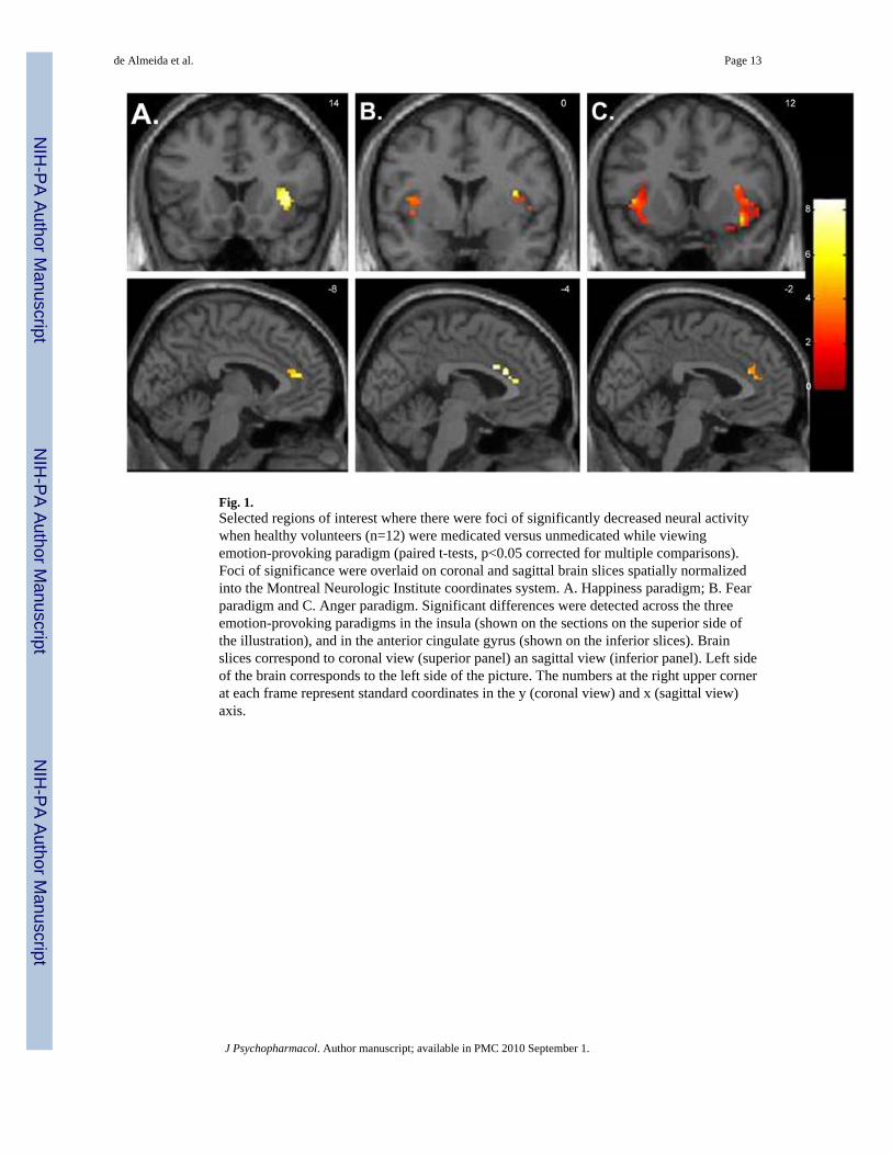
Fig. 1.Selected regions of interest where there were foci of significantly decreased neural activitywhen healthy volunteers (n=12) were medicated versus unmedicated while viewingemotion-provoking paradigm (paired t-tests, p<0.05 corrected for multiple comparisons).Foci of significance were overlaid on coronal and sagittal brain slices spatially normalizedinto the Montreal Neurologic Institute coordinates system. A. Happiness paradigm; B. Fearparadigm and C. Anger paradigm. Significant differences were detected across the threeemotion-provoking paradigms in the insula (shown on the sections on the superior side ofthe illustration), and in the anterior cingulate gyrus (shown on the inferior slices). Brainslices correspond to coronal view (superior panel) an sagittal view (inferior panel). Left sideof the brain corresponds to the left side of the picture. The numbers at the right upper cornerat each frame represent standard coordinates in the y (coronal view) and x (sagittal view)axis.
de Almeida et al. Page 13
J Psychopharmacol. Author manuscript; available in PMC 2010 September 1.
NIH
-PA Author Manuscript
NIH
-PA Author Manuscript
NIH
-PA Author Manuscript
NIH
-PA Author Manuscript
NIH
-PA Author Manuscript
NIH
-PA Author Manuscript
de Almeida et al. Page 14
Tabl
e 1
Subj
ectiv
e Se
lf-ra
ting
Scor
es a
fter P
rese
ntat
ion
of E
mot
iona
l and
Neu
tral B
lock
s of P
ictu
res f
rom
the
Inte
rnat
iona
l Aff
ectiv
e Pi
ctur
e Sy
stem
Med
icat
ed st
atus
Unm
edic
ated
stat
usPl
anne
d co
mpa
riso
n
emot
ion
neut
ral
Scal
esem
otio
n (S
D)
neut
ral (
SD)
emot
ion
(SD
)ne
utra
l (SD
)t
pt
p
Hap
pine
ss P
arad
igm
A
nger
1.05
(0.1
2)1.
03 (0
.09)
1.07
(0.1
8)1.
11 (0
.36)
−0.27
0.79
−0.90
0.39
A
rous
al2.
83 (0
.82)
1.97
(0.8
5)2.
77 (0
.73)
1.64
(0.9
5)0.
370.
721.
910.
08
Fe
ar1.
82 (0
.49)
1.03
(0.0
9)1.
82 (0
.7)
1.05
(0.1
3)−0.05
0.96
−1.00
0.34
H
appi
ness
2.72
(0.8
6)1.
75 (0
.79)
2.87
(0.8
7)1.
75 (0
.96)
−1.06
0.31
<0.0
11.
00
Fear
Par
adig
m
A
nger
2.08
(0.5
8)1.
05 (0
.13)
1.97
(0.6
5)1.
17 (0
.3)
0.54
0.60
−1.48
0.17
A
rous
al3.
12 (0
.78)
1.86
(0.9
6)3.
17 (0
.82)
1.58
(0.8
3)−0.37
0.72
1.82
0.10
Fe
ar3.
05 (0
.8)
1.03
(0.0
9)3.
08 (0
.75)
1.03
(0.0
9)−0.30
0.77
<0.0
11.
00
H
appi
ness
1.13
(0.2
7)1.
44 (0
.64)
1.15
(0.2
3)1.
47 (0
.72)
−0.17
0.87
−0.13
0.90
Ang
er P
arad
igm
A
nger
2.72
(0.4
8)1.
36 (0
.39)
2.68
(0.7
6)1.
08 (0
.21)
0.16
0.87
2.06
0.06
A
rous
al3.
23 (0
.73)
2.11
(0.8
8)3.
23 (0
.85)
1.92
(0.8
8)−0.03
0.98
0.69
0.51
Fe
ar2.
66 (0
.83)
1.28
(0.4
)2.
61 (0
.74)
1.22
(0.2
9)0.
440.
671.
000.
34
H
appi
ness
1.02
(0.0
6)1.
53 (0
.48)
1.08
(0.2
)1.
64 (0
.63)
−1.08
0.30
−0.57
0.58
Val
ues a
re m
eans
scor
es; S
D =
stan
dard
dev
iatio
n;
J Psychopharmacol. Author manuscript; available in PMC 2010 September 1.
NIH
-PA Author Manuscript
NIH
-PA Author Manuscript
NIH
-PA Author Manuscript
de Almeida et al. Page 15
Tabl
e 2
Tab
le 2
a: A
nato
mic
al L
ocat
ions
of t
he P
lann
ed C
ompa
riso
ns d
urin
g H
appi
ness
Par
adig
m
MN
I coo
rdin
ates
Reg
ion
Side
XY
ZK
Tz
p
Hap
py p
ictu
res
Clo
mip
ram
ine
< W
asho
ut
vm
PFC
(BA
11)
R28
42−8
263.
382.
740.
003
In
sula
R40
148
773.
432.
770.
003
R32
−26
1067
2.76
2.35
0.00
9
A
nter
ior C
ingu
late
(BA
24)
R4
2822
293.
052.
540.
005
A
nter
ior C
ingu
late
(BA
32)
L−8
4214
803.
853
0.00
1
C
auda
te b
ody
L−16
−14
2615
74.
113.
130.
001
R16
−14
2413
25.
53.
74<0
.001
m
edia
l PFC
(BA
10)
L−16
4214
323.
192.
630.
004
Pu
tam
enL
−14
210
662.
342.
070.
019
Tab
le 2
b: A
nato
mic
al L
ocat
ions
of t
he P
lann
ed C
ompa
riso
ns d
urin
g Fe
ar P
arad
igm
MN
I coo
rdin
ates
Reg
ion
Side
XY
ZK
Tz
p
Fear
pic
ture
s
Clo
mip
ram
ine
< W
asho
ut
vL
PFC
(BA
47)
L−32
16−2
243.
222.
610.
005
D
LPFC
(BA
46)
L−46
3616
303.
252.
620.
004
A
myg
dala
L−28
−8
−12
122.
832.
370.
009
In
sula
L−44
−16
1897
5.06
3.49
<0.0
01
R38
014
186
6.46
3.97
<0.0
01
Pa
rahi
ppoc
ampa
l Gyr
usR
20−34
−14
184
6.2
3.89
<0.0
01
D
LPFC
(BA
9)R
4222
3647
2.69
2.28
0.01
1
L−52
236
243.
622.
830.
002
Th
alam
usR
20−14
625
34.
313.
17<0
.001
Pu
tam
enR
28−16
−4
114
4.65
3.32
<0.0
01
Neu
tral p
ictu
res
J Psychopharmacol. Author manuscript; available in PMC 2010 September 1.
NIH
-PA Author Manuscript
NIH
-PA Author Manuscript
NIH
-PA Author Manuscript
de Almeida et al. Page 16T
able
2b:
Ana
tom
ical
Loc
atio
ns o
f the
Pla
nned
Com
pari
sons
dur
ing
Fear
Par
adig
m
MN
I coo
rdin
ates
Reg
ion
Side
XY
ZK
Tz
p
Clo
mip
ram
ine
> W
asho
ut
vm
PFC
(BA
11)
L−24
44−6
82.
221.
960.
025
Clo
mip
ram
ine
< W
asho
ut
A
nter
ior C
ingu
late
(BA
24)
L−4
2222
242.
552.
190.
014
A
myg
dala
R28
−8
−14
653.
582.
810.
002
Tab
le 2
c: A
nato
mic
al L
ocat
ions
of t
he P
lann
ed C
ompa
riso
ns d
urin
g A
nger
Par
adig
m
MN
I coo
rdin
ates
Reg
ion
Side
XY
ZK
Tz
p
Ang
er p
ictu
res
Clo
mip
ram
ine
< W
asho
ut
vL
PFC
(BA
47)
R36
30−2
223.
492.
80.
003
R26
16−12
423.
292.
70.
004
R44
182
473.
032.
50.
006
L−32
16−12
728.
494.
6<0
.001
D
LPFC
(BA
46)
R42
3616
534.
153.
20.
001
A
myg
dala
R28
−4
−16
613.
022.
50.
006
In
sula
L−32
16−10
529
6.62
4.1
<0.0
01
L−50
−38
2066
2.73
2.3
0.01
R38
12−10
824.
783.
4<0
.001
R32
1412
524
4.34
3.3
<0.0
01
R42
−40
1886
3.82
30.
001
A
nter
ior C
ingu
late
(BA
24)
R4
3024
353.
612.
90.
002
L−2
3218
412.
822.
40.
008
A
nter
ior C
ingu
late
(BA
32)
R6
3226
354.
373.
30.
001
L−2
3222
352.
442.
10.
016
Pa
rahi
ppoc
ampa
l Gyr
usR
16−48
214
33.
092.
60.
005
L−20
−34
−8
270
32.
50.
006
H
ippo
cam
pus
L−26
−36
−4
92.
732.
30.
01
D
LPFC
(BA
9)R
446
2837
3.45
2. 8
0.00
3
J Psychopharmacol. Author manuscript; available in PMC 2010 September 1.
NIH
-PA Author Manuscript
NIH
-PA Author Manuscript
NIH
-PA Author Manuscript
de Almeida et al. Page 17T
able
2c:
Ana
tom
ical
Loc
atio
ns o
f the
Pla
nned
Com
pari
sons
dur
ing
Ang
er P
arad
igm
MN
I coo
rdin
ates
Reg
ion
Side
XY
ZK
Tz
p
R34
2034
129
3.31
2.7
0.00
3
Th
alam
usR
8−22
−2
621
5.02
3.5
<0.0
01
L−18
−22
028
26.
724.
1<0
.001
C
auda
te b
ody
R8
128
173
6.02
3.9
<0.0
01
C
auda
te h
ead
R8
186
169
6.31
4.0
<0.0
01
L−8
60
642.
652.
30.
011
Pu
tam
enR
22−2
055
25.
43.
7<0
.001
L−18
4−8
256
4.33
3.2
0.00
1
Neu
tral p
ictu
res
Clo
mip
ram
ine
< W
asho
ut
In
sula
L−40
−12
612
24.
723.
4<0
.001
R40
−20
672
4.56
3.3
<0.0
01
vmPF
C: v
entro
med
ial p
refr
onta
l cor
tex;
R, r
ight
; L, l
eft;
BA
, Bro
dman
n ar
ea; C
oord
inat
es c
orre
spon
d to
the
ster
eota
xic
arra
y of
Mon
treal
Neu
rolo
gic
Inst
itute
; K: c
lust
er si
ze; P
FC, p
refr
onta
l cor
tex
vLPF
C: v
entro
late
ral p
refr
onta
l cor
tex
DLP
FC: d
orso
late
ral p
refr
onta
l cor
tex;
R, r
ight
; L, l
eft;
BA
, Bro
dman
n ar
ea; C
oord
inat
es c
orre
spon
d to
the
ster
eota
xic
arra
y of
Mon
treal
Neu
rolo
gic
Inst
itute
; K:
clus
ter s
ize;
vm
PFC
, ven
trom
edia
l pre
fron
tal c
orte
x
vLPF
C: v
entro
late
ral p
refr
onta
l cor
tex
DLP
FC: d
orso
late
ral p
refr
onta
l cor
tex;
Coo
rdin
ates
cor
resp
ond
to th
e st
ereo
taxi
c ar
ray
of M
ontre
al N
euro
logi
c In
stitu
te; B
A, B
rodm
ann
area
; R, r
ight
; L, l
eft;
K:
clus
ter s
ize
J Psychopharmacol. Author manuscript; available in PMC 2010 September 1.
Related Documents