97:692-700, 2007. First published Nov 8, 2006; doi:10.1152/jn.00759.2006 J Neurophysiol Marat Minlebaev, Yehezkel Ben-Ari and Rustem Khazipov the Neonatal Rat Barrel Cortex In Vivo Network Mechanisms of Spindle-Burst Oscillations in You might find this additional information useful... 68 articles, 34 of which you can access free at: This article cites http://jn.physiology.org/cgi/content/full/97/1/692#BIBL including high-resolution figures, can be found at: Updated information and services http://jn.physiology.org/cgi/content/full/97/1/692 can be found at: Journal of Neurophysiology about Additional material and information http://www.the-aps.org/publications/jn This information is current as of February 6, 2007 . http://www.the-aps.org/. American Physiological Society. ISSN: 0022-3077, ESSN: 1522-1598. Visit our website at (monthly) by the American Physiological Society, 9650 Rockville Pike, Bethesda MD 20814-3991. Copyright © 2005 by the publishes original articles on the function of the nervous system. It is published 12 times a year Journal of Neurophysiology on February 6, 2007 jn.physiology.org Downloaded from

Welcome message from author
This document is posted to help you gain knowledge. Please leave a comment to let me know what you think about it! Share it to your friends and learn new things together.
Transcript
97:692-700, 2007. First published Nov 8, 2006; doi:10.1152/jn.00759.2006 J NeurophysiolMarat Minlebaev, Yehezkel Ben-Ari and Rustem Khazipov the Neonatal Rat Barrel Cortex In Vivo Network Mechanisms of Spindle-Burst Oscillations in
You might find this additional information useful...
68 articles, 34 of which you can access free at: This article cites http://jn.physiology.org/cgi/content/full/97/1/692#BIBL
including high-resolution figures, can be found at: Updated information and services http://jn.physiology.org/cgi/content/full/97/1/692
can be found at: Journal of Neurophysiologyabout Additional material and information http://www.the-aps.org/publications/jn
This information is current as of February 6, 2007 .
http://www.the-aps.org/.American Physiological Society. ISSN: 0022-3077, ESSN: 1522-1598. Visit our website at (monthly) by the American Physiological Society, 9650 Rockville Pike, Bethesda MD 20814-3991. Copyright © 2005 by the
publishes original articles on the function of the nervous system. It is published 12 times a yearJournal of Neurophysiology
on February 6, 2007
jn.physiology.orgD
ownloaded from
Network Mechanisms of Spindle-Burst Oscillations in the Neonatal Rat BarrelCortex In Vivo
Marat Minlebaev, Yehezkel Ben-Ari, and Rustem KhazipovThe Mediterranean Institute of Neurobiology, Institut National de la Sante et de la Recherche Medicale U29, Universite Mediterreneen,Marseille, France
Submitted 21 July 2006; accepted in final form 6 November 2006
Minlebaev M, Ben-Ari Y, Khazipov R. Network mechanisms ofspindle-burst oscillations in the neonatal rat barrel cortex in vivo. JNeurophysiol 97: 692–700, 2007. First published November 8, 2006;doi:10.1152/jn.00759.2006. Early in development, cortical networksgenerate particular patterns of activity that participate in corticaldevelopment. The dominant pattern of electrical activity in the neo-natal rat neocortex in vivo is a spatially confined spindle-burst. Here,we studied network mechanisms of generation of spindle-bursts in thebarrel cortex of neonatal rats using a superfused cortex preparation invivo. Both spontaneous and sensory-evoked spindle-bursts werepresent in the superfused barrel cortex. Pharmacological analysisrevealed that spindle-bursts are driven by glutamatergic synapses witha major contribution of AMPA/kainate receptors, but slight participa-tion of NMDA receptors and gap junctions. Although GABAergicsynapses contributed minimally to the pacing the rhythm of spindle-burst oscillations, surround GABAergic inhibition appeared to becrucial for their compartmentalization. We propose that local spindle-burst oscillations, driven by glutamatergic synapses and spatiallyconfined by GABAergic synapses, contribute to the development ofbarrel cortex during the critical period of developmental plasticity.
I N T R O D U C T I O N
During development, cortical neuronal networks generateparticular patterns of activity that participate in cortical devel-opment (Ben-Ari 2001; Fox 2002; Katz and Shatz 1996;Khazipov and Luhmann 2006; Moody and Bosma 2005;O’Donovan 1999). The dominant pattern of electrical activityin the neonatal rat neocortex in vivo is a spindle-burst(Hanganu et al. 2006; Khazipov et al. 2004b). Spindle-burstsare spatially confined spindle-shape oscillations at alpha-betafrequency, associated with phase-locked neuronal firing andactivation of the glutamatergic and GABAergic synapses.Spindle-bursts constitute a self-organizing pattern that persistsafter deafferentation. In the intact animal, compartmentalizedspindle-bursts in somatosensory cortex are triggered by sen-sory feedback resulting from spontaneous movements in asomatotopic manner (Khazipov et al. 2004b). In visual cortex,spindle-bursts are triggered by spontaneous retinal waves in aneye-specific manner (Hanganu et al. 2006). However, thenetwork mechanisms underlying the generation of corticalspindle-bursts are poorly understood. Several patterns of cor-related neuronal activity sharing some common features withthe spindle-bursts in vivo were previously described in post-natal rodent neocortical and hippocampal slices and intactpreparations in vitro. These include “synchronized-via-gap-junctions neuronal domains” (Kandler and Katz 1995 1998a,b;Yuste et al. 1992, 1995) and calcium waves (Peinado 2000,
2001), gap-junction– and NMDA-receptor–based cholinergicoscillations (Dupont et al. 2006), giant depolarizing potentials(GDPs), and early network oscillations driven by glutamatergicand excitatory GABAergic synapses (Agmon et al. 1996;Ben-Ari et al. 1989; Garaschuk et al. 1998, 2000; Khazipov etal. 1997; Leinekugel et al. 1997, 1998). Consistent with the invitro findings, generation of the early hippocampal in vivopattern of sharp waves (Buhl and Buzsaki 2005; Leinekugel etal. 2002) was also shown to involve excitatory action of�-aminobutyric acid (GABA) (Sipila et al. 2006). However, towhat extent these developmentally regulated mechanisms con-tribute to generation of the neocortical in vivo pattern ofspindle-bursts remains largely unknown.
In the present study, we studied the mechanisms of spindle-bursts in the neonatal rat barrel cortex in vivo using a super-fused neocortex preparation initially developed for the adoles-cent rat hippocampus (Khazipov and Holmes 2003). We foundthat the physiological pattern of spindle-bursts is preserved inthe superfused barrel cortex. Pharmacological profiling ofspindle-bursts indicated that 1) generation of spindle-bursts isprimarily based on glutamatergic synapses, with a major roleof �-amino-3-hydroxy-5-methyl-4-isoxazolepropionic acid(AMPA) receptors and a slight contribution of N-methyl-D-aspartate (NMDA) receptors and gap junctions; and that 2)GABAergic synapses are not directly involved in the genera-tion of spindle-burst oscillations, although they play an impor-tant role in their spatial compartmentalization.
M E T H O D S
This study followed INSERM guidelines on animal care, withapproval from the animal care and use local committee. Wistar rats ofboth sexes from postnatal day (P) 1 to P7 were used. P0 was the dayof birth. During the surgical procedure, rats were anesthetized with acombination of 0.5–1.5 g/kg urethane injected intraperitoneally andice-cooling. The skin and periosteum were removed from the skull,which was then covered with a layer of dental acrylic, except for twoareas about 7 mm in diameter above the right and left barrel cortices(Fig. 1A). The rat was positioned in the stereotaxic apparatus; the skullwas attached to the nose (nasal bones) and ear bars (occipital bone)with dental acrylic. A 5-mm-diameter burr hole was drilled in theskull above the right barrel cortex, the dura was cut and removed, andthe cortical surface was covered with 0.9% NaCl during the procedureto prevent it from drying. The perfusion chamber was prepared asdescribed previously (Khazipov and Holmes 2003) with some modi-fications. In brief, a 2-mm-long cylinder was cut from a plastic tube(ID 3.5 mm, OD 4.5 mm) and glued to stretched nylon mesh withcyanoacrylamide glue. The chamber was positioned at the cortical
Address for reprint requests and other correspondence: R. Khazipov,INMED/INSERM U29,163 Avenue de Luminy, B.P. 13, 13273 Marseille,France (E-mail [email protected]).
The costs of publication of this article were defrayed in part by the paymentof page charges. The article must therefore be hereby marked “advertisement”in accordance with 18 U.S.C. Section 1734 solely to indicate this fact.
J Neurophysiol 97: 692–700, 2007.First published November 8, 2006; doi:10.1152/jn.00759.2006.
692 0022-3077/07 $8.00 Copyright © 2007 The American Physiological Society www.jn.org
on February 6, 2007
jn.physiology.orgD
ownloaded from
surface so that the mesh gently pressed onto the cortex. The chamberwas then fixed to the skull by dental acrylic. Chlorided silver wire wasinserted into the cerebellum and served as a ground electrode.
During recordings, rats were heated by a thermal pad (37°C). Thechamber was perfused with oxygenated (95% O2-5% CO2) artificialcerebrospinal fluid (ACSF) of the following composition (in mM):126 NaCl, 3.5 KCl, 2.0 CaCl2, 1.3 MgCl2, 25 NaHCO3, 1.2 NaH2PO4,and 11 glucose (pH 7.4 at a rate of 2 ml/min). Temperature in thechamber was kept at 35–37°C using an automatic temperature con-troller (TC-344B; Warner Instruments, Hamden, CT). Extracellularfield potential recordings (�1,000; 3- to 3,000-Hz band-pass) wereperformed using arrays of metal electrodes of 50 �m in diameter(California Fine Wire, Grover Beach, CA). Patch-clamp recordingswere performed using a Axopatch 200A amplifier (Axon Instruments,Union City, CA) using a patching technique similar to that describedin vivo (Leinekugel et al. 2002; Margrie et al. 2002). The pipetteswere filled with a solution of the following composition (in mM): 135Cs-gluconate (or methylsulfate), 2 MgCl2, 0.1 CaCl2, 1 EGTA, and 10HEPES (pH 7.25). Membrane potential values were corrected forliquid junction potential of �12 mV. Afferent stimulation was per-formed by applying electrical pulses (60 V, 50 �s, 0.03 s�1) throughpairs of electrodes inserted into the whisker pads and glued to the skinwith super glue.
Data were digitized at 10 kHz using a Digidata 1322A interface(Axon Instruments) and analyzed off-line using an Axon package
(Axon Instruments), MiniAnalysis (Synaptosoft, Decatur, GA), Ori-gin (Microcal Software, Northampton, MA), and Matlab (The Math-Works, Natick, MA). Group measures are expressed as means � SE.The statistical significance of differences was assessed with theStudent’s t-test. The level of significance was set at P � 0.05.
The drugs gabazine, 6-cyano-7-nitroquinoxaline-2,3-dione(CNQX) and D-2-amino-5-phosphonovaleric acid (D-APV) were pur-chased from Tocris Neuramin (Bristol, UK), diazepam from Roche(Basel, Switzerland), and all other compounds from Sigma (St. Louis,MO).
R E S U L T S
To study the mechanisms of spindle-bursts in the barrelcortex of neonatal rats (postnatal days P1–P7), we used atechnique of superfused neocortex in vivo that was originallydeveloped for adult hippocampus (Khazipov and Holmes2003). The photograph and the experimental setup are sche-matically shown in Fig. 1A. In brief, under anesthesia, a roundwindow was cut in the scull above the barrel cortex and durawas removed. A cylinder with fine mesh at its bottom wasgently pressed onto the surface of the barrel cortex (meshprevents pulsations and allows application of drugs directlyonto the cortical surface). In addition to the ease of pharma-
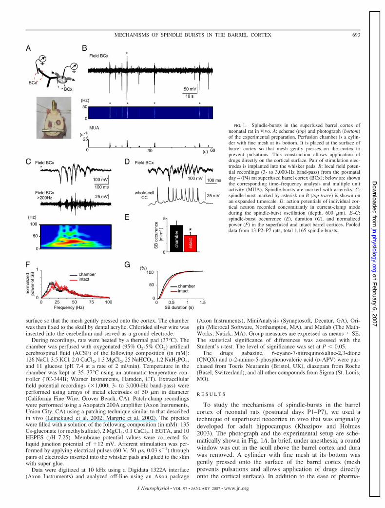
FIG. 1. Spindle-bursts in the superfused barrel cortex ofneonatal rat in vivo. A: scheme (top) and photograph (bottom)of the experimental preparation. Perfusion chamber is a cylin-der with fine mesh at its bottom. It is placed at the surface ofbarrel cortex so that mesh gently presses on the cortex toprevent pulsations. This construction allows application ofdrugs directly on the cortical surface. Pair of stimulation elec-trodes is implanted into the whisker pads. B: local field poten-tial recordings (3- to 3,000-Hz band-pass) from the postnatalday 4 (P4) rat superfused barrel cortex (BCx); below are shownthe corresponding time–frequency analysis and multiple unitactivity (MUA). Spindle-bursts are marked with asterisks. C:spindle-burst marked by asterisk on B (top trace) is shown onan expanded timescale. D: action potentials of individual cor-tical neuron recorded concomitantly in current-clamp modeduring the spindle-burst oscillation (depth, 600 �m). E–G:spindle-burst occurrence (E), duration (G), and normalizedpower (F) in the superfused and intact barrel cortices. Pooleddata from 13 P2–P7 rats; total 1,165 spindle-bursts.
693MECHANISMS OF SPINDLE BURSTS IN THE BARREL CORTEX
J Neurophysiol • VOL 97 • JANUARY 2007 • www.jn.org
on February 6, 2007
jn.physiology.orgD
ownloaded from
cological manipulations, this technique is also convenient forplacement of electrodes. The contralateral barrel cortex re-mained intact and served as a control.
We first determined whether the normal physiological pat-tern of spindle-bursts and sensory-evoked responses are pre-served in the superfused barrel cortex by comparing the activ-ity in the barrel cortex superfused with normal ACSF with theintact contralateral barrel cortex using local field potentialrecordings (3- to 3,000-Hz band-pass). Spontaneous activity inboth superfused and intact barrel cortices was similar andcharacterized by highly discontinuous periods of activity. Vir-tually all multiple unit activity (Fig. 1, B and C) and actionpotentials of individual cortical neurons recorded concomi-tantly in current-clamp mode (n � 9 cells; Fig. 1D) weresynchronized to intermittent spindle-shape oscillations. Thesewere similar to the spindle-bursts described previously in theprimary somatosensory cortex for areas of body representation(Khazipov et al. 2004b) as well as in visual cortex (Hanganu etal. 2006). Spindle-bursts had similar durations of 344 � 15 and370 � 15 ms and peak power of oscillations at 15 � 3 and21 � 3 Hz, and reversed in polarity at about 1 mm depth (in thesuperfused and intact barrel cortices) (Fig. 1, F and G; n � 13rats; P2–P7). Spindle-bursts occurred more frequently in thesuperfused barrel cortex (4.2 � 0.4 min�1) than in the intactbarrel cortex (2.5 � 0.4 min�1; P � 0.05; Fig. 1E).
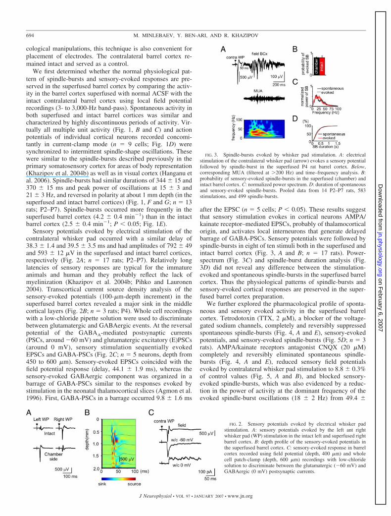
Sensory potentials evoked by electrical stimulation of thecontralateral whisker pad occurred with a similar delay of38.3 � 1.4 and 39.5 � 3.5 ms and had amplitudes of 792 � 49and 593 � 12 �V in the superfused and intact barrel cortices,respectively (Fig. 2A; n � 17 rats; P2–P7). Relatively longlatencies of sensory responses are typical for the immatureanimals and human and they probably reflect the lack ofmyelinization (Khazipov et al. 2004b; Pihko and Lauronen2004). Transcortical current source density analysis of thesensory-evoked potentials (100-�m-depth increment) in thesuperfused barrel cortex revealed a major sink in the middlecortical layers (Fig. 2B; n � 3 rats; P4). Whole cell recordingswith a low-chloride pipette solution were used to discriminatebetween glutamatergic and GABAergic events. At the reversalpotential of the GABAA-mediated postsynaptic currents(PSCs, around �60 mV) and glutamatergic excitatory (E)PSCs(around 0 mV), sensory stimulation sequentially evokedEPSCs and GABA-PSCs (Fig. 2C; n � 5 neurons, depth from450 to 600 �m). Sensory-evoked EPSCs coincided with thefield potential response (delay, 44.1 � 1.9 ms), whereas thesensory-evoked GABAergic component was organized in abarrage of GABA-PSCs similar to the responses evoked bystimulation in the neonatal thalamocortical slices (Agmon et al.1996). First, GABA-PSCs in a barrage occurred 9.8 � 1.6 ms
after the EPSC (n � 5 cells; P � 0.05). These results suggestthat sensory stimulation evokes in cortical neurons AMPA/kainate receptor–mediated EPSCs, probably of thalamocorticalorigin, and activates local interneurons that generate delayedbarrage of GABA-PSCs. Sensory potentials were followed byspindle-bursts in eight of ten stimuli both in the superfused andintact barrel cortex (Fig. 3, A and B; n � 17 rats). Power-spectrum (Fig. 3C) and spindle-burst duration analysis (Fig.3D) did not reveal any difference between the stimulation-evoked and spontaneous spindle-bursts in the superfused barrelcortex. Thus the physiological patterns of spindle-bursts andsensory-evoked cortical responses are preserved in the super-fused barrel cortex preparation.
We further explored the pharmacological profile of sponta-neous and sensory evoked activity in the superfused barrelcortex. Tetrodotoxin (TTX, 2 �M), a blocker of the voltage-gated sodium channels, completely and reversibly suppressedspontaneous spindle-bursts (Fig. 4, A and E), sensory-evokedpotentials, and sensory-evoked spindle-bursts (Fig. 5D; n � 3rats). AMPA/kainate receptors antagonist CNQX (20 �M)completely and reversibly eliminated spontaneous spindle-bursts (Fig. 4, A and E), reduced sensory field potentialsevoked by contralateral whisker pad stimulation to 8.8 � 0.3%of control values (Fig. 5, A and B), and blocked sensory-evoked spindle-bursts, which was also evidenced by a reduc-tion in the power of activity at the dominant frequency of theevoked spindle-burst oscillations (18 � 2 Hz) from 49.4 �
FIG. 2. Sensory potentials evoked by electrical whisker padstimulation. A: sensory potentials evoked by the left ant rightwhisker pad (WP) stimulation in the intact left and superfused rightbarrel cortex. B: depth profile of the sensory-evoked potentials inthe superfused barrel cortex. C: sensory-evoked response in barrelcortex recorded using field potential (depth, 400 �m) and wholecell patch-clamp (depth, 600 �m) recordings with low-chloridesolution to discriminate between the glutamatergic (�60 mV) andGABAergic (0 mV) postsynaptic currents.
FIG. 3. Spindle-bursts evoked by whisker pad stimulation. A: electricalstimulation of the contralateral whisker pad (arrow) evokes a sensory potentialfollowed by spindle-burst in the superfused P4 rat barrel cortex. Below,corresponding MUA (filtered at �200 Hz) and time–frequency analysis. B:probability of sensory-evoked spindle-bursts in the superfused (chamber) andintact barrel cortex. C: normalized power spectrum. D: duration of spontaneousand sensory-evoked spindle-bursts. Pooled data from 14 P2–P7 rats, 583stimulations, and 499 spindle-bursts.
694 M. MINLEBAEV, Y. BEN-ARI, AND R. KHAZIPOV
J Neurophysiol • VOL 97 • JANUARY 2007 • www.jn.org
on February 6, 2007
jn.physiology.orgD
ownloaded from
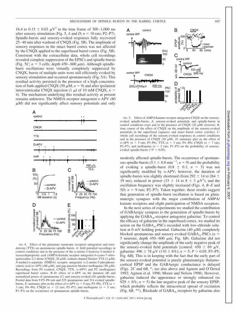
16.4 to 0.15 � 0.03 �V2 in the time frame of 300–1,000 msafter sensory stimulation (Fig. 5, A and D; n � 10 rats; P2–P7).Spindle-bursts and sensory-evoked responses fully recovered25–40 min after washout of CNQX (Fig. 5B). The amplitude ofsensory responses in the intact barrel cortex was not affectedby the CNQX applied to the superfused barrel cortex (Fig. 5B).Consistent with the extracellular data, whole cell recordingsrevealed complete suppression of the EPSCs and spindle-bursts(Fig. 5C; n � 3 cells; depth 450–600 �m). Although spindle-burst oscillations were virtually completely suppressed byCNQX, bursts of multiple units were still efficiently evoked bysensory stimulation and occurred spontaneously (Fig. 5A). Thisresidual activity persisted in the presence of a high concentra-tion of bath-applied CNQX (50 �M; n � 9) and after ipsilateralintraventricular CNQX injection (1 �l of 10 mM CNQX, n �4). The mechanism underlying this residual activity at presentremains unknown. The NMDA receptor antagonist D-APV (80�M) did not significantly affect sensory potentials and only
modestly affected spindle-bursts. The occurrence of spontane-ous spindle-bursts (5.1 � 0.8 min�1; n � 9) and the probabilityof evoking a spindle-burst (0.8 � 0.1; n � 5) was notsignificantly modified by D-APV; however, the duration ofspindle-bursts was slightly shortened (from 292 � 14 to 264 �19 ms), reduced in power (33 � 14 to 8 � 3 �V2), and theoscillation frequency was slightly increased (Figs. 4, B–E and5D; n � 9 rats; P2–P7). Taken together, these results suggestthat generation of spindle-burst oscillation is based on gluta-matergic synapses with the major contribution of AMPA/kainate receptors and slight participation of NMDA receptors.
In the next series of experiments we studied the contributionof GABAergic synapses to the generation of spindle-bursts byapplying the GABAA-receptor antagonist gabazine. To controlthe efficacy of gabazine in the superfused cortex, we studied itsaction on the GABAA-PSCs recorded with low-chloride solu-tion at 0 mV holding potential. Gabazine (40 �M) completelyblocked spontaneous and sensory-evoked GABAA-PSCs (n �5 neurons; depth 450–600 �m; Fig. 6B). Gabazine did notsignificantly change the amplitude of the early negative peak ofthe sensory-evoked field potentials [control: 450 � 69 �V,gabazine: 496 � 78 �V (110 � 6%); n � 5; P � 0.05; P3–P5;Fig. 6B]. This is in keeping with the fact that the early part ofthe sensory-evoked potential is purely glutamatergic thalamo-cortical EPSP and the GABAergic conductance is delayed(Figs. 2C and 6B, *; see also above and Agmon and O’Dowd1992; Agmon et al. 1996; Moore and Nelson 1998). However,gabazine induced the appearance or strongly enhanced (to929 � 6%; n � 5) the late negative peak of the sensory EPSP,which probably reflects the intracortical spread of excitation(Fig. 6B, **). Blockade of GABAA receptors by gabazine also
FIG. 4. Effect of the glutamate ionotropic receptors antagonists and tetro-dotoxin (TTX) on spontaneous spindle-bursts. A: field potential recordings incontrol conditions and in the presence of the �-amino-3-hydroxy-5-methyl-4-isoxazolepropionic acid (AMPA)/kainate receptor antagonist 6-cyano-7-nitro-quinoxaline-2,3-dione (CNQX, 20 �M), sodium channel blocker TTX (2 �M),N-methyl-D-aspartate (NMDA) receptor antagonist D-2-amino-5-phosphono-valeric acid (D-APV) (80 �M), and gap-junction blocker mefloquine (50 �M).Recordings from P4 (control, CNQX, TTX, D-APV) and P2 (mefloquine)superfused barrel cortex. B–D: effect of D-APV on the duration (B) andnormalized power of spontaneous (C) and sensory-evoked (D) spindle-bursts.Pooled data from 9 P3–P6 rats and 225 spontaneous and 214 evoked spindle-bursts. E: summary plot on the effect of D-APV (n � 9 rats; P3–P6), TTX (n �3 rats; P4–P6), CNQX (n � 12 rats; P2–P7), and mefloquine (n � 5 rats;P1–P3) on the occurrence of spontaneous spindle-bursts.
FIG. 5. Effect of AMPA/kainate receptor antagonist CNQX on the sensory-evoked spindle-bursts. A: sensory-evoked potentials and spindle-bursts incontrol conditions (top) and in the presence of CNQX (20 �M) (bottom). B:time course of the effect of CNQX on the amplitude of the sensory-evokedpotentials in the superfused (squares) and intact barrel cortex (circles). C:whole cell recordings of the sensory-evoked responses in control conditionsand in the presence of CNQX (20 �M). D: summary plot on the effect ofD-APV (n � 5 rats; P3–P6), TTX (n � 3 rats; P4–P6), CNQX (n � 7 rats;P2–P7), and mefloquine (n � 5 rats; P1–P3) on the probability of sensory-evoked spindle-bursts (*P � 0.05).
695MECHANISMS OF SPINDLE BURSTS IN THE BARREL CORTEX
J Neurophysiol • VOL 97 • JANUARY 2007 • www.jn.org
on February 6, 2007
jn.physiology.orgD
ownloaded from
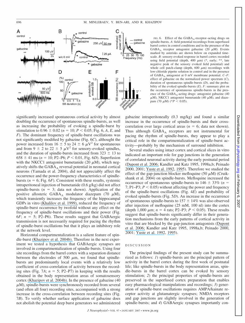
significantly increased spontaneous cortical activity by almostdoubling the occurrence of spontaneous spindle-bursts, as wellas increasing the probability of evoking a spindle-burst bystimulation to 0.96 � 0.02 (n � 10; P � 0.05; Fig. 6, A, E, andF). The dominant frequency of spindle-burst oscillations wasnot significantly modified by gabazine (Fig. 6C), althought thepower increased from 16 � 5 to 24 � 6 �V2 for spontaneousand from 9 � 2 to 22 � 5 �V2 for sensory-evoked spindles,and the duration of spindle-bursts increased from 323 � 13 to658 � 41 ms (n � 10; P2–P6; P � 0.01, Fig. 6D). Superfusionwith the NKCC1 antagonist bumetanide (20 �M), which neg-atively shifts the GABAA reversal potential in neonatal corticalneurons (Yamada et al. 2004), did not appreciably affect theoccurrence and the power-frequency characteristics of spindle-bursts (n � 6; Fig. 6F). Consistent with these results, systemicintraperitoneal injection of bumetanide (0.6 g/kg) did not affectspindle-bursts (n � 3; data not shown). Application of thepositive allosteric GABAA modulator diazepam (70 �M),which transiently increases the frequency of the hippocampalGDPs in vitro (Khalilov et al. 1999), reduced the frequency ofspindle-burst occurrence by twofold without any effect on thefrequency of spindle-burst oscillations and their power (Fig.6F; n � 5; P2–P6). These results suggest that GABAergictransmission is not necessarily required for pacing the rhythmof spindle-burst oscillations but that it plays an inhibitory roleat the network level.
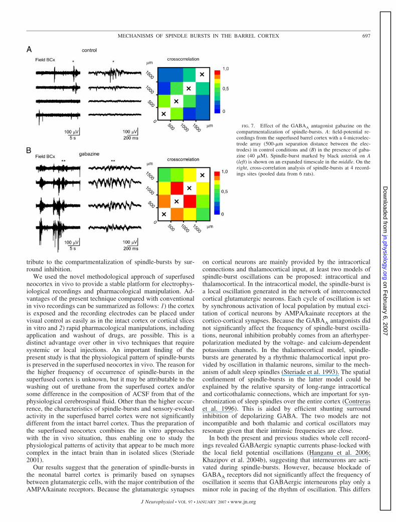
Horizontal compartmentalization is a salient feature of spin-dle-burst (Khazipov et al. 2004b). Therefore in the next exper-iment we tested a hypothesis that GABAergic synapses areinvolved in compartmentalization of spindle-bursts. Using foursite recordings from the barrel cortex with a separation distancebetween the electrodes of 500 �m, we found that spindle-bursts are predominantly local events with a relatively lowcoefficient of cross-correlation of activity between the record-ing sites (Fig. 7A; n � 5; P2–P7) in keeping with the resultsobtained in the body representation areas of somatosensorycortex (Khazipov et al. 2004b). In the presence of gabazine (40�M), spindle-bursts were synchronously recorded from several(and often all four) recording sites, accompanied with a strongincrease in the cross-correlation between recording sites (Fig.7B). To verify whether surface application of gabazine doesnot abolish the potential deep burst generators we administered
gabazine intraperitoneally (0.3 mg/kg) and found a similarincrease in the occurrence of spindle-bursts and their cross-correlation over large cortical areas (n � 6; data not shown).Thus although GABAA receptors are not instrumental forpacing the rhythm of spindle-bursts, they appear to play acritical role in the compartmentalization of spindle-burst ac-tivity—probably by the mechanism of surround inhibition.
Several studies using intact cortex and cortical slices in vitroindicated an important role for gap junctions in the generationof correlated neuronal activity during the early postnatal period(Dupont et al. 2006; Kandler and Katz 1995, 1998a,b; Peinado2000, 2001; Yuste et al. 1992, 1995). Therefore we studied theeffect of the gap-junction blocker mefloquine (50 �M) (Cruik-shank et al. 2004) on spindle-bursts. Mefloquine increased theoccurrence of spontaneous spindle-bursts to 146 � 33% (n �7; P1–P3; P � 0.05) without affecting the power and frequencyof the spindle-burst oscillations (Fig. 4E) and probability ofevoked spindle-bursts (Fig. 5D). An increase in the occurrenceof spontaneous spindle-bursts to 157 � 14% was also observedafter injection of mefloquine (25 mM, 100 nl) into the cortex(depth 1,000 �m; n � 4 rats; P2–P3; P � 0.05). These resultssuggest that spindle-bursts significantly differ in their genera-tion mechanisms from the early patterns of cortical activity invitro that are blocked by the gap-junction antagonists (Dupontet al. 2006; Kandler and Katz 1995, 1998a,b; Peinado 2000,2001; Yuste et al. 1992, 1995).
D I S C U S S I O N
The principal findings of the present study can be summa-rized as follows: 1) spindle-bursts are the principal pattern ofactivity in the barrel cortex during the first week of postnatallife; like spindle-bursts in the body representation areas, spin-dle-bursts in the barrel cortex can be evoked by sensorystimulation; 2) the principal properties of spindle-bursts arepreserved in the superfused cortex preparation that enableseasy pharmacological manipulations and recordings; 3) gener-ation of spindle-burst oscillations requires AMPA/kainate re-ceptor activity at glutamatergic synapses; NMDA receptorsand gap junctions are slightly involved in the generation ofspindle-bursts; and 4) GABAergic synapses importantly con-
FIG. 6. Effect of the GABAA-receptor–acting drugs onspindle-bursts. A: field potential recordings from superfusedbarrel cortex in control conditions and in the presence of theGABAA receptor antagonist gabazine (20 �M). Eventsmarked by asterisks are shown below on expanded time-scale. B: sensory-evoked response in barrel cortex recordedusing field potential (depth, 400 �m) (*, early; **, latenegative peak of the sensory evoked field potential) andwhole cell patch-clamp (depth, 600 �m) recordings withlow-chloride pipette solution in control and in the presenceof GABAA antagonist at 0 mV membrane potential. C–F:effect of gabazine on the normalized power spectrum (C),duration of spontaneous spindle-bursts (D), and the proba-bility of the evoked spindle-bursts (E). F: summary plot onthe occurrence of spontaneous spindle-bursts in the pres-ence of the GABAA-acting drugs: antagonist gabazine (40�M), NKCC1 antagonist bumetanide (40 �M), and diaze-pam (70 �M) (*P � 0.05).
696 M. MINLEBAEV, Y. BEN-ARI, AND R. KHAZIPOV
J Neurophysiol • VOL 97 • JANUARY 2007 • www.jn.org
on February 6, 2007
jn.physiology.orgD
ownloaded from
tribute to the compartmentalization of spindle-bursts by sur-round inhibition.
We used the novel methodological approach of superfusedneocortex in vivo to provide a stable platform for electrophys-iological recordings and pharmacological manipulation. Ad-vantages of the present technique compared with conventionalin vivo recordings can be summarized as follows: 1) the cortexis exposed and the recording electrodes can be placed undervisual control as easily as in the intact cortex or cortical slicesin vitro and 2) rapid pharmacological manipulations, includingapplication and washout of drugs, are possible. This is adistinct advantage over other in vivo techniques that requiresystemic or local injections. An important finding of thepresent study is that the physiological pattern of spindle-burstsis preserved in the superfused neocortex in vivo. The reason forthe higher frequency of occurrence of spindle-bursts in thesuperfused cortex is unknown, but it may be attributable to thewashing out of urethane from the superfused cortex and/orsome difference in the composition of ACSF from that of thephysiological cerebrospinal fluid. Other than the higher occur-rence, the characteristics of spindle-bursts and sensory-evokedactivity in the superfused barrel cortex were not significantlydifferent from the intact barrel cortex. Thus the preparation ofthe superfused neocortex combines the in vitro approacheswith the in vivo situation, thus enabling one to study thephysiological patterns of activity that appear to be much morecomplex in the intact brain than in isolated slices (Steriade2001).
Our results suggest that the generation of spindle-bursts inthe neonatal barrel cortex is primarily based on synapsesbetween glutamatergic cells, with the major contribution of theAMPA/kainate receptors. Because the glutamatergic synapses
on cortical neurons are mainly provided by the intracorticalconnections and thalamocortical input, at least two models ofspindle-burst oscillations can be proposed: intracortical andthalamocortical. In the intracortical model, the spindle-burst isa local oscillation generated in the network of interconnectedcortical glutamatergic neurons. Each cycle of oscillation is setby synchronous activation of local population by mutual exci-tation of cortical neurons by AMPA/kainate receptors at thecortico-cortical synapses. Because the GABAA antagonists didnot significantly affect the frequency of spindle-burst oscilla-tions, neuronal inhibition probably comes from an afterhyper-polarization mediated by the voltage- and calcium-dependentpotassium channels. In the thalamocortical model, spindle-bursts are generated by a rhythmic thalamocortical input pro-vided by oscillation in thalamic neurons, similar to the mech-anism of adult sleep spindles (Steriade et al. 1993). The spatialconfinement of spindle-bursts in the latter model could beexplained by the relative sparsity of long-range intracorticaland corticothalamic connections, which are important for syn-chronization of sleep spindles over the entire cortex (Contreraset al. 1996). This is aided by efficient shunting surroundinhibition of depolarizing GABA. The two models are notincompatible and both thalamic and cortical oscillators mayresonate given that their intrinsic frequencies are close.
In both the present and previous studies whole cell record-ings revealed GABAergic synaptic currents phase-locked withthe local field potential oscillations (Hanganu et al. 2006;Khazipov et al. 2004b), suggesting that interneurons are acti-vated during spindle-bursts. However, because blockade ofGABAA receptors did not significantly affect the frequency ofoscillation it seems that GABAergic interneurons play only aminor role in pacing of the rhythm of oscillation. This differs
FIG. 7. Effect of the GABAA antagonist gabazine on thecompartmentalization of spindle-bursts. A: field-potential re-cordings from the superfused barrel cortex with a 4-microelec-trode array (500-�m separation distance between the elec-trodes) in control conditions and (B) in the presence of gaba-zine (40 �M). Spindle-burst marked by black asterisk on A(left) is shown on an expanded timescale in the middle. On theright, cross-correlation analysis of spindle-bursts at 4 record-ings sites (pooled data from 6 rats).
697MECHANISMS OF SPINDLE BURSTS IN THE BARREL CORTEX
J Neurophysiol • VOL 97 • JANUARY 2007 • www.jn.org
on February 6, 2007
jn.physiology.orgD
ownloaded from
from the adult brain in which the major patterns of activity aresignificantly influenced by interneurons (Freund and Buzsaki1996; Fuentealba and Steriade 2005). GABAergic inhibitionundergoes significant developmental changes during the firstpostnatal week, during which time GABA—acting by GABAAreceptors—depolarizes immature neocortical neurons becauseof elevated intracellular [Cl�]i (LoTurco et al. 1995; Luhmannand Prince 1991; Owens et al. 1996; Yamada et al. 2004; Yusteand Katz 1991). Interestingly, in the hippocampus, GDPs invitro and sharp waves in vivo are blocked by the NKCC1antagonist bumetanide, which shifts the reversal potential ofthe GABAA-mediated responses toward negative values(Dzhala et al. 2005; Sipila et al. 2006). Although a similareffect of bumetanide on the GABAA reversal potential wasfound in the neocortical neonatal neurons (Yamada et al.2004), we did not observe any effect of bumetanide on spindle-bursts. Therefore it appears that early hippocampal patterns ofsharp waves and GDPs are more dependent on the depolariz-ing/excitatory GABA than the neocortical pattern of spindle-burst, which is consistent with the observations made in vitro(Garaschuk et al. 2000).
Although GABAergic interneurons are not directly involvedin setting the rhythm of spindle-burst oscillations, they playimportant role in their horizontal compartmentalization. Block-ade of GABAA receptors significantly increased the area ofactivation during spindle-bursts, evidenced by increases in theamplitude and power of oscillations, duration of spindle-bursts,and their horizontal spread. Thus compartmentalization ofspindle-bursts is determined not only by the vertical segrega-tion of the sensory feedback-driven essentially AMPA/kainite-receptor–mediated somatotopic excitation (Agmon et al. 1996;Bureau et al. 2004; Ferezou et al. 2006; Higashi et al. 2002;Khazipov et al. 2004b; Kidd and Isaac 1999; Petersen andSakmann 2001) but also by surround GABAergic inhibition,which prevents the horizontal spread of activity by long-rangeglutamatergic cortical connections, a pattern observed in theadult neocortex (Chagnac-Amitai and Connors 1989; Fox et al.2003; Sun et al. 2006). The inhibitory action of GABA at thenetwork level is probably explained by the shunting mecha-nisms amplified by the activation of the voltage-gated potas-sium channels and inactivation of sodium channels (Borg-Graham et al. 1998; Gao et al. 1998; Gulledge and Stuart 2003;Lu and Trussell 2001). These results are in general agreementwith the findings that administration of GABAA antagonistsinduces hypersynchronous seizurelike activity in the neocortexin vivo by P3 (Baram and Snead 1990) and in vitro by P2(Wells et al. 2000).
Comparing various neonatal patterns and mechanisms ofneuronal synchronization described in vitro and in the presentstudy in vivo, it appears that the in vivo and in vitro patternsshare some common features, although none of the patternsdescribed in vitro fully matches spindle-bursts, probably be-cause the in vitro models cannot fully reproduce the in vivoconditions (Steriade 2001). Studies using cortical preparationsin vitro emphasized the role of several developmentally regu-lated mechanisms of neuronal synchronization in the develop-ing cortex, including 1) gap junctions (Dupont et al. 2006;Kandler and Katz 1995, 1998a,b; Peinado 2000, 2001; Yuste etal. 1992, 1995), 2) NMDA receptors (Ben-Ari et al. 1989;Dupont et al. 2006; Leinekugel et al. 1997), and 3) depolariz-ing GABA (Ben-Ari et al. 1989; Garaschuk et al. 1998, 2000;
Khazipov et al. 1997, 2004a ; Leinekugel et al. 1997; Sipila etal. 2005, 2006). Our results suggest that the generation of thein vivo neocortical pattern of spindle-bursts relies on a rather“mature” mechanism based on AMPA/kainite-receptor–medi-ated synaptic transmission. It should be noted that AMPA/kainate antagonists were also efficient in suppressing sometypes of cortical network activity in the neonatal period,including spontaneous—but not stimulation-evoked—GDPs(Ben-Ari et al. 1989; Bolea et al. 1999; Khazipov et al. 1997;Lamsa et al. 2000), polysynaptic events in barrel cortex evokedby thalamic stimulation (Agmon et al. 1996), and neocorticalearly network oscillations (Garaschuk et al. 2000).
In the rodent somatosensory cortex, activity-dependent cor-tical plasticity is maximal over an early “critical” postnataldevelopmental period, which is characterized by enhancedsynaptic plasticity and by the potential for profound alterationsof anatomical and functional organization of the barrel cortexby manipulation of the sensory input (Crair and Malenka 1995;Erzurumlu and Kind 2001; Feldman et al. 1998, 1999; Fox2002; Fox and Wong 2005; Katz and Crowley 2002; Katz andShatz 1996; Van der Loos and Woolsey 1973). Glutamatereceptor blockade during the first postnatal week was previ-ously shown to disrupt the topographic refinement of thalamo-cortical connectivity and columnar organization of the barrelcortex (Elias et al. 2003; Fox et al. 1996) and impairs formationof the intracortical connectivity (Dagnew et al. 2003). Wepropose that local spindle-burst oscillations, driven by gluta-matergic synapses and compartmentalized by GABAergic syn-apses, contribute to development of the barrel cortex during thecritical period of developmental plasticity.
A C K N O W L E D G M E N T S
We thank G. Buzsaki, A. Sirota, M. Colonnese, M. Milh, R. Tyzio, I.Hanganu, and A. Zakharov for helpful comments on the manuscript.
G R A N T S
This work was supported by grants from the North Atlantic Treaty Orga-nization and Universite Mediterreneen to M. Minlebaev, Institut National de laSante et de la Recherche Medicale to Y. Ben-Ari, and Fondation pour laRecherche Medicale and Agence National de la Recherche to R. Khazipov.
R E F E R E N C E S
Agmon A, Hollrigel G, O’Dowd DK. Functional GABAergic synaptic con-nection in neonatal mouse barrel cortex. J Neurosci 16: 4684–4695, 1996.
Agmon A, O’Dowd DK. NMDA receptor-mediated currents are prominent inthe thalamocortical synaptic response before maturation of inhibition. J Neu-rophysiol 68: 345–349, 1992.
Baram TZ, Snead OC. Bicuculline induced seizures in infant rats: ontogenyof behavioral and electrocortical phenomena. Dev Brain Res 57: 291–295, 1990.
Ben-Ari Y. Developing networks play a similar melody. Trends Neurosci 24:353–360, 2001.
Ben-Ari Y, Cherubini E, Corradetti R, Gaıarsa J-L. Giant synaptic poten-tials in immature rat CA3 hippocampal neurones. J Physiol 416: 303–325,1989.
Bolea S, Avignone E, Berretta N, Sanchez-Andres JV, Cherubini E.Glutamate controls the induction of GABA-mediated giant depolarizingpotentials through AMPA receptors in neonatal rat hippocampal slices.J Neurophysiol 81: 2095–2102, 1999.
Borg-Graham LJ, Monier C, Fregnac Y. Visual input evokes transient andstrong shunting inhibition in visual cortical neurons. Nature 393: 369–373,1998.
Buhl DL, Buzsaki G. Developmental emergence of hippocampal fast-field“ripple” oscillations in the behaving rat pups. Neuroscience 134: 1423–1430, 2005.
Bureau I, Shepherd GM, Svoboda K. Precise development of functional andanatomical columns in the neocortex. Neuron 42: 789–801, 2004.
698 M. MINLEBAEV, Y. BEN-ARI, AND R. KHAZIPOV
J Neurophysiol • VOL 97 • JANUARY 2007 • www.jn.org
on February 6, 2007
jn.physiology.orgD
ownloaded from

Chagnac-Amitai Y, Connors BW. Horizontal spread of synchronized activityin neocortex and its control by GABA-mediated inhibition. J Neurophysiol61: 747–758, 1989.
Contreras D, Destexhe A, Sejnowski TJ, Steriade M. Control of spatiotem-poral coherence of a thalamic oscillation by corticothalamic feedback.Science 274: 771–774, 1996.
Crair MC, Malenka RC. A critical period for long-term potentiation atthalamocortical synapses. Nature 375: 325–328, 1995.
Cruikshank SJ, Hopperstadt M, Younger M, Connors BW, Spray DC,Srinivas M. Potent block of Cx36 and Cx50 gap junction channels bymefloquine. Proc Natl Acad Sci USA 101: 12364–12369, 2004.
Dagnew E, Latchamsetty K, Erinjeri JP, Miller B, Fox K, Woolsey TA.Glutamate receptor blockade alters the development of intracortical connec-tions in rat barrel cortex. Somatosens Mot Res 20: 77–84, 2003.
Dupont E, Hanganu IL, Kilb W, Hirsch S, Luhmann HJ. Rapid develop-mental switch in the mechanisms driving early cortical columnar networks.Nature 439: 79–83, 2006.
Dzhala VI, Talos DM, Sdrulla DA, Brumback AC, Mathews GC, BenkeTA, Delpire E, Jensen FE, Staley KJ. NKCC1 transporter facilitatesseizures in the developing brain. Nat Med 11: 1205–1213, 2005.
Elias DY, Latchamsetty K, Erinjeri JP, Miller B, Fox K, Woolsey TA.Glutamate receptor blockade alters the development of intracortical connec-tions in rat barrel cortex. Somatosens Mot Res 20: 77–84, 2003.
Erzurumlu RS, Kind PC. Neural activity: sculptor of “barrels” in theneocortex. Trends Neurosci 24: 589–595, 2001.
Feldman DE, Nicoll RA, Malenka RC. Synaptic plasticity at thalamocorticalsynapses in developing rat somatosensory cortex: LTP, LTD, and silentsynapses. J Neurobiol 41: 92–101, 1999.
Feldman DE, Nicoll RA, Malenka RC, Isaac JT. Long-term depression atthalamocortical synapses in developing rat somatosensory cortex. Neuron21: 347–357, 1998.
Ferezou I, Bolea S, Petersen CCH. Visualizing the cortical representation ofwhisker touch: voltage-sensitive dye imaging in freely moving mice. Neuron50: 617–629, 2006.
Fox K. Anatomical pathways and molecular mechanisms for plasticity in thebarrel cortex. Neuroscience 111: 799–814, 2002.
Fox K, Schlaggar BL, Glazewski S, O’Leary DD. Glutamate receptorblockade at cortical synapses disrupts development of thalamocortical andcolumnar organization in somatosensory cortex. Proc Natl Acad Sci USA 93:5584–5589, 1996.
Fox K, Wong ROL. A comparison of experience-dependent plasticity in thevisual and somatosensory systems. Neuron 48: 465–477, 2005.
Fox K, Wright N, Wallace H, Glazewski S. The origin of cortical surroundreceptive fields studied in the barrel cortex. J Neurosci 23: 8380–8391,2003.
Freund T, Buzsaki G. Interneurons of the hippocampus. Hippocampus 6:345–470, 1996.
Fuentealba P, Steriade M. The reticular nucleus revisited: intrinsic andnetwork properties of a thalamic pacemaker. Prog Neurobiol 75: 125–141,2005.
Gao XB, Chen G, van den Pol AN. GABA-dependent firing of glutamate-evoked action potentials at AMPA/kainate receptors in developing hypo-thalamic neurons. J Neurophysiol 79: 716–726, 1998.
Garaschuk O, Hanse E, Konnerth A. Developmental profile and synapticorigin of early network oscillations in the CA1 region of rat neonatalhippocampus. J Physiol 507: 219–236, 1998.
Garaschuk O, Linn J, Eilers J, Konnerth A. Large-scale oscillatory calciumwaves in the immature cortex. Nat Neurosci 3: 452–459, 2000.
Gulledge AT, Stuart GJ. Excitatory actions of GABA in the cortex. Neuron37: 299–309, 2003.
Hanganu IL, Ben-Ari Y, Khazipov R. Retinal waves trigger spindle bursts inthe neonatal rat visual cortex. J Neurosci 26: 6728–6736, 2006.
Higashi S, Molnar Z, Kurotani T, Toyama K. Prenatal development ofneural excitation in rat thalamocortical projections studied by optical re-cording. Neuroscience 115: 1231–1246, 2002.
Kandler K, Katz LC. Neuronal coupling and uncoupling in the developingnervous system. Curr Opin Neurobiol 5: 98–105, 1995.
Kandler K, Katz LC. Coordination of neuronal activity in developing visualcortex by gap junction-mediated biochemical communication. J Neurosci18: 1419–1427, 1998a.
Kandler K, Katz LC. Relationship between dye coupling and spontaneousactivity in developing ferret visual cortex. Dev Neurosci 20: 59–64, 1998b.
Katz LC, Crowley JC. Development of cortical circuits: lessons from oculardominance columns. Nat Rev Neurosci 3: 34–42, 2002.
Katz LC, Shatz CJ. Synaptic activity and the construction of cortical circuits.Science 274: 1133–1138, 1996.
Khalilov I, Dzhala V, Ben-Ari Y, Khazipov R. Dual role of GABA in theneonatal rat hippocampus. Dev Neurosci 21: 310–319, 1999.
Khazipov R, Holmes GL. Synchronization of kainate-induced epileptic ac-tivity via GABAergic inhibition in the superfused rat hippocampus in vivo.J Neurosci 23: 5337–5341, 2003.
Khazipov R, Khalilov I, Tyzio R, Morozova E, Ben-Ari Y, Holmes GL.Developmental changes in GABAergic actions and seizure susceptibility inthe rat hippocampus. Eur J Neurosci 19: 590–600, 2004a.
Khazipov R, Leinekugel X, Khalilov I, Gaıarsa J-L, Ben-Ari Y. Synchro-nization of GABAergic interneuronal network in CA3 subfield of neonatalrat hippocampal slices. J Physiol 498: 763–772, 1997.
Khazipov R, Luhmann HJ. Early patterns of electrical activity in thedeveloping cerebral cortex of humans and rodents. Trends Neurosci 29:414–418, 2006.
Khazipov R, Sirota A, Leinekugel X, Holmes GL, Ben-Ari Y, Buzsaki G.Early motor activity drives spindle bursts in the developing somatosensorycortex. Nature 432: 758–761, 2004b.
Kidd FL, Isaac JT. Developmental and activity-dependent regulation ofkainate receptors at thalamocortical synapses. Nature 400: 569–573, 1999.
Lamsa K, Palva JM, Ruusuvuori E, Kaila K, Taira T. Synaptic GABA(A)activation inhibits AMPA-kainate receptor-mediated bursting in the new-born (P0–P2) rat hippocampus. J Neurophysiol 83: 359–366, 2000.
Leinekugel X, Khalilov I, Ben-Ari Y, Khazipov R. Giant depolarizingpotentials: the septal pole of the hippocampus paces the activity of thedeveloping intact septohippocampal complex in vitro. J Neurosci 18: 6349–6357, 1998.
Leinekugel X, Khazipov R, Cannon R, Hirase H, Ben-Ari Y, Buzsaki G.Correlated bursts of activity in the neonatal hippocampus in vivo. Science296: 2049–2052, 2002.
Leinekugel X, Medina I, Khalilov I, Ben-Ari Y, Khazipov R. Ca2�
oscillations mediated by the synergistic excitatory actions of GABAA andNMDA receptors in the neonatal hippocampus. Neuron 18: 243–255, 1997.
LoTurco JJ, Owens DF, Heath MJ, Davis MB, Kriegstein AR. GABA andglutamate depolarize cortical progenitor cells and inhibit DNA synthesis.Neuron 15: 1287–1298, 1995.
Lu T, Trussell LO. Mixed excitatory and inhibitory GABA-mediated trans-mission in chick cochlear nucleus. J Physiol 535: 125–131, 2001.
Luhmann HJ, Prince DA. Postnatal maturation of the GABAergic system inrat neocortex. J Neurophysiol 247–263, 1991.
Margrie TW, Brecht M, Sakmann B. In vivo, low-resistance, whole-cellrecordings from neurons in the anaesthetized and awake mammalian brain.Pfluegers Arch 444: 491–498, 2002.
Moody WJ, Bosma MM. Ion channel development, spontaneous activity, andactivity-dependent development in nerve and muscle cells. Physiol Rev 85:883–941, 2005.
Moore CI, Nelson SB. Spatio-temporal subthreshold receptive fields in thevibrissa representation of rat primary somatosensory cortex. J Neurophysiol80: 2882–2892, 1998.
O’Donovan MJ. The origin of spontaneous activity in developing networks ofthe vertebrate nervous system. Curr Opin Neurobiol 9: 94–104, 1999.
Owens DF, Boyce LH, Davis MB, Kriegstein AR. Excitatory GABA re-sponses in embryonic and neonatal cortical slices demonstrated by grami-cidin perforated-patch recordings and calcium imaging. J Neurosci 16:6414–6423, 1996.
Peinado A. Traveling slow waves of neural activity: a novel form of networkactivity in developing neocortex. J Neurosci 20: RC54, 2000.
Peinado A. Immature neocortical neurons exist as extensive syncitial networkslinked by dendrodendritic electrical connections. J Neurophysiol 85: 620–629, 2001.
Petersen CCH, Sakmann B. Functionally independent columns of rat so-matosensory barrel cortex revealed with voltage-sensitive dye imaging.J Neurosci 21: 8435–8446, 2001.
Pihko E, Lauronen L. Somatosensory processing in healthy newborns. ExpNeurol 190, Suppl. 1: S2–S7, 2004.
Sipila ST, Huttu K, Soltesz I, Voipio J, Kaila K. Depolarizing GABA actson intrinsically bursting pyramidal neurons to drive giant depolarizingpotentials in the immature hippocampus. J Neurosci 25: 5280–5289, 2005.
Sipila ST, Schuchmann S, Voipio J, Yamada J, Kaila K. The cation-chloride cotransporter (NKCC1) promotes sharp waves in the neonatal rathippocampus. J Physiol 573: 765–773, 2006.
Steriade M. Impact of network activities on neuronal properties in cortico-thalamic systems. J Neurophysiol 86: 1–39, 2001.
699MECHANISMS OF SPINDLE BURSTS IN THE BARREL CORTEX
J Neurophysiol • VOL 97 • JANUARY 2007 • www.jn.org
on February 6, 2007
jn.physiology.orgD
ownloaded from
Steriade M, McCormick DA, Sejnowski TJ. Thalamocortical oscillations inthe sleeping and aroused brain. Science 262: 679–685, 1993.
Sun QQ, Huguenard JR, Prince DA. Barrel cortex microcircuits: thalamo-cortical feedforward inhibition in spiny stellate cells is mediated by a smallnumber of fast-spiking interneurons. J Neurosci 26: 1219–1230, 2006.
Van der Loos H, Woolsey TA. Somatosensory cortex: structural alterationsfollowing early injury to sense organs. Science 179: 395–398, 1973.
Wells JE, Porter JT, Agmon A. GABAergic inhibition suppresses paroxys-mal network activity in the neonatal rodent hippocampus and neocortex.J Neurosci 20: 8822–8830, 2000.
Yamada J, Okabe A, Toyoda H, Kilb W, Luhmann HJ, Fukuda A.Cl-uptake promoting depolarizing GABA actions in immature rat neocorti-cal neurones is mediated by NKCC1. J Physiol 557: 829–841, 2004.
Yuste R, Katz LC. Control of postsynaptic Ca2� influx in developingneocortex by excitatory and inhibitory neurotransmitters. Neuron 6: 333–344, 1991.
Yuste R, Nelson DA, Rubin WW, Katz LC. Neuronal domains in developingneocortex: mechanisms of coactivation. Neuron 14: 7–17, 1995.
Yuste R, Peinado A, Katz LC. Neuronal domains in developing neocortex.Science 257: 665–669, 1992.
700 M. MINLEBAEV, Y. BEN-ARI, AND R. KHAZIPOV
J Neurophysiol • VOL 97 • JANUARY 2007 • www.jn.org
on February 6, 2007
jn.physiology.orgD
ownloaded from
Related Documents