NESFATIN-1 IN HUMAN AND MURINE CARDIOMYOCYTES: SYNTHESIS, SECRETION, AND MOBILIZATION OF GLUT-4 Sandra Feijóo-Bandín, 1 , Diego Rodríguez-Penas, 1 , Vanessa García-Rúa, 1 , Ana Mosquera-Leal, 1 , Manuel Francisco Otero, 1 , Eva Pereira, 1 , José Rubio, 1 , Isabel Martínez, 2 , Luisa María Seoane, 3 , Oreste Gualillo, 4 , Manuel Calaza, 5 , Tomás García-Caballero, 6 , Manuel Portolés, 8 , Esther Roselló-Lletí, 8 , Carlos Diéguez, 7 , Miguel Rivera, 8 , José Ramón González-Juanatey JR, 1 , Francisca Lago. 1¶ Cellular and Molecular Cardiology Research Unit and Department of Cardiology 1 , Biostatistical Research Unit 2 , Endocrine Pathophysiology Research Group, 3 Neuroendocrine Interactions in Rheumatic Disease Laboratory 4 , Rheumatology Laboratory, 5 Department of Pathology 6 and Department of Physiology 7 of the Institute of Biomedical Research (IDIS) and University Clinical Hospital (CHUS-SERGAS), Santiago de Compostela, Spain; and La Fe University Hospital, 8 Valencia, Spain. Nesfatin-1, a satiety-inducing peptide identified in hypothalamic regions that regulate energy balance, is an integral regulator of energy homeostasis and a putative glucose-dependent insulin coadjuvant. We investigated its production by human cardiomyocytes and its effects on glucose uptake, in the main cardiac glucose transporter GLUT-4 and in intracellular signaling. Quantitative RT-PCR, western blots, confocal immunofluorescence microscopy and ELISA of human and murine cardiomyocytes and/or cardiac tissue showed that cardiomyocytes can synthesize and secrete nes- fatin-1. Confocal microscopy of cultured cardiomyocytes following GLUT-4 labelling showed that nesfatin-1 mobilizes this glucose transporter to cell peripherals. Rate of 2-[ 3 H]deoxy-D-glucose incorporation demonstrated that nesfatin-1 induces glucose uptake by HL-1 cells and cultured cardiomyocytes. Nesfatin-1 induced dose- and time-dependent increases in the phosphorylation of ERK1/2, AKT and AS160. In murine and human cardiac tissue, nesfatin-1 levels varied with diet and coronary health. In conclusion, human and murine cardiomyocytes can synthesize and secrete nesfatin-1, that is able to induce glucose uptake and the mobilization of the glucose transporter GLUT-4 in these cells. Nesfatin-1 cardiac levels are regulated by diet and coronary health. N esfatin-1 is an 82-aminoacid peptide derived from nucleobindin 2 (NUCB2) through processing by pro- hormone convertases, the enzymes that process proinsulin (1). First found in hypothalamic regions that regulate en- ergy balance (1), it appears to act there (2) as an integral regulator of energy homeostasis, circadian feeding rhythm and related endocrine functions (3, 4). It also circulates in the bloodstream, plasma levels depending on nutritional state and other metabolic circumstances (5–9). It reduces food and water intake and bodyweight gain in rodents (10 –15); increases spontaneous physical activity and whole body fat oxidation (13); and raises body tempera- ture (14). These multiple metabolic effects, and the re- ported association between plasma glucose and nesfatin-1 levels in rats and type 2 diabetes patients (5, 8, 16) suggest that it is a glucose-dependent coadjuvant of insulin (13, 17–20), while its dependence on metabolic state has sug- gested that plasma nesfatin-1 levels may be regulated by sustained changes in adipose tissue mass and/or inflam- matory status (6, 21), two of the most important deter- minants of metabolic syndrome. ISSN Print 0013-7227 ISSN Online 1945-7170 Printed in U.S.A. Copyright © 2013 by The Endocrine Society Received May 30, 2013. Accepted September 10, 2013. Abbreviations: RENAL-CARDIAC-VASCULAR doi: 10.1210/en.2013-1497 Endocrinology endo.endojournals.org 1 Endocrinology. First published ahead of print September 24, 2013 as doi:10.1210/en.2013-1497 Copyright (C) 2013 by The Endocrine Society

Welcome message from author
This document is posted to help you gain knowledge. Please leave a comment to let me know what you think about it! Share it to your friends and learn new things together.
Transcript
NESFATIN-1 IN HUMAN AND MURINECARDIOMYOCYTES: SYNTHESIS, SECRETION, ANDMOBILIZATION OF GLUT-4
Sandra Feijóo-Bandín,1, Diego Rodríguez-Penas,1, Vanessa García-Rúa,1,Ana Mosquera-Leal,1, Manuel Francisco Otero,1, Eva Pereira,1, José Rubio,1,Isabel Martínez,2, Luisa María Seoane,3, Oreste Gualillo,4, Manuel Calaza,5,Tomás García-Caballero,6, Manuel Portolés,8, Esther Roselló-Lletí,8,Carlos Diéguez,7, Miguel Rivera,8, José Ramón González-Juanatey JR,1,Francisca Lago.1
Cellular and Molecular Cardiology Research Unit and Department of Cardiology1, Biostatistical ResearchUnit2, Endocrine Pathophysiology Research Group,3 Neuroendocrine Interactions in Rheumatic DiseaseLaboratory4, Rheumatology Laboratory,5 Department of Pathology6 and Department of Physiology7 ofthe Institute of Biomedical Research (IDIS) and University Clinical Hospital (CHUS-SERGAS), Santiago deCompostela, Spain; and La Fe University Hospital,8 Valencia, Spain.
Nesfatin-1, a satiety-inducing peptide identified in hypothalamic regions that regulate energybalance, is an integral regulator of energy homeostasis and a putative glucose-dependent insulincoadjuvant. We investigated its production by human cardiomyocytes and its effects on glucoseuptake, in the main cardiac glucose transporter GLUT-4 and in intracellular signaling. QuantitativeRT-PCR, western blots, confocal immunofluorescence microscopy and ELISA of human and murinecardiomyocytes and/or cardiac tissue showed that cardiomyocytes can synthesize and secrete nes-fatin-1. Confocal microscopy of cultured cardiomyocytes following GLUT-4 labelling showed thatnesfatin-1 mobilizes this glucose transporter to cell peripherals. Rate of 2-[3H]deoxy-D-glucoseincorporation demonstrated that nesfatin-1 induces glucose uptake by HL-1 cells and culturedcardiomyocytes. Nesfatin-1 induced dose- and time-dependent increases in the phosphorylation ofERK1/2, AKT and AS160. In murine and human cardiac tissue, nesfatin-1 levels varied with diet andcoronary health. In conclusion, human and murine cardiomyocytes can synthesize and secretenesfatin-1, that is able to induce glucose uptake and the mobilization of the glucose transporterGLUT-4 in these cells. Nesfatin-1 cardiac levels are regulated by diet and coronary health.
Nesfatin-1 is an 82-aminoacid peptide derived fromnucleobindin 2 (NUCB2) through processing by pro-
hormone convertases, the enzymes that process proinsulin(1). First found in hypothalamic regions that regulate en-ergy balance (1), it appears to act there (2) as an integralregulator of energy homeostasis, circadian feeding rhythmand related endocrine functions (3, 4). It also circulates inthe bloodstream, plasma levels depending on nutritionalstate and other metabolic circumstances (5–9). It reducesfood and water intake and bodyweight gain in rodents(10–15); increases spontaneous physical activity and
whole body fat oxidation (13); and raises body tempera-ture (14). These multiple metabolic effects, and the re-ported association between plasma glucose and nesfatin-1levels in rats and type 2 diabetes patients (5, 8, 16) suggestthat it is a glucose-dependent coadjuvant of insulin (13,17–20), while its dependence on metabolic state has sug-gested that plasma nesfatin-1 levels may be regulated bysustained changes in adipose tissue mass and/or inflam-matory status (6, 21), two of the most important deter-minants of metabolic syndrome.
ISSN Print 0013-7227 ISSN Online 1945-7170Printed in U.S.A.Copyright © 2013 by The Endocrine SocietyReceived May 30, 2013. Accepted September 10, 2013.
Abbreviations:
R E N A L - C A R D I A C - V A S C U L A R
doi: 10.1210/en.2013-1497 Endocrinology endo.endojournals.org 1
Endocrinology. First published ahead of print September 24, 2013 as doi:10.1210/en.2013-1497
Copyright (C) 2013 by The Endocrine Society
That peripheral sources of nesfatin-1 exist is suggestedby its ability to cross the blood-brain barrier and by re-ports of NUCB2 production - dependent on nutritionalstatus and/or sympathetic nerve activity - by pituitarygland, gastric endocrine cells, endocrine pancreas, liver,subcutaneous and visceral adipose tissue, skeletal muscle,and testis (6, 16, 22–24). However, the only reported ob-servations of endogenous fully-processed nesfatin-1 ap-pear to be those of Oh-I et al (1) in rat cerebroespinal fluidand Angelone et al (25) in rat cardiac extracts.
Like many other peptides that regulate and/or are reg-ulated by feeding and metabolism, nesfatin-1 can influ-ence cardiovascular function. In rat studies it has inducednegative inotropism and modulated lusitropism in per-fused hearts (25) and increased sympathetic activity, meanarterial pressure and heart rate (26–28). To the best of ourknowledge, fully-processed nesfatin-1 peptide has beendetected in the human circulation using a sandwich-typeELISA (7) but it has never been identified in other humantissues. In this study we investigated its presence in humancardiac tissue, its influence on correlated signaling pro-teins (ERK1/2, AKT and AS160) and on GLUT-4 mobi-lization, and the effects of diet and cardiovascular diseaseon its levels in cardiomyocytes.
Materials and Methods
All reagents were from Sigma Chemical Co (MO, USA) unlessotherwise stated.
EthicsThe study protocol was approved by the Galician Clinical
Research Ethics Committee (2007/304). All acquisitions of hu-man tissues were performed with the informed consents and inaccordance with the Declaration of Helsinki 2008 (and, whenpertinent, the European Convention of Human Rights and Bio-medicine (ETS 164) and the UK Human Tissue Act 2004) fol-lowing approval by local medical/health research ethics commit-tees. All animals were maintained and euthanized followingprotocols approved by the Animal Care Committee of the Uni-versity of Santiago de Compostela (USC) in accordance with EUDirective 2010/63.
Human tissue samplesRight atrial appendages were obtained from 268 patients un-
dergoing valve surgery or coronary artery bypass grafting (pa-tient characteristics in Table 1). Ventricular tissue samples fromcardiac transplant patients (29) were donated by the UniversityHospital La Fé, Valencia, Spain. Samples of human stomachs(mucosa) were obtained from the Department of Digestive Pa-thology of the University Clinical Hospital, Santiago deCompostela.
AnimalsThree/four-week-old male Sprague-Dawley rats were fed for
18 weeks with standard diet [3.85 kcal/g; 10% fat (5.6% soy-
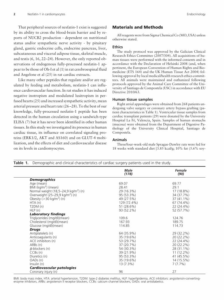
Table 1. Demographic and clinical characteristics of cardiac surgery patients used in the study.
Male(178)
Female(90)
DemographicsAge (mean) 69.07 69.77BMI (kg/m2) (mean) 28.41 29.1Normal weight (18,5–24,9 kg/m2) (n) 29 (16.3%) 17 (18.8%)Overweight (25–29,9 kg/m2) (n) 95 (53.3%) 34 (37.7%)Obesity (�30 kg/m2) (n) 49 (27.5%) 37 (41.1%)HTA (n) 129 (72.4%) 67 (74.4%)T2DM (n) 51 (28.6%) 22 (24.4%)HLP (n) 93 (52.2%) 52 (57.7%)Laboratory findingsTriglycerides (mg/dl)(mean) 109.6 124.76Cholesterol (mg/dl)(mean) 167.93 189.75Glucose (mg/dl)(mean) 114.85 114.73DrugsAntiplatelets (n) 64 (35.9%) 29 (32.2%)Anticoagulants (n) 35 (19.6%) 20 (22.2%)ACE inhibitors (n) 53 (29.7%) 22 (24.4%)ARBs (n) 37 (20.7%) 20 (22.2%)�-blockers (n) 54 (30.3%) 28 (31.1%)CCBs (n) 39 (21.9%) 11 (12.2%)Diuretics (n) 95 (53.3%) 41 (45.5%)OADs (n) 35 (19.6%) 14 (15.5%)Insulin (n) 13 (7.3%) 7 (7.7%)Cardiovascular patologiesCoronary injury (n) 96 27
BMI: body mass index, HTA: arterial hypertension, T2DM: type-2 diabetes mellitus, HLP: hyperlipidemia, ACE inhibitors: angiotensin-converting-enzyme inhibitors, ARBs: angiotensin II receptor blockers, CCBs: calcium channel blockers, OADs: oral antidiabetics.
2 Nesfatin-1 in cardiomyocytes Endocrinology
bean oil and 4.4% lard), 20% protein, 70% carbohydrate (35%sucrose, 31% cornstarch and 4% maltodextrin); Research Diets,Inc. USA] or high-fat diet [4.73 kcal/g; 45% fat (5.6% soybeanoil and 39.4% lard), 20% protein, 35% carbohydrate (17%sucrose, 8% cornstarch and 10% maltodextrin); Research Diets,Inc. USA] and euthanized by decapitation. Hearts were collectedand quickly frozen at –80°C. Neonatal (1- to 3-day-old)Sprague-Dawley rats were killed by cervical dislocation and theirhearts used for establishment of primary cardiomyocyte cultures(see below).
Cell culturesCardiomyocytes from human atrial appendages and neonatal
rat hearts and adult mouse atrial HL-1 cardiomyocytes (a gift ofDr. W.C. Claycomb of Louisiana State University Medical Cen-ter, USA), were all cultured as previously described (30). Adulthuman ventricular cardiomyocytes (Promocell GmbH; Heidel-berg, Germany) were cultured according to the supplier’sinstructions.
qPCRQuantitative reverse-transcription real time PCR for NUCB2
was performed on RNA extracted with the RNeasy Total RNAExtraction Kit (Quiagen, UK) using the master mix and specificprimers provided by SABioscience (rat NUCB2, 145 bp, Cat. No.PPR49435A, reference position 1245, GenBankNM 021663.2; rat GAPDH, 172 bp, Cat. No. PPR06557A,reference position 363, GenBank NM 017008.3; humanNUCB2, 126 bp, Cat. No. PPH19934A, reference position 894,GenBank NM 005013.2; human GAPDH, 175 bp, Cat. No.PPH00150E, reference position 1287, GenBankNM 002046.3; mouse NUCB2, 95 bp, Cat. No. PPM25549A,reference position 1314, GenBank NM 016773.3; mouseGADPH, 140 bp, Cat. No. PPM02946E, reference position 309,GenBank NM 008084.2). We also performed one-step reverse-transcription real-time PCR with Brilliant III SYBR Green QRT-PCR Master Mix Kit (Stratagene, USA) for the following specificprimers: rat CD36, 92 bp, GCGACATGATTAATGGCACA(forward) and CCTGCAAATGTCAGAGGAAA (reverse), Gen-Bank NM 031561.2; rat GADPH, 224 bp, GCTCATGACCA-CAGTCCATG (forward) and GGCATGTCAGATCCACAAC(reverse), GenBank NM 017008.4. Results were analyzed usingMxPro v4 software (Stratagene, USA).
Western blottingCultured cardiomyocytes (50,000 cells/cm2) or heart tissue
were lysed and subjected to SDS-PAGE/western blotting as de-scribed previously (31), using antibodies against nesfatin-1 res-idues 1–28 (Phoenix Pharmaceuticals, USA), ERK1/2 and phos-phoERK1/2 (Thermo Fisher Scientific Inc, USA), AKT andSer473-phosphorylated AKT (Cell Signaling Technology Inc,USA), AS160 and phosphoAS160 (Cell Signaling TechnologyInc, USA), �-actin and GAPDH (Thermo Fisher Scientific Inc,USA), all at 1:1000 dilution unless AS160 (1:500).
ImmunocytochemistryUsing a Dako Link 48 Autostainer, primary cultures of hu-
man and neonatal rat cardiomyocytes were stained with FLEXmouse antidesmin antibodies (clone D33, Dako) and rabbit an-tinesfatin-1 antibodies (Phoenix Pharmaceuticals, Inc., USA),
both at 1:200, followed by EnVision FLEX/HRP Dako for de-tection. Negative controls: normal goat serum or antinesfatin-1antibody preadsorbed by nesfatin-1 peptide (Phoenix Pharma-ceuticals Inc, USA).
Immunocytofluorescent labeling and confocalmicroscopy
Primary cultures of neonatal rat cardiomyocytes were de-prived of serum for 12 hours before treatment with nesfatin-1(0.01–1 �M for 10 minutes or 1 �M for 5–30 minutes) or 100nM insulin. Cells (10,000 per A 12 mm circular coverslip) andtissues were processed as previously described (31) using rabbitantibodies: anti-GLUT-4 (Abcam, UK) at 1:100 dilution, anti-nesfatin-1 (Phoenix Pharmaceuticals, USA.) at 1:500, or anti-prohormone convertase 1/3 (Abcam, UK) at 1:100. Quantitativeconfocal microscopy was performed using a Leica DMIRE2 con-focal microscope and software to compare GLUT-4 concentra-tions in the cell peripherals with those in the cytoplasm andperinuclear region, as other authors have described previouslyfor cardiomyocytes in culture (31–34). A total of 216 cells wereanalyzed. For each coverslip, a minimum of six distinct and ran-domly chosen visual fields were photographed and analyzed asz-stacks using a Leica DMIRE2 confocal microscope and soft-ware. Mean intensity of Cy3 fluorescence was taken to indicaterelative expression level of the GLUT-4 glucose transporter. Tomeasure intensity of fluorescence in the cytoplasm-perinuclearregion vs the cell peripherals-cytoplasmic membrane, ROIs (Re-gions of Interest) of equal sizes were first set up manually, andthen the cytoplasm-perinuclear region and the cell peripherals-cytoplasmic membrane ROIs for each of several cells per fieldchosen randomly and measured automatically. Cells with any ofthe following characteristics were excluded from analysis: atyp-ically large or small cells; perceptibly in the G1 or later phase ofthe cell cycle; not in contact with at least one neighbor; having anouter cytoplasmic border not clearly distinguishable from thoseof its neighbors. Great care was taken to localize the ROIs wellwithin the cell cytoplasmic-perinuclear and cell peripherals areas(as indicated by presence or absence of TO-PRO-3 fluorescence,respectively). To maximize the number of cells measured and theobjectivity with which ROI localization was carried out, the pro-cess was carried out twice, by two independent observers. Thusover 70 ratios were obtained for each experimental variable.
Glucose uptake experimentsSerum-deprived HL-1 cells or neonatal rat cardiomyocytes
(104 per P24 well) were treated for 2.5 hours with nesfatin-1(0.1–100 nM). Treatment for 1 hour with insulin (100 nM) wasused as a positive control. Cells were washed twice in glucose-free HEPES-buffered saline solution at pH7.4 (140 mM NaCl, 5mM KCl, 2.5 mM MgSO4, 1 mM CaCl2, 20 mM HEPES) andglucose uptake determined as described previously using [3H]2-deoxy-D-glucose (Perkin Elmer Inc, USA) (13). Nonspecificglucose uptake was determined in the presence of 10 �M cy-tochalasin B, and the value obtained subtracted from all othermeasurements.
Enzyme-linked immunosorbent assaysPlasma NUCB2/nesfatin-1 levels of male Sprague-Dawley
rats fed for 18 weeks with standard or high-fat diet were deter-mined by ELISA (Phoenix Pharmaceuticals Inc, USA; minimum
doi: 10.1210/en.2013-1497 endo.endojournals.org 3
detection limit: 0.8 ng/ml; range: 0.1–1000 ng/ml; linear range:0.8–14.1 ng/ml; intra-assay error:�10%; interassay error:�15%). NUCB2/nesfatin-1 secretion by neonatal rat cardiomy-ocytes was measured in the culture medium in nine 6-well platesin which cells had been cultured for 3 days at a density of 4.5 �105 cells/well.
Statistical analysesAll experimental data were obtained from at least three in-
dependent experiments. The comparisons between groups ofdata were done with the appropriate test depending on the nor-mality of the data. We used t test with gaussian data and Mann-Whitney U, Kruskal-Wallis and Wilcoxon signed rank tests withnongaussian data. All the correlation coefficients were measuredas Spearman r. All the analysis were done with IBM SPSS 15.0(USA) or Prism 5 GraphPad (USA) softwares.
Results
NUCB2 gene expression in human and murinecardiomyocytes
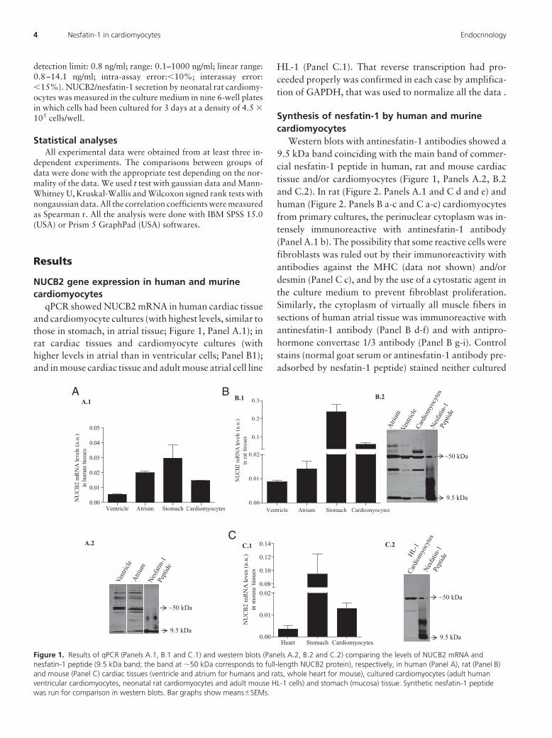
qPCR showed NUCB2 mRNA in human cardiac tissueand cardiomyocyte cultures (with highest levels, similar tothose in stomach, in atrial tissue; Figure 1, Panel A.1); inrat cardiac tissues and cardiomyocyte cultures (withhigher levels in atrial than in ventricular cells; Panel B1);and in mouse cardiac tissue and adult mouse atrial cell line
HL-1 (Panel C.1). That reverse transcription had pro-ceeded properly was confirmed in each case by amplifica-tion of GAPDH, that was used to normalize all the data .
Synthesis of nesfatin-1 by human and murinecardiomyocytes
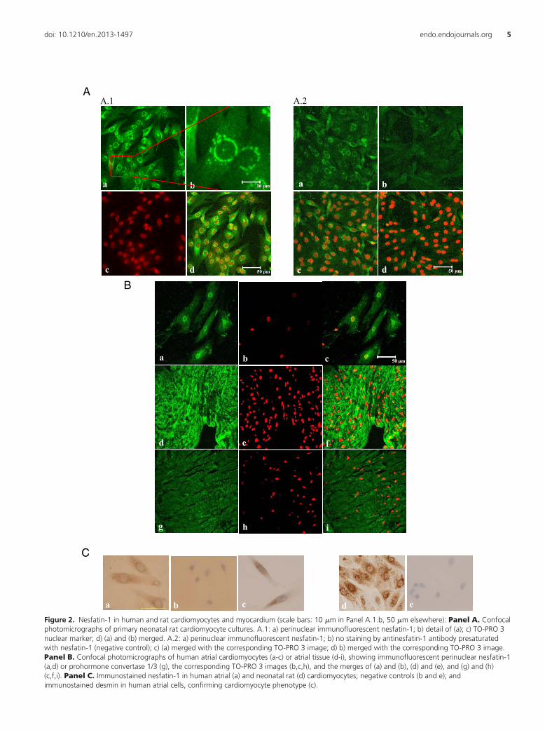
Western blots with antinesfatin-1 antibodies showed a9.5 kDa band coinciding with the main band of commer-cial nesfatin-1 peptide in human, rat and mouse cardiactissue and/or cardiomyocytes (Figure 1, Panels A.2, B.2and C.2). In rat (Figure 2. Panels A.1 and C d and e) andhuman (Figure 2. Panels B a-c and C a-c) cardiomyocytesfrom primary cultures, the perinuclear cytoplasm was in-tensely immunoreactive with antinesfatin-1 antibody(Panel A.1 b). The possibility that some reactive cells werefibroblasts was ruled out by their immunoreactivity withantibodies against the MHC (data not shown) and/ordesmin (Panel C c), and by the use of a cytostatic agent inthe culture medium to prevent fibroblast proliferation.Similarly, the cytoplasm of virtually all muscle fibers insections of human atrial tissue was immunoreactive withantinesfatin-1 antibody (Panel B d-f) and with antipro-hormone convertase 1/3 antibody (Panel B g-i). Controlstains (normal goat serum or antinesfatin-1 antibody pre-adsorbed by nesfatin-1 peptide) stained neither cultured
Ventricle Atrium Stomach Cardiomyocytes0.00
0.01
0.02
0.1
0.2
0.3
NUC
B2 m
RNA
levels
(a.u
.)in
rat t
issue
s
Heart Stomach Cardiomyocytes0.00
0.01
0.020.08
0.10
0.12
0.14
NU
CB
2 m
RN
A le
ves
(a.u
.)in
mou
se ti
ssue
s
A B
C
Ventricle Atrium Stomach Cardiomyocytes0.00
0.01
0.02
0.03
0.04
0.05
NU
CB
2 m
RN
A le
vels
(a.u
.)in
hum
an ti
ssue
s
A.1
A.2
B.1 B.2
C.1 C.2
~50 kDa
9.5 kDa
~50 kDa
9.5 kDa
~50 kDa
9.5 kDa
Figure 1. Results of qPCR (Panels A.1, B.1 and C.1) and western blots (Panels A.2, B.2 and C.2) comparing the levels of NUCB2 mRNA andnesfatin-1 peptide (9.5 kDa band; the band at �50 kDa corresponds to full-length NUCB2 protein), respectively, in human (Panel A), rat (Panel B)and mouse (Panel C) cardiac tissues (ventricle and atrium for humans and rats, whole heart for mouse), cultured cardiomyocytes (adult humanventricular cardiomyocytes, neonatal rat cardiomyocytes and adult mouse HL-1 cells) and stomach (mucosa) tissue. Synthetic nesfatin-1 peptidewas run for comparison in western blots. Bar graphs show means�SEMs.
4 Nesfatin-1 in cardiomyocytes Endocrinology
A
a
c
A.1 A.2
a
c
bb
dd d
B
a b c
ed f
h ig
c
f
C
da ecbFigure 2. Nesfatin-1 in human and rat cardiomyocytes and myocardium (scale bars: 10 �m in Panel A.1.b, 50 �m elsewhere): Panel A. Confocalphotomicrographs of primary neonatal rat cardiomyocyte cultures. A.1: a) perinuclear immunofluorescent nesfatin-1; b) detail of (a); c) TO-PRO 3nuclear marker; d) (a) and (b) merged. A.2: a) perinuclear immunofluorescent nesfatin-1; b) no staining by antinesfatin-1 antibody presaturatedwith nesfatin-1 (negative control); c) (a) merged with the corresponding TO-PRO 3 image; d) b) merged with the corresponding TO-PRO 3 image.Panel B. Confocal photomicrographs of human atrial cardiomyocytes (a-c) or atrial tissue (d-i), showing immunofluorescent perinuclear nesfatin-1(a,d) or prohormone convertase 1/3 (g), the corresponding TO-PRO 3 images (b,c,h), and the merges of (a) and (b), (d) and (e), and (g) and (h)(c,f,i). Panel C. Immunostained nesfatin-1 in human atrial (a) and neonatal rat (d) cardiomyocytes; negative controls (b and e); andimmunostained desmin in human atrial cells, confirming cardiomyocyte phenotype (c).
doi: 10.1210/en.2013-1497 endo.endojournals.org 5
cells (Figure 2. Panels A.2 and C b and e) nor cardiactissues (data not shown).
Secretion of nesfatin-1 by primary cultures of ratcardiomyocytes
Secretion of NUCB2/nesfatin-1 into the culture me-dium was measured only in primary cultures of neonatalrat cardiomyocytes due to the limited cardiomyocyte pop-ulations afforded by human right atrial appendages in cul-ture. In culturemedia supportingapproximately4.5�105
confluent starving cells the ELISA-measured NUCB2/nes-fatin-1 concentration was 4.55 � 0.52 ng/mg of cellularprotein (n�21).
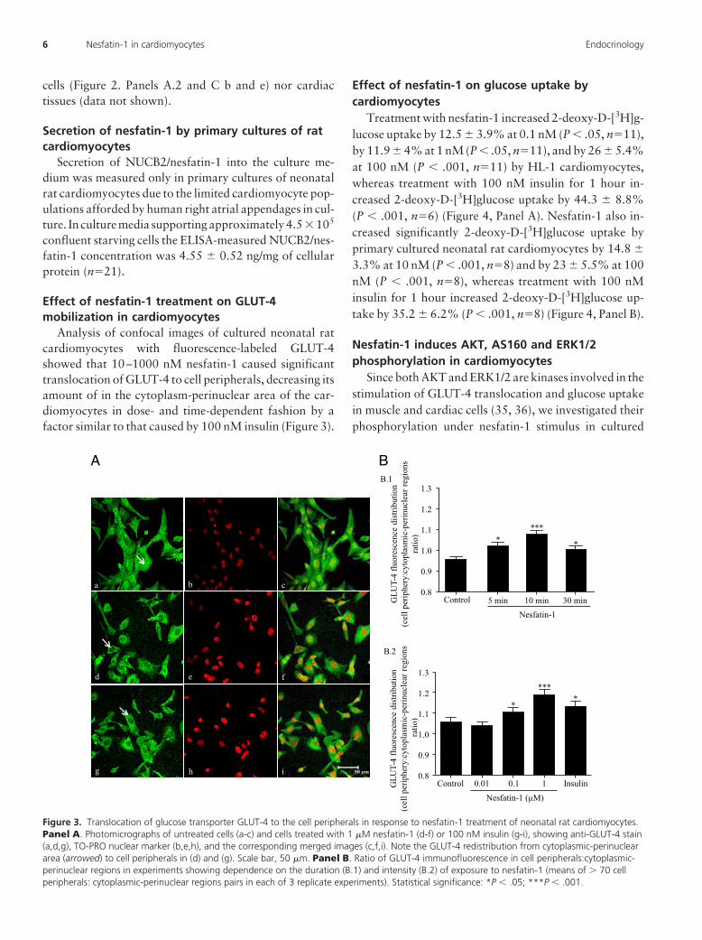
Effect of nesfatin-1 treatment on GLUT-4mobilization in cardiomyocytes
Analysis of confocal images of cultured neonatal ratcardiomyocytes with fluorescence-labeled GLUT-4showed that 10–1000 nM nesfatin-1 caused significanttranslocation of GLUT-4 to cell peripherals, decreasing itsamount of in the cytoplasm-perinuclear area of the car-diomyocytes in dose- and time-dependent fashion by afactor similar to that caused by 100 nM insulin (Figure 3).
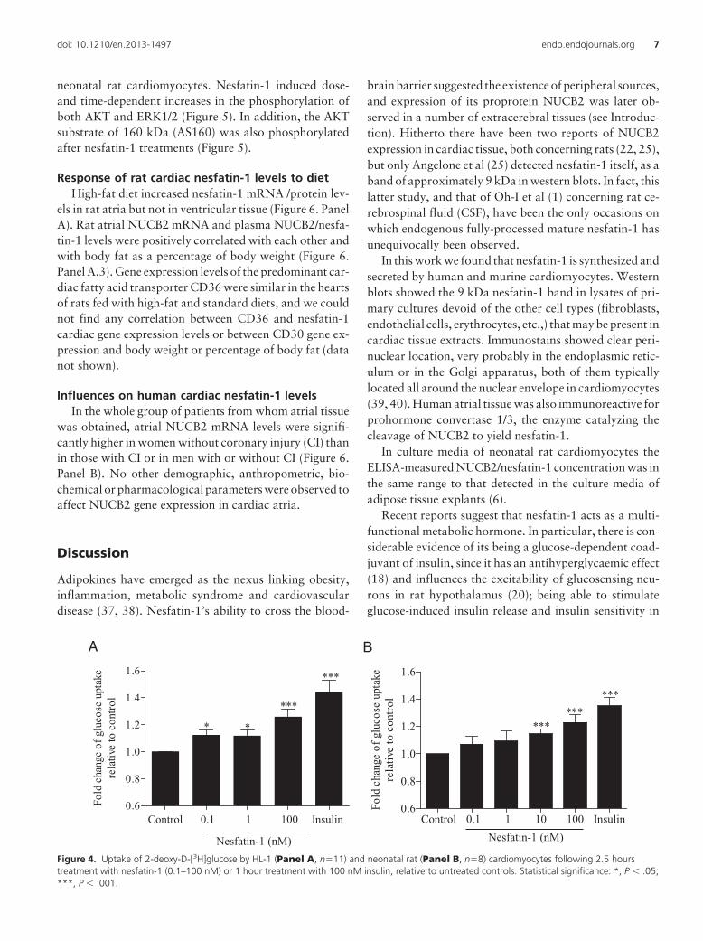
Effect of nesfatin-1 on glucose uptake bycardiomyocytes
Treatment with nesfatin-1 increased 2-deoxy-D-[3H]g-lucose uptake by 12.5 � 3.9% at 0.1 nM (P � .05, n�11),by 11.9 � 4% at 1 nM (P � .05, n�11), and by 26 � 5.4%at 100 nM (P � .001, n�11) by HL-1 cardiomyocytes,whereas treatment with 100 nM insulin for 1 hour in-creased 2-deoxy-D-[3H]glucose uptake by 44.3 � 8.8%(P � .001, n�6) (Figure 4, Panel A). Nesfatin-1 also in-creased significantly 2-deoxy-D-[3H]glucose uptake byprimary cultured neonatal rat cardiomyocytes by 14.8 �
3.3% at 10 nM (P � .001, n�8) and by 23 � 5.5% at 100nM (P � .001, n�8), whereas treatment with 100 nMinsulin for 1 hour increased 2-deoxy-D-[3H]glucose up-take by 35.2 � 6.2% (P � .001, n�8) (Figure 4, Panel B).
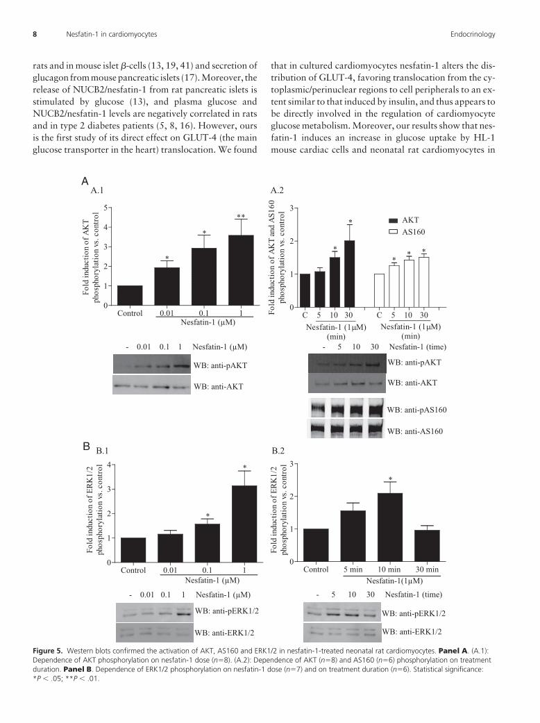
Nesfatin-1 induces AKT, AS160 and ERK1/2phosphorylation in cardiomyocytes
Since both AKT and ERK1/2 are kinases involved in thestimulation of GLUT-4 translocation and glucose uptakein muscle and cardiac cells (35, 36), we investigated theirphosphorylation under nesfatin-1 stimulus in cultured
a cb
d e f
g h i
A B
Control 5 min 10 min 30 min 0.8
0.9
1.0
1.1
1.2
1.3
*
****
GLU
T-4
fluor
esce
nce
dist
ribut
ion
( cel
l per
iphe
ry:c
ytop
lasm
ic-p
erin
ucle
ar re
gion
sra
tio)
B.1
B.2
Nesfatin-1
Control 0.01 0.1 1 Insulin0.8
0.9
1.0
1.1
1.2
1.3
****
*
Nesfatin-1 (µM)
i
GLU
T-4
fluor
esce
nce
dist
ribut
ion
(cel
l per
iphe
ry:c
ytop
lasm
ic-p
erin
ucle
ar re
gion
sra
tio)
Figure 3. Translocation of glucose transporter GLUT-4 to the cell peripherals in response to nesfatin-1 treatment of neonatal rat cardiomyocytes.Panel A. Photomicrographs of untreated cells (a-c) and cells treated with 1 �M nesfatin-1 (d-f) or 100 nM insulin (g-i), showing anti-GLUT-4 stain(a,d,g), TO-PRO nuclear marker (b,e,h), and the corresponding merged images (c,f,i). Note the GLUT-4 redistribution from cytoplasmic-perinucleararea (arrowed) to cell peripherals in (d) and (g). Scale bar, 50 �m. Panel B. Ratio of GLUT-4 immunofluorescence in cell peripherals:cytoplasmic-perinuclear regions in experiments showing dependence on the duration (B.1) and intensity (B.2) of exposure to nesfatin-1 (means of � 70 cellperipherals: cytoplasmic-perinuclear regions pairs in each of 3 replicate experiments). Statistical significance: *P � .05; ***P � .001.
6 Nesfatin-1 in cardiomyocytes Endocrinology
neonatal rat cardiomyocytes. Nesfatin-1 induced dose-and time-dependent increases in the phosphorylation ofboth AKT and ERK1/2 (Figure 5). In addition, the AKTsubstrate of 160 kDa (AS160) was also phosphorylatedafter nesfatin-1 treatments (Figure 5).
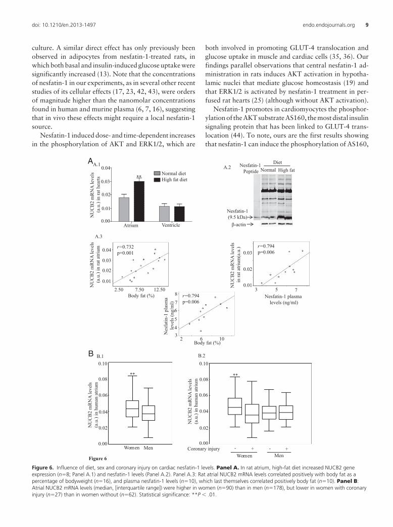
Response of rat cardiac nesfatin-1 levels to dietHigh-fat diet increased nesfatin-1 mRNA /protein lev-
els in rat atria but not in ventricular tissue (Figure 6. PanelA). Rat atrial NUCB2 mRNA and plasma NUCB2/nesfa-tin-1 levels were positively correlated with each other andwith body fat as a percentage of body weight (Figure 6.Panel A.3). Gene expression levels of the predominant car-diac fatty acid transporter CD36 were similar in the heartsof rats fed with high-fat and standard diets, and we couldnot find any correlation between CD36 and nesfatin-1cardiac gene expression levels or between CD30 gene ex-pression and body weight or percentage of body fat (datanot shown).
Influences on human cardiac nesfatin-1 levelsIn the whole group of patients from whom atrial tissue
was obtained, atrial NUCB2 mRNA levels were signifi-cantly higher in women without coronary injury (CI) thanin those with CI or in men with or without CI (Figure 6.Panel B). No other demographic, anthropometric, bio-chemical or pharmacological parameters were observed toaffect NUCB2 gene expression in cardiac atria.
Discussion
Adipokines have emerged as the nexus linking obesity,inflammation, metabolic syndrome and cardiovasculardisease (37, 38). Nesfatin-1’s ability to cross the blood-
brain barrier suggested the existence of peripheral sources,and expression of its proprotein NUCB2 was later ob-served in a number of extracerebral tissues (see Introduc-tion). Hitherto there have been two reports of NUCB2expression in cardiac tissue, both concerning rats (22, 25),but only Angelone et al (25) detected nesfatin-1 itself, as aband of approximately 9 kDa in western blots. In fact, thislatter study, and that of Oh-I et al (1) concerning rat ce-rebrospinal fluid (CSF), have been the only occasions onwhich endogenous fully-processed mature nesfatin-1 hasunequivocally been observed.
In this work we found that nesfatin-1 is synthesized andsecreted by human and murine cardiomyocytes. Westernblots showed the 9 kDa nesfatin-1 band in lysates of pri-mary cultures devoid of the other cell types (fibroblasts,endothelial cells, erythrocytes, etc.,) that may be present incardiac tissue extracts. Immunostains showed clear peri-nuclear location, very probably in the endoplasmic retic-ulum or in the Golgi apparatus, both of them typicallylocated all around the nuclear envelope in cardiomyocytes(39, 40). Human atrial tissue was also immunoreactive forprohormone convertase 1/3, the enzyme catalyzing thecleavage of NUCB2 to yield nesfatin-1.
In culture media of neonatal rat cardiomyocytes theELISA-measured NUCB2/nesfatin-1 concentration was inthe same range to that detected in the culture media ofadipose tissue explants (6).
Recent reports suggest that nesfatin-1 acts as a multi-functional metabolic hormone. In particular, there is con-siderable evidence of its being a glucose-dependent coad-juvant of insulin, since it has an antihyperglycaemic effect(18) and influences the excitability of glucosensing neu-rons in rat hypothalamus (20); being able to stimulateglucose-induced insulin release and insulin sensitivity in
Control 0.1 1 100 Insulin 0.6
0.8
1.0
1.2
1.4
1.6
Fold
cha
nge
of g
luco
se u
ptak
ere
lativ
e to
con
trol
A
Nesfatin-1 (nM)
* *
***
***
Nesfatin-1 (nM)
B
***
Control 0.1 1 10 100 Insulin0.6
0.8
1.0
1.2
1.4
1.6
Fold
cha
nge
of g
luco
se u
ptak
ere
lativ
e to
con
trol
******
Figure 4. Uptake of 2-deoxy-D-[3H]glucose by HL-1 (Panel A, n�11) and neonatal rat (Panel B, n�8) cardiomyocytes following 2.5 hourstreatment with nesfatin-1 (0.1–100 nM) or 1 hour treatment with 100 nM insulin, relative to untreated controls. Statistical significance: *, P � .05;***, P � .001.
doi: 10.1210/en.2013-1497 endo.endojournals.org 7
rats and in mouse islet �-cells (13, 19, 41) and secretion ofglucagon from mouse pancreatic islets (17). Moreover, therelease of NUCB2/nesfatin-1 from rat pancreatic islets isstimulated by glucose (13), and plasma glucose andNUCB2/nesfatin-1 levels are negatively correlated in ratsand in type 2 diabetes patients (5, 8, 16). However, oursis the first study of its direct effect on GLUT-4 (the mainglucose transporter in the heart) translocation. We found
that in cultured cardiomyocytes nesfatin-1 alters the dis-tribution of GLUT-4, favoring translocation from the cy-toplasmic/perinuclear regions to cell peripherals to an ex-tent similar to that induced by insulin, and thus appears tobe directly involved in the regulation of cardiomyocyteglucose metabolism. Moreover, our results show that nes-fatin-1 induces an increase in glucose uptake by HL-1mouse cardiac cells and neonatal rat cardiomyocytes in
Control 0.01 0.1 10
1
2
3
4 *
*
Fold
indu
ctio
n of
ERK
1/2
phos
phor
ylat
ion
vs. c
ontro
l
B B.1 B.2
- 0.01 0.1 1 Nesfatin-1 (µM)
WB: anti-pERK1/2
WB: anti-ERK1/2
Nesfatin-1 (µM)Control 5 min 10 min 30 min
0
1
2
3
Fold
indu
ctio
n of
ERK
1/2
phos
phor
ylat
ion
vs. c
ontro
l
*
Nesfatin-1(1µM)
WB: anti-pERK1/2
WB: anti-ERK1/2
- 5 10 30 Nesfatin-1 (time)
A
Control 0.01 0.1 10
1
2
3
4
5
*
*
Fold
indu
ctio
n of
AK
Tph
osph
oryl
atio
n vs
. con
trol
**
A.1 A.2
Nesfatin-1 (µM)
- 5 10 30 Nesfatin-1 (time)
WB: anti-pAKT
WB: anti-AKT
C 5 10 30 C 5 10 300
1
2
3
Nesfatin-1 (1µM)(min)
Nesfatin-1 (1µM)(min)
*
*
***
Fold
indu
ctio
n of
AK
T an
d A
S160
phos
phor
ylat
ion
vs. c
ontr
ol
- 0.01 0.1 1 Nesfatin-1 (µM)
WB: anti-pAKT
WB: anti-AKT
AKTAS160
WB: anti-pAS160
WB: anti-AS160
Figure 5. Western blots confirmed the activation of AKT, AS160 and ERK1/2 in nesfatin-1-treated neonatal rat cardiomyocytes. Panel A. (A.1):Dependence of AKT phosphorylation on nesfatin-1 dose (n�8). (A.2): Dependence of AKT (n�8) and AS160 (n�6) phosphorylation on treatmentduration. Panel B. Dependence of ERK1/2 phosphorylation on nesfatin-1 dose (n�7) and on treatment duration (n�6). Statistical significance:*P � .05; **P � .01.
8 Nesfatin-1 in cardiomyocytes Endocrinology
culture. A similar direct effect has only previously beenobserved in adipocytes from nesfatin-1-treated rats, inwhich both basal and insulin-induced glucose uptake weresignificantly increased (13). Note that the concentrationsof nesfatin-1 in our experiments, as in several other recentstudies of its cellular effects (17, 23, 42, 43), were ordersof magnitude higher than the nanomolar concentrationsfound in human and murine plasma (6, 7, 16), suggestingthat in vivo these effects might require a local nesfatin-1source.
Nesfatin-1 induced dose- and time-dependent increasesin the phosphorylation of AKT and ERK1/2, which are
both involved in promoting GLUT-4 translocation andglucose uptake in muscle and cardiac cells (35, 36). Ourfindings parallel observations that central nesfatin-1 ad-ministration in rats induces AKT activation in hypotha-lamic nuclei that mediate glucose homeostasis (19) andthat ERK1/2 is activated by nesfatin-1 treatment in per-fused rat hearts (25) (although without AKT activation).
Nesfatin-1 promotes in cardiomyocytes the phosphor-ylation of the AKT substrate AS160, the most distal insulinsignaling protein that has been linked to GLUT-4 trans-location (44). To note, ours are the first results showingthat nesfatin-1 can induce the phosphorylation of AS160,
1 20.00
0.01
0.02
0.03
0.04
Nesfatin-1(9.5 kDa)
β-actin
Nesfatin-1Peptide Normal High fat
DietAA.1 A.2
A.3
Figure 6
B
MenWomen0.00
0.02
0.04
0.06
0.08
0.10
NU
CB
2 m
RN
A le
vels
(a
.u.)
in h
uman
atri
um
**
B.1 B.2
Men
0.00
0.02
0.04
0.06
0.08
0.10
NU
CB
2 m
RN
A le
vels
(a
.u.)
in h
uman
atri
um
**
WomenCoronary injury - -+ +
NU
CB
2 m
RN
A le
vels
(a.u
.) in
rat h
earts
VentricleAtrium
**Normal dietHigh fat diet
NU
CB
2 m
RN
A le
vels
(a.u
.) in
rat a
trium
r=0.732p=0.001
0.01
0.02
0.03
0.04
Body fat (%)2.50 7.50 12.50 N
UC
B2
mR
NA
leve
ls
in ra
t atri
um(a
.u.)
Nesfatin-1 plasmalevels (ng/ml)
3 5 70.01
0.02
0.03r=0.794p=0.006
Nes
fatin
-1 p
lasm
ale
vels
(ng/
ml)
43
5
67
8
Body fat (%)2 6 10
r=0.794p=0.006
0.01
0.02
0.03
0.04
0.00
Figure 6. Influence of diet, sex and coronary injury on cardiac nesfatin-1 levels. Panel A. In rat atrium, high-fat diet increased NUCB2 geneexpression (n�8; Panel A.1) and nesfatin-1 levels (Panel A.2). Panel A.3: Rat atrial NUCB2 mRNA levels correlated positively with body fat as apercentage of bodyweight (n�16), and plasma nesfatin-1 levels (n�10), which last themselves correlated positively body fat (n�10). Panel B:Atrial NUCB2 mRNA levels (median, [interquartile range]) were higher in women (n�90) than in men (n�178), but lower in women with coronaryinjury (n�27) than in women without (n�62). Statistical significance: **P � .01.
doi: 10.1210/en.2013-1497 endo.endojournals.org 9
and this could indeed help to clarify the physiological/pathophysiological roles of nesfatin-1 in glucose and en-ergy metabolism suggested previously by others (45). Un-der basal conditions, the Rab GTPase activating proteindomain of AS160 stimulates hydrolysis of Rab-associatedGTP and the formation of the inactive GDP-bound Rab,which restrains GLUT-4 exocytosis; however, insulin-stimulated phosphorylation of AS160 by AKT relieves thisrestraint by leading to an increase in the GTP-bound Rabwhich, in turn, favors greater GLUT-4 exocytosis to cell-surface membranes (44, 46). AS160 is considered to be apoint of convergence for coordinating physiological reg-ulation of GLUT-4 membrane recruitment in cardiomy-ocytes (47).
In rodents, synthesis of nesfatin-1 (or at least NUCB2)appears to respond to diet or nutritional status both cen-trally, in the hypothalamic paraventricular nucleus (1,48), and in peripheral tissues such as the stomach (49),adipose tissue (6) and heart (this work), where it may helpcontrol energy expenditure and may respond to develop-mental and hormonal signals as well as to metabolic sta-tus. The finding that cardiac and plasma NUCB2/nesfa-tin-1 levels are positively correlated, and that bothcorrelate with body fat, is in keeping with a report thatplasma NUCB2 concentrations are higher in mice withdiet-induced obesity than in nonobese mice (6).
Obesity imposes a metabolic stress to the heart whichmay ultimately lead to cardiac metabolic inflexibility andlipotoxicity, being the fatty acid transporter CD36 theresponsible of the chronic increasing uptake of fatty acidsby the cardiac muscle under those circumstances (36). Inthis work, high-fat diet did not induce changes in cardiacCD36 gene expression, and there was no correlation be-tween cardiac CD36 and nesfatin-1 levels; however, wecan not exclude that nesfatin-1 could affect the surfacepresence of CD36 and its translocation from intracellularstorage compartments, and future experiments shouldclarify a possible involvement of this peptide in the com-plex interplay between signaling pathways and traffickingcomponents known to be involved in the regulation ofGLUT-4 and CD36 translocation (36).
Only two previous studies have investigated NUCB2/nesfatin-1 regulation in human cells or tissues. Yamada etal (50) found that troglitazone stabilizes NUCB2 mRNAthrough ERK1/2 activation in a human medulloblastomacell line, and Riva et al (17) that NUCB2 deficiency in thepancreatic islets of type 2 diabetes patients can be reme-died by culture in glucolipotoxic conditions. In the presentstudy atrial NUCB2 mRNA levels were significantlyhigher in women without coronary lesions than in otherpatients, suggesting a sex-dependent relationship betweencardiac nesfatin-1 levels and cardiovascular disease.
In conclusion, nesfatin-1 is synthesized, and helps toregulate glucose metabolism, in the hearts of humans andexperimental animals, where its levels vary with diet andischemic cardiomyopathy.
Acknowledgments
We thank Patricia Viaño for expert technical assistance.
Address all correspondence and requests for reprints to:¶CORRESPONDENCE: Dr Francisca Lago, Laboratorio 7, In-stituto de Investigaciones Sanitarias de Santiago de Compostela(IDIS), Hospital Clínico Universitario, Travesía Choupana s/n,15706 Santiago de Compostela, Spain., Tel: � 34 981 950 902Fax: �34 981 950 905 Email: [email protected].
Disclosure Summary: The authors have nothing to disclose.This work was supported by: This work was funded by the
Health Research Fund of the Carlos III Health Institute (FIS11/00497) and the Spanish Society of Cardiology; VG-R is fundedby the University Professional Development Program (FPU) ofthe Spanish Ministry of Education; MFO by the Rio HortegaProgram of the Spanish Ministry of Economy and Competitive-ness; DR-P by the International PhD School, USC; SF-B andAM-L by the IDICHUS Research Foundation of USC ClinicalHospital; and LMS, OG and FL by SERGAS.
References
1. Oh-I S, Shimizu H, Satoh T, Okada S, Adachi S, Inoue K Eguchi H,Yamamoto M, Imaki T, Hashimoto K, Tsuchiya T, Monden T,Horiguchi K, Yamada M, Mori M. Identification of nesfatin-1 as asatiety molecule in the hypothalamus. Nature. 2006;443:709–712.
2. Yosten GLC, Samson WK. The anorexigenic and hypertensive ef-fects of nesfatin-1 are reversed by pretreatment with an oxytocinreceptor antagonist. Am J Physiol Regul Integr Comp Physiol. 2010;298:R1642–R1647.
3. García-Galiano D, Navarro VM, Gaytan F, Tena-Sempere M. Ex-panding roles of NUCB2/Nesfatin-1 in neuroendocrine regulation.J Mol Endocrinol. 2010;45:281–290.
4. Sedbazar U, Maejima Y, Nakata M, Mori M, Yada T. Paraven-tricular NUCB2/nesfatin-1 rises in synchrony with feeding suppres-sion during early light phase in rats. Biochem Biophys Res Commun.2013;434:434–438.
5. Li QC, Wang HY, Chen X, Guan HZ, Jiang ZY. Fasting plasmalevels of nesfatin-1 in patients with type 1 and type 2 diabetes mel-litus and the nutrients-related fluctuation of nesfatin-1 level in nor-mal humans. Regul Pept. 2010;159:72–77.
6. Ramanjaneya M, Chen J, Brown JE, Tripathi G, Hallschmid M,Patel S, Kern W, Hillhouse EW, Lehnert H, Tan BK, Randeva HS.Identification of nesfatin-1 in human and murine adipose tissue: anovel depot-specific adipokine with increased levels in obesity. En-docrinology. 2010;151:3169–3180.
7. Tsuchiya T, Shimizu H, Yamada M, Osaki A, OH-I S, Ariyama Y,Takahashi H, Okada S, Hashimoto K, Satoh T, Kojima M, Mori M.Fasting concentrations of nesfatin-1 are negatively correlated withbody mass index in non-obese males. Clin Endocrinol (Oxf). 2010;73:484–490.
8. Zhang Z, Li L, Yang M, Liu H, Boden G, Yang G. Increased plasma
10 Nesfatin-1 in cardiomyocytes Endocrinology
levels of nesfatin-1 in patients with newly diagnosed type 2 diabetesmellitus. Exp Clin Endocrinol Diabetes. 2012;120:91–95.
9. Basar O, Akbal E, Köklü S, Koçak E, Tuna Y, Ekiz F, Gültuna S,Y�lmaz FM, Aydogan T. A novel appetite peptide, nesfatin-1 inpatients with non-alcoholic fatty liver disease. Scand J Clin LabInvest. 2012;72:479–483.
10. Shimizu H, OH-I S, Hashimoto K, Nakata M, Yamamoto S, Yo-shida N, Eguchi H, Kato I, Inoue K, Satoh T, Okada S, Yamada M,Yada T, Mori M. Peripheral administration of nesfatin-1 reducesfood intake in mice: the leptin-independent mechanism. Endocri-nology. 2009;150:662–671.
11. Maejima Y, Sedbazar U, Suyama S, Kohno D, Onaka T, Takano E,Yoshida N, Koike M, Uchiyama Y, Fujiwara K, Yashiro T, HorvathTL, Dietrich MO, Tanaka S, Dezaki K, OH-I S, Hashimoto K, Shi-mizu H, Nakata M, Mori M, Yada T. Nesfatin-1-regulated oxyto-cinergic signaling in the paraventricular nucleus causes anorexiathrough a leptin-independent melanocortin pathway. Cell Metab.2009;10:355–365.
12. Stengel A, Goebel M, Taché Y. Nesfatin-1: a novel inhibitory reg-ulator of food intake and body weight. Obes Rev. 2011;12:261–271.
13. Gonzalez R, Perry RL, Gao X, Gaidhu MP, Tsushima RG, CeddiaRB, Unniappan S. Nutrient responsive nesfatin-1 regulates energybalance and induces glucose-stimulated insulin secretion in rats. En-docrinology. 2011;152:3628–3637.
14. Könczöl K, Pintér O, Ferenczi S, Varga J, Kovács K, Palkovits M,Zelena D, Tóth ZE. Nesfatin-1 exerts long-term effect on food in-take and body temperature. Int J Obes (Lond). 2012;36:1514–1521.
15. Miyata S, Yamada N, Kawada T. Possible involvement of hypotha-lamic nucleobindin-2 in hyperphagic feeding in Tsumura SuzukiObese Diabetes Mice. Biol Pharm Bull. 2012;35:1784–1793.
16. Foo KS, Brauner H, Ostenson CG, Broberger C. Nucleobindin-2/nesfatin in the endocrine pancreas:distribution and relationship toglycaemic state. J Endocrinol. 2010;204:255–263.
17. Riva M, Nitert MD, Voss U, Sathanoori R, Lindqvist A, Ling C,Wierup N. Nesfatin-1 stimulates glucagon and insulin secretion andbeta cell NUCB2 is reduced in human type 2 diabetic subjects. CellTissue Res. 2011;346:393–405.
18. Su Y, Zhang J, Tang Y, Bi F, Liu JN. The novel function of nesfa-tin-1: Anti-hyperglycemia. Biochem Biophys Res Commun. 2010;391:1039–1042.
19. Yang M, Zhang Z, Wang C, Li K, Li S, Boden G, Li L, Yang G.Nesfatin-1 action in the brain increases insulin sensitivity throughAkt/AMPK/TORC2 pathway in diet-induced insulin resistance. Di-abetes. 2012;61:1959–1968.
20. Chen X, Dong J, Jiang ZY. Nesfatin-1 influences the excitability ofglucosensing neurons in the hypothalamic nuclei and inhibits thefood intake. Regul Pept. 2012;177:21–26.
21. Stengel A, Goebel-Stengel M, Jawien J, Kobelt P, Taché Y, Lam-brecht NW. Lipopolysaccharide increases gastric and circulatingNUCB2/nesfatin-1 concentrations in rats. Peptides. 2011;32:1942–1947.
22. Stengel A, Goebel M, Yakubov I, Wang L, Witcher D, Coskun T,Taché Y, Sachs G, Lambrecht NW. Identification and characteriza-tion of nesfatin-1 immunoreactivity in endocrine cell types of the ratgastric oxyntic mucosa. Endocrinology. 2009;150:232–238.
23. García-Galiano D, Pineda R, Ilhan T, Castellano JM, Ruiz-Pino F,Sánchez-Garrido MA, Vazquez MJ, Sangiao-Alvarellos S, Romero-Ruiz A, Pinilla L, Diéguez C, Gaytán F, Tena-Sempere M. Cellulardistribution, regulated expression, and functional role of the an-orexigenic peptide, NUCB2/nesfatin-1, in the testis. Endocrinology.2012;153:1959–1971.
24. Osaki A, Shimizu H, Ishizuka N, Suzuki Y, Mori M, Inoue S. En-hanced expression of nesfatin/nucleobindin-2 in white adipose tis-sue of ventromedial hypothalamus-lesioned rats. Neurosci Lett.2012;521:46–51.
25. Angelone T, Filice E, Pasqua T, Amodio N, Galluccio M, Monte-santi G, Quintieri AM, Cerra MC. Nesfatin-1 as a novel cardiacpeptide: identification, functional characterization, and protectionagainst ischemia/reperfusion injury. Cell Mol Life Sci. 2013;70:495–509.
26. Yosten GLC, Samson WK. Nesfatin-1 exerts cardiovascular actionsin the brain: possible interaction with the central melanocortin sys-tem. Am J Physiol Regul Integr Comp Physiol. 2009;297:R330–R336.
27. Yamawaki H, Takahashi M, Mukohda M, Morita T, Okada M,Hara Y. A novel adipocytokine, nesfatin-1 modulates peripheralarterial contractility and blood pressure in rats. Biochem BiophysRes Commun. 2012;418:676–681.
28. Mimee A, Smith PM, Ferguson AV. Nesfatin-1 influences the ex-citability of neurons in the nucleus of the solitary tract and regulatescardiovascular function. Am J Physiol Regul Integr Comp Physiol.2012;302:R1297–R1304.
29. García-Rúa V, Otero MF, Lear PV, Rodríguez-Penas D, Feijóo-Bandín S, Noguera-Moreno T, Calaza M, Álvarez-Barredo M,Mosquera-Leal A, Parrington J, Brugada J, Portolés M, Rivera M,González-Juanatey JR, Lago F. Increased expression of fatty-acidand calcium metabolism genes in failing human heart. PLoS One.2012;7:e37505.
30. González-Juanatey JR, Iglesias MJ, Alcaide C, Piñeiro R, Lago F.Doxazosin induces apoptosis in cardiomyocytes cultured in vitro bya mechanism that is independent of alpha1-adrenergic blockade.Circulation. 2003;107:127–131.
31. Lear PV, Iglesias MJ, Feijóo-Bandín S, Rodríguez-Penas D, Mos-quera-Leal A, García-Rúa V, Gualillo O, Ghè C, Arnoletti E, Muc-cioli G, Diéguez C, González-Juanatey JR, Lago F. Des-acyl ghrelinhas specific binding sites and different metabolic effects from ghrelinin cardiomyocytes. Endocrinology. 2010;151:3286–3298.
32. Slot JW, Garruti G, Martin S, Oorschot V, Posthuma G, KraegenEW, Laybutt R, Thibault G, James DE. Glucose transporter(GLUT-4) is targeted to secretory granules in rat atrial cardiomyo-cytes. J Cell Biol. 1997;137:1243–1254.
33. Lu H, Buchan RJ, Cook SA. MicroRNA-223 regulated Glut4 ex-pression and cardiomyocyte glucose metabolism. Cardiovasc Res.2010;86:410–420.
34. Waller AP, George M, Kalyanasundaram A, Kang C, Periasamy M,Hu K, Lacombe VA. GLUT12 functions as a basal and insulin-independent glucose transporter in the heart. Biochim Biophys Acta.2013;1832:121–127.
35. Pu J, Peng G, Li L, Na H, Liu Y, Liu P. Palmitic acid acutely stim-ulates glucose uptake via activation of Akt and ERK1/2 in skeletalmuscle cells. J Lipid Res. 2011;52:1319–1327.
36. Steinbusch LK, Schwenk RW, Ouwens DM, Diamant M, Glatz JF,Luiken JJ. Subcellular trafficking of the substrate transportersGLUT4 and CD36 in cardiomyocytes. Cell Mol Life Sci. 2011;68:2525–2538.
37. Lago F, Dieguez C, Gómez-Reino J, Gualillo O. Adipokines asemerging mediators of immune response and inflammation. NatClin Pract Rheumatol. 2007;3:716–724.
38. Turer AT, Hill JA, Elmquist JK, Scherer PE. Adipose tissue biologyand cardiomyopathy: translational implications. Circ Res. 2012;111:1565–1577.
39. Kronebusch PJ, Singer SJ. The microtubule-organizing complex andthe Golgi apparatus are co-localized around the entire nuclear en-velope of interphase cardiac myocytes. J Cell Sci. 1987;88:25–37.
40. McFarland TP, Milstein ML, Cala SE. Rough endoplasmic reticu-lum to junctional sarcoplasmic reticulum trafficking of calsequestrinin adult cardiomyocytes. J Mol Cell Cardiol. 2010;49:556–564.
41. Nakata M, Manaka K, Yamamoto S, Mori M, Yada T. Nesfatin-1enhances glucose-induced insulin secretion by promoting Ca(2�)influx through L-type channels in mouse islet �-cells. Endocr J.2011;58:305–313.
42. Brailoiu GC, Dun SL, Brailoiu E, Inan S, Yang J, Chang JK, Dun NJ.
doi: 10.1210/en.2013-1497 endo.endojournals.org 11
Nesfatin-1: distribution and interaction with a G protein-coupledreceptor in the rat brain. Endocrinology. 2007;148:5088–5094.
43. Tagaya Y, Osaki A, Miura A, Okada S, Ohshima K, Hashimoto K,Yamada M, Satoh T, Shimizu H, Mori M. Secreted nucleobindin-2inhibits 3T3–L1 adipocyte differentiation. Protein Pept Lett. 2012;19:997–1004.
44. Cartee GD, Wojtaszewski FP. Role of AKT substrate of 160 kDa ininsulin-stimulated and contraction-stimulated glucose transport.Appl Physiol Nutr Metab. 2007;32:557–566.
45. Nakata M, Yada T 2013 Role of NUBC2/Nesfatin-1 in glucosecontrol: diverse functions in islets, adipocytes and brain. Curr PharmDes (in press).
46. Bruss MD, Arias EB, Lienhard GE, Cartee GD. Increased phos-phorylation of AKT substrate of 160 kDa (AS160) in rat skeletalmuscle in response to insulin or contractile activity. Diabetes. 2005;54:41–50.
47. Samovski D, Su X, Xu Y, Abumrad NA, Stahl PD. Insulin and
AMPK regulate FA translocase/CD36 plasma membrane recruit-ment in cardiomyocytes via Rab GAP AS160 and Rab8a Rab GT-Pase. J Lipid Res. 2012;53:709–717.
48. Kohno D, Nakata M, Maejima Y, Shimizu H, Sedbazar U, YoshidaN, Dezaki K, Onaka T, Mori M, Yada T. Nesfatin-1 neurons inparaventricular and supraoptic nuclei of the rat hypothalamus co-express oxytocin and vasopressin and are activated by refeeding.Endocrinology. 2008;149:1295–1301.
49. Li Z, Xu G, Li Y, Zhao J, Mulholland MW, Zhang W. mTOR-dependent modulation of gastric nesfatin-1/NUCB2. Cell PhysiolBiochem. 2012;29:493–500.
50. Yamada M, Horiguchi K, Umezawa R, Hashimoto K, Satoh T,Ozawa A, Shibusawa N, Monden T, Okada S, Shimizu H, Mori M.Troglitazone, a ligand of peroxisome proliferator-activated recep-tor-gamma, stabilizes NUCB2 (Nesfatin) mRNA by activating theERK1/2 pathway:isolation and characterization of the humanNUCB2 gene. Endocrinology. 2010;151:2494–2503.
12 Nesfatin-1 in cardiomyocytes Endocrinology
Related Documents