NERVOUS TISSUE NERVOUS TISSUE

NERVOUS TISSUE. Histology 1- Neuron 2- Neuroglia: a- Astrocytes b- microglia b- microglia c- Schwann cell c- Schwann cell d- Oligodendrocytes e- Ependymal.
Dec 20, 2015
Welcome message from author
This document is posted to help you gain knowledge. Please leave a comment to let me know what you think about it! Share it to your friends and learn new things together.
Transcript

NERVOUS TISSUENERVOUS TISSUE

HistologyHistology
1- Neuron1- Neuron
2- Neuroglia:2- Neuroglia:
a- Astrocytesa- Astrocytes
b- microgliab- microglia
c- Schwann cellc- Schwann cell
d- Oligodendrocytesd- Oligodendrocytes
e- Ependymal cellse- Ependymal cells

1- Neuron1- Neuron

Neuron:Neuron:
Neurons: RolesNeurons: Roles
- generate impulsesgenerate impulses
- Integrate information (impulses)Integrate information (impulses)
- Transmit impulsesTransmit impulses

NeuronsNeurons

Neurons: close upNeurons: close up
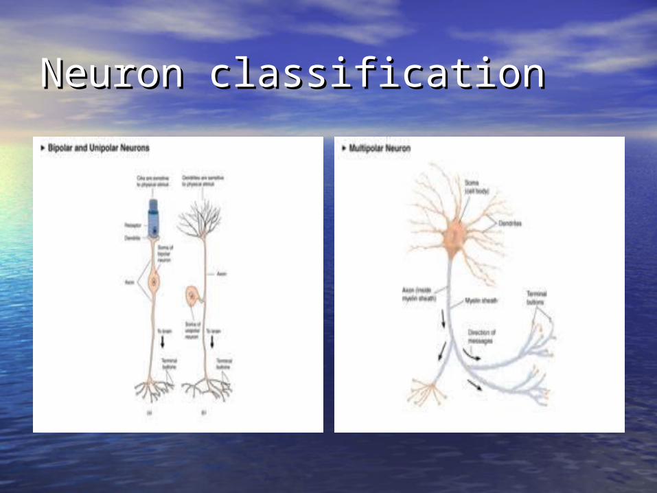
Neuron classificationNeuron classification
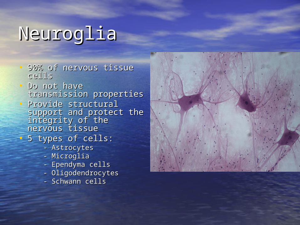
NeurogliaNeuroglia
• 90% of nervous tissue 90% of nervous tissue cellscells
• Do not have transmission Do not have transmission propertiesproperties
• Provide structural support Provide structural support and protect the integrity and protect the integrity of the nervous tissueof the nervous tissue
• 5 types of cells:5 types of cells: - Astrocytes- Astrocytes - Microglia- Microglia - Ependyma cells- Ependyma cells - Oligodendrocytes- Oligodendrocytes - Schwann cells- Schwann cells

AstrocytesAstrocytes
• Provide physical support Provide physical support for neuronsfor neurons
• Control movements of Control movements of nutrients and wastes to nutrients and wastes to and from neuronsand from neurons
• Help recycle and process Help recycle and process some neurotransmitterssome neurotransmitters
• Play a role in the Blood-Play a role in the Blood-Brain Barrier (BBB)Brain Barrier (BBB)
• Play a role in synapse Play a role in synapse formationformation
• Maintain constant brain Maintain constant brain ECFECF

Blood-brain barrierBlood-brain barrier
• Protects the brain from Protects the brain from “external” influences“external” influences
• Inability of some Inability of some compounds to cross from compounds to cross from the blood into the brainthe blood into the brain
• Due to tight junctions Due to tight junctions between endocytes of he between endocytes of he capillary wallcapillary wall
• Maybe due to astrocytesMaybe due to astrocytes• Glucose, gases can passGlucose, gases can pass• Some medications and Some medications and
other compounds can not other compounds can not passpass

MicrogliaMicroglia
• Derived from Derived from macrophagesmacrophages
• Defense of the Defense of the brainbrain
• Clean up old, sick Clean up old, sick and dead cellsand dead cells
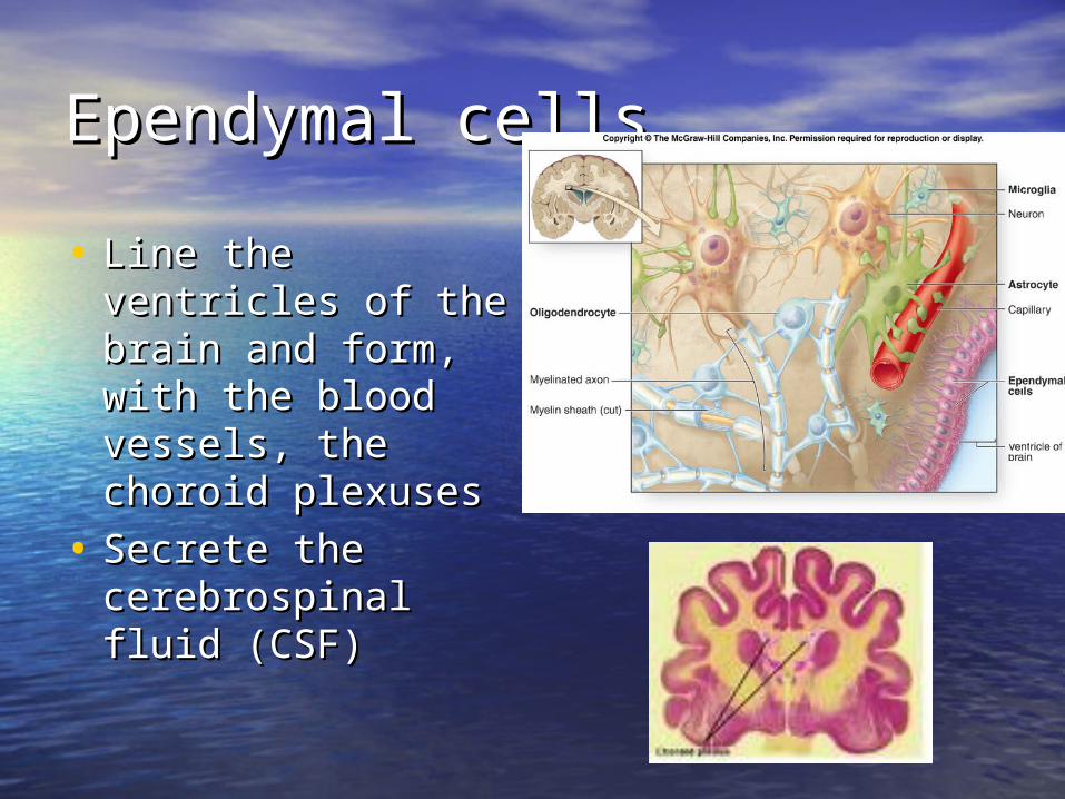
Ependymal cellsEpendymal cells
• Line the ventricles Line the ventricles of the brain and of the brain and form, with the form, with the blood vessels, the blood vessels, the choroid plexuseschoroid plexuses
• Secrete the Secrete the cerebrospinal fluid cerebrospinal fluid (CSF)(CSF)
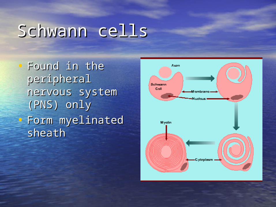
Schwann cellsSchwann cells
• Found in the Found in the peripheral nervous peripheral nervous system (PNS) onlysystem (PNS) only
• Form myelinated Form myelinated sheathsheath

Myelin sheath and Schwann Myelin sheath and Schwann cellcell

OligodendrocytesOligodendrocytes
• Present in the Present in the white matter of the white matter of the CNS onlyCNS only
• One cell sends One cell sends cytoplasmic cytoplasmic extensions extensions containing myelin containing myelin toward several toward several neuronsneurons
• Each extension Each extension wraps around a wraps around a portion of the portion of the neuronal axonneuronal axon

Neuron physiologyNeuron physiology
1- Resting membrane potential1- Resting membrane potential
2- Action potential2- Action potential
3- Graded potential3- Graded potential
- Excitatory potential- Excitatory potential
- Inhibitory potential- Inhibitory potential

Neuron physiologyNeuron physiology
1- Resting membrane potential1- Resting membrane potential
2- Action potential2- Action potential
3- Graded potential3- Graded potential
- Excitatory potential- Excitatory potential
- Inhibitory potential- Inhibitory potential

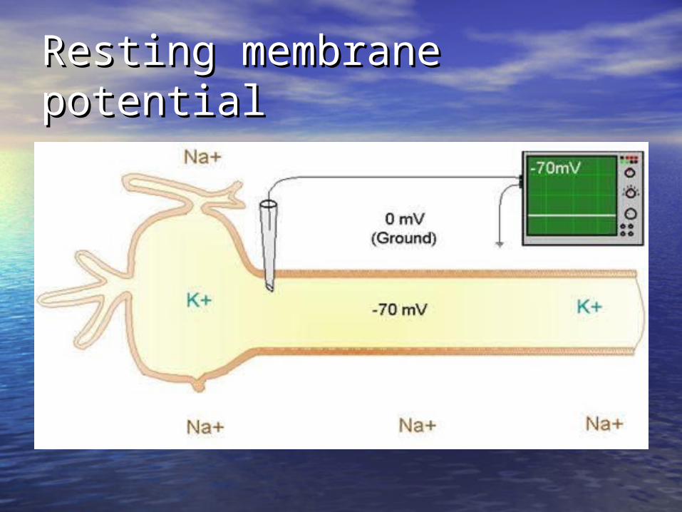
Resting membrane potentialResting membrane potential
Due to:Due to:• a large number of sodium ions located a large number of sodium ions located
outside of the celloutside of the cell• A large number of potassium ions located A large number of potassium ions located
inside the cellinside the cell• Large amount of proteins inside the cell Large amount of proteins inside the cell
gives it a negative charge while the gives it a negative charge while the outside is positively chargedoutside is positively charged
• The difference between the 2 sides of the The difference between the 2 sides of the cell membrane is -70 mV cell membrane is -70 mV
Resting membrane potentialResting membrane potential
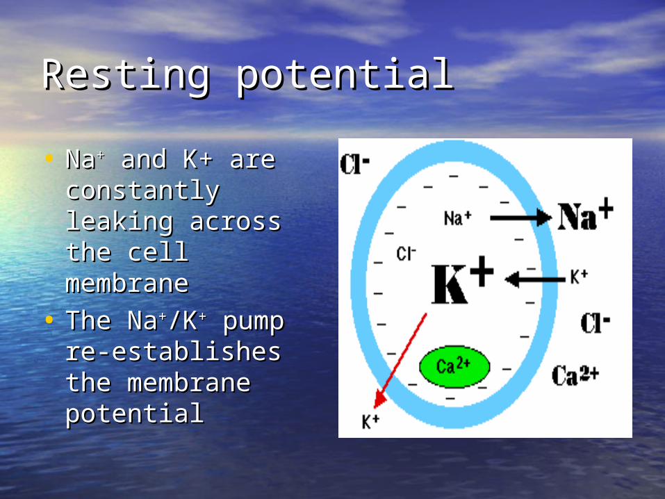
Resting potentialResting potential
• NaNa++ and K+ are and K+ are constantly leaking constantly leaking across the cell across the cell membranemembrane
• The NaThe Na++/K/K++ pump pump re-establishes the re-establishes the membrane membrane potentialpotential

Neuron physiologyNeuron physiology
1- Resting membrane potential1- Resting membrane potential
2- Action potential2- Action potential
3- Graded potential3- Graded potential
- Excitatory potential- Excitatory potential
- Inhibitory potential- Inhibitory potential
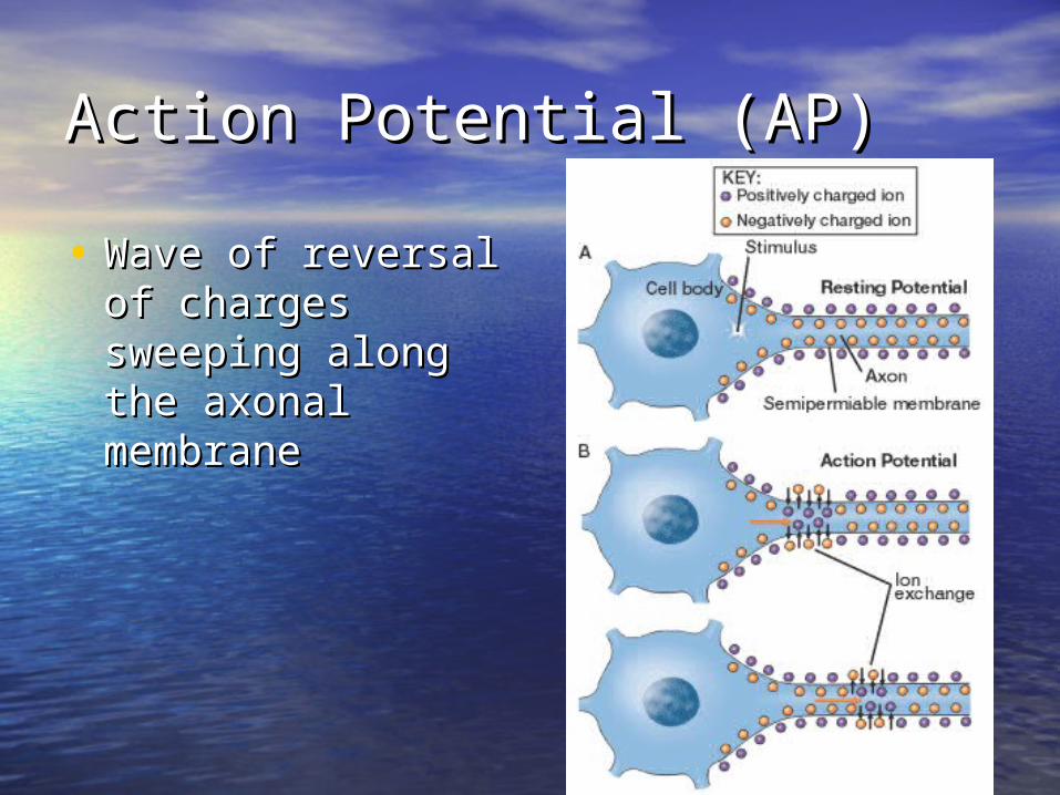
Action Potential (AP)Action Potential (AP)
• Wave of reversal of Wave of reversal of charges sweeping charges sweeping along the axonal along the axonal membranemembrane

Action PotentialAction Potential
• Initiated at the hillock of the neuronInitiated at the hillock of the neuron
• The hillock “sums up” the information The hillock “sums up” the information coming from the dendrites and neuronal coming from the dendrites and neuronal body.body.
• ““Summing up” for a neuron means adding Summing up” for a neuron means adding up all excitatory and inhibitory impulses. up all excitatory and inhibitory impulses. If the membrane potential (the sum) If the membrane potential (the sum) reaches threshold (-55 mV) or above), an reaches threshold (-55 mV) or above), an AP is triggeredAP is triggered
Action PotentialAction Potential
• The difference in potential activates The difference in potential activates voltage gated channels voltage gated channels channels channels that open and close as a function of that open and close as a function of the current, regional membrane the current, regional membrane potentialpotential

Action PotentialAction Potential
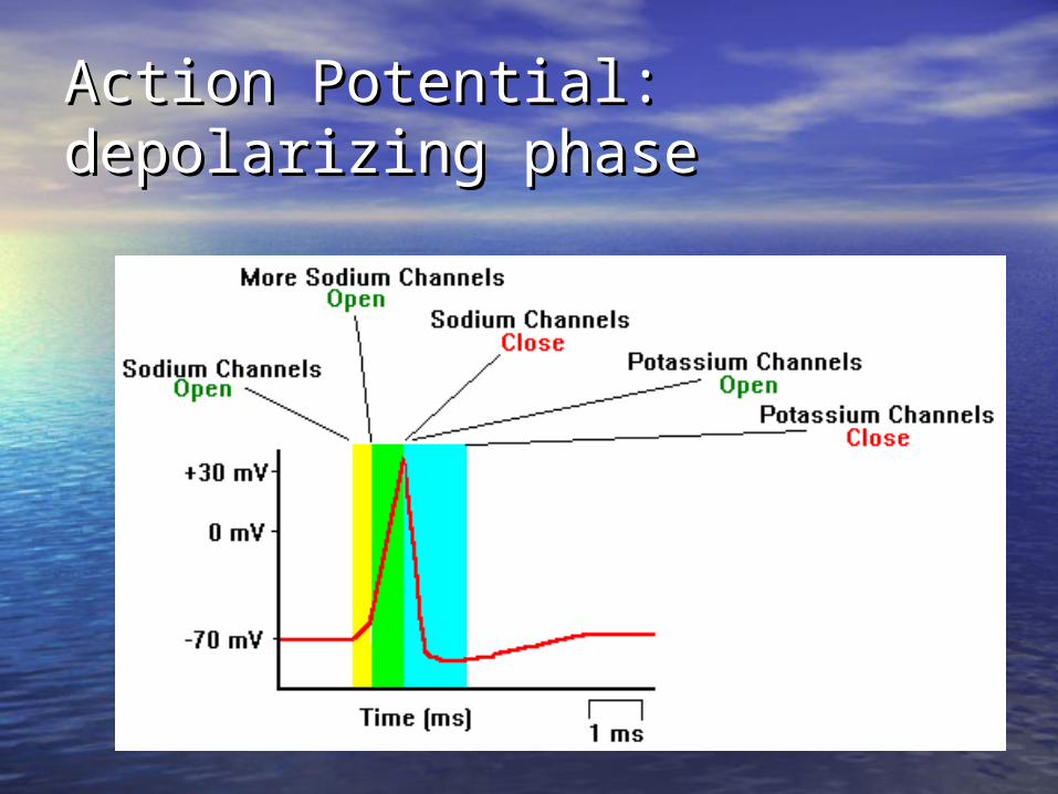
Action Potential: depolarizing Action Potential: depolarizing phasephase

Action PotentialAction Potential
• All-or None effect: Once the All-or None effect: Once the threshold is reached, the AP is threshold is reached, the AP is triggered. All APs are identical.triggered. All APs are identical.

Action Potential: Refractory Action Potential: Refractory periodsperiods
• Absolute refractory Absolute refractory period: all the period: all the depolarizing phase + depolarizing phase + most of repolarizing most of repolarizing phase:phase:
• Relative refractory Relative refractory period: end of period: end of repolarization until the repolarization until the Na/K pump reestablish Na/K pump reestablish resting state resting state
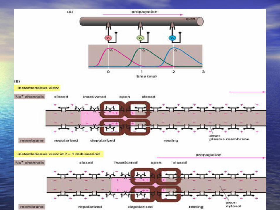
Action PotentialAction Potential
• Note: the AP travels always in 1 direction, Note: the AP travels always in 1 direction, from the soma toward the axonal bulbfrom the soma toward the axonal bulb
• The AP cannot travel toward the soma The AP cannot travel toward the soma because of the presence of the refractory because of the presence of the refractory period: the voltage-gated sodium channels period: the voltage-gated sodium channels are still closed (because of the inner gate) are still closed (because of the inner gate) while the neighboring channels are being while the neighboring channels are being activated and conduct the AP down the activated and conduct the AP down the axonaxon

Neuron physiologyNeuron physiology
1- Resting membrane potential1- Resting membrane potential
2- Action potential2- Action potential
3- Graded potential3- Graded potential
- Excitatory potential- Excitatory potential
- Inhibitory potential- Inhibitory potential

Graded PotentialsGraded Potentials
- They are small changes in membrane - They are small changes in membrane potential across the cell membranepotential across the cell membrane
- They occur on the dendrites and the - They occur on the dendrites and the neuron somaneuron soma
- They are controlled by ligand-gated - They are controlled by ligand-gated channelschannels

• Some compounds such as novocaine Some compounds such as novocaine and tetrodotoxin (TTX) from the and tetrodotoxin (TTX) from the puffer fish bind to voltage gated puffer fish bind to voltage gated sodium channels.sodium channels.
• What will be the consequences?What will be the consequences?

Ligand gated channelsLigand gated channels
• The ligand binding to these channels The ligand binding to these channels are various neurotransmitters are various neurotransmitters present in the NSpresent in the NS
• Ligand binding is specific and related Ligand binding is specific and related to the receptor shapeto the receptor shape
• Receptors for ligand A usually cannot Receptors for ligand A usually cannot bind to ligand Bbind to ligand B

Excitatory post synaptic Excitatory post synaptic potential (EPSP)potential (EPSP)
• A sodium channel opens in response A sodium channel opens in response to the binding of a ligand on its to the binding of a ligand on its receptor.receptor.
• Sodium ions move inside the cell, Sodium ions move inside the cell, therefore depolarizing ittherefore depolarizing it
• The amount of sodium entering the The amount of sodium entering the cell is not enough for the membrane cell is not enough for the membrane to reach thresholdto reach threshold
EPSPEPSP
• Any time a positively charged ions Any time a positively charged ions (cations) move into the cell, the (cations) move into the cell, the membrane depolarizes. Any other membrane depolarizes. Any other ECF cations will have the same effectECF cations will have the same effect
• Negatively charged ions (anions) Negatively charged ions (anions) from the ICF moving toward the ECF from the ICF moving toward the ECF will have the same effect (excitation)will have the same effect (excitation)

Inhibitory post synaptic Inhibitory post synaptic potential (IPSP)potential (IPSP)
• Ligand gated potassium channels open Ligand gated potassium channels open in response to the binding of a ligand in response to the binding of a ligand on its receptor. on its receptor.
• Potassium ions leave the cell Potassium ions leave the cell the the membrane potential become more membrane potential become more negative (-90mV) negative (-90mV) it is moving away it is moving away from the threshold from the threshold it is even more it is even more difficult to depolarize the membrane difficult to depolarize the membrane inhibitioninhibition
IPSPIPSP
• Any positive charged ions moving Any positive charged ions moving from the cell toward the IF will have from the cell toward the IF will have an inhibitory effectan inhibitory effect
• Any negatively charged ions moving Any negatively charged ions moving from the IF toward the cell will also from the IF toward the cell will also have an inhibitory effect (ex: chloride have an inhibitory effect (ex: chloride ions)ions)

SummationSummation
• The impulses arriving at the neuron The impulses arriving at the neuron are summed at the hillock. If the are summed at the hillock. If the result produce a voltage greater than result produce a voltage greater than the threshold, an AP will be triggeredthe threshold, an AP will be triggered

Temporal summationTemporal summation
• Two impulses must arrive at the same Two impulses must arrive at the same location at very close time intervalslocation at very close time intervals
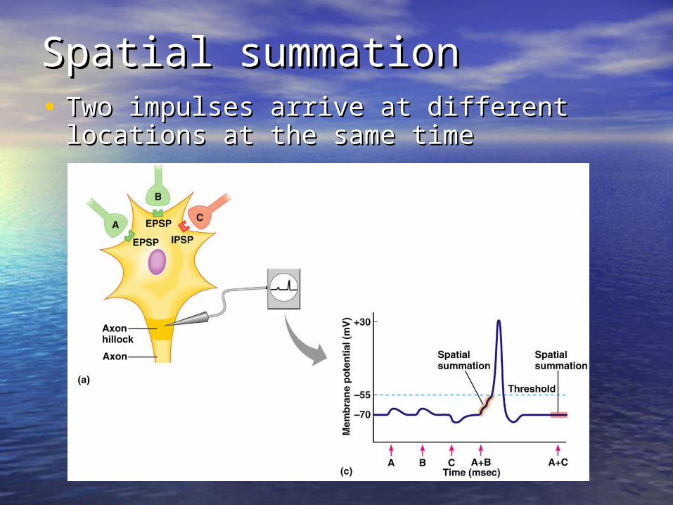
Spatial summationSpatial summation• Two impulses arrive at different locations Two impulses arrive at different locations
at the same timeat the same time

EPSP & IPSP & EPSP & IPSP & summationsummation
Graded potentialsGraded potentials
• Immediately after a graded potential, Immediately after a graded potential, Na/K pumps reestablish resting Na/K pumps reestablish resting membrane potentialmembrane potential

Impulse conductionImpulse conduction
• Saltatory conduction along the Saltatory conduction along the myelinated axonsmyelinated axons
• Continuous conduction along the Continuous conduction along the unmyelinated axonsunmyelinated axons

Saltatory Saltatory conductionconduction
+
+
+
+
+
+
+
+
+
+
+
+
+
+
+
+
+
+
+
+
+
+
+
+
+
+
+
+
+
+
+
+
Myelinated axon
Myelinsheath
Node ofRanvier
Extracellular fl uid
Direction of action potential propagation
Intracellular fl uid
– –
+ + +
+ + +
+ + + + + +
– +
–
+
–
+
–
– – –
– – –
–
–
– – –
– – –
– – –
– – –
– – –
– – –
– – –
– – –
–
–
–
–
–
– – – –
+
–
+
–
+
–
+
+
+
+
+
+
+
+
+
+
+
+
+
+
+
+
+
+
+
+
+
+
+
+
+
+
+
+
+
+
+
+
– –
+ + +
+ + +
+ + +
+ + +
– + + +
– – –
– – –
– – –
–
–
– – –
– – –
– – –
– – –
– – –
– – –
– – –
– – –
–
–
–
–
–
– – – –
+
–
+
–
+
–
Extracellular fl uid
Axon hillock

Continuous Continuous conductionconduction
• Along the axonAlong the axon
• In one direction In one direction only (due to the only (due to the refractory period)refractory period)
Unmyelinated axon
++
+ +
+
+
+ + + + + + + + + + + + + + +
+ + + + + + + + + + + + + + +
Extracellular fluid
Extracellular fluid
RestingExtracellular fluid
Plasmamembrane
Intracellular fluid
– – – – – – – – – – – – – – –
– – – – – – – – – – – – – – –
–
++
+ +–– – –
––
– – –
++
+ +
+
+
+ + +
+ + + + + + + + + + + +
+ + +
+ + + + + + + + + + + +
Initiation
Site oforiginalactionpotential
–– – –
– – – – – – – – – – – –
–– – –
– – – – – – – – – – – –
++
+ +–– – –
–– – –
Site A
Region ofdepolarization
Site B
Axon hillock
Direction of action potential propagation
++
+ +
+ + + +
+ + + + + + + +
+ + + +
+ + + + + + + +
Propagation
– – – –
– – – – – – – –
– – – –
– – – – – – – –
++
+ +–– – –
––
– –
Site A
Refractorystate
Site B Site C
+
+
+ + +
+ + +
– – –
– – –
–
–
++
+ +
+ + + +
+ + + + + + + +
+ + + +
+ + + + + + + +
– – – –
– – – – – – – –
– – – –
– – – – – – – –
++
+ +–– – –
–– – –
Site A
Refractorystate
Site B Site C
Region ofrepolarization(resting state)
Site D
+
+
+ + +
+ + +
– – –
– – –
–
–
Region ofdepolarization
Region ofdepolarization
Propagationcontinues
(a)
(b)
(c)
(d)
Factors affecting the speed of Factors affecting the speed of AP transmissionAP transmission
• State of myelination of the axon – State of myelination of the axon –
Myelinated axon transmit much Myelinated axon transmit much
faster than unmyelinated axonsfaster than unmyelinated axons
• The size of the axon: the larger the The size of the axon: the larger the axon, the faster the APaxon, the faster the AP

Readings:Readings:
• Chp. 7: p. 167-195Chp. 7: p. 167-195
• Chp. 8, p. 197-213Chp. 8, p. 197-213
• Clinical connections: p. 180, p. 193, Clinical connections: p. 180, p. 193, p. 210, p. 212.p. 210, p. 212.
• Not expected: Toolbox, p.174, p. Not expected: Toolbox, p.174, p. 178, p. 192, 178, p. 192,
Related Documents