Nervous system by Dr. Dalia Mohamed Ali

Nervous system
Jan 01, 2016
Nervous system. by Dr. Dalia Mohamed Ali. Divisions of the Nervous System. The nervous system (N.S) is divided into two main parts : 1) The central nervous system(C.N.S ) 2) The peripheral nervous system(P.N.S). The peripheral nervous system. - PowerPoint PPT Presentation
Welcome message from author
This document is posted to help you gain knowledge. Please leave a comment to let me know what you think about it! Share it to your friends and learn new things together.
Transcript
Nervous system
by Dr. Dalia Mohamed
Ali

2
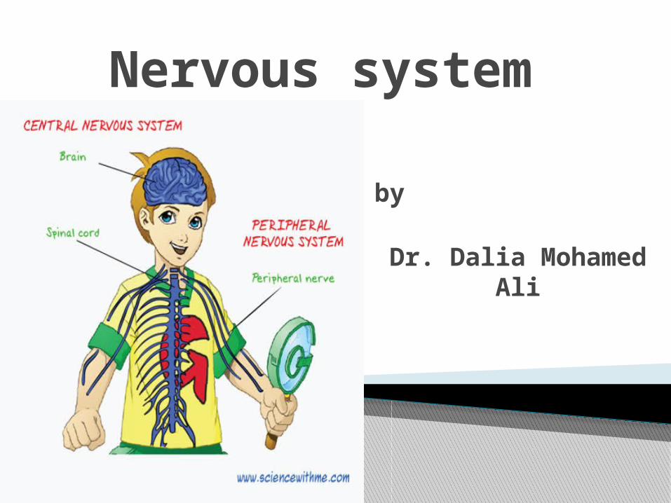
The nervous system (N.S) is divided into two main parts:
1) The central nervous system(C.N.S)
2) The peripheral nervous system(P.N.S)
Divisions of the Nervous System

3
It is formed of:- Cranial nerves arise from brain- Spinal nerves arise from spinal cord It is divided functionally into :a-Somatic nervous system : controls skeletal muscles .
b-Autonomic nervous system :Controls smooth muscle ,heart &glands (sympathetic & parasympathetic) .
The peripheral nervous system

1-somatic nervous system
2-Autonomic nervous system
The somatic nervous system is concerned with somatic functions.
It includes the nerves supplying the skeletal muscles.
Controls the voluntary movements of the body by acting on the skeletal muscles .
The autonomic nervous system is concenrned with regulation of visceral or vegetative functions.
involuntary nervous system.
The autonomic nervous system consists of two divisions:
* sympathetic division * parasympathetic
division.
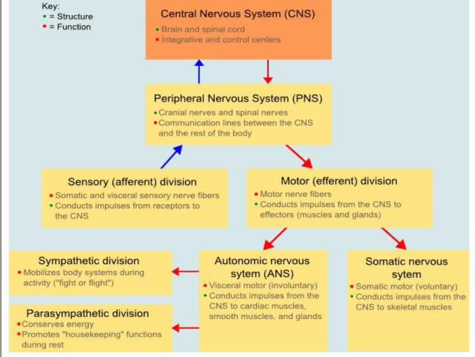
It includes: -Brain -Spinal cord
It is formed of:-Highly specialized nerve cells (neurons)-Supporting cells (neuralgia)
The structures of brain and spinal cord are arranged in two layers: the gray matter and white matter.
In brain the white matter is centrally placed and gray matter is in the outer part.
In spinal cord white matter is in the outer part and gray matter is in inner part.
Central nervous system


Brain is situated in the skull.
It is continued as spinal cord in the vertebral canal through the foramen magnum of the skull bone.
Brain and spinal cord are surrounded by three layers of meninges:
• the outer dura mater• middle arachnoid • inner pia mater.
The space between the arachnoid mater and pia mater is known as subarachnoid space.
This space is filled with a fluid called cerebrospinal fluid (CSF).
The brain and spinal cord are actually suspended in CSF.

9

10
It is formed of : a) cerebrum ( the 2 cerebral
hemispheres + the interbrain). b) Brain stem: which includes:
◦Midbrain ( upper part)◦Pons (middle part)◦medulla oblongata (lower part)
c)cerebellum
A) The brain

11
12
The functional & structural unit of the Nervous System is the neuron.
Neuron is called the nerve cellNerve cell body: Irregular in shape mass of cytoplasm called neuroplasm which is covered by a cell membrane.
The cytoplasm contains a large nucleus, Nissel bodies, neurofibrils, mitochondria and Golgi apparatus.
Nissel bodies and neurofibrils are found only in nerve cell and not in other cell
Neuron & Neurolgia
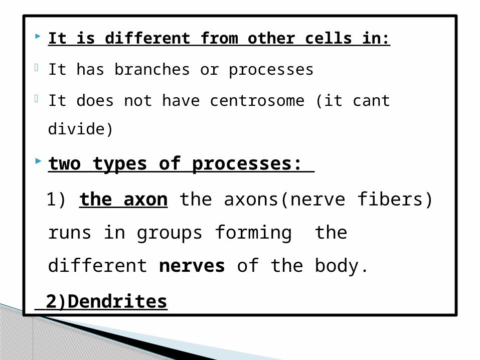
It is different from other cells in:
- It has branches or processes
- It does not have centrosome (it cant divide)
two types of processes:
1) the axon the axons(nerve fibers) runs in
groups forming the different nerves of
the body.
2)Dendrites
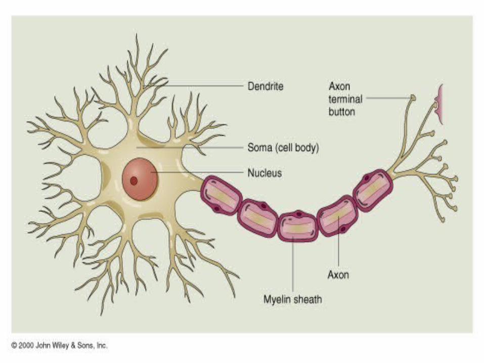

1) Axon
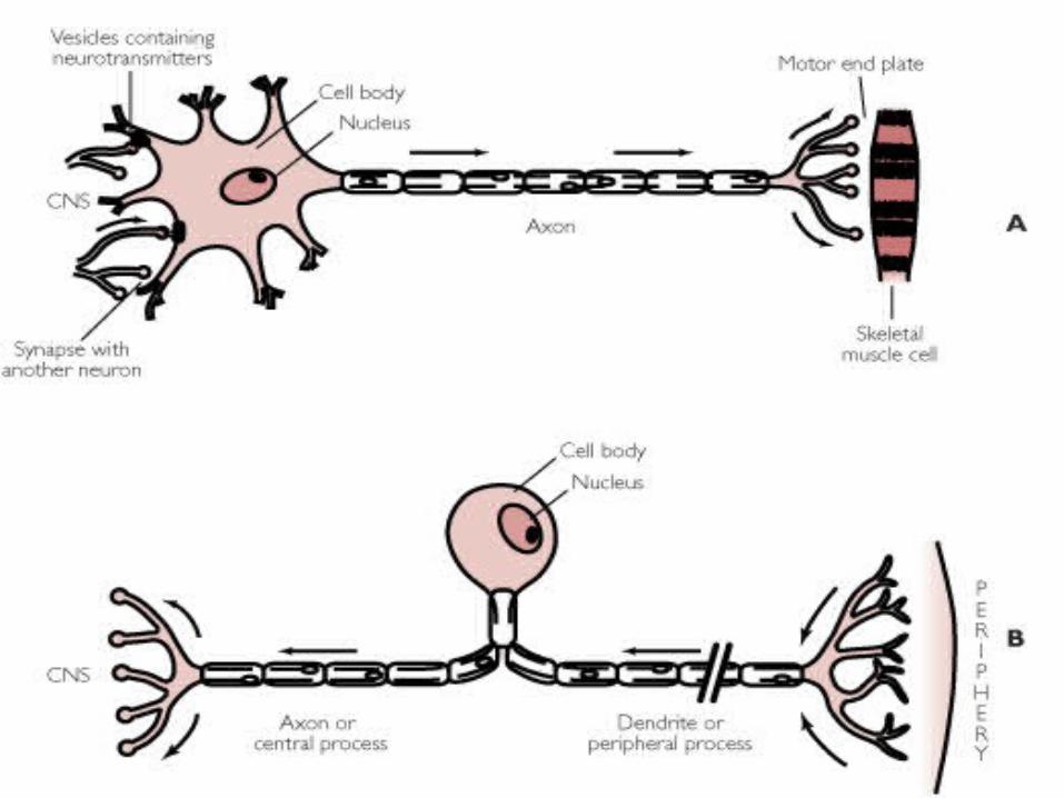
• single• the longest process of the cell body • carries nervous impulses away from the cell body. out side the CNS• the axons(nerve fibers) runs in groups (fasciculus) forming the nerves of the
body
2)Dendrites
• are multiple• the short processes of the cell body• which carry impulses towards the nerve cell body

Organization of nerve : Many axons together form a bundle called
fasciculus. many fasciculi together form a nerve.

Myelin sheath myelin sheath is a thick lipoprotein sheath that insulates the
nerve fiber. it is absent at regular intervals. myelin sheath is responsible for the white color of the nerve
fibers
-Myelinated nerve fiberThe nerve fibers which are insulated by myelin sheath.
-Nonmyelinated nerve fiberThe nerve fiber is not covered by myelin sheath.
Function of Myelin sheath:1- Faster conduction faster conduction of impulse2- Insulating capacityrestricts the nerve impulse within the
single nerve fiber

18

Classification of neuron
I- depending on number of poles: 1- unipolar neuronshave only one pole from which both the axon
and dendrite aries
2- Bipolar neuronshave two poles.axon arises from one pole and
dendrites aries from the other pole
3- Multipolar neuronshave many poles. One of the poles gives rise to
the axon and all the other poles give rise to dendrites
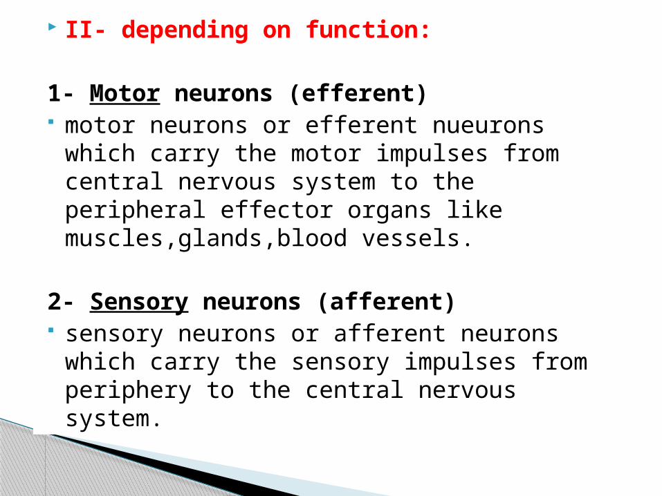
II- depending on function:
1- Motor neurons (efferent) motor neurons or efferent nueurons which
carry the motor impulses from central nervous system to the peripheral effector organs like muscles,glands,blood vessels.
2- Sensory neurons (afferent) sensory neurons or afferent neurons which
carry the sensory impulses from periphery to the central nervous system.
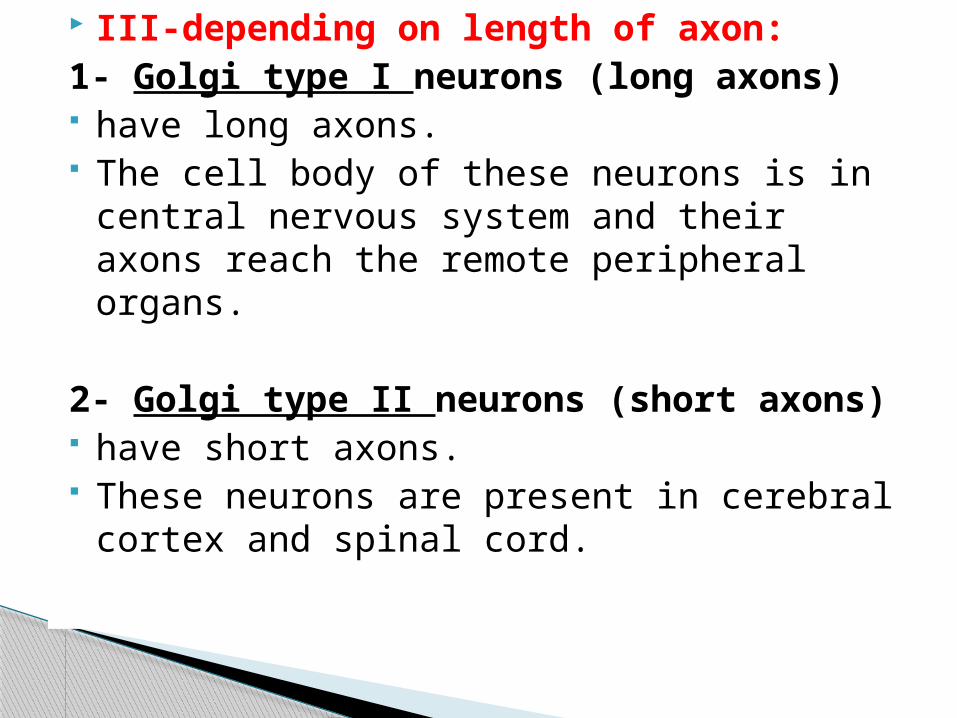
III-depending on length of axon:1- Golgi type I neurons (long axons) have long axons. The cell body of these neurons is in central
nervous system and their axons reach the remote peripheral organs.
2- Golgi type II neurons (short axons) have short axons. These neurons are present in cerebral
cortex and spinal cord.

I- depending on structure:-Myelinated nerve fibers-NonMyelinated nerve fibers II-depending on distribution-somatic nerve fibers-autonomic nerve fibers III-depending on origin:-cranial nerve: arise from brain-spinal nerve: arise from spinal cord
Classification of nerve fibers:

IV-depending on function:1-sensory or afferent nerve fibers which carry
sensory impulses from different parts of the body to the central nervous system
2-motor or efferent nerve fibers which carry motor impulses from central nervous system to different parts of the body.
V-depending on neurotransmitter:1. Adrenergic nerve fibers that secrete
noradrenaline 2. cholinergic nerve fibers that secrete
acetylcholine
ExcitabilityConductivityRefractory period: -absolute RP -relative RPAdaptationSummation Infatigability
Properties of nerve fiber

Excitabilitythe physiochemical change that occurs in a
tissue when a stimulus is applied
Conductivitythe ability of nerve fibers to transmit the impulse
from the area of stimulation to the other areas
Refractory periodthe period at which the nerve does not give any
response to a stimulus
-absolute RP-relative RP

Adaptation While stimulating a nerve fiber the excitability of
the nerve fiber is greater in the beginning. continuous stimulation Later the response
decreases slowly and finally the nerve fiber does not show any response at all
Infatigability A nerve fiber cannot be fatigued, even if it is stimulated
continuously for a long time
Summation When one subliminal stimulus is applied it does not
produce any response in the nerve fiber . if two or more subliminal stimuli are applied within a short
interval the response is produced

Neuroglia or glia cell is supporting cells It is nonexcitable not transmit impulses Classification:1- central glia cells:-astrocytes-microglia-oligodendrocytes2- peripheral glia cells:-schwan cells-satellite cells
Neuroglia
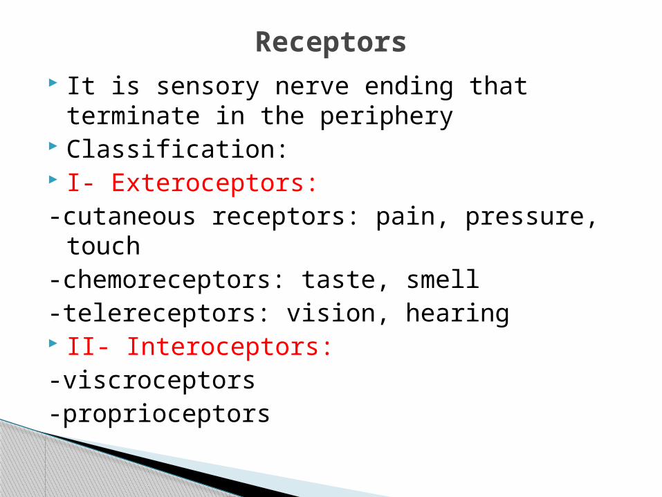
It is sensory nerve ending that terminate in the periphery
Classification: I- Exteroceptors:-cutaneous receptors: pain, pressure, touch-chemoreceptors: taste, smell-telereceptors: vision, hearing II- Interoceptors:-viscroceptors-proprioceptors
Receptors
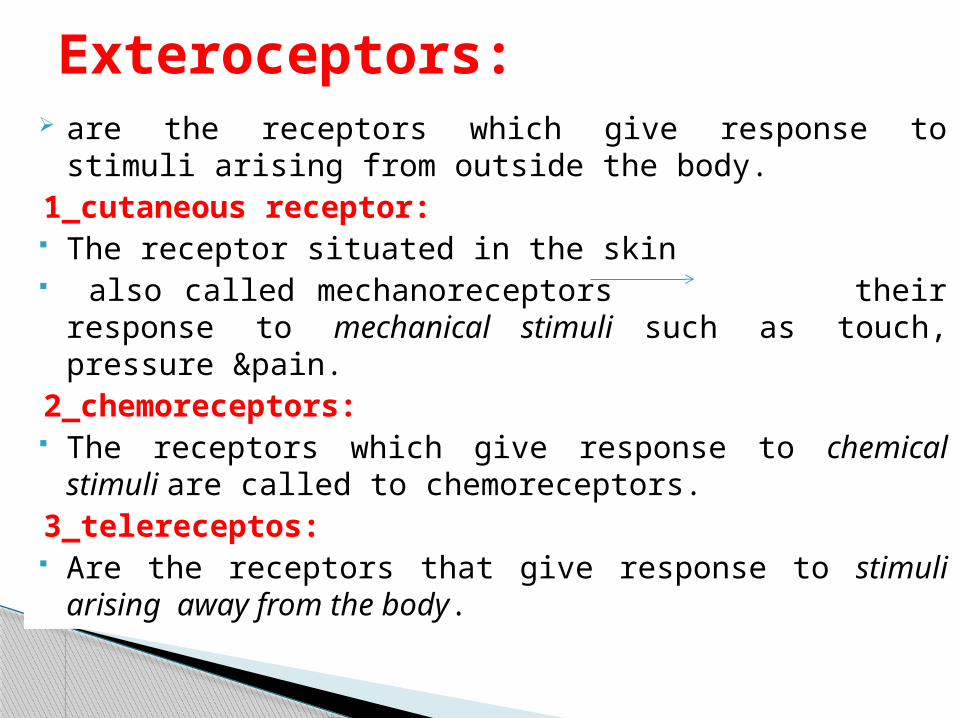
are the receptors which give response to stimuli arising from outside the body.
1_cutaneous receptor: The receptor situated in the skin also called mechanoreceptors their response
to mechanical stimuli such as touch, pressure &pain.2_chemoreceptors: The receptors which give response to chemical
stimuli are called to chemoreceptors.3_telereceptos: Are the receptors that give response to stimuli
arising away from the body.
Exteroceptors:
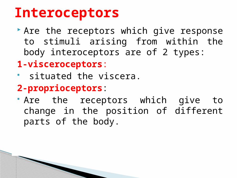
Are the receptors which give response to stimuli arising from within the body interoceptors are of 2 types:
1-visceroceptors: situated the viscera.2-proprioceptors: Are the receptors which give to change in
the position of different parts of the body.
Interoceptors

Exteroceptors
Chemoreceptors
Taste buds
smell
olfactory
receptors
telereceptors
Vision
rods and
cones in
retina
Hearing
hair cells
in organ
of corti
cutaneous receptors
Touch
Meisnner's
pacinian corpuscle
Pain
free nerve ending nociceptor
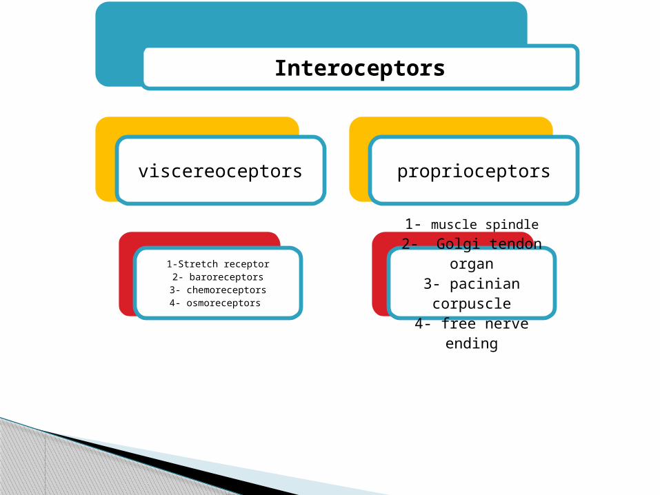
Interoceptors
viscereoceptors
1-Stretch receptor2- baroreceptors
3- chemoreceptors4- osmoreceptors
proprioceptors
1- muscle spindle2- Golgi tendon
organ3- pacinian corpuscle
4- free nerve ending
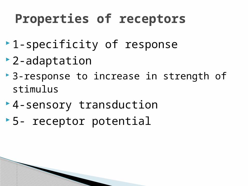
1-specificity of response 2-adaptation 3-response to increase in strength of stimulus 4-sensory transduction 5- receptor potential
Properties of receptors

1-specificity of response the response given by a particular types of
receptor to a specific sensation.
2-adaptation It is the decrease in the response when a
receptor is stimulated continuously with constant strength.
Properties of receptors
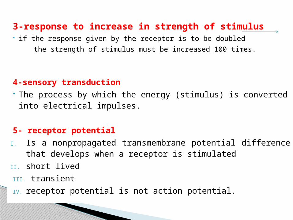
3-response to increase in strength of stimulus if the response given by the receptor is to be doubled the strength of stimulus must be increased 100 times.
4-sensory transduction The process by which the energy (stimulus) is converted
into electrical impulses.
5- receptor potentialI. Is a nonpropagated transmembrane potential
difference that develops when a receptor is stimulated II. short livedIII. transient IV. receptor potential is not action potential.

Synapse: It is the junction between the two neurons.Classification:I- Anatomical:1-axaoaxnic synapse in which axon of one neuron terminates on axon
of another neuron.2-axodendritic synapse in which axon of one neuron terminates on
dendrite of another neuron.3-axosomatic synapse in which axon of one neuron ends on soma(cell
body)of another neuron.
Synapse

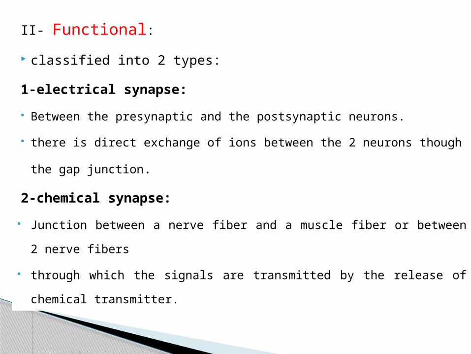
II- Functional:
classified into 2 types:
1-electrical synapse:
Between the presynaptic and the postsynaptic neurons.
there is direct exchange of ions between the 2 neurons
though the gap junction.
2-chemical synapse:
Junction between a nerve fiber and a muscle fiber or between
2 nerve fibers
through which the signals are transmitted by the release of
chemical transmitter.
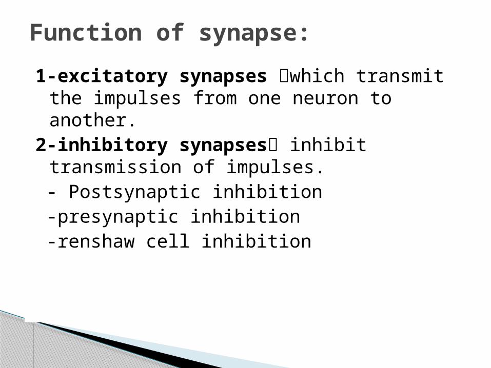
1-excitatory synapses which transmit the impulses from one neuron to another.
2-inhibitory synapses inhibit transmission of impulses.
- Postsynaptic inhibition -presynaptic inhibition -renshaw cell inhibition
Function of synapse:
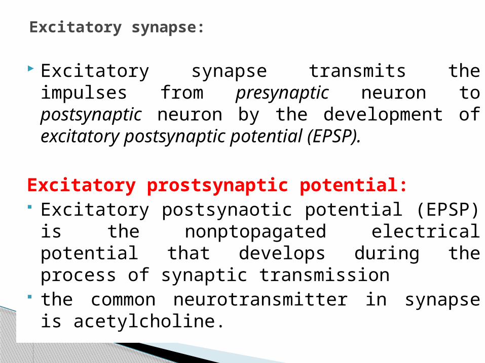
Excitatory synapse transmits the impulses from presynaptic neuron to postsynaptic neuron by the development of excitatory postsynaptic potential (EPSP).
Excitatory prostsynaptic potential: Excitatory postsynaotic potential (EPSP) is the
nonptopagated electrical potential that develops during the process of synaptic transmission
the common neurotransmitter in synapse is acetylcholine.
Excitatory synapse:

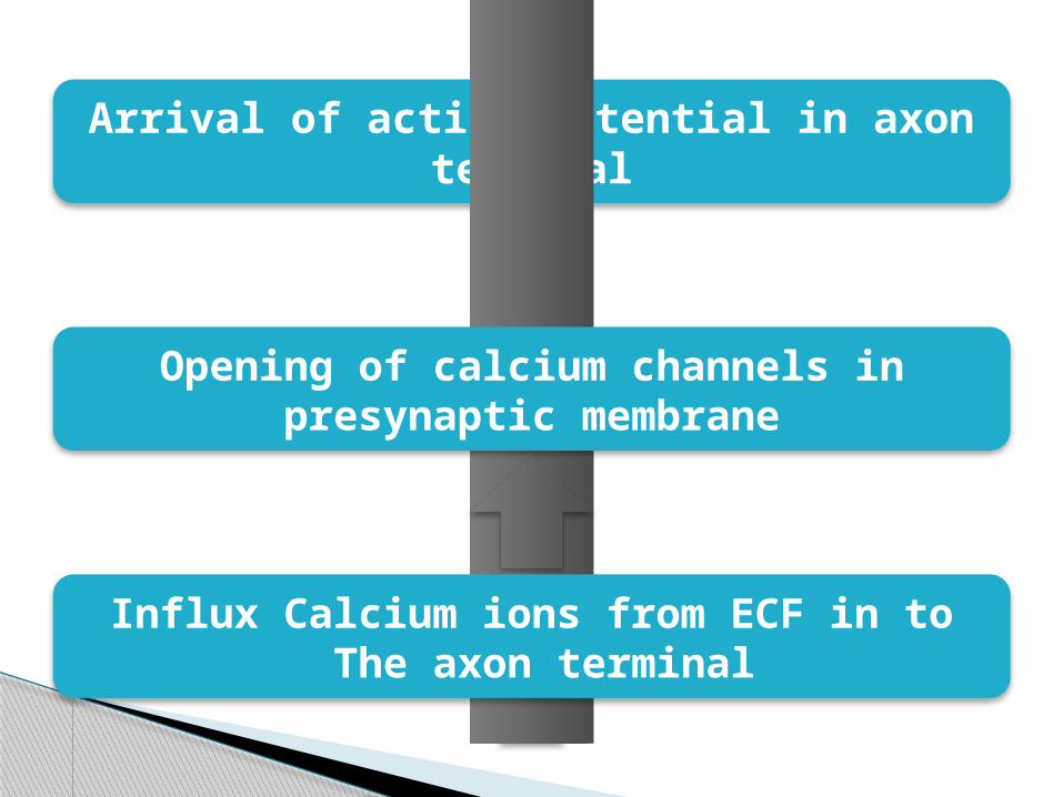
Arrival of action potential in axon terminal
Opening of calcium channels in presynaptic membrane
Influx Calcium ions from ECF in to The axon terminal

Formation of Ach-receptor complex
Opening of sodium channels and influx of sodium ions from ECF
Development of EPSP
Opening of sodium channels in initial segment of axon
Influx of sodium ions from ECF and development of action potential
Spread of action potential.
Passage of Ach through synaptic cleft


Inhibition of synaptic transmission is classified into 3 types:
1-postsynaptic inhibition.2-presynaptic inhibition.3-renshaw cell inhibiton
Inhibitory synapse:
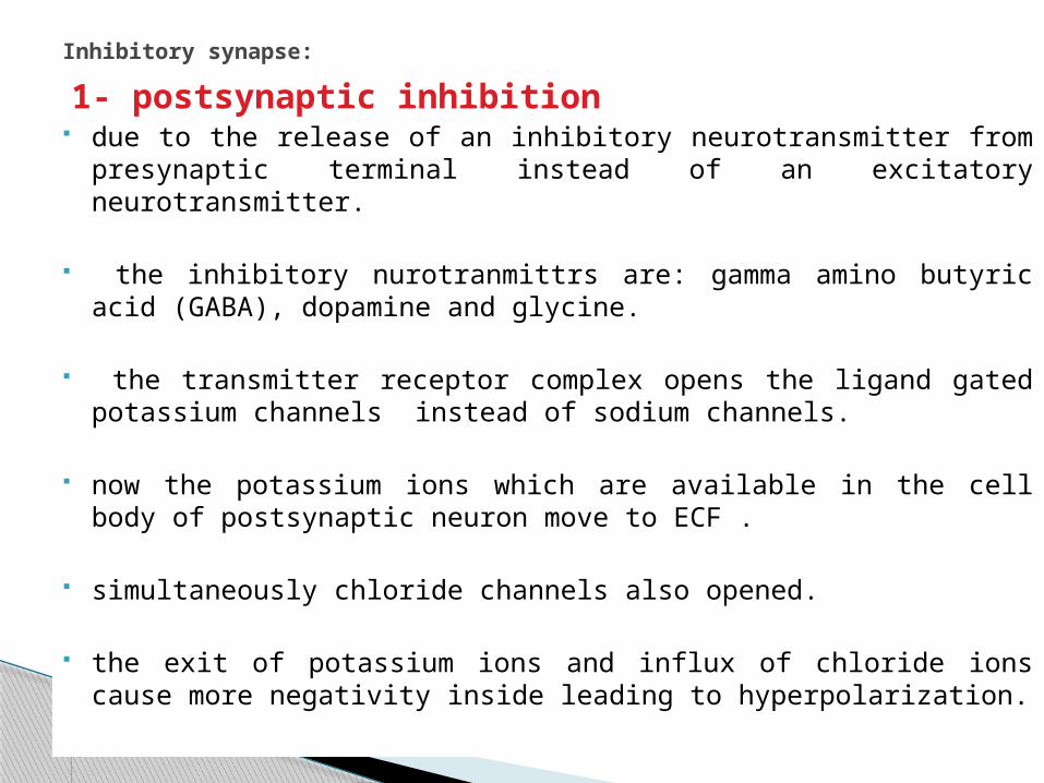
1- postsynaptic inhibition due to the release of an inhibitory neurotransmitter from
presynaptic terminal instead of an excitatory neurotransmitter.
the inhibitory nurotranmittrs are: gamma amino butyric acid (GABA), dopamine and glycine.
the transmitter receptor complex opens the ligand gated potassium channels instead of sodium channels.
now the potassium ions which are available in the cell body
of postsynaptic neuron move to ECF .
simultaneously chloride channels also opened.
the exit of potassium ions and influx of chloride ions cause more negativity inside leading to hyperpolarization.
Inhibitory synapse:
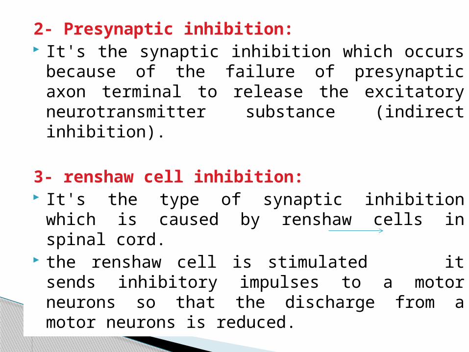
2- Presynaptic inhibition: It's the synaptic inhibition which occurs
because of the failure of presynaptic axon terminal to release the excitatory neurotransmitter substance (indirect inhibition).
3- renshaw cell inhibition: It's the type of synaptic inhibition which is
caused by renshaw cells in spinal cord. the renshaw cell is stimulated it sends
inhibitory impulses to a motor neurons so that the discharge from a motor neurons is reduced.
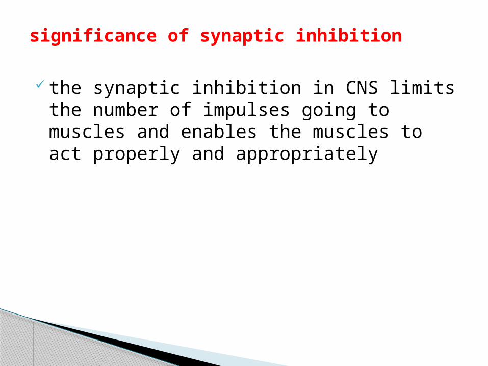
the synaptic inhibition in CNS limits the number of impulses going to muscles and enables the muscles to act properly and appropriately
significance of synaptic inhibition

1-one way conduction : Impulses are transmitted only in one direction
(from presynaptic neuron to postsynaptic neuron).
2-the synaptic delay: Synaptic delay is a short delay that occurs during
the transmission of impulses through the synapse it's due to the time taken for:
i. release of neurotransmitter.ii. passage of neurotransmitter from axon
terminal to postsynaptic membrane.iii. action of the neurotransmitter to open the ionic
channels in postsynaptic membrane.
Properties of synapse
3-fatigue: the fatigue at the synapse is due to the depletion of
neurotransmitter substance acetylcholine.
4-summation: it's the progressive increase in the excitatory postsynaptic
potential(EPSP) in postsynaptic neuron when: * many presynaptic excitatory terminals are stimulated
simultaneously * or when single presynaptic terminal is stimulated
repeatedly.
5-electricale property: The electrical properties of the synapse are the EPSP and
IPSP which are already.

Neurotransmitters: It is chemical substance that acts as the
mediator for the transmission of nerve impulse from one neuron to another through synapse.
Classification:I- depending on chemical nature:1- aminoacids2- amines3- othersII-depending on function:1- excitatory neurotransmitters2- inhibitory neurotransmitters
Neurotransmitters

Excitatory and inhibitory actions:
Inhibitory Excitatory
1-noradrenaline.2-adrenaline.
1-GABA.2-glycine.3-dopamine.
1-acetylcholine.2-histamine.

It is the response to peripheral nervous stimulation that occurs without our consciousness.
Reflex arc:1-Receptor It is the end organ , which receives the stimulus 2-affrent nerve Afferent or sensory nerve transmits sensory impulses from the
receptor to the center 3-center The center is located in the brain or spinal cord 4-Efferent nerve Efferent or motor nerve transmits motor impulses from the center
the effectors organ 5- Effector organ The effector organ is the structure such as the muscle or gland
where the activity occures in response to the stimulus
Reflex activity

Classification of reflexes:I- depending on inborn or acquired-unconditioned reflexes-conditioned reflexesII- depending on site of the center:-cerebellar reflexes-cortical reflexes-midbrain reflexes-Medullary reflexes-spinal reflexes
III- depending on the purpose:-protective reflexes The protective reflexes are the relaxes which protect the body
from nociceptic (harmful) the flexor muscles contract during these reflexes resulting in
flexion at joints-antigravity reflexes The body against the gravitational force the extensor muscles contract during these reflexes resulting
in extension at joints
IV- depending on the number of synapse:-monosynaptic reflexesreflexes having only one synapse in the reflex arc are called
monosynaptic reflexes
-polysynaptic reflexesReflexes having more than one synapse in the reflex are called
polysynaptic reflexes.

V- depending on clinical base:1- superficial reflexes Are the reflex which are elicited from the surface
of the body . superficial reflex are of 2 type mucous
membrane reflex and skin reflex 2-deep reflexes Are elicited from the deeper structures beneath
the skin like tendon 3-visceral reflex Arising from the pupil and the visceral organs.4-pathological reflex Elicited only in pathological condition .

Superficial mucous membrane
response Stimulus Reflex
gagging Irritation of Pharyngeal mucous membrane
Pharyngeal reflex
Raising of Uvular Irritation of Uvular Uvular reflex
Deep reflex
response Stimulus Reflex
Closure of mouth Tapping middle of the chin
Jaw jerk
Extension of leg Percussion of patellar ligament
Knee jerk of patellar tendon reflex

59
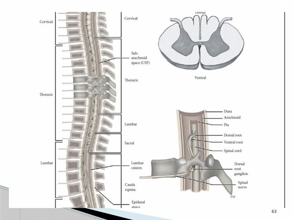
Covered by 3 meninges like the brain.
Contains cavity called the central canal .
It is extends from foramen magnum where it is continuous with medulla oblongata
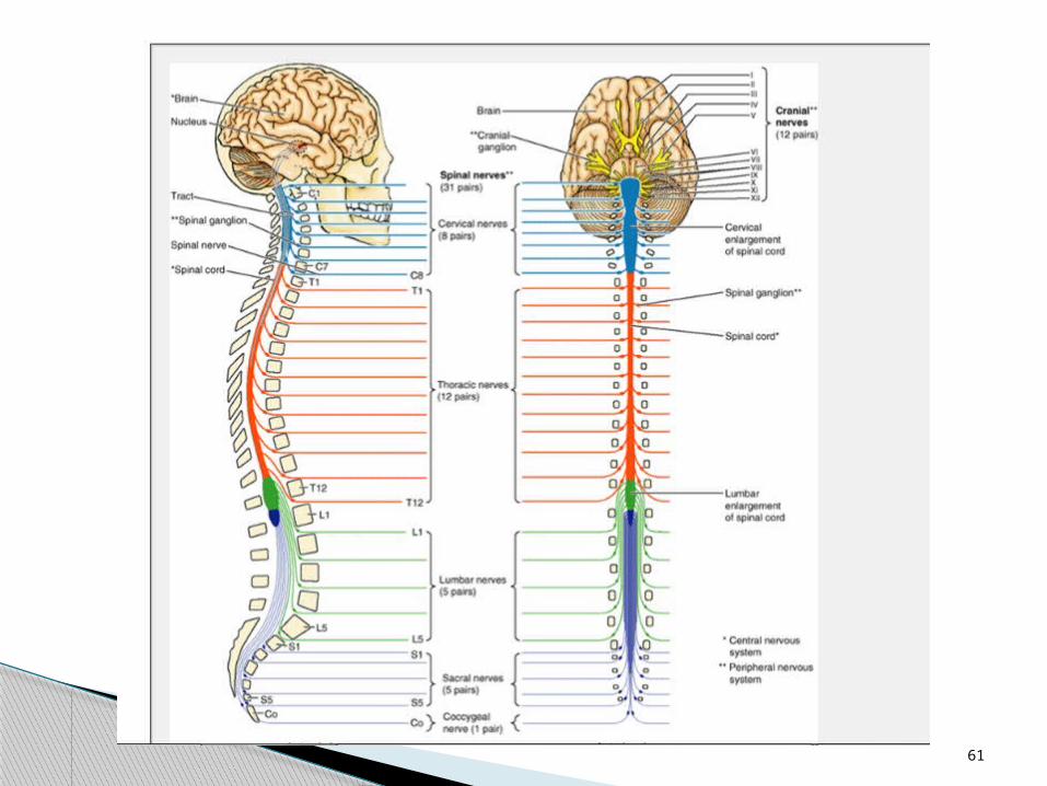
The cord is divided into segments (31):
a-cervical segments: 8b-Thoracic segments :12c-Lumber segments :5d- Sacral segments :5e- Single Coccygeal 1
B) The spinal cord

Spinal nerves The segments Of spinal cord correspond to
the 31 pairs of spinal nerves in a symmetrical manner
Cervical spinal nerves = 8Thoracic spinal nerves = 12 Lumbar spinal nerves = 5 Sacral spinal nerves = 5 Coccygeal nerves = 1
61

62
3 -In cross section ,it shows:a) Grey matter: it is arranged in the shape of butter fly or
like letter H. It projects as: 2 dorsal horns (contain sensory neurons ) & 2 ventral horns ( contain motor neurons) . A third lateral horn is found only in the
segments which give autonomic outflow. B) White matter : surrounds the grey
matter and contains nerve fibers which run
as tracts.
63
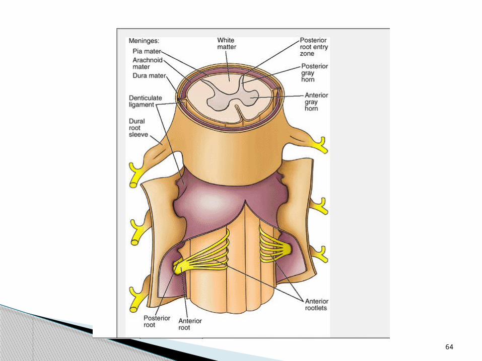
64

Tract in spinal cord Tract of the spinal cord are collection of nerve
fibers passing through the spinal cord. Spinal tract are divided into two main groups :1. Short tract2. Long tractLong tract two type : Ascending tract which carry sensory impulses from the spinal
cord to brain Descending tract which carry motor impulses from brain to spinal
cord

Ascending tract of spinal cord
function tract situation
Touch
Pain and temperature
1. Anterior spinothalamic tract
2. Lateral spinothalamic tract
Anterior white column

Descending tracts of spinal cord The descending Of the spinal cord are
formed by motor nerve fibers arising from brain and descend in to the spinal cord
The descending tracts of spinal cord are two types :
1-pyramidal tracts 2- extrapyramidal tracts

Pyramidal tracts These tracts of spinal cord and concerned with
voluntary motor activities of the body.Function The pyramidal tract are concerned with voluntary
movement of the body. fibers of the pyramidal tract transmit motor impulses
from motor area of cerebral cortex to the anterior motor neurons of the spinal cord .
these two tract are responsible for fine, skilled movements .
The lesion in the neurons of motor cortex and the fibers of pyramidal tract is called the upper motor neuron lesion
Extrapyramidal tract the Descending tract of spinal cord other than
pyramidal tract are called Extrapyramidal tract
somatosensory systemsomatosensory system is defined as sensory system
associated with different parts of the body.
sensations are of two type :1. Somatic sensations2. Special sensationsSomatic sensation : Somatic sensation are sensations arising from skin ,
muscles, tendons and joint . these sensation have specific receptors Special sensation : Special sensation are the complex sensation , sensation of vision ,hearing, taste and smell are the
Special sensation
Somatosensory & somatomotor
system
Epicretic sensation The mild or light sensations. epicretic sensation are :1. Fine touch or tactile sensation2. Tactile localization Protopathic sensation protopathic sensation are the crude sensationprotopathic sensation are :1. Pressure sensation2. Pain sensationDeep sensation : Deep sensation are the sensation arising from the
deeper structures beneath the skin and the visceral organs .
Type of Somatic sensation

The Sensory pathways are two types :1. Pathways of somatosensory system2. Pathways viscerosensory system The Pathways of somatosensory system convey
the information from the sensory receptors in skin , skeletal muscles and joints.
the pathways of viscerosensory system convey the information from the receptors of the viscera
Sensory pathways
pyramidal tracts extrapyramidal tracts
the extrapyramidal tracts are concerned regulation of tone, posture and equilibrium .
upper motor neuron : upper motor neuron are the neurons in the higher center of
brain ,which control the lower motor neurons . there are three type of upper motor neuron :
1. motor neuron in the cerebral cortex2. neurons in basal ganglia3. neurons in the cerebellum
lower motor neuron : lower motor neuron are the anterior gray horn in spinal cord
and motor neurons of the cranial nerve nuclei situated in brainstem which innervate the muscles directly .
classification of motor pathways
components of pain sensation: A pain stimulus produces two pain
sensation1.fast pain 2. slow pain Fast pain is the first sensation whenever a
pain stimulus is applied. It is experienced as a bright, sharp and
localized pain . The receptors for both the components of pain
are the samepathway of pain sensation from faceCarried by trigeminal nerve.
Pain

Definition: is the pain that is perceived at a site adjacent
to or away from the site of origin.
examples of referred pain1. cardiac pain is felt at the inner part of left arm
and left shoulder.2. pain in ovary is referred to umbilicus.
Referred pain

Dermatomal rule
According to dermatomal rule, pain is referred to a structure , which is developed from the same dermatome from which the pain producing structure.
A dermatome includes all the structures or parts of the body , which are innervated by afferent nerve fibers of one dorsal root.
analgesia system The pain control system.
the body has its own analgesia system in brain which provides a short term relief from pain . it is also called endogenous analgesia.
The analgesia system has got its own pathway through which it blocks
the synaptic transmission of pain sensation in spinal cord and suppresses the pain sensation.
mechanism of referred pain
Related Documents