Nerve Tissue Al-Maarefa College

Nerve Tissue Al-Maarefa College. Objective Understand the microscopic difference between excitable and non-excitable cells present in the nervous system.
Jan 18, 2016
Welcome message from author
This document is posted to help you gain knowledge. Please leave a comment to let me know what you think about it! Share it to your friends and learn new things together.
Transcript

Nerve Tissue
Al-Maarefa College

Objective
• Understand the microscopic difference between excitable and non-excitable cells present in the nervous system.
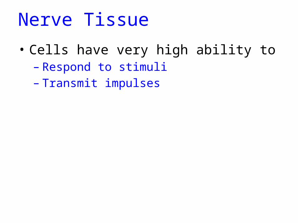
Nerve Tissue
• Cells have very high ability to – Respond to stimuli– Transmit impulses
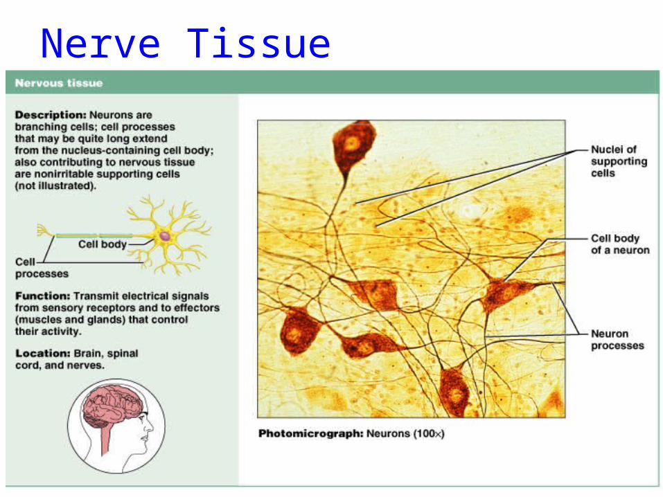
Nerve Tissue

Nerve Tissue
• NEURON is the main nerve cell)

Nerve Tissue
• NEURON is the main nerve cell– Cell Body(3)– Dendrites (5)– Axon(1)

Motor Neurone

Motor Neurone
• Cell body• Dendrites• Axon– Covered by Myelin
sheath

Neurones

Neuron
• Multipolar
Kuehnel, Color Atlas of Cytology, Histology, and Microscopic Anatomy

Neuron's – cerebral cortex
Kuehnel, Color Atlas of Cytology, Histology, and Microscopic Anatomy

Neuron's – spinal cord
Kuehnel, Color Atlas of Cytology, Histology, and Microscopic Anatomy

Nerve and Reflex Arc
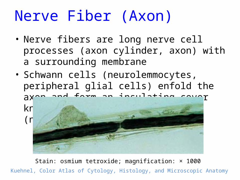
Nerve Fiber (Axon)• Nerve fibers are long nerve cell processes (axon
cylinder, axon) with a surrounding membrane• Schwann cells (neurolemmocytes, peripheral glial
cells) enfold the axon and form an insulating cover known as Schwann’s sheath (neurolemma).
Kuehnel, Color Atlas of Cytology, Histology, and Microscopic Anatomy
Stain: osmium tetroxide; magnification: × 1000

Nerve Fiber (Axon)• Myelinated nerve fiber, i.e., the axon is covered by a
myelin sheath, which is rich in lipids.• Every 0.8 to 1.0 mm, a node of Ranvier subdivides
the myelin sheath into segments or internodes.
Kuehnel, Color Atlas of Cytology, Histology, and Microscopic Anatomy
Stain: osmium tetroxide; magnification: × 1000

Sciatic nerve – cross section• 1 Fascicle (nerve fiber bundle)• 2 Perineurium• 3 Epineurium• 4 Artery• 5 Vein• 6 Adipose
tissue
Stain: alum hematoxylin; magnification: × 10Kuehnel, Color Atlas of Cytology, Histology, and Microscopic Anatomy

Glial Cells
• Glial cells (Neuroglia) or (Glia)• (Greek "glue”• Non-neuronal cells that maintain homeostasis,
form myelin, and provide support and protection for the brain's neurons
• They occupy the entire space between neurons and separate nerve cells from blood vessels

Glial Cells - Functions
1. Surround neurons and hold them in place2. Supply nutrients and oxygen to neurons3. Insulate one neuron from another4. Destroy pathogens and remove dead neurons

Glial Cells - Types
• Astrocytes (macroglia)• Oligodendrocytes• Microgliocytes

Astrocytes
Kuehnel, Color Atlas of Cytology, Histology, and Microscopic Anatomy
• Most abundant• Deal wit homeostasis – relate to vessels
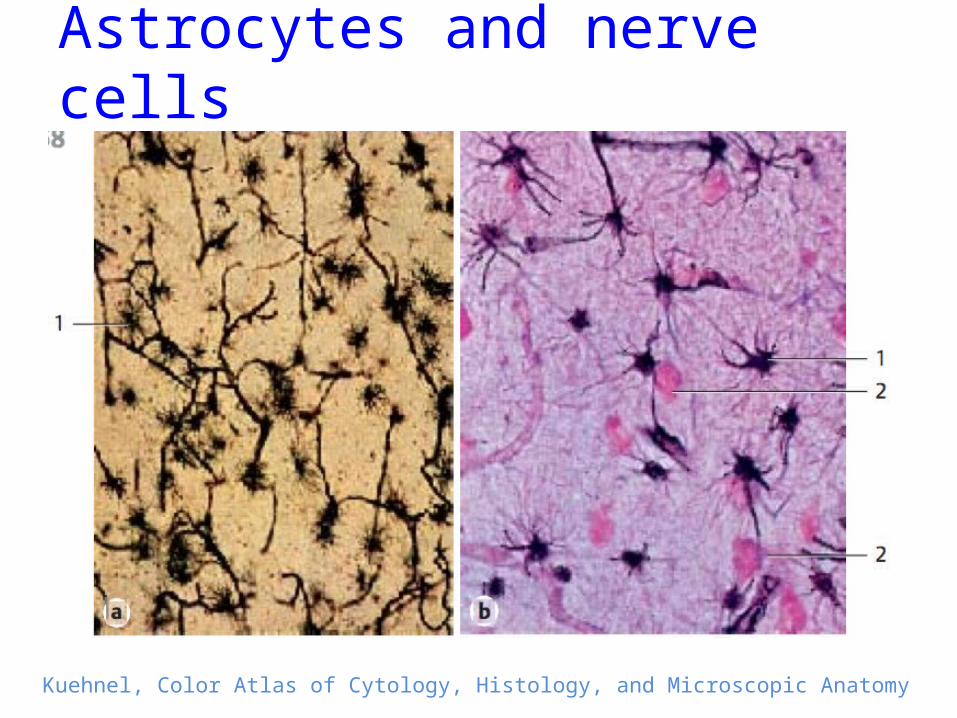
Astrocytes and nerve cells
Kuehnel, Color Atlas of Cytology, Histology, and Microscopic Anatomy

Microglia

Microglia
• Oligodendrocytes:– Closely related to
neurons– Provide myelin
protection for CNS neurons

Neurons and glial cells
Kuehnel, Color Atlas of Cytology, Histology, and Microscopic Anatomy
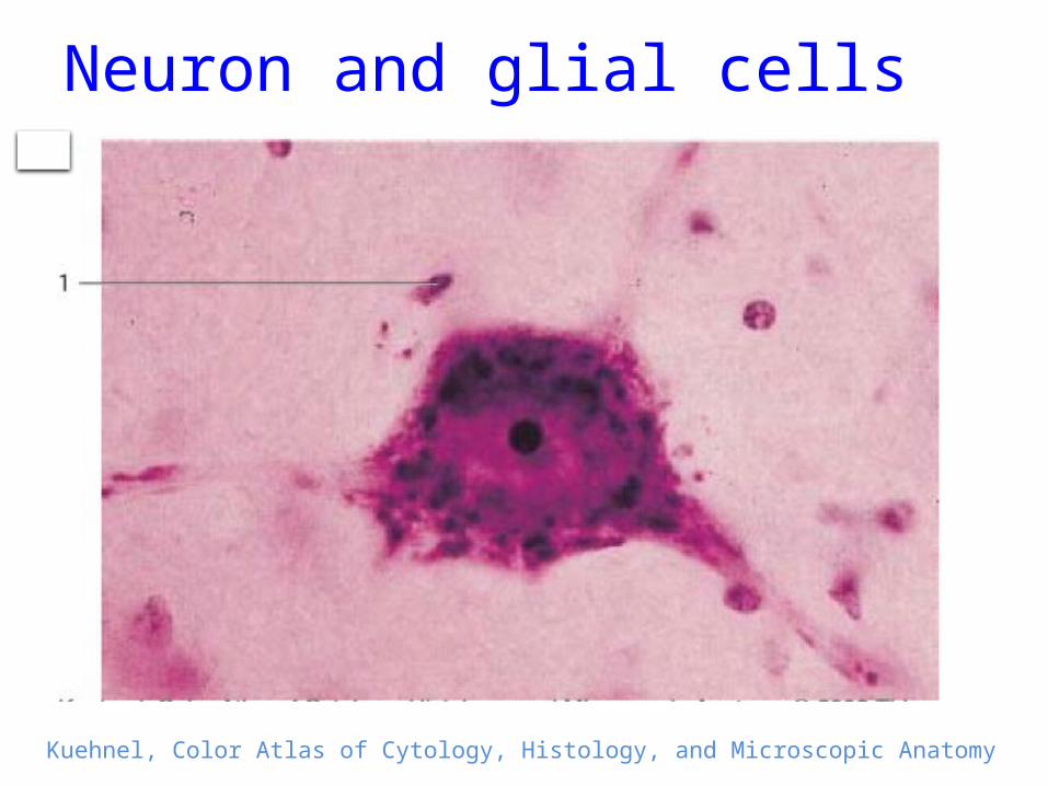
Neuron and glial cells
Kuehnel, Color Atlas of Cytology, Histology, and Microscopic Anatomy

Summary
• Nerve Tissue Cells:– Neurons:• Myelinated• Non-myelinated

Summary
• Nerve Tissue Cells:– Neurons:• Myelinated• Non-myelintaed
– Neurolial cells:• Astrocytes • Microglia• Oligodendrocytes
Related Documents