NERVE ENTRAPMENTS FREQUENTLY SEEN & COMMONLY MISSED Phillip M. Steele, MD CAQ Diagnostic Musculoskeletal Ultrasound (RMSK) Boarded Family Practice CAQ Sports Medicine Performance Injury Care & Sports Medicine

Welcome message from author
This document is posted to help you gain knowledge. Please leave a comment to let me know what you think about it! Share it to your friends and learn new things together.
Transcript

NERVE ENTRAPMENTS FREQUENTLY SEEN & COMMONLY MISSED
Phillip M. Steele, MD
CAQ Diagnostic Musculoskeletal Ultrasound (RMSK)
Boarded Family Practice
CAQ Sports Medicine
Performance Injury Care & Sports Medicine

DISCLOSUREI have no financial
relationship with any
diagnostic equipment utilized
in this talk.
Faculty for American Medical
Society for Sports Medicine
advanced diagnostic
ultrasound courses.
Faculty for MSKUS cadaver
diagnostic and injection
courses.
Dorsal Compartments SAX
Carpal Tunnel SAX
LEARNING OBJECTIVE
Awareness of common nerve pain syndromes.
Describe common nerve entrapment syndrome physical
exam findings.
Awareness of cutaneous nerve entrapment symptoms that
masquerade as other diagnosis.

BASIC NERVE ANATOMY

TYPES OF NERVE INJURY

HOW INJURED?
Etiologies
Isolated contusion
Repetitive compression
Stretch injury
Surgical injury
Vibration
Viral
Compressive bracing/casting
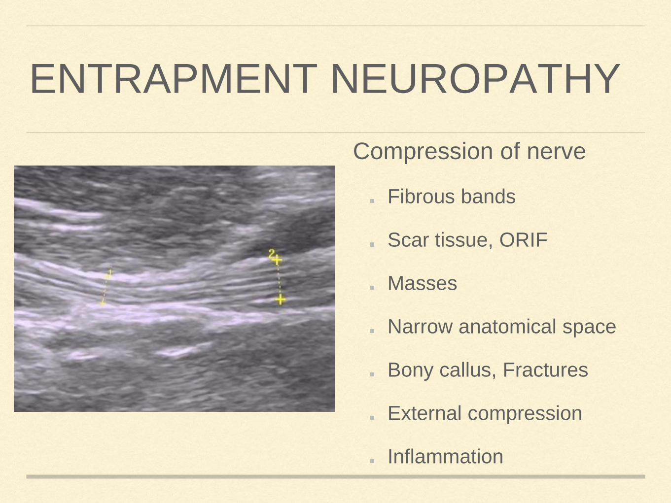
ENTRAPMENT NEUROPATHY
Compression of nerve
Fibrous bands
Scar tissue, ORIF
Masses
Narrow anatomical space
Bony callus, Fractures
External compression
Inflammation

THE WORK UP?
AWARENESS
HISTORY
PHYSICAL EXAM
SENSORY EXAM
EMG/NCT
MRI
ULTRASOUND

WHAT THE HISTORY TELLS YOU!
History is vague and non localizing in early disease making a diagnosis
challenging.
Muscle weakness or atrophy is present in late stage disease.
Most injuries will have subtle features of a more “classical” nerve
entrapment syndrome.
Our training provides little experience recognizing, diagnosing or
treating many of the sensory cutaneous branch entrapments.
Best diagnostic tool for entrapment neuropathy is good anatomical
knowledge cutaneous nerve distribution.

PHYSICAL EXAM FINDINGS
PE findings of decreased sensation
of sensory nerve distribution.
Pain distribution.
Muscle fatigue with prolonged
contraction.
Weakness of motor nerves distal to
injury site.
Tinel’s over entrapment.
Pain with compression maneuvers.
Special test for some nerves.
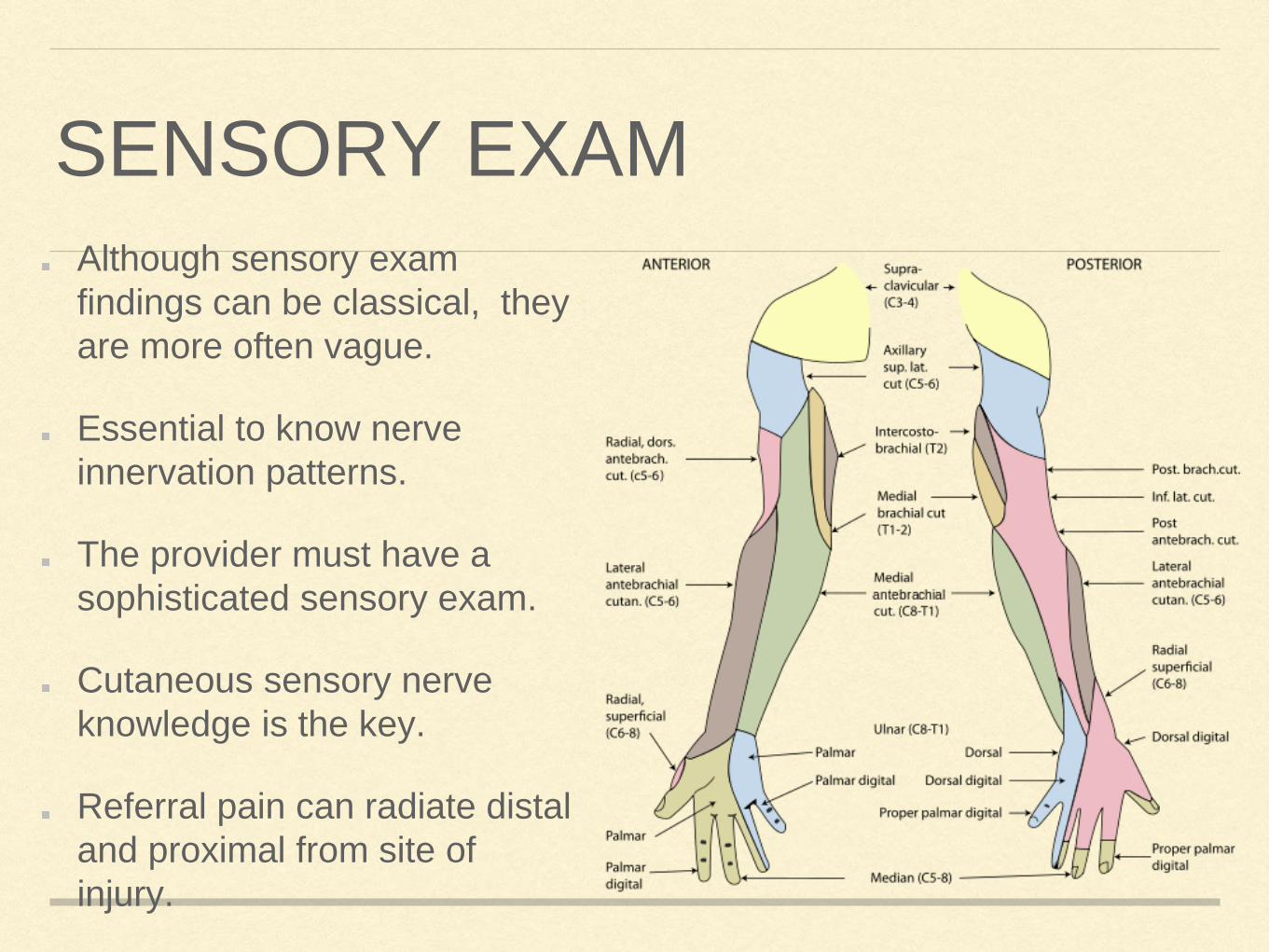
SENSORY EXAM
Although sensory exam
findings can be classical, they
are more often vague.
Essential to know nerve
innervation patterns.
The provider must have a
sophisticated sensory exam.
Cutaneous sensory nerve
knowledge is the key.
Referral pain can radiate distal
and proximal from site of
injury.

EMG/NCT
Electrodiagnostic studies include two components:
Nerve conduction and needle electromyography.
Nerve conduction studies evaluate how fast a nerve
conducts electricity and how much electricity reaches
the final destination.
In general, a decrease in nerve conduction velocity
suggests a demyelinating injury to the nerve at the site
of slowing.
A decrease in the amount of electricity that reaches
the final destination suggests either a conduction block
from a demyelinating injury or axonal injury.
Needle electromyography assist in differentiating
between demyelinating and axonal injuries, grading
injury severity, and determining injury chronicity.

EXCEPTIONS TO EMG/NCT
Must have demyelination of the nerve or significant axonal damage.
At least 3-6 weeks after injury. EMG -
First Degree or neurapraxia injury not well identified. EMG -
Second Degree injury or axonotmesis not well identified. EMG -
Third Degree = injury to the axon & endoneurium. EMG + helpful.
Fourth Degree = injury to axon, endoneurium, perineurium. EMG +
Fifth Degree = injury to axon, endoneurium, perineurium & epineurium. EMG +
CTS SENSITIVITY = 85%
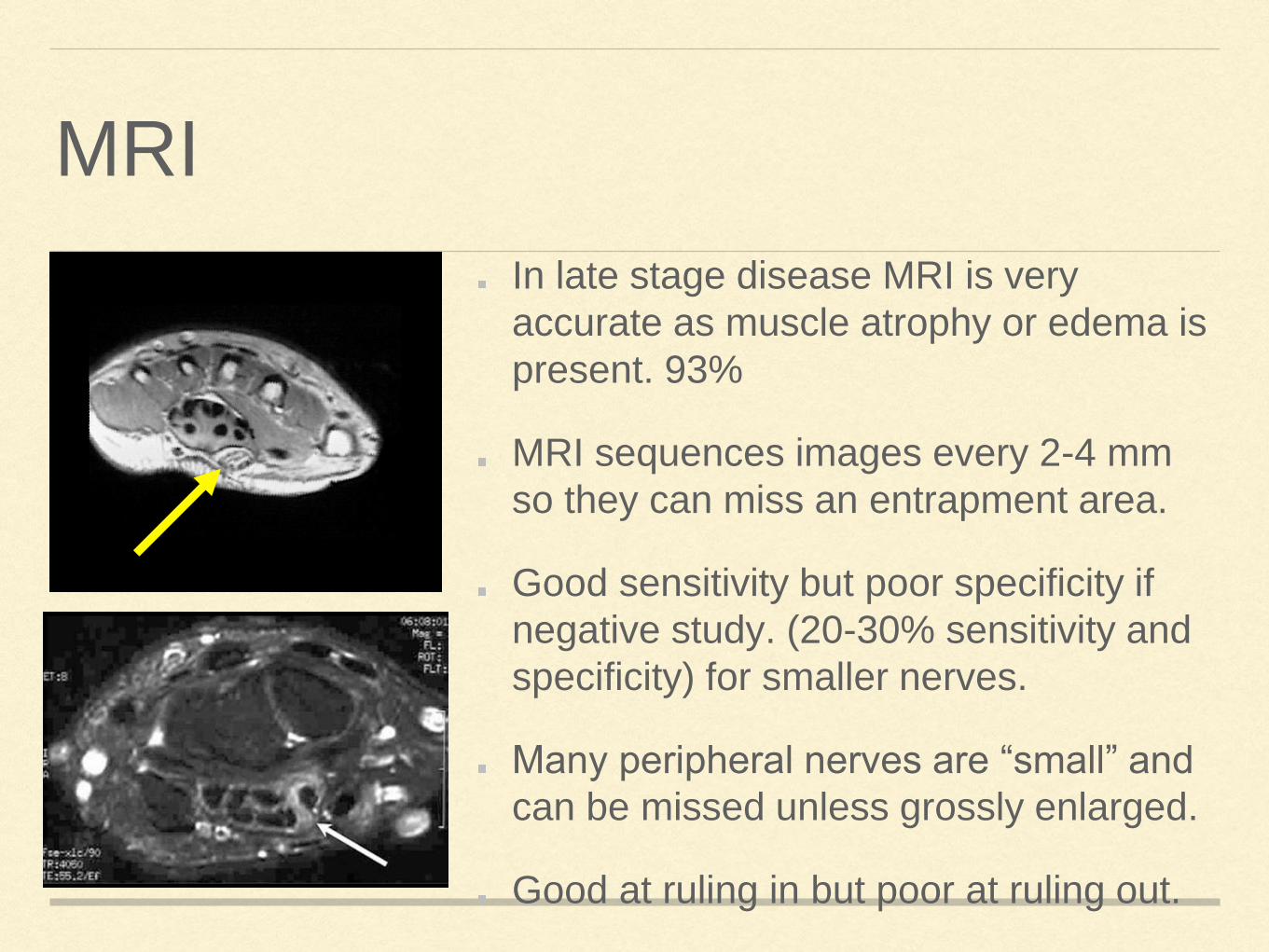
MRI
In late stage disease MRI is very
accurate as muscle atrophy or edema is
present. 93%
MRI sequences images every 2-4 mm
so they can miss an entrapment area.
Good sensitivity but poor specificity if
negative study. (20-30% sensitivity and
specificity) for smaller nerves.
Many peripheral nerves are “small” and
can be missed unless grossly enlarged.
Good at ruling in but poor at ruling out.

MRI FINDINGS
Hyper-intense signal of
the nerve suggest
edema nerve damage.
60% of asymptomatic
individuals have hyper-
intense signal of the ulna
nerve.
Superior view for deep
structures.
Patient size?

MSK ULTRASOUND EVALUATION OF NERVES

MSKUS EVALUATIONMSKUS can show the
constricting tissue which
is creating a loss of
normal nerve
architecture.
Proximal swelling with
distal tapering.
Sensitivity and
specificity typically >
92%.
Increase of greater than
2mm circumferential
area.
Ulna nerve LAX
Functional Median Nerve Exam

FUNCTIONAL EXAMINATION
Functional static and
dynamic exam.
Power and color flow
doppler to identify
inflammation, infection and
vascular structures.
Evaluation of surrounding
structures for atrophy, fatty
infiltration, scarring,
instability.
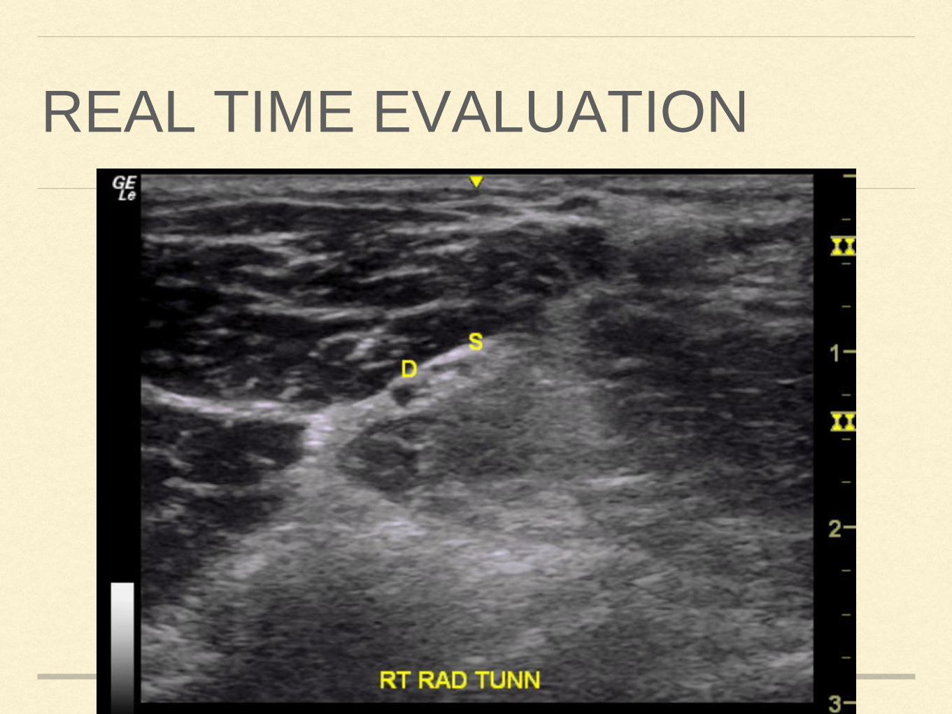
REAL TIME EVALUATION

NEUROHYDROLYSIS
Real time visualization.
Diagnostic and
therapeutic nerve block.
Stretch out the
constriction.
Minimal side effects.
Can be used for some
chronic pain diagnosis.
Superficial Cutaneous Radial Nv
SCNV Thumb
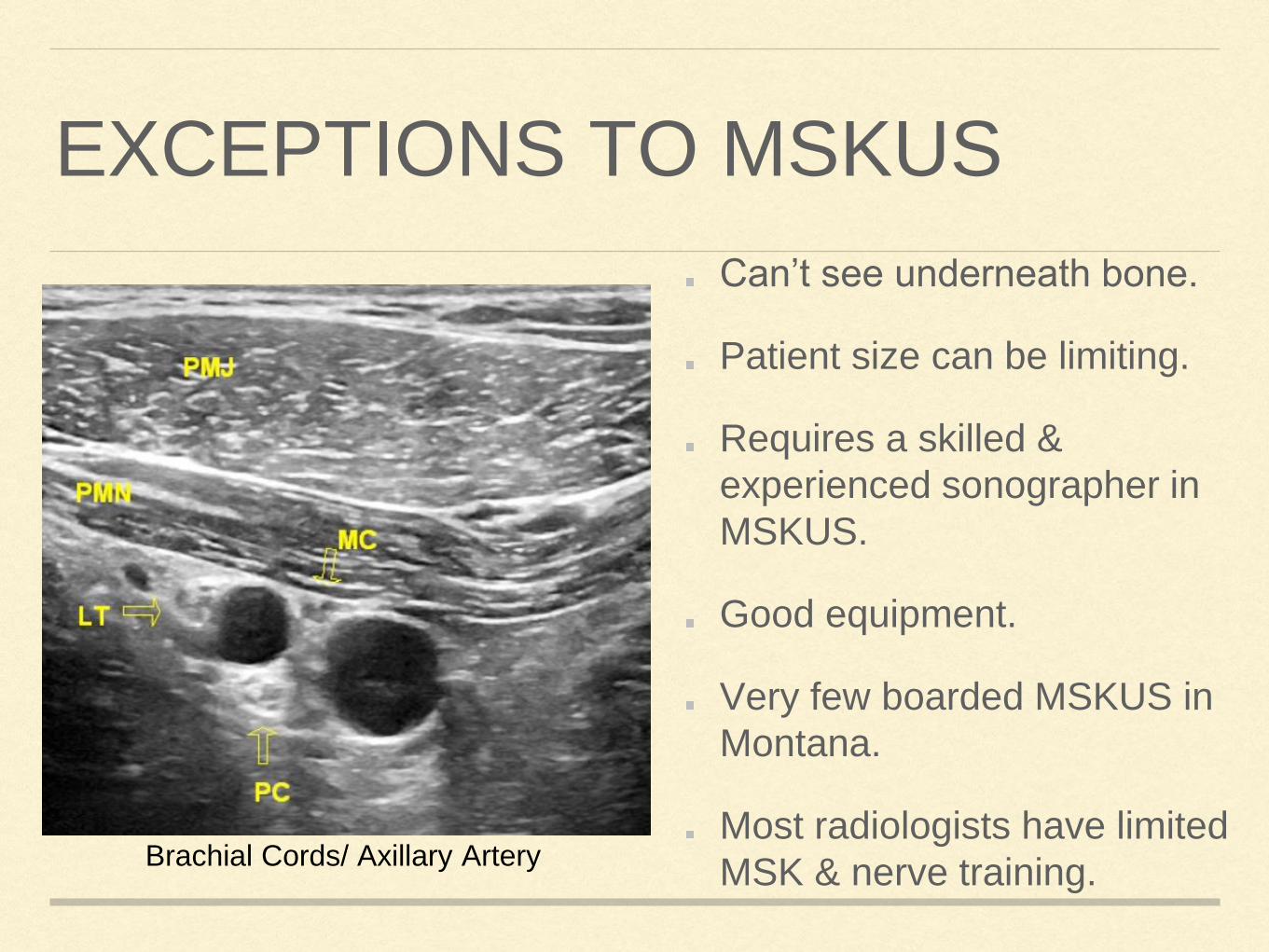
EXCEPTIONS TO MSKUS
Can’t see underneath bone.
Patient size can be limiting.
Requires a skilled &
experienced sonographer in
MSKUS.
Good equipment.
Very few boarded MSKUS in
Montana.
Most radiologists have limited
MSK & nerve training. Brachial Cords/ Axillary Artery

A WORD ABOUT TREATMENTThe hardest part is making the diagnosis!
PT is more effect if they have the correct
diagnosis.
Avoid deep tissue work around the entrapment
site.
Avoid aggressive stretching of the nerve.
Avoid dry needling, scraper, deep myofascial
release, manual trigger point release, &
compressive bracing.
Dose pack, NSAIDS oral and topical,
iontophoresis & correction of structural and
ergonomic issues.
Neurohydrolysis and or surgical release if failed
conservative therapy.

BEWARE!
Patients tend to dig into nerve
entrapments.
Hurts good?
Makes it feel better for a short
period of time.
They become the problem.
Like a knuckle cracker, they just
can’t help themselves.
Stop the digging is your first
therapy goal.

IS IT SHOULDER PAIN OR IS IT NECK PAIN?
Nerve entrapments to the neck
& shoulder are common.
Challenging to diagnosis.
Scapula motion is the key.!
Suprascapular nerve
Dorsal scapular nerve
Axillary nerve
Spinal accessory nerve
Supraclavicular nerve

SUPRASCAPULAR NERVE
Paralabral cyst thought to be
most common?
28% of full thickness RTC tear
also include nerve entrapment.
Osteoarthritis association.
Iatrogenically injured with RTC
repair.
Consider in the setting of pain
with minimal MRI findings.

SUPRASCAPULAR NERVE ENTRAMPMENT
The most commonly injured
branch of the brachial plexus in
sports.
Hallmark finding is painless
weakness to resisted external
rotation.
Most common symptom is a
vague lateral shoulder pain
Posterolateral dull, burning, deep
or diffuse ache that is worse with
overhead.

OVERHEAD WORK OR SPORT
Common in the over head
athlete.
Seen in 35-45% of professional
volleyball athletes on the
serving arm
Loss of throwing or hitting
power or velocity.
Not well documented in the
overhead worker but may
present similar to thoracic outlet
with weakness during overhead
work.

SUPRASCAPULAR NOTCH
Nerve courses through the suprascapular
notch.
Notch is bridged by a thick transverse
scapular ligament.
Entrapment occurs as the nerve is
relatively fixed at the notch.
Maximal stretching of the nerve with cross
body adduction or protracted forward
flexion.
Causes weakness of both abduction and
external rotation.
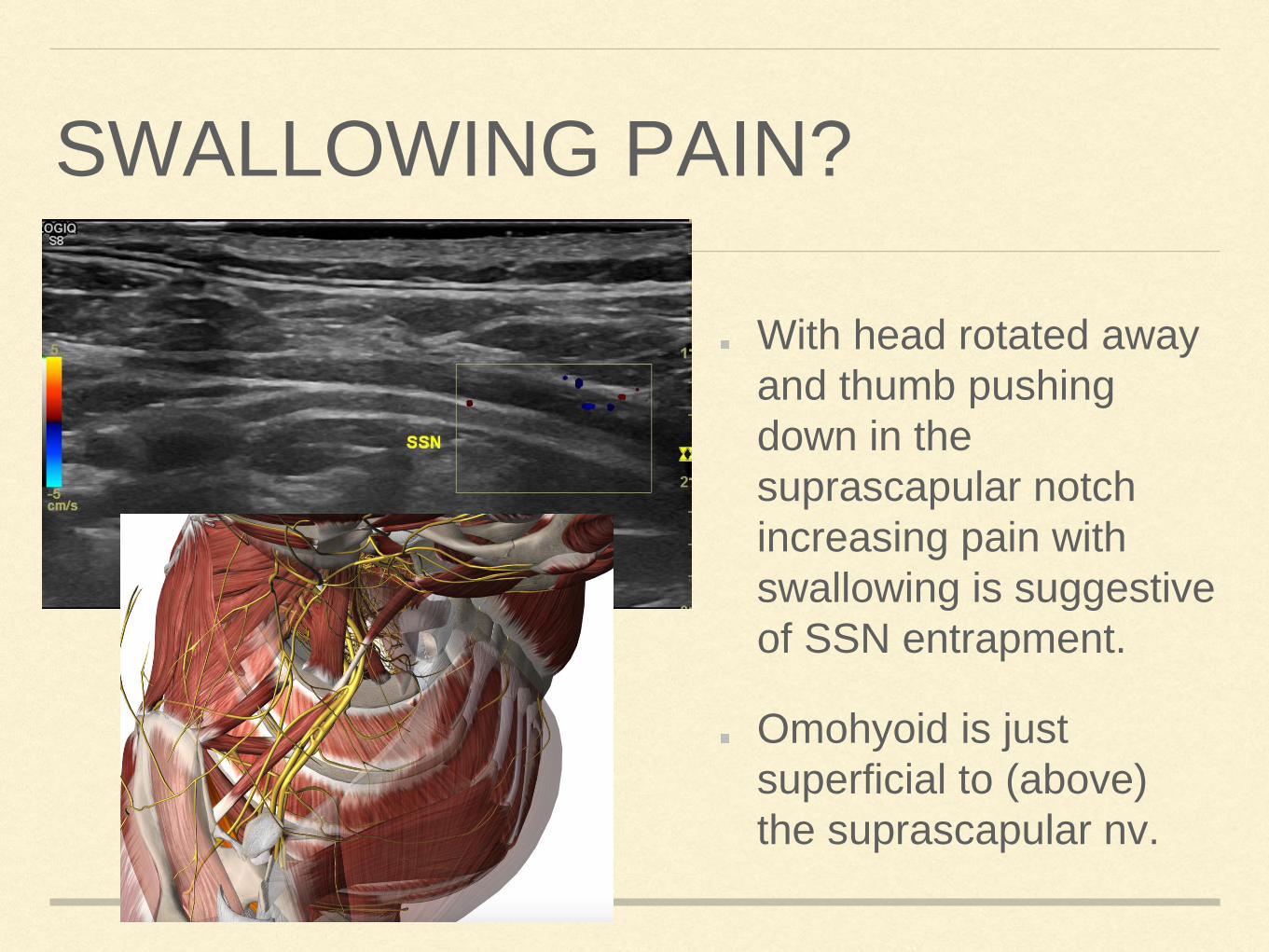
SWALLOWING PAIN?
With head rotated away
and thumb pushing
down in the
suprascapular notch
increasing pain with
swallowing is suggestive
of SSN entrapment.
Omohyoid is just
superficial to (above)
the suprascapular nv.
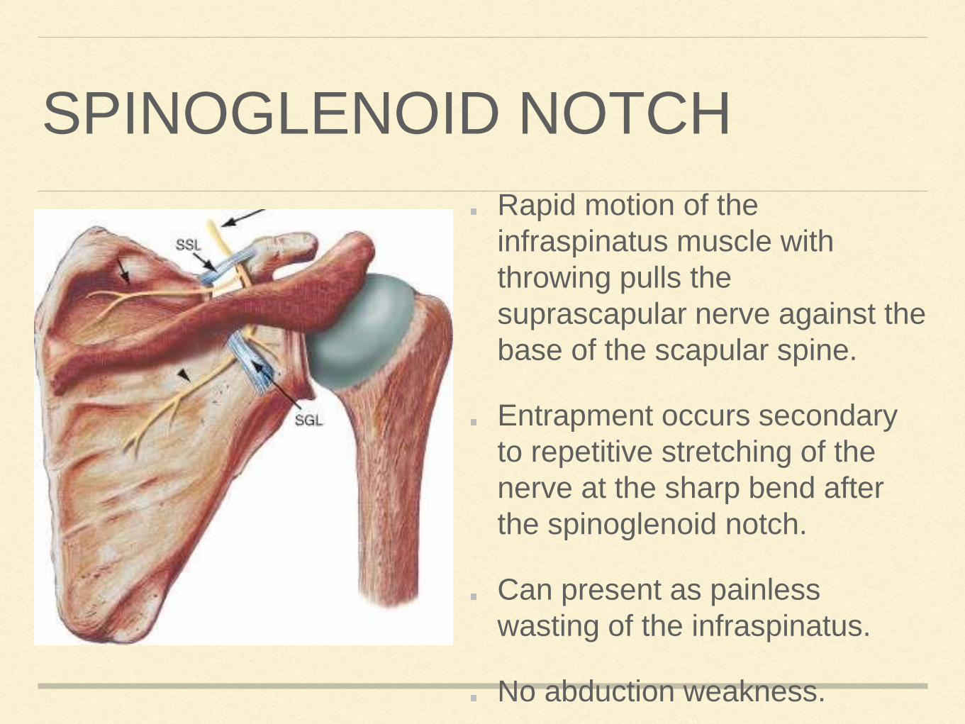
SPINOGLENOID NOTCH
Rapid motion of the
infraspinatus muscle with
throwing pulls the
suprascapular nerve against the
base of the scapular spine.
Entrapment occurs secondary
to repetitive stretching of the
nerve at the sharp bend after
the spinoglenoid notch.
Can present as painless
wasting of the infraspinatus.
No abduction weakness.
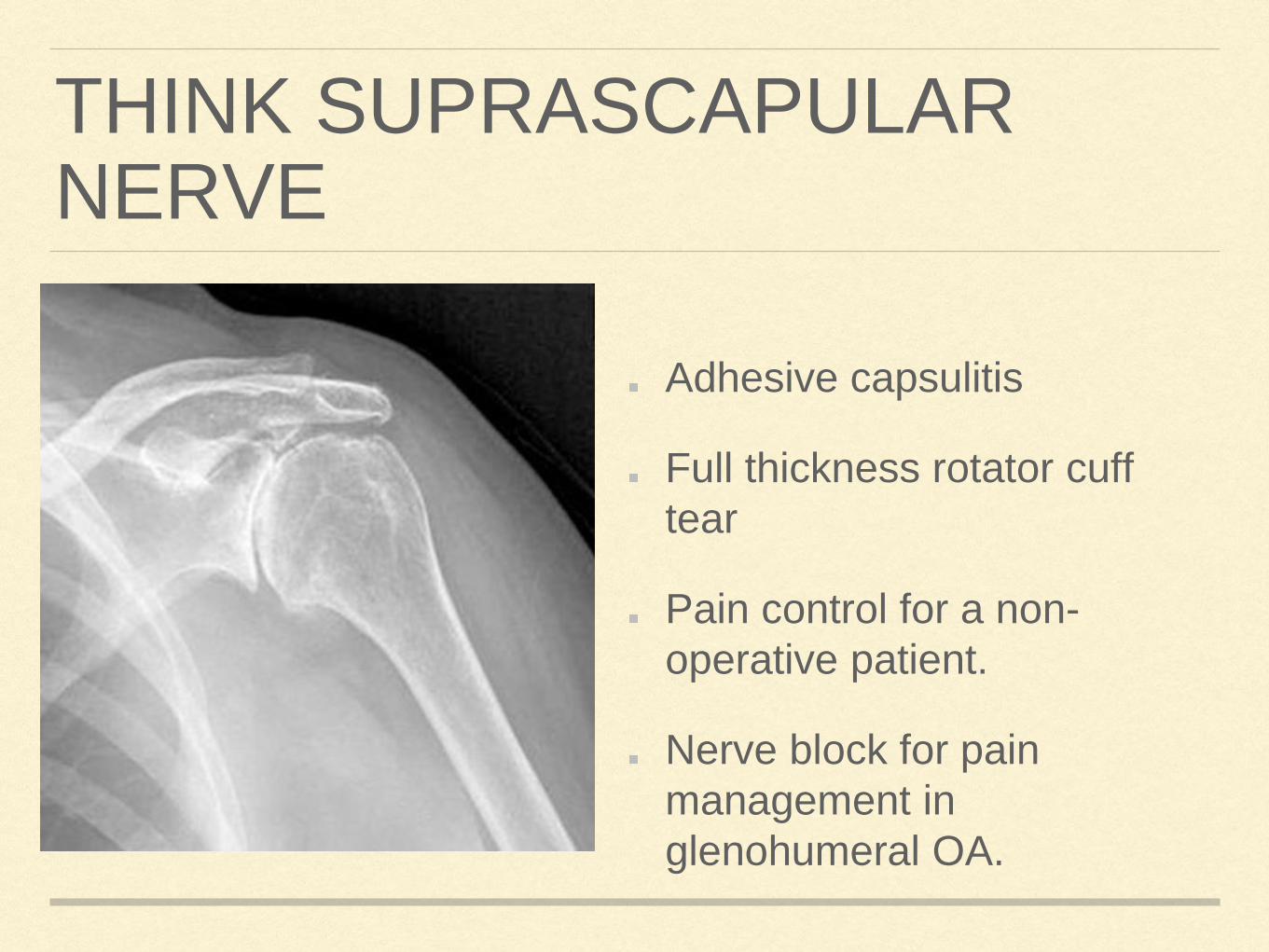
THINK SUPRASCAPULAR NERVE
Adhesive capsulitis
Full thickness rotator cuff
tear
Pain control for a non-
operative patient.
Nerve block for pain
management in
glenohumeral OA.

SUPERFICIAL CERVICAL PLEXUS?
Supraclavicular nerve
(SCN)
Spinal Accessory nerve
(SAN)
Lesser Occipital nerve (LON)
Transverse Cervical nerve
(TCN)
Greater Auricular nerve (GAN)
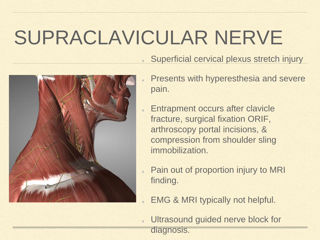
SUPRACLAVICULAR NERVESuperficial cervical plexus stretch injury
Presents with hyperesthesia and severe
pain.
Entrapment occurs after clavicle
fracture, surgical fixation ORIF,
arthroscopy portal incisions, &
compression from shoulder sling
immobilization.
Pain out of proportion injury to MRI
finding.
EMG & MRI typically not helpful.
Ultrasound guided nerve block for
diagnosis.

HYPERESTHESIA AFTER SHOULDER INJURY
Tented clavicle fractures cause
impingement of the SCN.
Symptoms may extend beyond
anatomical zone and include the proximal
deltoid and posterolateral scapula
Branch locations are highly variable.
Surgically there is no clinically relevant
safe zone.
Horizontal incisions result in greatest risk.

SUPRACLAVICULAR NERVE BLOCKS FOR CLAVICLE FRACTURE
For acute fracture
pain control with long
acting anesthetics.
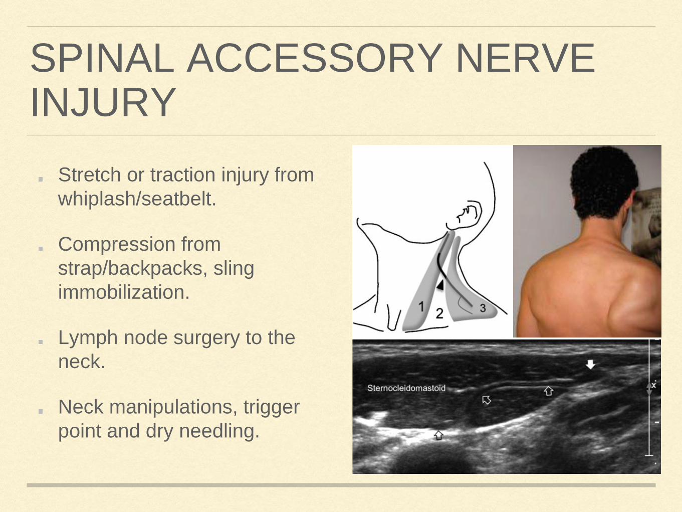
SPINAL ACCESSORY NERVE INJURY
Stretch or traction injury from
whiplash/seatbelt.
Compression from
strap/backpacks, sling
immobilization.
Lymph node surgery to the
neck.
Neck manipulations, trigger
point and dry needling.
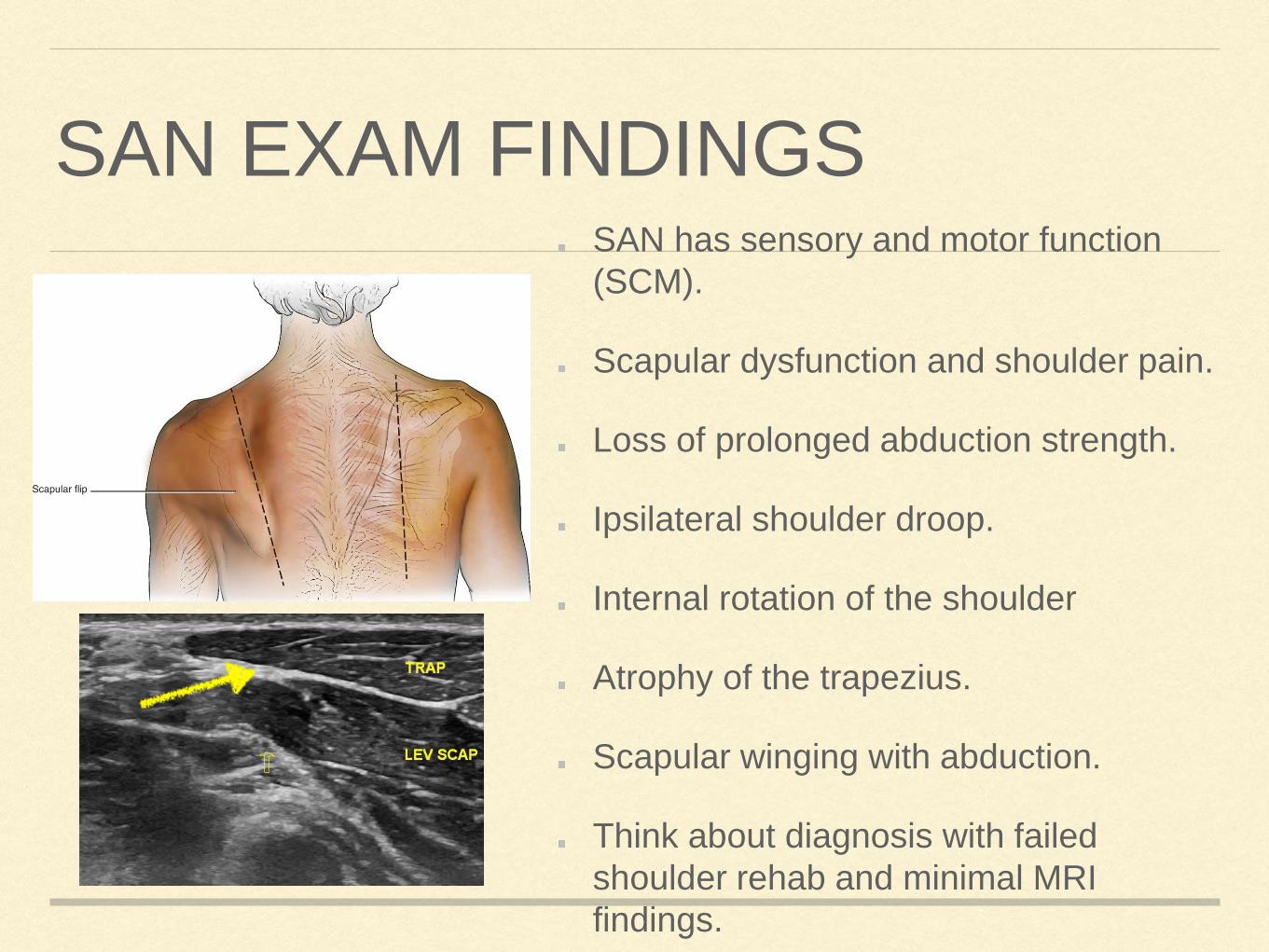
SAN EXAM FINDINGSSAN has sensory and motor function
(SCM).
Scapular dysfunction and shoulder pain.
Loss of prolonged abduction strength.
Ipsilateral shoulder droop.
Internal rotation of the shoulder
Atrophy of the trapezius.
Scapular winging with abduction.
Think about diagnosis with failed
shoulder rehab and minimal MRI
findings.

SAN WORK UP
Best identified by a good
physical exam.
EMG correlates poorly with
shoulder dysfunction or pain.
EMG helpful if shoulder
weakness or atrophy.
MRI is sensitive if atrophy.
MSKUS for nerve block for
confirmation of pain generation.

SAN ENTRAPMENT CAUSES OTHER SYMPTOMS?
Adhesive capsulitis
Shoulder
impingement
Muscle spasms
Torticollis
Shoulder pain
refractory to PT and
minimal MRI
findings.
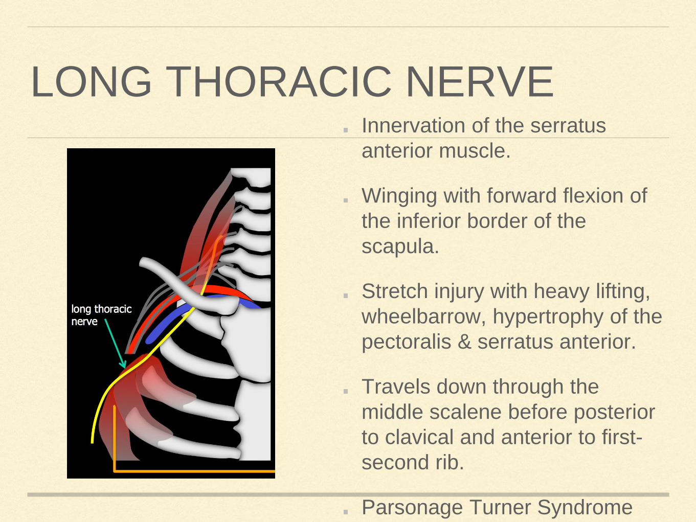
LONG THORACIC NERVEInnervation of the serratus
anterior muscle.
Winging with forward flexion of
the inferior border of the
scapula.
Stretch injury with heavy lifting,
wheelbarrow, hypertrophy of the
pectoralis & serratus anterior.
Travels down through the
middle scalene before posterior
to clavical and anterior to first-
second rib.
Parsonage Turner Syndrome
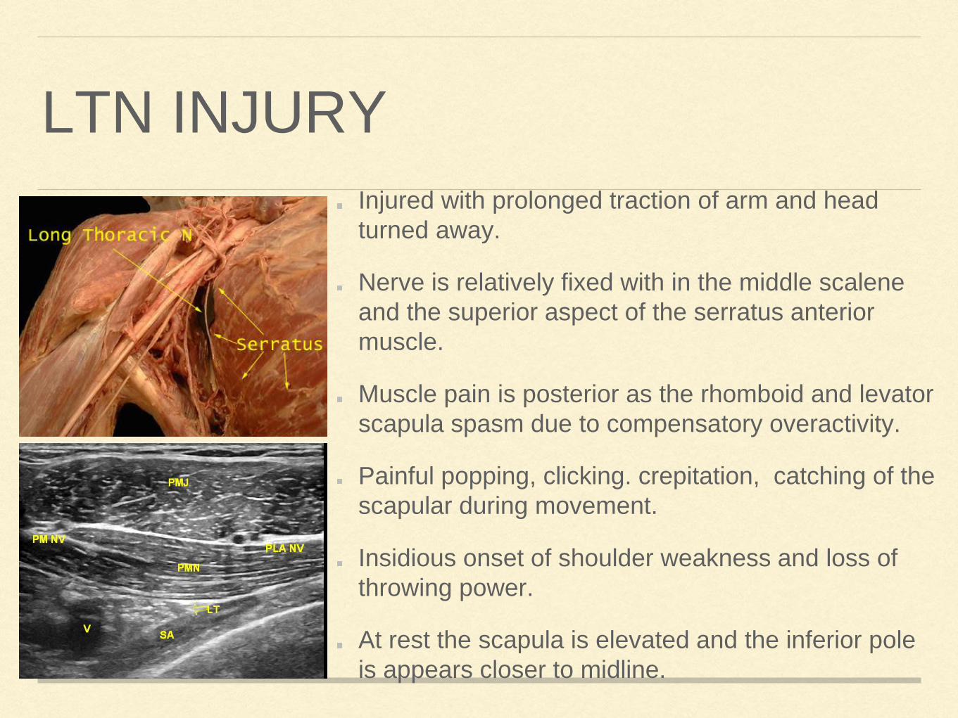
LTN INJURY
Injured with prolonged traction of arm and head
turned away.
Nerve is relatively fixed with in the middle scalene
and the superior aspect of the serratus anterior
muscle.
Muscle pain is posterior as the rhomboid and levator
scapula spasm due to compensatory overactivity.
Painful popping, clicking. crepitation, catching of the
scapular during movement.
Insidious onset of shoulder weakness and loss of
throwing power.
At rest the scapula is elevated and the inferior pole
is appears closer to midline.

DORSAL SCAPULA NERVE

DSN INJURY
Pure motor nerve derived from the C5
nerve root.
Pierces the middle scalene muscle and
travels between the posterior scalene and
serratus posterior.
Innervates the levator scapulae &
rhomboid major and minor
Injured by compression (straps),
hypertrophy of middle scalene, shoulder
disolcation, & whiplash stretch injury.
Can share a common trunk with Long
thoracic.
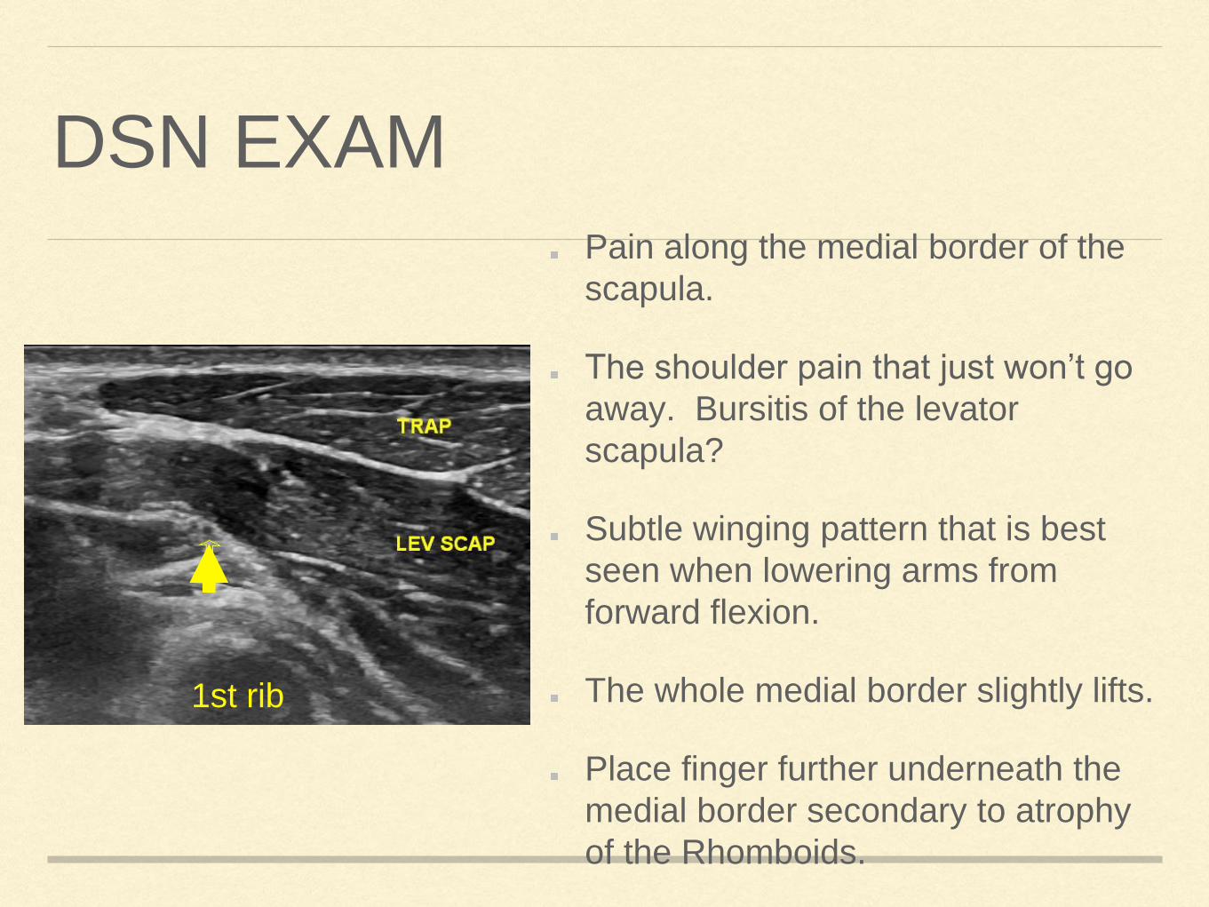
DSN EXAM
Pain along the medial border of the
scapula.
The shoulder pain that just won’t go
away. Bursitis of the levator
scapula?
Subtle winging pattern that is best
seen when lowering arms from
forward flexion.
The whole medial border slightly lifts.
Place finger further underneath the
medial border secondary to atrophy
of the Rhomboids.
1st rib

AXILLARY NERVE ENTRAPMENT
History of shoulder dislocation or
hyperlaxity.
Overhead workers complains of
weakness and fatigue.
May occur with severe motor
findings without sensory findings.
Subtle numbness to lateral
shoulder (deltoid patch).
Hertel sign (extension lag).
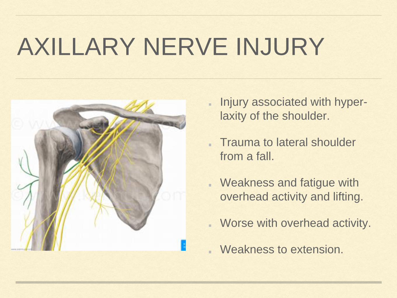
AXILLARY NERVE INJURY
Injury associated with hyper-
laxity of the shoulder.
Trauma to lateral shoulder
from a fall.
Weakness and fatigue with
overhead activity and lifting.
Worse with overhead activity.
Weakness to extension.

QUADRILATERAL SPACE SYNDROME
Compression of axillary nv &
circumflex artery.
Pain is usually vague &
nonspecific.
Deltoid & teres minor weakness.
Dead arm with posterior lateral
pain.
Non dermatomal pattern.
Point tenderness at QS.
Pain with abduction and external
rotation.
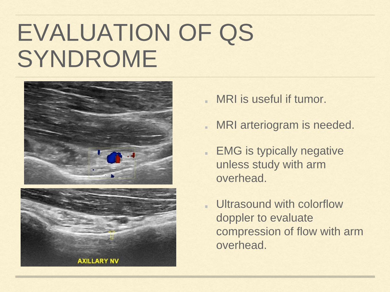
EVALUATION OF QS SYNDROME
MRI is useful if tumor.
MRI arteriogram is needed.
EMG is typically negative
unless study with arm
overhead.
Ultrasound with colorflow
doppler to evaluate
compression of flow with arm
overhead.

THE FORGOTTEN NERVE
MUSCULOCUTANEOUSC5-C6 (lateral cord)
Innervation of the bicep, brachialis, coracobrachialis
Superficial sensory after elbow = lateral antebrachial cutaneous
nerve

MCN SYMPTOMS
Vague upper arm pain.
Forearm pain or numbness during
flexion.
Weakness to bicep & brachialis is a
late finding.
Check for atrophy or asymmetry of
muscle contraction.
Radicular pain down the lateral
flexor surface of the forearm.
Repetitive lifting with supination/
pronation
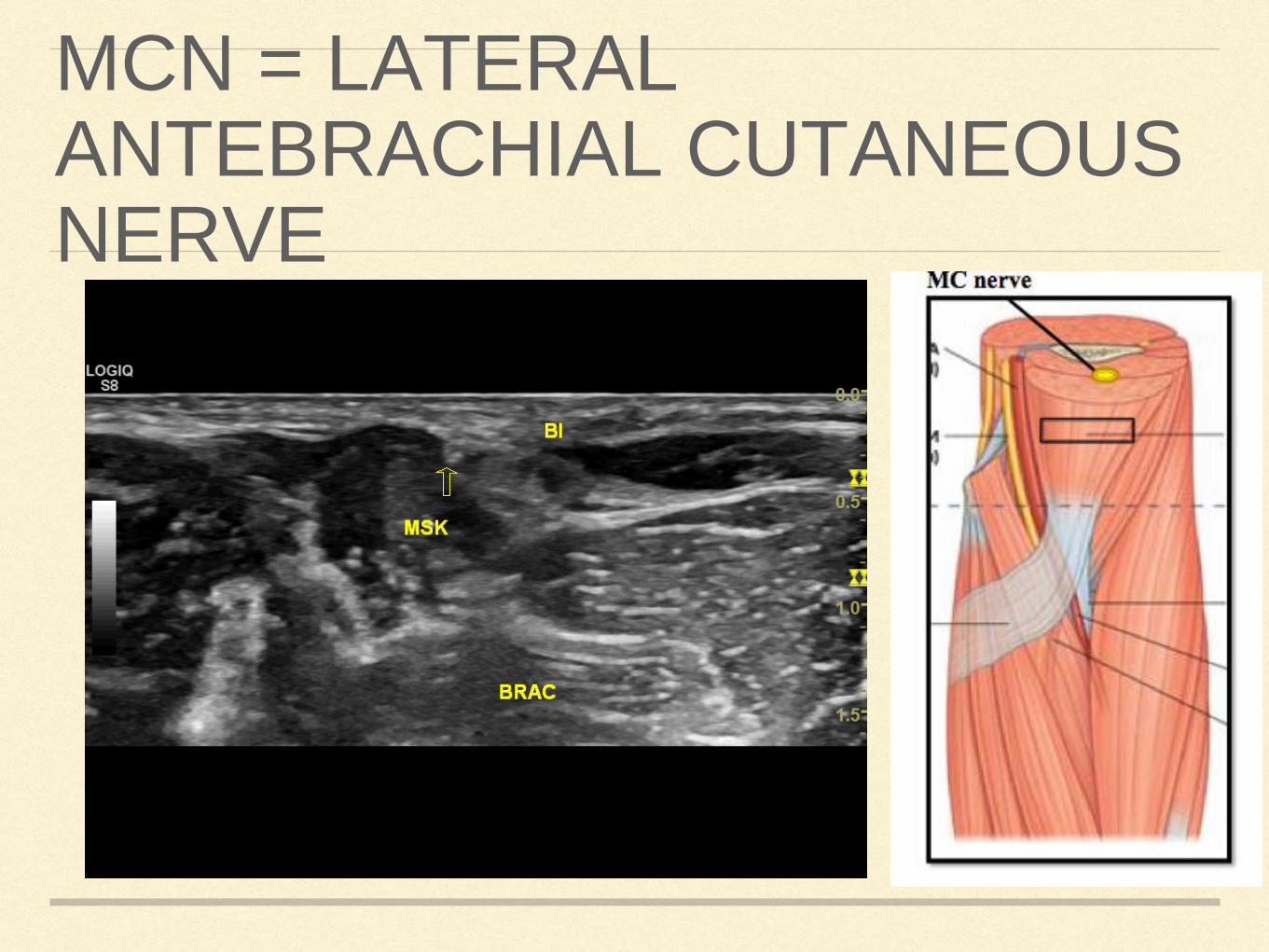
MCN = LATERAL ANTEBRACHIAL CUTANEOUS NERVE

LABCN
Lateral elbow pain 3-5 cm proximal elbow crease.
Associated with repetitive activity.
Forearm paresthesia.
Think of patient with forearm radiculopathy with
minimal C-Spine MRI.
Painful Brachioradialis.
Numbness increases with lateral bicep tendon
pressure at elbow crease during
pronation/supination.
EMG not helpful unless atrophy.
Nerve block for diagnosis.
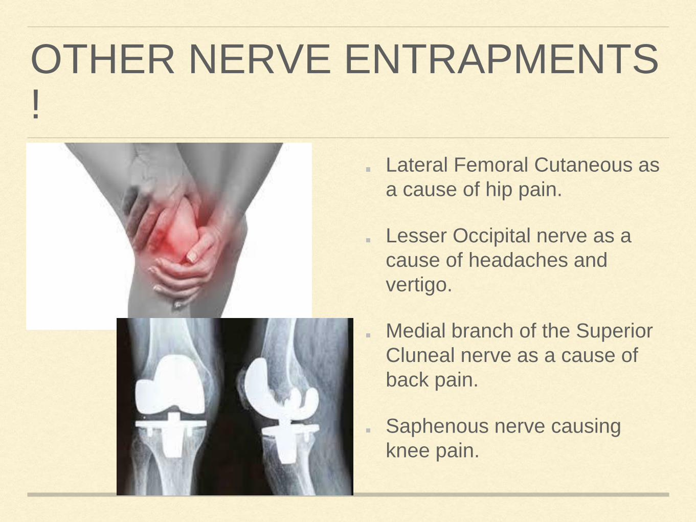
OTHER NERVE ENTRAPMENTS !
Lateral Femoral Cutaneous as
a cause of hip pain.
Lesser Occipital nerve as a
cause of headaches and
vertigo.
Medial branch of the Superior
Cluneal nerve as a cause of
back pain.
Saphenous nerve causing
knee pain.

OCCIPITAL HEADACHES?
Not all occipital = GON
Lesser occipital nerve typically runs
through the SCM and is found lateral
to the trapezius insertion.
Radiates to the eye across the
temple
Can cause vertigo.
Can causes nausea.
Nerve block to confirm.

BACKPAIN?Medial branch of the Superior
Cluneal nerve.
Entrapped in a fibrossous tunnel.
10-15% of all back pain.
7-8 cm from spinous process (L1)
along iliac crest.
Failed back program, minimal
MRI findings, radicular pattern
similar S1.
Hand to back of hip to relieve
pain.

LATERAL FEMORAL CUTANEOUS NERVE
Sensory nerve to lateral thigh.
Think about with hip OA exam
when x-ray or MRI is mild to
moderate (out of proportions to
pain level).
Exam finding include stiffness with
range of motion & guarding.
Painful ASIS
May be a burning groin pain.
The hyper-stretcher!

KNEE PAIN?SAPHENOUS NERVE ENTRAPMENT OF THE INFRAPATELLAR BRACH.Can mimic: including lumbar
radiculopathy, patellofemoral d/o,
suprapatellar plica, medial meniscus
tear, tibial stress fx, pes anserine
tendonopathy/ bursitis, synovitis and
CRPS.
Can be injured during surgery (1-20%)
risk.
Seen with OA and TKA.
Entrapped by a fibrous band spanning
between the vastus medialis and
adductor magnus (Hunters canal).
10cm proximal to medial femoral
condyle.

COMMON FIBULAR NERVE AKA
COMMON PERONEUS NERVE
Painful lateral lower leg.
S/P TKA
May snap with
flexion/extension of the
knee.
Pain posterior fibula just
medial to hamstring
insertion.
Weakness is a late
finding.
EMG typically negative.
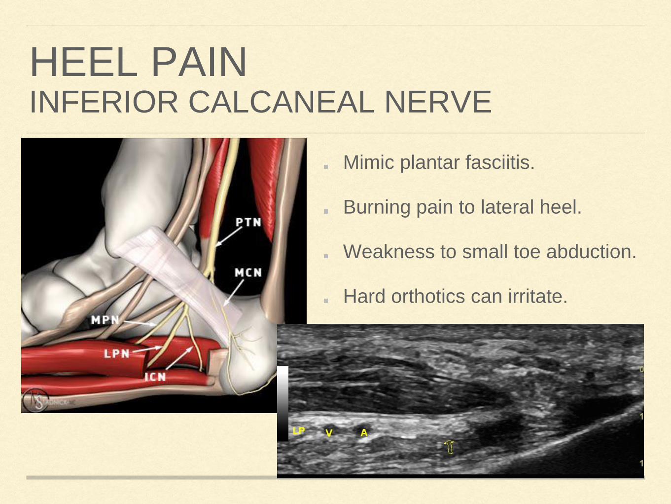
HEEL PAININFERIOR CALCANEAL NERVE
Mimic plantar fasciitis.
Burning pain to lateral heel.
Weakness to small toe abduction.
Hard orthotics can irritate.
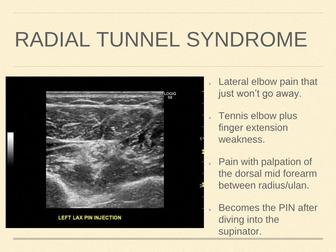
RADIAL TUNNEL SYNDROME
Lateral elbow pain that
just won’t go away.
Tennis elbow plus
finger extension
weakness.
Pain with palpation of
the dorsal mid forearm
between radius/ulan.
Becomes the PIN after
diving into the
supinator.

FINAL THOUGHTS/ QUESTIONS?
Not all radiculopathy is
cervical.
Not all shoulder pain is a
rotator cuff tear.
MRI with minimal findings and
pain out of proportion to
clinical findings is a nerve
entrapment syndrome until
proven otherwise.


WHY NEUROPATHY?
Prolonged pressure causes
ischemia due to compression
of vasa nervorum.
Impairment of axonal
transport.
Proliferation of intra-neural
connective tissue.

A WORD ON SENSITIVITY & SPECIFICITY
EMG/NCT is considered the gold standard. Studies of sensitivity & specificity
depends on which nerve was studied.
A clear understanding that the nerve must be physiologically affected for EMG/NCT
testing to be clinically relevant.
The correlation of pain and physiological changes is not clearly understood.
EMG has only moderate sensitivity and specificity for many nerve entrapment to the
upper and lower extremity.
It is critical to remember that a normal study does not rule out the presence of
cervical or lumbar radiculopathy.
EMG correlation to symptoms is reported anywhere from 55% to 86% sensitivity.
Related Documents


