Neonatal Hypothyroidism Affects the Timely Expression of Myelin-associated Glycoprotein in the Rat Brain Angeles Rodriguez-Petia, Nieves Ibarrola, Miguel Angel lIfiguez, Alberto Muihoz, and Juan Bernal Instituto de Investigaciones Biomedicas, Consejo Superior de Investigaciones Cientificas, Facultad de Medicina, Universidad Autonoma de Madrid, 28029 Madrid, Spain Abstract Congenital hypothyroidism strongly affects myelination. To as- sess the role of thyroid hormone on myelin gene expression, we have studied the effect of hypothyroidism on the steady state levels of myelin-associated glycoprotein (MAG) and its mRNA in rat brain during the first postnatal month. As studied by immunoblot analysis of several brain regions, MAG in- creased from days 10-15 onwards, reaching constant levels by days 20-25. Hypothyroid samples showed a delay in the accu- mulation of MAG that was more severe in rostral regions, such as cortex and hippocampus. The effect of hypothyroidism on the accumulation of the protein correlated with mRNA levels. MAG mRNA started to accumulate in the cerebrum of normal animals by postnatal day 7, reaching maximal levels by day 20. Hypothyroid rats showed a delay of several days in the onset of mRNA expression, increasing thereafter at the same rate as in normal animals, and eventually reaching similar values. When individual brain regions were analyzed, we found strong re- gional differences in the effect of hypothyroidism. The cerebral cortex was most affected, with messenger levels lower than in normal animals at all ages. In more caudal regions differences between control and hypothyroid rats were evident only at the earlier stages of myelination, with spontaneous recovery at later ages. By run on analysis, we found no differences in tran- scriptional activities of the MAG gene in normal, hypothyroid, or T4-treated rats. Therefore, the effects of hypothyroidism on MAG mRNA and protein levels were most likely caused by decreased mRNA stability. We propose that thyroid hormone contributes to enhanced myelin gene expression by affecting the stability of newly transcribed mRNA in the early phases of myelination. (J. Clin. Invest. 1993.812-818.) Key words: mye- lination * development * cretinism * oligodendrocytes - mRNA stabilization Introduction Among the most dramatic and less understood actions of thy- roid hormones are those exerted on brain development. Con- Portions of this work have been presented in part at the Keystone Symposium for Cellular and Molecular Biology, Tammaron, CO, 8-15 March 199 1, and at the 19th Meeting of the European Thyroid Associa- tion, Hannover, Germany, 25-30 August 1991. Address correspondence to Dr. Juan Bernal, Instituto de Investiga- ciones Biom~dicas, C/Arturo Duperier 4, 28029 Madrid, Spain. Received for publication 8 November 1991 and in revised form 5 October 1992. genital hypothyroidism in the human being may lead to mental deficiency and other neurological abnormalities. In experimen- tal animals such as the rat, the deleterious effects of thyroid hormone deprivation during the fetal and neonatal periods on brain maturation have also been extensively documented (for reviews, see references 1-4). The molecular basis of thyroid hormone action on the brain remain largely ignored. The physiological effects of thy- roid hormone are the end result of the regulation of a series of specific genes (5, 6) mediated through receptors of the erbA family of nuclear proteins acting as ligand-dependent tran- scription factors (5-9). Both receptor binding activity and the mRNAs coding for two of the three known forms of thyroid hormone receptor, namely a- 1 and 3-1 c- erbAs have been dem- onstrated in rat brain, and a specific temporal and regional distribution pattern has been described by hybridization histo- chemistry in adult and developing animals ( 10, 11). Therefore it is likely that the brain effects of thyroid hormone, as in other organs, are also caused by the control of expression of specific brain genes. However, until recently, no genes expressed in the nervous system have been shown to be regulated at the pre- translational level by thyroid hormone ( 12). In particular, de- spite the known effects of thyroid hormone on myelination ( 1-4, 13-18), there are few data on the role and mechanism of thyroid hormone action in the regulation of myelin gene ex- pression in vivo. Our laboratory has recently shown that genes encoding myelin proteins are affected by hypothyroidism at the pretranslational level ( 12). For one of the abundant struc- tural proteins of myelin, the basic protein (MBP),' thyroid hormone has been proposed to act at the transcriptional ( 19), or posttranscriptional levels (20). Myelin is a multilamelar membrane structure surrounding many axons in the central and peripheral nervous system (21- 24). Central myelin is produced by differentiated oligodendro- cytes and consists mainly of lipids (75%) and proteins (25%). A minor component of these proteins is myelin-associated gly- coprotein (MAG), which comprises -1% of central myelin proteins, and is present in periaxonal membranes of uncom- pacted myelin. MAG is a member of the immunoglobulin su- perfamily, with strong homologies with cell adhesion mole- cules such as N-CAM, L l (Ng-CAM), and J l (21, 25, 26). In hypomyelinating mutant mice (quaking), a splicing defect of the MAG gene has been found (27), and direct proof that binding of oligodendrocytes to neurons is mediated by MAG has been obtained (28). From its cell adhesion properties, the early expression of MAG is proposed to play a critical role in myelination, by establishing homotypic interactions among 1. Abbreviations used in this paper: MAG, myelin-associated glycopro- tein; MBP, myelin basic protein; MMI, methylmercaptoimidazol; P5, postnatal day 5. 812 A. Rodriguez-Penia, N. Ibarrola, M. A. Intiguez, A. Mufloz, and J. Bernal J. Clin. Invest. ©O The American Society for Clinical Investigation, Inc. 0021-9738/93/03/0812/07 $2.00 Volume 91, March 1993, 812-818

Welcome message from author
This document is posted to help you gain knowledge. Please leave a comment to let me know what you think about it! Share it to your friends and learn new things together.
Transcript

Neonatal Hypothyroidism Affects the Timely Expressionof Myelin-associated Glycoprotein in the Rat BrainAngeles Rodriguez-Petia, Nieves Ibarrola, Miguel Angel lIfiguez, Alberto Muihoz, and Juan BernalInstituto de Investigaciones Biomedicas, Consejo Superior de Investigaciones Cientificas,Facultad de Medicina, Universidad Autonoma de Madrid, 28029 Madrid, Spain
Abstract
Congenital hypothyroidism strongly affects myelination. To as-sess the role of thyroid hormone on myelin gene expression, wehave studied the effect of hypothyroidism on the steady statelevels of myelin-associated glycoprotein (MAG) and itsmRNA in rat brain during the first postnatal month. As studiedby immunoblot analysis of several brain regions, MAG in-creased from days 10-15 onwards, reaching constant levels bydays 20-25. Hypothyroid samples showed a delay in the accu-mulation ofMAG that was more severe in rostral regions, suchas cortex and hippocampus. The effect of hypothyroidism onthe accumulation of the protein correlated with mRNA levels.MAG mRNA started to accumulate in the cerebrum of normalanimals by postnatal day 7, reaching maximal levels by day 20.Hypothyroid rats showed a delay of several days in the onset ofmRNA expression, increasing thereafter at the same rate as innormal animals, and eventually reaching similar values. Whenindividual brain regions were analyzed, we found strong re-gional differences in the effect of hypothyroidism. The cerebralcortex was most affected, with messenger levels lower than innormal animals at all ages. In more caudal regions differencesbetween control and hypothyroid rats were evident only at theearlier stages of myelination, with spontaneous recovery atlater ages. By run on analysis, we found no differences in tran-scriptional activities of the MAG gene in normal, hypothyroid,or T4-treated rats. Therefore, the effects of hypothyroidism onMAG mRNA and protein levels were most likely caused bydecreased mRNA stability. We propose that thyroid hormonecontributes to enhanced myelin gene expression by affectingthe stability of newly transcribed mRNA in the early phases ofmyelination. (J. Clin. Invest. 1993.812-818.) Key words: mye-lination * development * cretinism * oligodendrocytes - mRNAstabilization
Introduction
Among the most dramatic and less understood actions of thy-roid hormones are those exerted on brain development. Con-
Portions of this work have been presented in part at the KeystoneSymposium for Cellular and Molecular Biology, Tammaron, CO, 8-15March 199 1, and at the 19th Meeting ofthe European Thyroid Associa-tion, Hannover, Germany, 25-30 August 1991.
Address correspondence to Dr. Juan Bernal, Instituto de Investiga-ciones Biom~dicas, C/Arturo Duperier 4, 28029 Madrid, Spain.
Received for publication 8 November 1991 and in revisedform 5October 1992.
genital hypothyroidism in the human being may lead to mentaldeficiency and other neurological abnormalities. In experimen-tal animals such as the rat, the deleterious effects of thyroidhormone deprivation during the fetal and neonatal periods onbrain maturation have also been extensively documented (forreviews, see references 1-4).
The molecular basis of thyroid hormone action on thebrain remain largely ignored. The physiological effects of thy-roid hormone are the end result of the regulation of a series ofspecific genes (5, 6) mediated through receptors of the erbAfamily of nuclear proteins acting as ligand-dependent tran-scription factors (5-9). Both receptor binding activity and themRNAs coding for two of the three known forms of thyroidhormone receptor, namely a- 1 and 3-1 c- erbAs have been dem-onstrated in rat brain, and a specific temporal and regionaldistribution pattern has been described by hybridization histo-chemistry in adult and developing animals ( 10, 11). Thereforeit is likely that the brain effects of thyroid hormone, as in otherorgans, are also caused by the control of expression of specificbrain genes. However, until recently, no genes expressed in thenervous system have been shown to be regulated at the pre-translational level by thyroid hormone ( 12). In particular, de-spite the known effects of thyroid hormone on myelination( 1-4, 13-18), there are few data on the role and mechanism ofthyroid hormone action in the regulation of myelin gene ex-pression in vivo. Our laboratory has recently shown that genesencoding myelin proteins are affected by hypothyroidism atthe pretranslational level ( 12). For one of the abundant struc-tural proteins of myelin, the basic protein (MBP),' thyroidhormone has been proposed to act at the transcriptional ( 19),or posttranscriptional levels (20).
Myelin is a multilamelar membrane structure surroundingmany axons in the central and peripheral nervous system (21-24). Central myelin is produced by differentiated oligodendro-cytes and consists mainly of lipids (75%) and proteins (25%).A minor component of these proteins is myelin-associated gly-coprotein (MAG), which comprises -1% of central myelinproteins, and is present in periaxonal membranes of uncom-pacted myelin. MAG is a member of the immunoglobulin su-perfamily, with strong homologies with cell adhesion mole-cules such as N-CAM, L l (Ng-CAM), and J l (21, 25, 26). Inhypomyelinating mutant mice (quaking), a splicing defect ofthe MAG gene has been found (27), and direct proof thatbinding of oligodendrocytes to neurons is mediated by MAGhas been obtained (28). From its cell adhesion properties, theearly expression ofMAG is proposed to play a critical role inmyelination, by establishing homotypic interactions among
1. Abbreviations used in this paper: MAG, myelin-associated glycopro-tein; MBP, myelin basic protein; MMI, methylmercaptoimidazol; P5,postnatal day 5.
812 A. Rodriguez-Penia, N. Ibarrola, M. A. Intiguez, A. Mufloz, and J. Bernal
J. Clin. Invest.©O The American Society for Clinical Investigation, Inc.0021-9738/93/03/0812/07 $2.00Volume 91, March 1993, 812-818
oligodendrocytes, or heterotypic interactions between oligo-dendrocytes and neurons (25, 26).
In view of the likely important role of MAG expression inthe initial steps of myelination, we have studied the temporalpattern ofMAG gene expression during the neonatal period innormal and hypothyroid rats. We find that in the absence ofthyroid hormone, as occurs in neonatal hypothyroidism, theaccumulation of MAG mRNA in the brain is delayed severaldays leading to a transient decrease ofMAG mRNA and pro-tein in most brain regions examined. The results suggest thatthyroid hormones are needed for the timely expression ofMAG acting at a posttranscriptional level. Among other possi-bilities, it is likely that thyroid hormone act by stabilizing newlytranscribed MAG mRNA during the early phases of myelina-tion.
Methods
Handling of animals and measurement of thyroid hormones. Wistarrats raised in our animal facilities have been used throughout thesestudies. To induce fetal and neonatal hypothyroidism, drinking watercontaining 0.02% methylmercaptoimidazol (MMI) was administeredto pregnant rats starting from the 9th d after conception and was con-tinued until the end ofthe experiments. In addition to MMI treatment,and to ensure cerebral hypothyroidism, the neonates were surgicallythyroidectomized on postnatal day 5 (P5).
Thyroid hormones were measured in individual cortices of normaland hypothyroid rats on P5 and P20. On P5, hypothyroidism wascaused by MMI treatment only, as described above, whereas on P20,hypothyroidism was the combined result ofMMI treatment plus surgi-cal thyroidectomy performed 15 d before. Tissue extraction and radio-immunoassay procedures were as described by Morreale de Escobar etal. (29).
Immunoblots. MAG protein abundance was estimated as follows:brain tissue was homogenized in the cold in 4 vol of PBS. To 0.5-mlaliquots ofthe homogenates was added 0.5 ml ofSDS extraction buffer(0.125 M Tris-HCl, pH 6.8, 4.6% SDS, and 10% beta mercaptoeth-anol), and the mixture was vortexed vigorously, boiled for 2 min, andcentrifuged in an Eppendorf centrifuge for 5 min. Aliquots of the su-pernatants containing 10 jg of protein were electrophoresed in 7.5%polyacrylamide gels by the Laemli method, and transferred to nitrocel-lulose using a semidry transfer apparatus (Pharmacia LKB Biotechnol-ogy Inc., Piscataway, NJ) at 1 mA/cm2 for 90 min. The filters werewashed and blocked with blotto (5% skimmed milk in PBS). The poly-clonal MAG antibody, P5, directed against the COOH-terminal por-tion of clone lB235/MAG (25) was a gift of Dr J. G. Sutcliffe (Re-search Institute ofScripps Clinic, La Jolla, CA). The antibody was usedat 1:5,000 dilution. For the color reaction we used the alkaline phos-phatase kit from Promega (Madison, WI) following the manufacturer'sinstruction.
Slot and Northern blots. Total RNA was prepared (30) from differ-ent brain regions at different times during postnatal development andpoly(A)+ RNA was further purified after oligo(dT) cellulose chroma-tography (3 1 ). Slot and Northern blots were performed on nylon (Ny-tran; Schleicher & Schuell, Inc., Keene, NH). For slot blotting, 1, 2,and 3 ,ug of poly(A)+ RNA were applied to the filters using a slot blotapparatus Bio Rad Laboratories Inc., Hercules, CA). Samples weretreated as described (32). For Northern blots, 20 ,g of total RNA wasfractionated on formaldehyde-agarose gels and transferred to the filtersusing standard techniques (32). RNA concentrations in the sampleswere measured by absorbance at 260 nm, and an aliquot of the RNApreparation was electrophoresed in agarose minigels and visualized byethidium bromide staining to control for RNA integrity. After transfer,the nylon filters were stained with methylene blue (33). The intensityof stained 18S and 28S ribosomal RNAs gave a direct comparison ofthe relative amounts ofRNA applied to the gels. In some experiments,
and to further control for integrity and amount ofRNA present in thefilters, these were hybridized with a cDNA probe encoding cyclophilin.Cyclophilin mRNA levels are constant in the brain during develop-ment (34) and thyroid manipulations have no effect ( 12). Filters werehybridized at 420C in 3 X SSC ( 1 X SSC = 0.15 M NaCl, 0.015 M Nacitrate), 5 X Denhardt's solution (1 x Denhardt's solution = 0.1%Ficoll, 0.1% polyvinylpirrolidone, 0.1% BSA) and 50% formamide for20 h, and washed at 650C in 0.2 x SSC, 0.5% SDS, before exposure onx-ray films.
As a probe, we used the MAG clone 1B236-18 (25) containing thefull-length rat cDNA; labeling was by either the random primingmethod or by nick translation following standard techniques (32).[32P]dCTP (sp act > 3,000 Ci/mmol; from New England Nuclear,Boston, MA, or Amersham Corp., Arlington Heights, IL) was used forthe labeling reactions. Specific activities ofthe radioactive probes wereabove 108 dpm//Ag DNA.
Run-on assays. To measure the transcriptional activity ofthe MAGgene under different thyroidal status, we isolated cell nuclei from cere-bral corteces or whole cerebra of normal, hypothyroid, and T4-treatedhypothyroid rats. Nuclei were isolated after homogenizing the tissuesin 0.25 M sucrose, 1 mM MgC12, 1 mM CaC12, 10 mM Tris-HCl, pH7.4, containing 0.5% NP-40 (Sigma Chemical Co., St. Louis, MO)using a Dounce homogenizer at 0C. Run-on reactions were per-formed basically according to (35) using [a- 32p]UTP and nuclei equiv-alent to 100 ,ug of DNA. The probes were 5 ,g of linearized MAG orcyclophilin plasmid DNA immobilized on nitrocellulose filters afterdenaturation in NaOH. Linearized pBR322 was also present on thefilters to control for background hybridization. Labeled RNA was puri-fied by phenol/chloroform extraction after addition of 3 vol of guani-dine isothiocyanate solution (30) and isopropanol precipitation. Unin-corporated label was removed by centrifugation through SephadexG-50 spun columns. Recovery as high molecular weight material fromthe columns was 7% of the total radioactivity present in the run-onreactions. Hybridizations were performed in 0.2 M sodium phosphatebuffer, pH 7.2, 1 mM EDTA, 7% SDS, and 45% formamide containing250 ,g/ ml Escherichia coli transfer RNA as a carrier, at 50°C for 2 d.After hybridization the filters were washed in 40 mM sodium phos-phate buffer, pH 7.2, containing 1% SDS at 65°C. Control of MAGRNA hybridization to the immobilized cDNA on the filters was carriedout by including a 3H-labeled MAG mRNA. This was synthesized bytranscribing a full-length MAG cDNA template cloned in pGEM4 withSP6 polymerase in the presence of [3H]UTP using standard methods(32). Synthesis ofthe transcript was assessed by TCA precipitation andagar gel electrophoresis. 50,000 cpm of the tritiated MAG mRNA wasmixed with the 32P-labeled RNA from each experimental group beforeadding to the filters. After hybridization and washing, the filters wereexposed to x-ray films, and the bands were cut out to measure 32pand 3H.
Statistical analysis. Data from MAG mRNA abundance as mea-sured by slot blots were submitted to two-way analysis of variance.Significance of differences between groups was assessed using the pro-tected least significance differences test, and considered significantwhen P < 0.05. All these calculations were performed as described bySnedecor and Cochran (36).
Results
T4 and T3 concentrations in the cerebral cortex ofnormal andhypothyroid rats. To determine whether our protocol for in-ducing cerebral hypothyroidism resulted in low concentrationsof thyroid hormones, we determined T4 and T3 concentra-tions by RIA in ethanolic extracts of total cerebral cortex fromnormal and hypothyroid rats killed at 5 and 20 d of age. T4 andT3 determinations were performed in the cerebral cortex, sinceT4 5'-deiodinase is abundant in this part of the brain, and itsactivity increases in hypothyroid animals (37). Therefore,
Myelin-associated Glycoprotein in Hypothyroidism 813

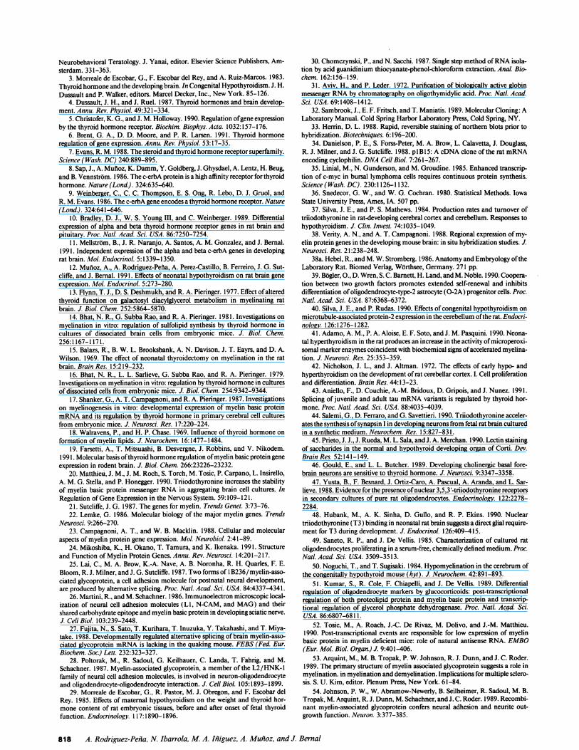
brain T3 levels may be normal even ifplasma and other tissuesshow low levels ofthe hormone. Fig. 1 shows that T4 increasedin normal animals from 0.9 ng/g on P5 to 2 ng/g on P20 (P< 0.001). After MMI treatment to the dams from the 9th dafter conception, T4 levels in the cerebral cortex of the off-springs 5 d after birth was 0.1 ng/g. T4 increased in hypothy-roid, P20 animals, but its concentration was still only 0.2 ng/g;i.e., 10-fold lower than in normal animals of the same age. T3concentrations increased in normal animals from 3.2 to 3.8ng/g (P < 0.01) from P5 to P20. In hypothyroid animals, T3concentrations were 0.14 and 0.21 ng/g, respectively. Theseresults demonstrate that the combined treatment of chemicaland surgical thyroidectomy ensured low tissue levels ofT4 andT3 throughout the developmental period studied.
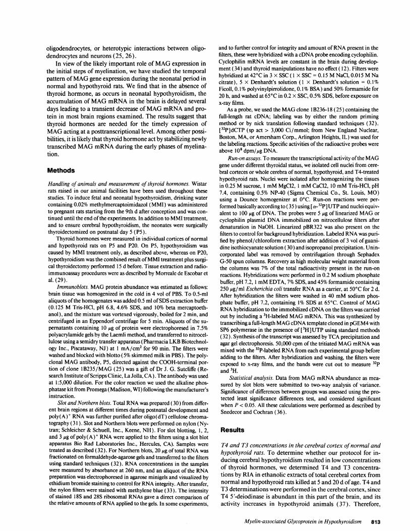
Developmental expression ofMAG mRNA. The expressionof the MAG gene during development was studied in slot blotsofpoly(A)' RNA samples isolated from cerebra ofnormal andhypothyroid animals at different times after birth. 1-, 2-, and3-,gg aliquots of RNA obtained from individual rats were ap-plied to the filters, which were hybridized with a MAG cDNAprobe. Fig. 2 shows densitometric measurements of the corre-sponding signals in the radioautograms from individual ani-mals. MAG mRNA was not detected during the first 5-7 dafter birth. In normal animals, it increased in abundance, there-after reaching maximal levels around day 20. In hypothyroidanimals, MAG mRNA accumulation in total brain also in-creased with age, but more slowly and with an apparent delaywhen compared with normal animals. The data points werecomputer-fitted to generate the curves depicted, with signifi-cance levels ofP < 0.01 for the control curve, and P < 0.001 forthe hypothyroid curve. Two-way analysis of variance taking asfactors age and thyroid status, indicated that as a whole the datafrom hypothyroid animals were different from the controls (Fratio = 9.0, P = 0.008). When individual time points wereconsidered, there were no significant differences between hypo-thyroid and control values beyond 20 d of age, when theplateau of MAG mRNA was attained.
0 I.~~~~09
U,
0 5 1 0 1 5 20 25 30Days after birth
Figure 2. Developmental pattern ofMAG mRNA expression in nor-mal and hypothyroid rats. MAG mRNA was measured in slot blotsusing poly(A)+ RNA purified from whole cerebra of normal (blacksquares and solid line) and hypothyroid (open dots and dotted line)animals. The data represented are individual values, or mean±SD ofdata from three different animals.
Temporal and regional effects ofhypothyroidism on MAGgene expression. The results described above suggest that a ma-jor effect ofhypothyroidism on MAG gene expression is a tran-sient delay in the normal developmental pattern. However,myelination has not the same timing in all brain regions, pro-ceeding normally from caudal to rostral areas (38). In otherwords, in the above experiment, we examined a mixture ofwidely heterogeneous regions at different stages of myelina-tion. Therefore, we examined the effect of hypothyroidism onthe regional pattern of expression of the MAG gene during thefirst month of life. Brains from normal and hypothyroid ani-mals were individually dissected into regions including cere-bral cortex, striatum, hippocampus, mesencephalon, and hypo-thalamus. RNA was isolated from individual cortices or frompooled regions as described, and the mRNA encoding MAG,
,~z
5
4
0l)I-
0)c
3
2i
01
oi
T4 T3
I
DAYS AFTER BIRTH
Figure 1. T4 and T3 concentrations in the cerebral cortices of normaland hypothyroid rats. T4 and T3 were measured by radioimmunoas-say in extracts from the cerebral cortices of normal (solid bars) andhypothyroid (hatched bars) rats at 5 and 20 d after birth. Differencesbetween normal and hypothyroid animals were statistically signifi-cant, with P < 0.001.
4 k
LL 31-
4 2 -
A ND
1 0
B-~
20 20
Age groups
B l A
2 5 3 0
Figure 3. Effect of hypothyroidism on MAG mRNA abundance inthe cerebral cortex. Total RNA was purified from individual corticesat 10, 20, and 30 d (experiment A) or at 20 and 25 d (experiment B)after birth, and analyzed by Northern blotting using a labeled MAGcDNA as a probe. The filter was rehybridized with a probe for cyclo-philin, an ubiquitous mRNA unaffected by hypothyroidism ( 12), andthe ratio MAG to cyclophilin was measured. In experiment A, weprocessed three individual samples from each group, and the data aremean±SD ofthree values. MAG mRNA was not detected on day 10(ND). In experiment B, we analyzed pools oftwo samples from eithernormal or hypothyroid animals.
814 A. Rodriguez-Pefna, N. Ibarrola, M. A. Ifliguez, A. Muiloz, and J. Bernal

was identified by Northern blotting. Fig. 3 shows the resultsobtained using cerebral cortices of normal and hypothyroidrats of different ages. (The rat cerebral cortex contains an esti-mated 1.1 million callosal fibers, from which about two thirdsare myelinated. (38a) These fibers enter the cortical platearound day 3 after birth, and the adult pattern is attained at theend ofthe 1st wk oflife. In our experiments, cortex MAG arisesfrom the cortex proper and from the subcortical white matter.)Two different sets of data are represented in the figure. Datalabeled A are the mean±SD of three individual cortices fromeach age group. Data labeled B are single determinations frompools of two different cortices. Results in each case are verysimilar. The MAG mRNA bands were quantified by densitom-etry after Northern blotting and related to the intensity of acyclophilin mRNA control. On day, 1O little MAG mRNA waspresent in normal cortex, and it was undetectable in the cortexof hypothyroid animals. At any subsequent time, hypothyroidlevels were reduced by - 80% or more compared to normalvalues.
Fig. 4 shows Northern blot patterns ofMAG mRNA in four
different regions of the brain from normal controls (C) andhypothyroid (H) rats throughout the first month of life, at 5,10, 15, 20, and 30 d of age. Pools of three samples were pro-cessed in these experiments. The results show that the effect ofhypothyroidism depends on the region examined and the ageof the animals. In the hippocampus, maximal amounts ofMAG mRNA were attained at around 25 d of age in normalanimals. In hypothyroid animals, however, lower levels werefound at all ages, and by 25 d, normal amounts were not yetattained. In contrast to the cortex and hippocampus, the effectof hypothyroidism in the striatum, mesencephalon, and hypo-thalamus was evident only at the earliest stages of myelination,from the 10th to the 15th postnatal days.
Patterns ofMAG expression in normal and hypothyroidanimals. Fig. 5 shows the results ofMAG expression in immu-noblots of different regions of normal and hypothyroid brains.Different ages along the neonatal period are represented in thefigure. The P5 antibody used for the immunoblots detected amain 100-1 16 kD protein band that was not present in thebrain before myelination. This band was not detected in con-trol experiments using extracts from other organs such as liveror kidney (not shown). The blots were overdeveloped to in-
10 15 20 25st C H C HC H CH4 _ a
HIPPOCAMPUS116 .- " _W84-58.-5- _48.5--
10 15 20 25 30st C H C H C HC H C H
_ U, 6l .o
_
- - -
_ _
011_bxM-V
STRIATUM
C C H C C H C C HT6 1 S -20 25 30
CORTEX
10 15 20 25st C H C H C H C H
*,
HIPPOCAMPUS
10 15 20 25 30st C H C H C H C H C H
116 -84-58-
48.5-
MESENCEPHALON
C C C C C H C1 25 1F-
HYPOTHALAMUS
C H CH _C R C H C H C H5 10 Z 025u
Figure 4. Developmental pattern of MAG mRNA in brain regions.Total RNA was purified from pooled (three samples for each pool)regions individually dissected from the brains of normal (C) and hy-pothyroid (H) rats at different days after birth. After transfer the ny-lon filters were stained to visualize ribosomal RNA to control for in-tegrity and amount ofRNA present in each lane (not shown). Thelanes contained similar amounts of RNA, except for C 15 and H 15 inthe striatum, which contained lower and higher amounts, respec-tively, and H15 in the hypothalamus that contained much loweramount of RNA than calculated.
STRATUM MESENCEPHALON10 15 25 30
C H C H C H C N
-MAG
CEREBELLUM
Figure 5. Developmental pattern ofMAG in normal and hypothyroidrats. Protein extracts from different brain regions at different ages
were electrophoresed in denaturing polyacrylamide gels and electro-blotted to nitrocellulose. The filters were probed with a polyclonalantibody directed against the COOH-terminal portion of MAG.Shown are the results obtained using three pooled samples from nor-
mal (C) or hypothyroid (H) killed at 10, 15, 20, 25, and 30 d ofpostnatal age. MAG was present as a broad protein band spanning insize from 100 to 116 kD.
Myelin-associated Glycoprotein in Hypothyroidism 815
0 #*%wow

crease the intensity of a nonspecific band of 58 kD that was notaltered by hypothyroidism, and thus served as control for theamount ofprotein loaded on the gels. In the cortex and hippo-campus of normal rats, MAG was almost undetectable on PO0,and was clearly present on P15. It increased subsequentlyreaching high levels on P25 and P30. In striatum and mesen-
cephalon, MAG was already present on PlO, and increasedthereafter reaching maximal levels on P20-P25. There was a
clear effect of hypothyroidism in the cerebral cortex, hippo-campus and striatum, with very low levels ofMAG up to P20compared with normal animals. In hypothyroid animals, levelsofMAG increased after P20 and, therefore, the differences withnormal animals tended to disappear. The effect ofhypothyroid-ism in the mesencephalon was less evident, and there was some-what less MAG in the hypothyroid samples of P15 and P20.The transient effect of hypothyroidism on MAG protein was
also evident in the cerebellum, where only on P10 there was
clearly a lower amount of protein in hypothyroid rats.Hyperthyroidism accelerates MAG mRNA expression. If
thyroid hormones are involved in the timing ofMAG expres-
sion, it is likely that contrary to the effect of hypothyroidism,thyroid hormone excess induced a precocious expression oftheMAG gene. To study this possibility, normal or hypothyroidanimals were administered 5 ,ug ofT4 per rat during 5 d beforedeath to evaluate the effect of hyperthyroidism. MAG mRNAwas examined by Northern blotting in the striatum on day 7and in the cerebral cortex on days 10 and 12 (Fig. 6). On day 7,no MAG mRNA was detected in untreated normal or hypothy-roid animals, but it was clearly present in both groups of T4-treated rats. According to previous results, very little MAG was
also detected in the cerebral cortex on day 10 in normal ani-mals, and again T4 treatment resulted in MAG mRNA accu-
mulation in both control and hypothyroid animals. On day 12,MAG was already clearly expressed in the cortex of normalrats, but was almost absent in hypothyroid animals. T4 treat-ment increased MAG expression in both groups. These resultsdemonstrate that thyroid hormone excess resulted in an acceler-ation ofMAG mRNA accumulation.
Thyroid hormones do not affect the rate oftranscription ofthe MAG gene. To examine the cause of the effects of thyroidhormone deficiency or excess, we performed run-on analysesusing nuclei from normal, hypothyroid, or T4-treated rats as
described above. The in vitro-labeled nuclear transcripts werehybridized with full-length cDNAs encoding MAG, cyclophi-lin as a thyroid hormone independent message, and pBR322 as
control for nonspecific hybridization. As a source of nuclei wefirst used cerebral corteces of 1 2-d-old animals as in the experi-
STRIATUM CORTEXC H C H C H
T4 -+- + + - + -+ -+
NORTHERN tsBLOTS
P7
C # -MAG
plo P12
Figure 6. Effect of hypothyroidism on MAG mRNA accumulation.Hypothyroid and control animals were treated (+) or untreated (-)with a single daily dose of 5 gg T4 during the last 5 d before death.Rats were killed at 7, 10, and 12 d and MAG mRNA was measuredby Northern blotting in striatum (7 d) or cerebral cortex ( 10 and 12 d).
ment illustrated in Fig. 6. A representative result from one oftwo independent experiments is illustrated in Fig. 7 A, showingno obvious significant differences among the three experimen-tal groups. The mean MAG/cyclophilin ratios obtained afterdensitometry for the two experiments were 1.1 (control), 1.3(hypothyroid) and 0.8 (T4 treated). We also used nuclei fromtotal cerebra of 13-d-old animals, which were expected to bemore actively transcribing the MAG gene. To better quantifythe results and to correct for eventual differences in the amountofMAG RNA hybridized to the respective probes, we includeda [3H ]MAGmRNA in the hybridization mixture. After hybrid-ization and exposure to x-ray films, the bands were cut out andradioactivity was measured. The results shown in Fig. 7 B,again did not reveal significant differences between normal andhypothyroid animals. The signal for T4-treated animals was
slightly higher, both in the MAG probe and the CF controlprobe. After quantification ofthe data, taking into account thetotal amount of 32p present in the hybridization mixtures ( 1.4,1.6, and 2.1 million cpm for control, hypothyroid, and T4treated, respectively) and the percentage [3H ]MAG mRNAhybridized (control = 4.27, hypothyroid = 2.52, T4 treated= 4.07), the percent of total 32P hybridized was 0.20 (controlrats), 0.24 (hypothyroid rats), and 0.18 (T4-treated rats).Taken together from the results of the three experiments, we
conclude that MAG mRNA changes with thyroid status werenot caused by altered MAG transcription.
Discussion
It is known that one of the most severe effects of neonatalhypothyroidism is a lower deposition of myelin in the centralnervous system. Hypothyroid brains have decreased amountsof cholesterol, cerebrosides, sulfatides, glycolipids, sulfolipids,and gangliosides in the myelin sheaths ( 13-18). These effectsappear to be the consequence of decreased activities of en-zymes involved in myelin lipid synthesis, such as cerebrosidesulfotransferase and galactosyl transferases ( 16). At present, itis not known whether thyroid hormone controls directly theactivity of these enzymes or some other key event leading tomyelination. Myelination is a highly regulated timely eventthat in the rat starts suddenly a few days after birth and depends
C H T4A MAG
CF
pBR
B MAG
CF
pBR
Cortex
Cerebrum
Figure 7. Run-on analysis of the effect of hypothyroidism and T4treatment on the MAG gene. Run-on analyses were performed usingnuclei from pooled (three rats per pool) cerebral cortices (A), or cere-
bra (B) ofnormal, hypothyroid or T4 treated rats as described above.Cortex nuclei were from 1 2-d-old animals and cerebral nuclei from1 3-d-old animals. As probes we used MAG, cyclophilin (CS), andpBR322. No hybridization was obtained using nuclei transcribed inthe presence of alpha-amanitin (not shown).
816 A. Rodriguez-Penia, N. Ibarrola, M. A. Iniguez, A. Munioz, and J. Bernal
-, - m- ~m.

on the proper differentiation of oligodendrocytes from its 02Aprecursor cells. Oligodendrocyte differentiation is determinedby the concerted actions of fibroblast and platelet-derivedgrowth factors (39). As shown in this paper, thyroid hormoneis also required for full expression of at least one of the differen-tiated properties of oligodendrocytes; i.e. expression of theMAG gene, and its main role is to ensure an accurate timing ofgene expression.
Hypothyroidism led to a delay in the pattern ofMAG accu-mulation in different brain regions. The regional effect of hypo-thyroidism reflects the fact that myelination does not proceedsimultaneously throughout the brain. Normally, the myelina-tion wave starts in caudal regions and proceeds towards rostralareas. This pattern has been deduced from studies of myelinmRNA accumulation by either Northern blots or in situ hybrid-ization (38). We show here that regions that myelinate last aremore affected by hypothyroidism than those of earlier myelina-tion. From the patterns of expression of MAG mRNA or pro-tein, we conclude that caudal regions such as cerebellum, hypo-thalamus, and mesencephalon present a transient alterationwith spontaneous recovery beyond days 15-20 after birth. Thispattern is similar to the effect of hypothyroidism on microtu-bule-associated protein-2 in the cerebellum (40). In contrast,normalization in the hippocampus and striatum did not occurbefore days 25-30, and myelination in the cerebral cortex wasimpaired beyond the first month of life.
The timing control by thyroid hormone is also supportedby the effect of hyperthyroidism. In the presence ofan excess ofthyroid hormone, MAG expression was accelerated in previ-ously normal or hypothyroid animals. This is in agreementwith other results reporting an acceleration of myelination inhypothyroid rats, with precocious increases of myelin enzymeactivities (41 ). A clear timing control by thyroid hormone alsooccurs during cerebellar development (42), in the transitionbetween the juvenile and adult forms of Tau (43), in the ex-pression of neuronal markers such as synapsin I in culture(44), during development of the organ of Corti (45), or in theappearance ofenzymes during development ofcholinergic neu-rons (46).
The action of thyroid hormone on MAG expression couldbe exerted directly by the interaction of the thyroid hormonereceptor with promoter sequences in the MAG gene. Althoughwe have detected no labeling in white matter by in situ hybrid-ization using c-erbA probes ( 11 ), oligodendrocytes in primarycultures have been shown to express the thyroid hormone re-ceptor (47). T3 receptors have recently been demonstrated inglial cells isolated from rat brain during the neonatal period(48). Highest receptor density was found in the cerebral cortex.The rest of brain regions contained much fewer amounts ofreceptor and, contrary to the cortex, receptor density declinedduring development, especially in the cerebellum. These dataagree with the transient dependency ofmyelin genes during theearly stages of myelination reported in this paper. Recently,Nikodem and associates have described the presence of se-quences in the promoter region of myelin basic protein thathave functional characteristics of a thyroid hormone respon-sive element ( 19). In contrast, we find that neither hypothy-roidism nor thyroid hormone treatment modified significantlythe rates oftranscription ofthe MAG gene. Therefore, hypothy-roidism could have led to a decreased stability of MAGmRNA. An increased degradation in the presence of an equalrate of synthesis would be predicted to result in a delay ofmRNA accumulation. Others have also shown an effect of thy-
roid hormone on MBP gene expression in aggregating braincell cultures, which is mediated through an action on mRNAstability rather than transcription (20). The fact that the rate oftranscription was not modified is also an argument against apossible effect on the number of oligodendrocytes, which is inagreement with the known lack ofeffect ofthyroid hormone onoligodendrocyte proliferation (49). Other reports have alsosuggested that the number ofoligodendrocytes does not changein hypothyroidism (50). Although an effect on mRNA trans-port from the nucleus to the cytoplasm cannot be discarded atpresent, it is likely that thyroid hormone is required to stabilizenewly transcribed MAG mRNA during the earlier phases ofmyelination to allow for a rapid accumulation of the tran-scripts and protein production. This is not surprising, in viewof the fact that although the main control on myelin gene ex-pression is exerted at the level of transcription by oligodendro-cyte-specific transcription factors (21-24), there is also controlat the posttranscriptional level. For example, glucocorticoidsposttranscriptionally regulate the expression of basic proteinand proteolipid protein (5 1 ). The deficits ofmyelination ofthemyelin deficient mice (mid) are also caused by posttranscrip-tional events (52).
As we have shown previously ( 12), other oligodendrocytespecific genes as MBP and proteolipid protein are also affectedby neonatal hypothyroidism. The question arises as to whetherthyroid hormone influences the expression ofeach myelin geneby a similar mechanism, or the effects are the end result of theregulation of an as yet unidentified key developmental genethat could be influenced by thyroid hormone itself, or by thy-roid hormone-dependent factors, including growth hormone.Whether thyroid hormone acts directly or indirectly, its effecton the expression ofMAG would be critical early in myelina-tion. Because of its cell-adhesion properties, its localization inthe periaxonal space and uncompacted myelin, and its timingof expression in relation to other myelin proteins (53), MAGhas been proposed to establish initial interactions between theoligodendrocyte surface and the axon to be myelinated, and inthe maintenance of this interaction.
Independently ofthe mechanism ofaction, a delay ofmyeli-nation, even iftransient, would result in serious disturbances ofbrain maturation, since the normal coordination between axongrowth and targeting and myelination will not occur. The al-tered pattern of myelination should have profound conse-quences on the hypothyroid brain in terms of neuron perfor-mance and development of fiber networks. In addition to itsrole in myelination, MAG has also neurite outgrowth activity(54), an impairment of which would constitute an additionalfactor contributing to the complexity and severity of the hypo-thyroid brain phenotype.
Acknowledgments
We thank Dr. Gabriella Morreale de Escobar for helpful discussionsand determinations of T4 and T3 by RIA. We also thank Dr. J. G.Sutcliffe for the MAG cDNA probe and the MAG antibody.
This work was supported by grants from Direccion General de In-vestigacion Cientifica y Tecnica (PM88-0006), from Antibi6ticos-Pharma S.A. (Madrid, Spain), and from the Community of Madrid(C254/90).
References1. Eayrs, J. T. 1968. Developmental relationships between brain and thyroid.
In Endocrinology and Human Behaviour. R. P. Michael, editor. Oxford Univer-sity Press, London. 239-255.
2. Legrand, J. 1984. Effects ofthyroid hormones on central nervous system. In
Myelin-associated Glycoprotein in Hypothyroidism 817
Neurobehavioral Teratology. J. Yanai, editor. Elsevier Science Publishers, Am-sterdam. 331-363.
3. Morreale de Escobar, G., F. Escobar del Rey, and A. Ruiz-Marcos. 1983.Thyroid hormone and the developing brain. In Congenital Hypothyroidism. J. H.Dussault and P. Walker, editors. Marcel Decker, Inc., New York. 85-126.
4. Dussault, J. H., and J. Ruel. 1987. Thyroid hormones and brain develop-ment. Annu. Rev. Physiol. 49:321-334.
5. Christofer, K. G., and J. M. Holloway. 1990. Regulation ofgene expressionby the thyroid hormone receptor. Biochim. Biophys. Acta. 1032:157-176.
6. Brent, G. A., D. D. Moore, and P. R. Larsen. 1991. Thyroid hormoneregulation of gene expression. Annu. Rev. Physiol. 53:17-35.
7. Evans, R. M. 1988. The steroid and thyroid hormone receptor superfamily.Science (Wash. DC) 240:889-895.
8. Sap, J., A. Mufioz, K. Damm, Y. Goldberg, J. Ghysdael, A. Lentz, H. Beug,and B. Vennstrom. 1986. The c-erbA protein is a high affinity receptor for thyroidhormone. Nature (Lond.). 324:635-640.
9. Weinberger, C., C. C. Thompson, E. S. Ong, R. Lebo, D. J. Gruol, andR. M. Evans. 1986. The c-erbA gene encodes a thyroid hormone receptor. Nature(Lond.). 324:641-646.
10. Bradley, D. J., W. S. Young III, and C. Weinberger. 1989. Differentialexpression of alpha and beta thyroid hormone receptor genes in rat brain andpituitary. Proc. Nati. Acad. Sci. USA. 86:7250-7254.
11. Mellstrbm, B., J. R. Naranjo, A. Santos, A. M. Gonzalez, and J. Bernal.1991. Independent expression of the alpha and beta c-erbA genes in developingrat brain. Mol. Endocrinol. 5:1339-1350.
12. Muftoz, A., A. Rodriguez-Pefia, A. Perez-Castillo, B. Ferreiro, J. G. Sut-cliffe, and J. Bernal. 1991. Effects of neonatal hypothyroidism on rat brain geneexpression. Mol. Endocrinol. 5:273-280.
13. Flynn, T. J., D. S. Deshmukh, and R. A. Pieringer. 1977. Effect ofalteredthyroid function on galactosyl diacylglycerol metabolism in myelinating ratbrain. J. Biol. Chem. 252:5864-5870.
14. Bhat, N. R., G. Subba Rao, and R. A. Pieringer. 1981. Investigations onmyelination in vitro: regulation of sulfolipid synthesis by thyroid hormone incultures of dissociated brain cells from embryonic mice. J. Biol. Chem.256:1167-1171.
15. Balazs, R., B. W. L. Brooksbank, A. N. Davison, J. T. Eayrs, and D. A.Wilson. 1969. The effect of neonatal thyroidectomy on myelination in the ratbrain. Brain Res. 15:219-232.
16. Bhat, N. R., L. L. Sarlieve, G. Subba Rao, and R. A. Pieringer. 1979.Investigations on myelination in vitro: regulation by thyroid hormone in culturesof dissociated cells from embryonic mice. J. Bio. Chem. 254:9342-9344.
17. Shanker, G., A. T. Campagnoni, and R. A. Pieringer. 1987. Investigationson myelinogenesis in vitro: developmental expression of myelin basic proteinmRNA and its regulation by thyroid hormone in primary cerebral cell culturesfrom embryonic mice. J. Neurosci. Res. 17:220-224.
18. Walravens, P., and H. P. Chase. 1969. Influence of thyroid hormone onformation of myelin lipids. J. Neurochem. 16:1477-1484.
19. Farsetti, A., T. Mitsuashi, B. Desvergne, J. Robbins, and V. Nikodem.1991. Molecular basis ofthyroid hormone regulation ofmyelin basic protein geneexpression in rodent brain. J. Biol. Chem. 266:23226-23232.
20. Matthieu, J. M., J. M. Roch, S. Torch, M. Tosic, P. Carpano, L. Insirello,A. M. G. Stella, and P. Honegger. 1990. Triiodothyronine increases the stabilityof myelin basic protein messenger RNA in aggregating brain cell cultures. InRegulation of Gene Expression in the Nervous System. 59:109-121.
21. Sutcliffe, J. G. 1987. The genes for myelin. Trends Genet. 3:73-76.22. Lemke, G. 1986. Molecular biology of the major myelin genes. Trends
Neurosci. 9:266-270.23. Campagnoni, A. T., and W. B. Macklin. 1988. Cellular and molecular
aspects of myelin protein gene expression. Mol. Neurobiol. 2:41-89.24. Mikoshiba, K., H. Okano, T. Tamura, and K. Ikenaka. 1991. Structure
and Function of Myelin Protein Genes. Annu. Rev. Neurosci. 14:201-217.25. Lai, C., M. A. Brow, K.-A. Nave, A. B. Noronha, R. H. Quarles, F. E.
Bloom, R. J. Milner, and J. G. Sutcliffe. 1987. Two forms of 1B236/myelin-asso-ciated glycoprotein, a cell adhesion molecule for postnatal neural development,are produced by alternative splicing. Proc. Nail. Acad. Sci. USA. 84:4337-4341.
26. Martini, R., and M. Schachner. 1986. Immunoelectron microscopic local-ization of neural cell adhesion molecules (LI, N-CAM, and MAG) and theirshared carbohydrate epitope and myelin basic protein in developing sciatic nerve.J. Cell Biol. 103:239-2448.
27. Fujita, N., S. Sato, T. Kurihara, T. Inuzuka, Y. Takahashi, and T. Miya-take. 1988. Developmentally regulated alternative splicing ofbrain myelin-asso-ciated glycoprotein mRNA is lacking in the quaking mouse. FEBS (Fed. Eur.Biochem. Soc.) Lett. 232:323-327.
28. Poltorak, M., R. Sadoul, G. Keilhauer, C. Landa, T. Fahrig, and M.Schachner. 1987. Myelin-associated glycoprotein, a member of the L2/HNK-1family of neural cell adhesion molecules, is involved in neuron-oligodendrocyteand oligodendrocyte-oligodendrocyte interaction. J. Cell Biol. 105:1893-1899.
29. Morreale de Escobar, G., R. Pastor, M. J. Obregon, and F. Escobar delRey. 1985. Effects of maternal hypothyroidism on the weight and thyroid hor-mone content of rat embryonic tissues, before and after onset of fetal thyroidfunction. Endocrinology. 117:1890-1896.
30. Chomczynski, P., and N. Sacchi. 1987. Single step method ofRNA isola-tion by acid guanidinium thiocyanate-phenol-chloroform extraction. Anal. Bio-chem. 162:156-159.
31. Aviv, H., and P. Leder. 1972. Purification of biologically active globinmessenger RNA by chromatography on oligothymidylic acid. Proc. Nall. Acad.Sci. USA. 69:1408-1412.
32. Sambrook, J., E. F. Fritsch, and T. Maniatis. 1989. Molecular Cloning: ALaboratory Manual. Cold Spring Harbor Laboratory Press, Cold Spring, NY.
33. Herrin, D. L. 1988. Rapid, reversible staining of northern blots prior tohybridization. Biotechniques. 6:196-200.
34. Danielson, P. E., S. Forss-Peter, M. A. Brow, L. Calavetta, J. Douglass,R. J. Milner, and J. G. Sutcliffe. 1988. plB15: A cDNA clone of the rat mRNAencoding cyclophilin. DNA Cell Biol. 7:261-267.
35. Linial, M., N. Gunderson, and M. Groudine. 1985. Enhanced transcrip-tion of c-myc in bursal lymphoma cells requires continuous protein synthesis.Science(Wash. DC). 230:1126-1132.
36. Snedecor, G. W., and W. G. Cochran. 1980. Statistical Methods. IowaState University Press, Ames, IA. 507 pp.
37. Silva, J. E., and P. S. Mathews. 1984. Production rates and turnover oftriiodothyronine in rat-developing cerebral cortex and cerebellum. Responses tohypothyroidism. J. Clin. Invest. 74:1035-1049.
38. Verity, A. N., and A. T. Campagnoni. 1988. Regional expression of my-elin protein genes in the developing mouse brain: in situ hybridization studies. J.Neurosci. Res. 2 1:238-248.
38a. Hebel, R., and M. W. Stromberg. 1986. Anatomy and Embryology oftheLaboratory Rat. Biomed Verlag, Worthsee, Germany. 271 pp.
39. Bbgler, O., D. Wren, S.C. Barnett, H. Land, andM. Noble. 1990. Coopera-tion between two growth factors promotes extended self-renewal and inhibitsdifferentiation of oligodendrocyte-type-2 astrocyte (0-2A) progenitor cells. Proc.Natl. Acad. Sci. USA. 87:6368-6372.
40. Silva, J. E., and P. Rudas. 1990. Effects of congenital hypothyroidism onmicrotubule-associated protein-2 expression in the cerebellum ofthe rat. Endocri-nology. 126:1276-1282.
41. Adamo, A. M., P. A. Aloise, E. F. Soto, and J. M. Pasquini. 1990. Neona-tal hyperthyroidism in the rat produces an increase in the activity of microperoxi-somal marker enzymes coincident with biochemical signs of accelerated myelina-tion. J. Neurosci. Res. 25:353-359.
42. Nicholson, J. L., and J. Altman. 1972. The effects of early hypo- andhyperthyroidism on the development of rat cerebellar cortex. I. Cell proliferationand differentiation. Brain Res. 44:13-23.
43. Aniello, F., D. Couchie, A.-M. Bridoux, D. Gripois, and J. Nunez. 1991.Splicing of juvenile and adult tau mRNA variants is regulated by thyroid hor-mone. Proc. Natl. Acad. Sci. USA. 88:40354039.
44. Salemi, G., D. Ferraro, and G. Savettieri. 1990. Triiodothyronine acceler-ates the synthesis of synapsin I in developing neurons from fetal rat brain culturedin a synthetic medium. Neurochem. Res. 15:827-831.
45. Prieto, J. J., J. Rueda, M. L. Sala, and J. A. Merchan. 1990. Lectin stainingof saccharides in the normal and hypothyroid developing organ of Corti. Dev.Brain Res. 52:141-149.
46. Gould, E., and L. L. Butcher. 1989. Developing cholinergic basal fore-brain neurons are sensitive to thyroid hormone. J. Neurosci. 9:3347-3358.
47. Yusta, B., F. Besnard, J. Ortiz-Caro, A. Pascual, A. Aranda, and L. Sar-lieve. 1988. Evidence for the presence of nuclear 3,5,3'-triiodothyronine receptorsin secondary cultures of pure rat oligodendrocytes. Endocrinology. 122:2278-2284.
48. Hubank, M., A. K. Sinha, D. Gullo, and R. P. Ekins. 1990. Nucleartriiodothyronine (T3) binding in neonatal rat brain suggests a direct glial require-ment for T3 during development. J. Endocrinol. 126:409-415.
49. Saneto, R. P., and J. De Vellis. 1985. Characterization of cultured ratoligodendrocytes proliferating in a serum-free, chemically defined medium. Proc.Nail. Acad. Sci. USA. 3509-3513.
50. Noguchi, T., and T. Sugisaki. 1984. Hypomyelination in the cerebrum ofthe congenitally hypothyroid mouse (hyt). J. Neurochem. 42:891-893.
51. Kumar, S., R. Cole, F. Chiapelli, and J. De Vellis. 1989. Differentialregulation of oligodendrocyte markers by glucocorticoids: post-transcriptionalregulation of both proteolipid protein and myelin basic protein and transcrip-tional regulation of glycerol phosphate dehydrogenase. Proc. Natl. Acad. Sci.USA. 86:6807-681 1.
52. Tosic, M., A. Roach, J.-C. De Rivaz, M. Dolivo, and J.-M. Matthieu.1990. Post-transcriptional events are responsible for low expression of myelinbasic protein in myelin deficient mice: role of natural antisense RNA. EMBO(Eur. Mol. Biol. Organ.) J. 9:401-406.
53. Arquint, M., M. B. Tropak, P. W. Johnson, R. J. Dunn, and J. C. Roder.1989. The primary structure of myelin associated glycoprotein suggests a role inmyelination. in myelination and demyelination. Implications for multiple sclero-sis. S. U. Kim, editor. Plenum Press, New York. 61-84.
54. Johnson, P. W., W. Abramow-Newerly, B. Seilheimer, R. Sadoul, M. B.Tropak, M. Arquint, R. J. Dunn, M. Schachner, andJ. C. Roder. 1989. Recombi-nant myelin-associated glycoprotein confers neural adhesion and neurite out-growth function. Neuron. 3:377-385.
818 A. Rodriguez-Peqla, N. Ibarrola, M. A. Ifliguez, A. Muiloz, and J. Bernal
Related Documents