Neonatal Electroencephalography: Normal and Abnormal Eli M. Mizrahi, M.D. Peter Kellaway Section of Neurophysiology Department of Neurology Section of Pediatric Neurology Department of Pediatrics Baylor College of Medicine St. Luke’s Episcopal Hospital and Texas Children’s Hospital Houston, Texas

Welcome message from author
This document is posted to help you gain knowledge. Please leave a comment to let me know what you think about it! Share it to your friends and learn new things together.
Transcript

Neonatal Electroencephalography:
Normal and AbnormalEli M. Mizrahi, M.D.
Peter Kellaway Section of NeurophysiologyDepartment of Neurology
Section of Pediatric NeurologyDepartment of Pediatrics
Baylor College of Medicine
St. Luke’s Episcopal Hospital andTexas Children’s Hospital
Houston, Texas

Learning Objectives
• Understand the clinical utility of neonatal electroencephalography
• Recognize normal developmental features of neonatal EEG
• Recognize abnormal features of neonatal EEG and their clinical implications
• Recognize electrographic seizure discharges

Disclosure Statement• Grant and Research Support
– National Institutes of Health, Bethesda, MD, USA– NeuroPace, Inc., Palo Alto, CA, USA
• Speaker’s Bureau– None
• Consultant– Michael E. DeBakey Veteran’s Affairs Medical
Center, Houston, TX, USA– SleepMed/Digitrace Care Services, Houston, TX,
USA• Major Stock Shareholder
– None• Other Financial/Material Support
– None
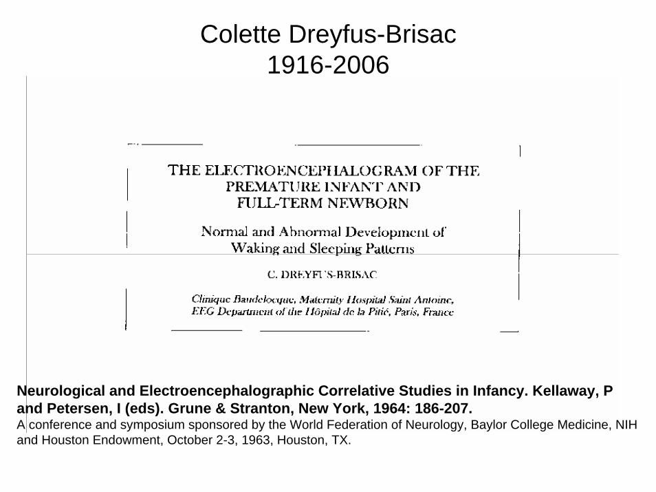
Colette Dreyfus-Brisac1916-2006
Neurological and Electroencephalographic Correlative Studies in Infancy. Kellaway, P and Petersen, I (eds). Grune & Stranton, New York, 1964: 186-207.A conference and symposium sponsored by the World Federation of Neurology, Baylor College Medicine, NIH and Houston Endowment, October 2-3, 1963, Houston, TX.

EEG in Assessment of the Neonate• General considerations
– Role of EEG relative to other testing modalities
– Technical considerations– Issues in interpretation
• Features of the normal and abnormal neonatal EEG
• Application to specific clinical problems

Neonatal EEG Best Utilized as a Correlative Study
• Infant’s medical history• Recording strategies based upon history and
physical findings• Clinical observation and stimulation during
recording• Relationship of EEG findings to other laboratory
findings• Consultation between neonatologist and
neurophysiologist
Application of Neonatal EEG in Consideration of Clinical Questions
• Determination of conceptional age• Assessment of diffuse brain injury
– Assessment of presence of brain death• Assessment of focal brain injury• Determination of timing of brain injury• Determination of prognosis• Diagnosis of neonatal seizures

Technical Considerations
• Electrode application• EEG channel selection• Polygraphic channels
– Respirations– Electrocardiogram– Electro-oculogram– Electromyogram

Recording Strategies
• Montage selection– Single montage– Inclusion of Cz and other midline regions
• Recording duration• Recording protocol
– Wake/sleep cycles– Suspected abnormal clinical events
Basis for Interpretation• The normal neonatal EEG undergoes rapid and
predictable changes as a consequence of brain growth and development.
• Abnormalities may be characterized by altered developmental characteristics as well as specific patterns or wave-forms.
• Patterns that are normal at one development stage may be abnormal at another.

Challenges in Interpretation
• The significance of some features of the neonatal EEG have not been determined.
• Certain abnormal EEG findings in the period immediately following brain injury may be transient– Their significance is related to change over
time– Underscores the need for serial recordings

Neonatal EEG and Brain Development
• Basic assumption– Brain development proceeds at the same rate
whether the infant is in the nursery or in utero• Cautions
– No intercurrent CNS injuries– Recently challenged by computer-based
analysis

Trends in Appearance of Neonatal EEG Parallel Brain Development
• Continuity• Synchrony• Waveforms• Wake/sleep cycles

Continuity• There is a gradual change from a
discontinuous pattern to a continuous pattern
27-28 wks CA
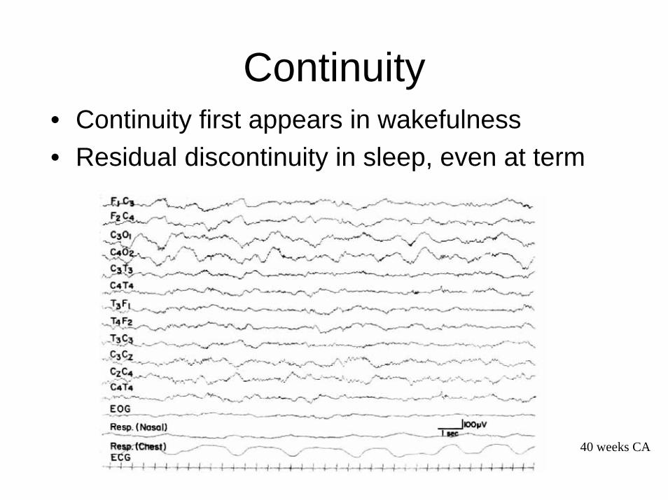
Continuity• Continuity first appears in wakefulness• Residual discontinuity in sleep, even at term
40 weeks CA

Developmental Trends of Discontinuity

Synchrony• Initial period of “hypersynchrony”• Associated with discontinuity
27-28 wks CA

Synchrony
• Initial period of synchrony– “hypersynchrony”
• Followed by asynchrony• Then gradual return to synchrony on the
two sides– Degree of synchrony increases with
conceptional age

Synchrony
29-30 weeks CA

Synchrony

Discontinuity and Asynchrony
29-30 weeks CA

Synchrony
40 weeks CA
Specific Waveforms• Emergence and disappearance of
specific waveforms– Specific character and location– Orderly progress
• “Grapho-elements”• Markers of conceptional age
– Beta-delta complexes: “brushes”– Temporal bursts: theta and alpha– Frontal sharp waves: “encoches frontales”

Grapho-elements

Beta-delta Complexes
• Hallmark of prematurity– Slow-wave with superimposed fast frequency
activity• Initially central• Eventually posterior• Onset: 29 weeks C.A.• Disappearance: 38 weeks
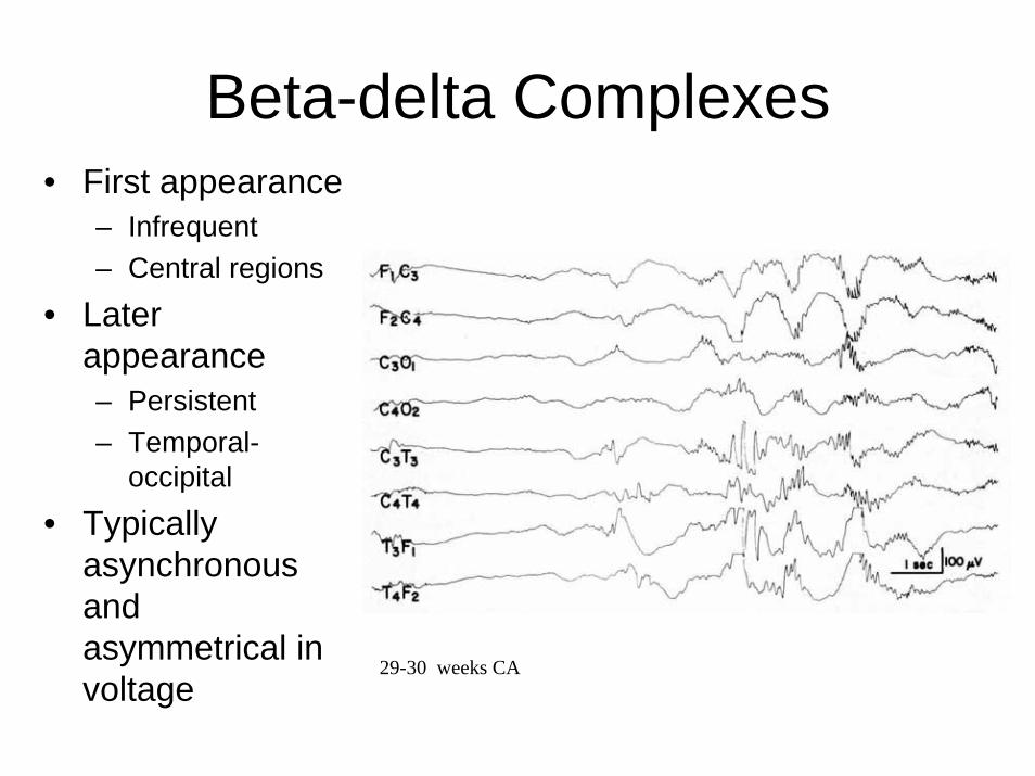
Beta-delta Complexes• First appearance
– Infrequent– Central regions
• Later appearance– Persistent– Temporal-
occipital
• Typically asynchronous and asymmetrical in voltage
29-30 weeks CA

Temporal Theta Bursts
30-32 weeks CA

Temporal Alpha Bursts
32-33 weeks CA

Frontal Sharp Transients• Synchronous, symmetrical• Onset: 34-35 weeks C.A.• Persist through term
36 weeks CA

Development of Reactivity and Wake/Sleep Cycles
• Changes in EEG activity in response to stimuli emerge at about 33-34 weeks C.A.– State-dependent
• Clear EEG differences between wakefulness and sleep emerge at about 36-37 weeks C.A.

Normal Features Waves
• There are some waveforms that are considered normal but not specifically considered age-dependent grapho-elements– Bifrontal delta in near-term and term infants

Bifrontal Delta
38 – 40 weeks CA

Waveforms of Uncertain Diagnostic Significance
• Midline sharp waves• Rhythmic theta and alpha activity

Midline Central Theta
40 weeks CA
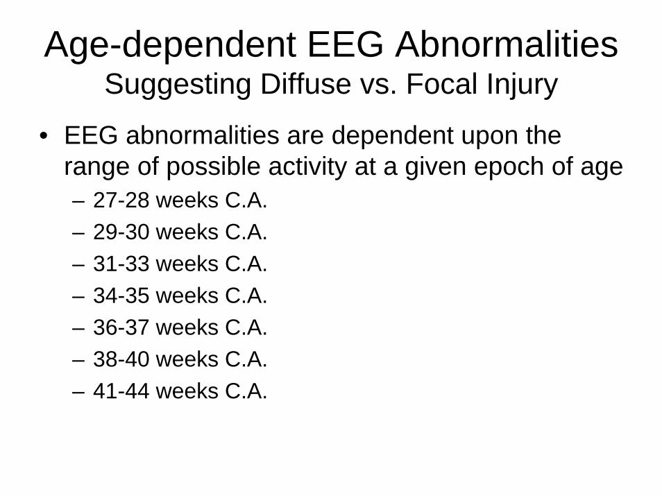
Age-dependent EEG AbnormalitiesSuggesting Diffuse vs. Focal Injury
• EEG abnormalities are dependent upon the range of possible activity at a given epoch of age– 27-28 weeks C.A.– 29-30 weeks C.A.– 31-33 weeks C.A.– 34-35 weeks C.A.– 36-37 weeks C.A.– 38-40 weeks C.A.– 41-44 weeks C.A.

Age-dependent Patterns ofDiffuse Brain Injury
• Prolonged interburst duration 27-28 wks C.A.• Depressed voltage 27-28 wks C.A.• Dyschronism 29-30 wks C.A.• Prolonged generalized voltage
attenuation 34-35 wks C.A.• Absence of sleep cycling 36-37 wks C.A.• Depressed and undifferentiated
background 36-37 wks C.A.• Suppression-burst activity 36-37 wks C.A.• Hypsarrhythmia 41-44 wks C.A

Dyschronism
• Determination of conceptional age depends upon the presence or absence of certain established developmental milestones characterized by specific EEG features.
• Dyschronism: the finding of developmental features that are inconsistent with age– Other EEG features– Infant’s actual age

Dyschronism
• External– Developmental
features in all wake/sleep states are immature for stated age
• EEG-age determination correct?
• Clinical-age determination correct?
– Immature EEG features evidence of delayed maturation
• Intrauterine cerebral insult
• Internal– Mixture of
developmental features present in different physiologic states
– Precise determination of conceptional age cannot be made
– Suggests diffuse dysfunction
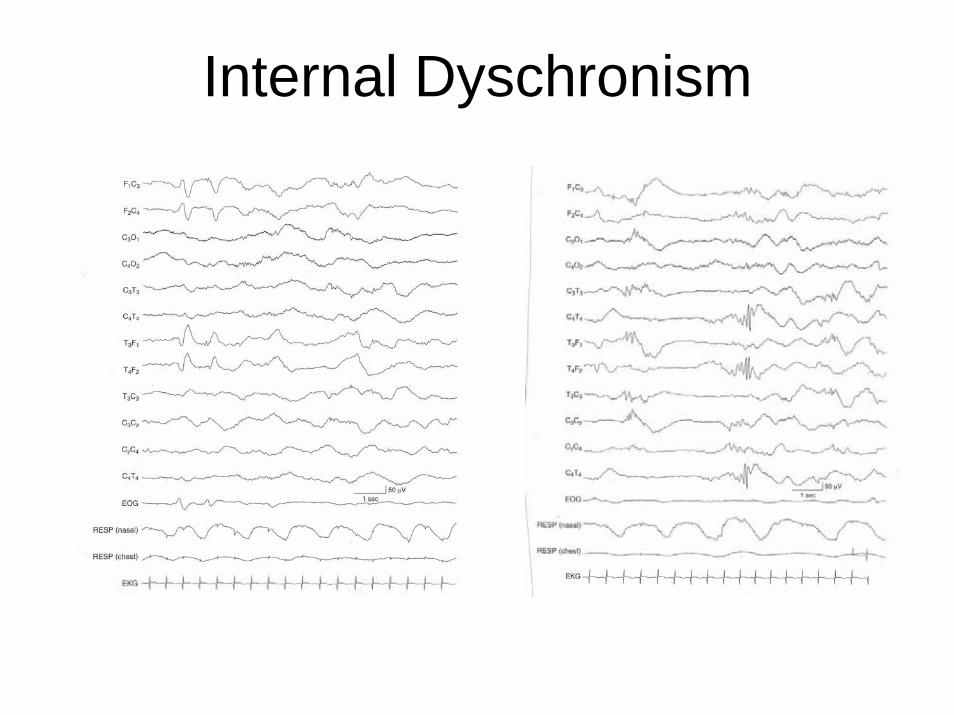
Internal Dyschronism

Depressed and UndifferentiatedBackground
40 weeks CA

Suppression-Burst Background
40 weeks CA

Suppression-burst Patterns
• Hypoxic-ischemic encephalopathy• In-born errors of metabolism

Suppression-burst Patterns

Suppression-burst Patterns

Age-dependent Patterns ofFocal Brain Injury
• Voltage depressionover one hemisphere 27-28 wks C.A.
• Central positive sharp waves 29-30 wks C.A.• Persistent focal sharp waves 31-33 wks C.A.• Electrical seizure activity 34-35 wks C.A.

Voltage DepressionOver One Hemisphere
40 weeks CA – subarachnoid hemorrhage

Central Positive Sharp Waves
• Postive rolandicsharp waves
• Initially associated with intraventricularhemorrhage
• Now more clearly associated with sequelae– Periventricular
leukomalacia

Central Positive Sharp Waves
29-30 weeks CA

Central Positive Sharp Waves
36 weeks CA - IVH

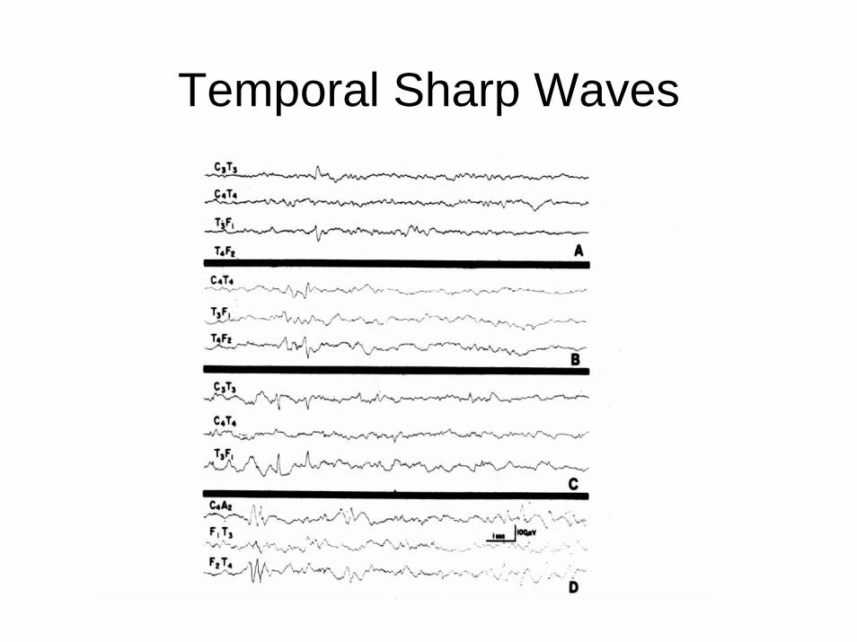
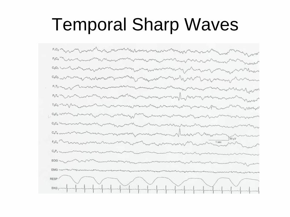
Temporal Sharp Waves
• May be present as normal activity• No clear criteria to differentiate normal
from abnormal temporal sharp waves• Consider
– Amplitude and duration– Occurrence– Complexity of waveform– Polarity– Changing states
Temporal Sharp Waves
Temporal Sharp Waves

Temporal Sharp Waves

Temporal Sharp Waves

Temporal Sharp Waves

Temporal Sharp Waves

Multifocal Sharp WavesFocal or diffuse brain injury?

Multifocal Sharp Waves
32 weeks CA
Clinical Utility of Neonatal EEG
• Best utilized in the context of a clinical question– What is the infant’s conceptional age?– Has the infant suffered a diffuse brain injury?– Is there evidence of a focal brain injury?– When did the CNS injury occur?– What is the prognosis?– Has the infant experienced a seizure?

What is the Infant’s ConceptionalAge?
• When dates are uncertain or there are conflicting data– Head ultrasound– Dates by history– Physical examination
• Neonatal EEG can be utilized to determine conceptional age based upon age-dependent features– Within 2 weeks

Has the Infant Suffered a Diffuse CNS Injury?
• Features that indicate diffuse dysfunction– Undifferentiated– Depressed and undifferentiated– Suppression-burst– Isoelectric
• Features that suggest diffuse dysfunction– Internal dyschronism– Multifocal sharp waves– Lack of wake-sleep cycling

Has the Infant Suffered a Diffuse CNS Injury?
• Generalized abnormalities may indicate the degree of diffuse disturbance
• EEG abnormalities, typically, are not representative of a specific neuropathologic process

Has the Infant Suffered a Diffuse CNS Injury?
• The EEG is typically not utilized in the neonate in the determination of brain death
• Brain death in this age group is a clinical diagnosis
• The time course in the determination of brain death may be longer than in older patients
• EEG data will be more clinically pertinent in relation to prognosis
Has the Infant Suffered a Diffuse CNS Injury?
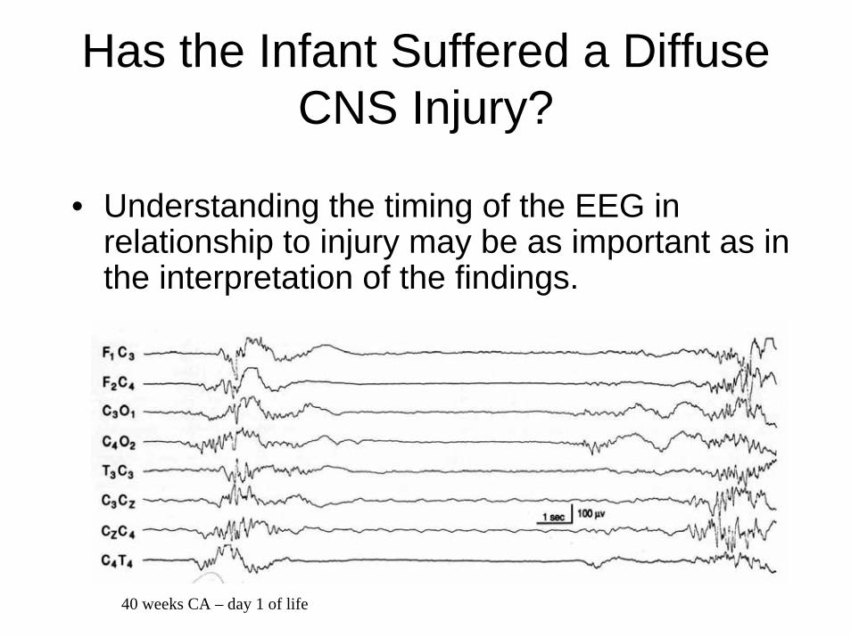
• Understanding the timing of the EEG in relationship to injury may be as important as in the interpretation of the findings.
40 weeks CA – day 1 of life

Has the Infant Suffered a Diffuse CNS Injury?
• Understanding the timing of the EEG in relationship to injury may be as important as in the interpretation of the findings.
4 days later

Is There Evidence of Focal CNS Injury?
• Persistent focal abnormalities suggest focal structural lesions– Sharp waves– Voltage asymmetry– Focal slow
• Differentiate normal from abnormal transients

Are Focal Abnormalites Specific To an Underlying Etiology?
• Attempts have been made to correlate focal findings with etiology– Positive central (rolandic) sharp waves
• Initially: intraventricular hemorrhage• Now: periventricular leukomalacia
– Periodic laterized epileptiform discharges (PLEDs)
• Initially: herpes simplex encephalitis• Now: seizure discharges of the depressed brain

When Did the Injury Occur?
• Most controversial aspect of neonatal EEG interpretation
• Finding of delayed maturation suggests that the injury occurred in utero– Dyschronism

What is the Prognosis?
• The greater the EEG abnormality, the more grave the prognosis
• Although the initial EEG may be very abnormal the accuracy of prognosis is based upon the evolution of the abnormality– Degree and rate of resolution
• A normal initial EEG reliably suggests a good prognosis– Within first 24 hours

EEG Features Thought to Have Prognostic Significance
• Developemental assessment• Character of the background activity• Focal features

Prognostic Value of Abnormal or Absent Developmental Features
• Possible association with neurological impairment (“at-risk”)– Dyschronism– Absence of expected grapho-elements– Delayed maturation in serial recordings

Prognostic Value of EEG Background Activity
• Association with significant neurological impairment or death– Electrocerebral
inactivity– Suppression-burst– Depressed and
undifferentiated– Prolonged interburst
intervals (pre-term)
• Association with possible neurological impairment– Depressed or
undifferentiated– Non-reactive– Aberrant sleep
architecture

Prognostic Value of Focal EEG Features
• Association with significant neurological impairment or death– Positive rolandic sharp
waves (>2/min)– Persistent voltage
asymmetry
• Association with possible neurological impairment– Positive rolandic sharp
waves (<2/min)– Multifocal sharp waves

Practical Considerations
• Know the timing of EEG in relation to suspected injury– Further from the injury with an abnormal EEG
the more likely it is to be persistent• Perform serial EEGs
– Abnormal EEGs early in the course of injury may improve, suggesting an improved prognosis

Clinical Utility of Neonatal EEG
• Best utilized in the context of a clinical question– What is the infant’s conceptional age?– Has the infant suffered a diffuse brain injury?– Is there evidence of a focal brain injury?– When did the CNS injury occur?– What is the prognosis?– Has the infant experienced a seizure?

Has the Infant Experienced a Seizure?

Clinical ImperativesConsequences of Neonatal Seizures
• Immediate– Alternations of
autonomic function• Heart rate• Respirations• Systemic blood
pressure
– Hypermetabolic state– Etiology of underlying
CNS dysfunction
• Long-term– High early death rate– Impairment later in life
• Developmental delay• Neurological
abnormalities• Post-neonatal epilepsy

Problems of Seizure Recognition
• Timing– Unpredictable in onset– Frequency
unpredictable– Duration is
unpredictable• Self-limited• Intractable
• Clinical Identification– Some features unique
to neonatal period– Recognition may be
difficult– Pathophysiology
differs by seizure type• Epileptic• Non-epileptic

Classification of Neonatal Seizures
• Methods of Classification– Clinical features
• Predominant clinical behavior• Components of an evolving seizure
– Temporal relationship of clinical features to EEG seizure activity
– Pathophysiology
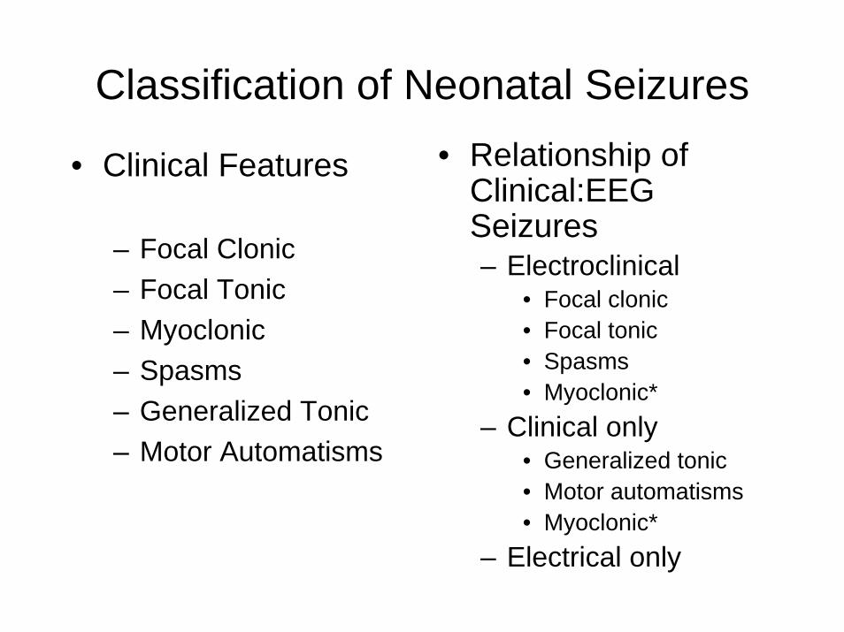
Classification of Neonatal Seizures• Relationship of
Clinical:EEGSeizures– Electroclinical
• Focal clonic• Focal tonic• Spasms• Myoclonic*
– Clinical only• Generalized tonic• Motor automatisms• Myoclonic*
– Electrical only
• Clinical Features
– Focal Clonic– Focal Tonic– Myoclonic– Spasms– Generalized Tonic– Motor Automatisms
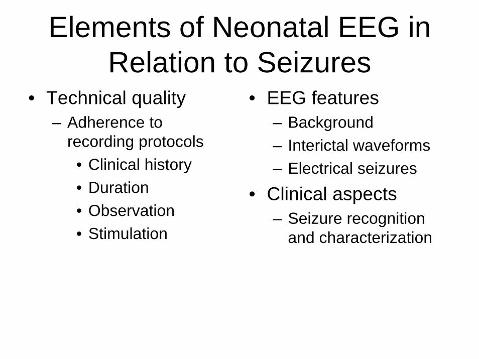
Elements of Neonatal EEG in Relation to Seizures
• Technical quality– Adherence to
recording protocols• Clinical history• Duration• Observation• Stimulation
• EEG features– Background– Interictal waveforms– Electrical seizures
• Clinical aspects– Seizure recognition
and characterization

Interictal Sharp Waves

Electrical Seizure Activity

Seizure Duration
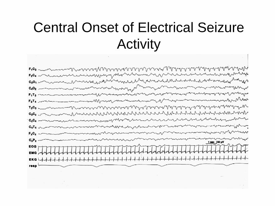
Central Onset of Electrical Seizure Activity

Temporal Onset of Electrical Seizure Activity

Frontal Onset of Electrical Seizure Activity

Midline Central Onset of ElectricalSeizure Activity

Midline Central Onset of ElectricalSeizure Activity

Occipital Onset of Electrical Seizure Activity

Multifocal Electrical Seizure Activity

Multifocal Electrical Seizure Activity

Low-Voltage Electrical Seizure Activity
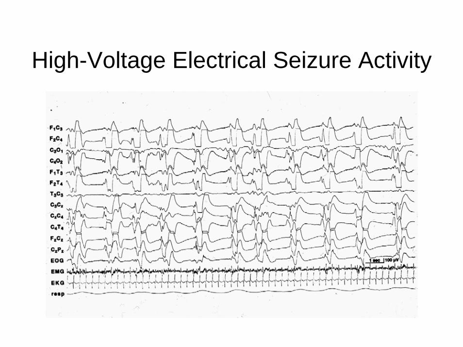
High-Voltage Electrical Seizure Activity

Spike Morphology of ElectricalSeizure Activity

Slow Wave Morphology of ElectricalSeizure Activity

Slow Wave Morphology of ElectricalSeizure Activity

Complex Morphology of ElectricalSeizure Activity

Complex Morphology of ElectricalSeizure Activity

Complex Morphology of ElectricalSeizure Activity

Complex Morphology of MultifocalElectrical Seizure Activity

Migration of Electrical Seizure Activity

Electrical Seizure Activity Not Associated with Clinical Seizures
• Seizure discharges of the depressed• Alpha seizure discharges• Seizure discharges in infants paralyzed for
respiratory care

Seizure Discharges of the Depressed Brain

Alpha Seizure Discharges

Alpha Seizure Discharges

Ictal Features of Epileptic Spasms

Sources of Artifacts that May Mimic Electrical Seizures
• Environmental interference– Electrical interference due to mechanical
devices• Alterations of electrode impedence• Movement-induced artifacts• Endogenous non-cerebral potentials


Environmental Interference:Mechanical Device – IV Pump


Environmental Interference:Mechanical Device – ECMO


Alterations of Electrode Impedence


Induced by Movement:Mechanical Ventilation


Induced by Movement:Spontaneous Tremors


Induced by Movement:Spontaneous Tremors

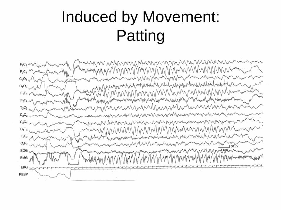
Induced by Movement:Patting


Induced by Movement:Patting


Endogenous Non-cerebral Potentials: EKG


Endogenous Non-cerebral Potentials: Sucking


Endogenous Non-cerebral Potentials: Jaw Tremor


Endogenous Non-cerebral Potentials: Eye Movements


Endogenous Non-cerebral Potentials: EMG


Endogenous Non-cerebral Potentials: EMG with Arousal

Practical Considerations in the Interpretation of Neonatal EEG
• Consider conceptional age– Parameters of the normal and abnormal EEG
are conceptional age dependent• Consider the EEG in relation to the clinical
problem– Try to address the clinical issues

Acknowledgements
Peter Kellaway Section of NeurophysiologyDepartment of Neurology Baylor College of MedicineHouston, Texas, USA
James D. Frost, Jr., M.D.Richard A. Hrachovy, M.D.
Peter Kellaway, Ph.D.(1920-2003)


Clinical ImperativesConsequences of Neonatal Seizures
• Immediate– Alternations of
autonomic function• Heart rate• Respirations• Systemic blood
pressure
– Hypermetabolic state– Etiology of underlying
CNS dysfunction
• Long-term– High early death rate– Impairment later in life
• Developmental delay• Neurological
abnormalities• Post-neonatal epilepsy

Problems of Seizure Recognition
• Timing– Unpredictable in onset– Frequency
unpredictable– Duration is
unpredictable• Self-limited• Intractable
• Clinical Identification– Some features unique
to neonatal period– Recognition may be
difficult– Pathophysiology
differs by seizure type• Epileptic• Non-epileptic
Classification of Neonatal Seizures
• Methods of Classification– Clinical features
• Predominant clinical behavior• Components of an evolving seizure
– Temporal relationship of clinical features to EEG seizure activity
– Pathophysiology

Classification of Neonatal Seizures• Relationship of
Clinical:EEGSeizures– Electroclinical
• Focal clonic• Focal tonic• Spasms• Myoclonic*
– Clinical only• Generalized tonic• Motor automatisms• Myoclonic*
– Electrical only
• Clinical Features
– Focal Clonic– Focal Tonic– Myoclonic– Spasms– Generalized Tonic– Motor Automatisms

Suppression-burst Patterns
• Hypoxic-ischemic encephalopathy• In-born errors of metabolism

Suppression-burst Patterns

Suppression-burst Patterns

Interictal Sharp Waves
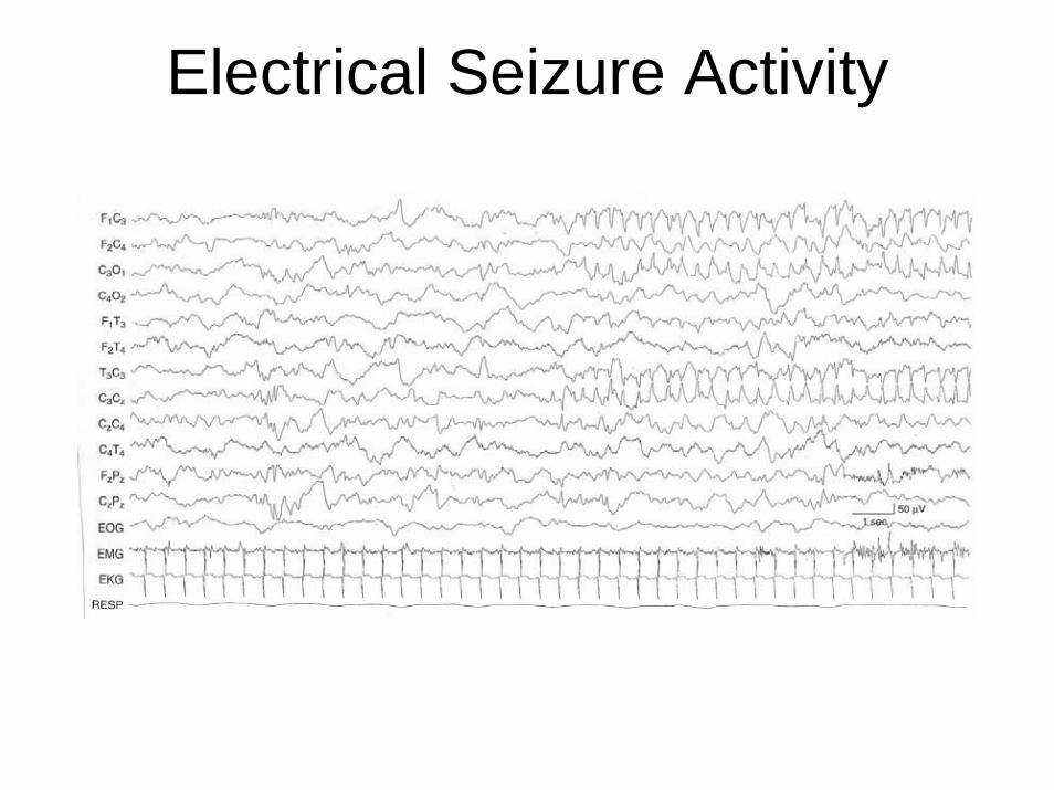
Electrical Seizure Activity

Seizure Duration

Ictal Features of Epileptic Spasms

Sources of Artifacts that May Mimic Electrical Seizures
• Environmental interference– Electrical interference due to mechanical
devices• Alterations of electrode impedence• Movement-induced artifacts• Endogenous non-cerebral potentials


Environmental Interference:Mechanical Device – IV Pump

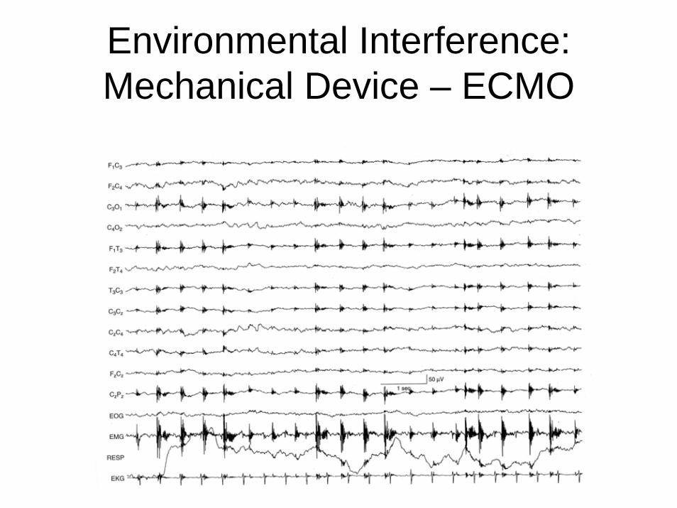
Environmental Interference:Mechanical Device – ECMO


Alterations of Electrode Impedence

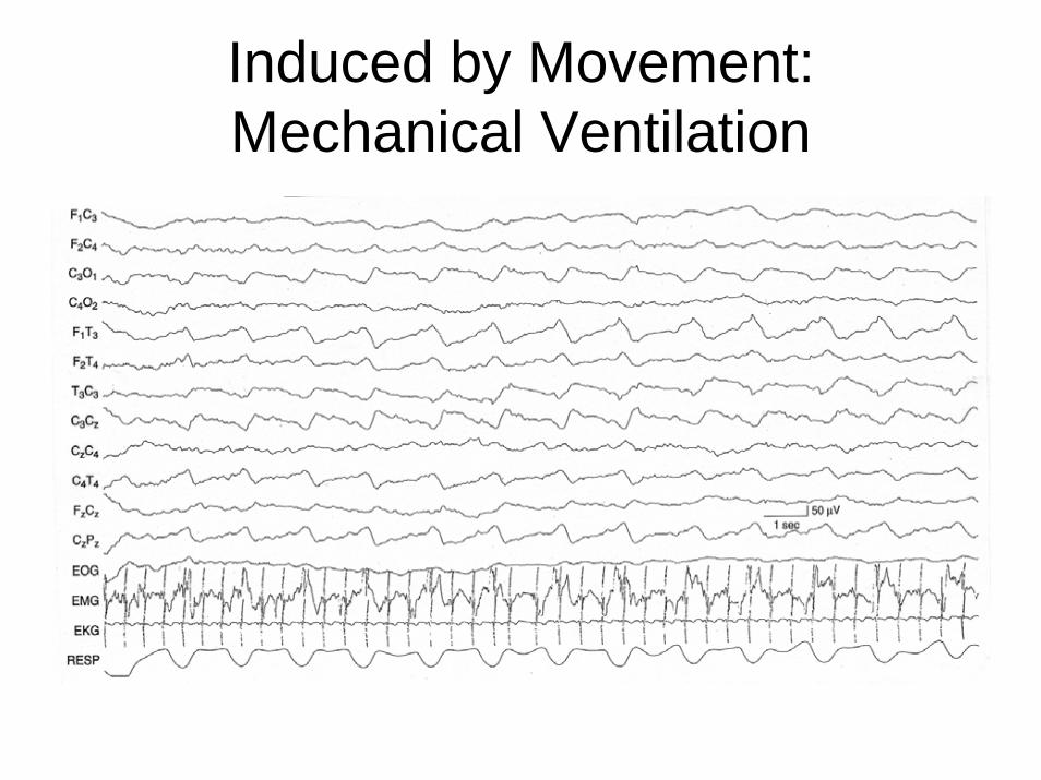
Induced by Movement:Mechanical Ventilation


Induced by Movement:Spontaneous Tremors

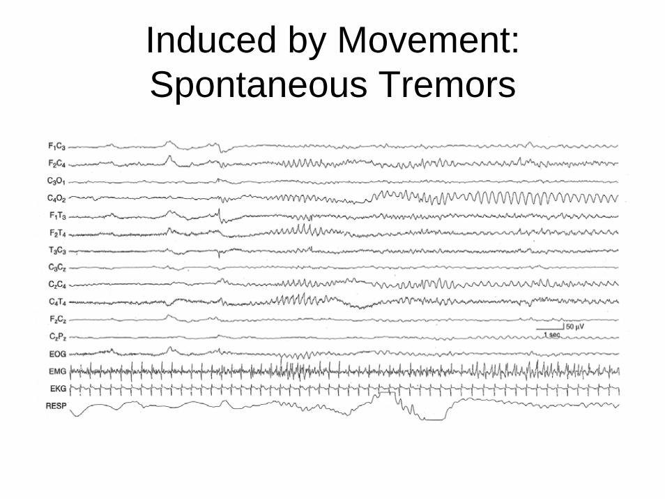
Induced by Movement:Spontaneous Tremors


Induced by Movement:Patting
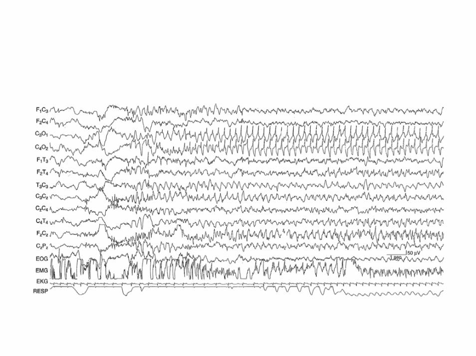
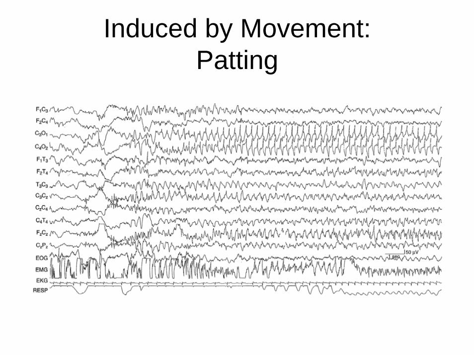
Induced by Movement:Patting


Endogenous Non-cerebral Potentials: EKG


Endogenous Non-cerebral Potentials: Sucking


Endogenous Non-cerebral Potentials: Jaw Tremor

Endogenous Non-cerebral Potentials: Eye Movements


Endogenous Non-cerebral Potentials: EMG


Endogenous Non-cerebral Potentials: EMG with Arousal

Related Documents