Insect Biochemistry and Molecular Biology 34 (2004) 477–483 www.elsevier.com/locate/ibmb Negative regulation of prophenoloxidase (proPO) activation by a clip-domain serine proteinase homolog (SPH) from endoparasitoid venom Guangmei Zhang a , Zhi-Qiang Lu b , Haobo Jiang b , Sassan Asgari c, a Insect Molecular Biology Laboratory, Plant and Pest Science, Waite Campus, University of Adelaide, Glen Osmond, SA 5064, Australia b Department of Entomology and Plant Pathology, Oklahoma State University, Stillwater, OK 74078, USA c Department of Zoology and Entomology, School of Life Sciences, University of Queensland, St. Lucia, Qld 4072, Australia Received 28 October 2003; accepted 20 February 2004 Abstract Most parasitic wasps inject maternal factors into the host hemocoel to suppress the host immune system and ensure successful development of their progeny. Melanization is one of the insect defence mechanisms against intruding pathogens or parasites. We previously isolated from the venom of Cotesia rubecula a 50 kDa protein that blocked melanization in the hemolymph of its host, Pieris rapae [Insect Biochem. Mol. Biol. 33 (2003) 1017]. This protein, designated Vn50, is a serine proteinase homolog (SPH) containing an amino-terminal clip domain. In this work, we demonstrated that recombinant Vn50 bound P. rapae hemolymph components that were recognized by antisera to Tenebrio molitor prophenoloxidase (proPO) and Manduca sexta proPO-activating proteinase (PAP). Vn50 is stable in the host hemolymph—it remained intact for at least 72 h after parasitization. Using M. sexta as a model system, we found that Vn50 efficiently down-regulated proPO activation mediated by M. sexta PAP-1, SPH-1, and SPH-2. Vn50 did not inhibit active phenoloxidase (PO) or PAP-1, but it significantly reduced the proteolysis of proPO. If recom- binant Vn50 binds P. rapae proPO and PAP (as suggested by the antibody reactions), it is likely that the molecular interactions among M. sexta proPO, PAP-1, and SPHs were impaired by this venom protein. A similar strategy might be employed by C. rubecula to negatively impact the proPO activation reaction in its natural host. # 2004 Elsevier Ltd. All rights reserved. Keywords: Phenoloxidase; Melanization; Insect immunity; Parasitoid; Cotesia rubecula; Manduca sexta; Pieris rapae 1. Introduction Introduction of maternal factors into the body cavity of their host insects is a common strategy evolved in endoparasitic wasps to manipulate host physiology for their own benefits. These include viruses or virus-like particles, such as polydnaviruses (PDVs), and venom fluid. PDVs, produced in the calyx region of the female reproductive organ, are essential for successful develop- ment of the parasitoids inside the host (Edson et al., 1981; Fleming and Summers, 1991). They disrupt the host cellular immune system and inhibit formation of hemocyte capsules around the parasitoid eggs. PDVs are accompanied by venom proteins in most parasit- oid–host systems, and in some cases are only effective when injected together with venom. Venom proteins are probably involved in uncoating of PDVs in vitro and virus persistence in vivo (Stoltz et al., 1988). In many instances, it has been shown that venom enhances the effects of PDVs (Kitano, 1986; Tanaka, 1987) or might provide protection for the eggs during the period between oviposition and expression of PDV genes (Webb and Luckhart, 1994). In addition to their synergistic effects together with PDVs, venom Abbreviations: PAGE, polyacrylamide gel electrophoresis; proPO and PO, prophenoloxidase and phenoloxidase; PAP, proPO- activating proteinase; SPH, serine proteinase homolog; PDV, polydnavirus; SDS, sodium dodecyl sulfate; TBS, Tris buffered saline; PBS, phosphate buffered saline; PTU, 1-phenyl-2-thiourea. Corresponding author. Tel.: +61-7-3365-2043; fax: +61-7-3365- 1655. E-mail address: [email protected] (S. Asgari). 0965-1748/$ - see front matter # 2004 Elsevier Ltd. All rights reserved. doi:10.1016/j.ibmb.2004.02.009

Welcome message from author
This document is posted to help you gain knowledge. Please leave a comment to let me know what you think about it! Share it to your friends and learn new things together.
Transcript
Abbreviations: PAGE, polyacryla
and PO, prophenoloxidase and
activating proteinase; SPH, serin
polydnavirus; SDS, sodium dodecyl s
PBS, phosphate buffered saline; PTU� Corresponding author. Tel.: +6
1655.
E-mail address: [email protected]
0965-1748/$ - see front matter # 200
doi:10.1016/j.ibmb.2004.02.009
mide gel electrophoresis; proPO
phenoloxidase; PAP, proPO-
e proteinase homolog; PDV,
ulfate; TBS, Tris buffered saline;
, 1-phenyl-2-thiourea.
1-7-3365-2043; fax: +61-7-3365-
u (S. Asgari).
4 Elsevier Ltd. All rights reserved.
Insect Biochemistry and Molecular Biology 34 (2004) 477–483
www.elsevier.com/locate/ibmbNegative regulation of prophenoloxidase (proPO) activationby a clip-domain serine proteinase homolog (SPH) from
endoparasitoid venom
Guangmei Zhang a, Zhi-Qiang Lu b, Haobo Jiang b, Sassan Asgari c,�
a Insect Molecular Biology Laboratory, Plant and Pest Science, Waite Campus, University of Adelaide, Glen Osmond, SA 5064, Australiab Department of Entomology and Plant Pathology, Oklahoma State University, Stillwater, OK 74078, USA
c Department of Zoology and Entomology, School of Life Sciences, University of Queensland, St. Lucia, Qld 4072, Australia
Received 28 October 2003; accepted 20 February 2004
Abstract
Most parasitic wasps inject maternal factors into the host hemocoel to suppress the host immune system and ensure successfuldevelopment of their progeny. Melanization is one of the insect defence mechanisms against intruding pathogens or parasites. Wepreviously isolated from the venom of Cotesia rubecula a 50 kDa protein that blocked melanization in the hemolymph of its host,Pieris rapae [Insect Biochem. Mol. Biol. 33 (2003) 1017]. This protein, designated Vn50, is a serine proteinase homolog (SPH)containing an amino-terminal clip domain. In this work, we demonstrated that recombinant Vn50 bound P. rapae hemolymphcomponents that were recognized by antisera to Tenebrio molitor prophenoloxidase (proPO) and Manduca sexta proPO-activatingproteinase (PAP). Vn50 is stable in the host hemolymph—it remained intact for at least 72 h after parasitization. Using M. sextaas a model system, we found that Vn50 efficiently down-regulated proPO activation mediated by M. sexta PAP-1, SPH-1, andSPH-2. Vn50 did not inhibit active phenoloxidase (PO) or PAP-1, but it significantly reduced the proteolysis of proPO. If recom-binant Vn50 binds P. rapae proPO and PAP (as suggested by the antibody reactions), it is likely that the molecular interactionsamong M. sexta proPO, PAP-1, and SPHs were impaired by this venom protein. A similar strategy might be employed byC. rubecula to negatively impact the proPO activation reaction in its natural host.# 2004 Elsevier Ltd. All rights reserved.
Keywords: Phenoloxidase; Melanization; Insect immunity; Parasitoid; Cotesia rubecula; Manduca sexta; Pieris rapae
1. Introduction
Introduction of maternal factors into the body cavity
of their host insects is a common strategy evolved in
endoparasitic wasps to manipulate host physiology for
their own benefits. These include viruses or virus-like
particles, such as polydnaviruses (PDVs), and venom
fluid. PDVs, produced in the calyx region of the female
reproductive organ, are essential for successful develop-
ment of the parasitoids inside the host (Edson et al.,
1981; Fleming and Summers, 1991). They disrupt the
host cellular immune system and inhibit formation of
hemocyte capsules around the parasitoid eggs. PDVs
are accompanied by venom proteins in most parasit-
oid–host systems, and in some cases are only effective
when injected together with venom. Venom proteins
are probably involved in uncoating of PDVs in vitro
and virus persistence in vivo (Stoltz et al., 1988).
In many instances, it has been shown that venom
enhances the effects of PDVs (Kitano, 1986; Tanaka,
1987) or might provide protection for the eggs during
the period between oviposition and expression of
PDV genes (Webb and Luckhart, 1994). In addition to
their synergistic effects together with PDVs, venom
478 G. Zhang et al. / Insect Biochemistry and Molecular Biology 34 (2004) 477–483
components affect host physiology and development(Digilio et al., 2000; Gupta and Ferkovich, 1998).In other endoparasitoids that do not produce PDVs,venom becomes the only factor in suppression or regu-lation of the host immune system (Richards andParkinson, 2000).Inhibition of melanization following parasitism has
been reported from several systems. Melanin formationinvolves the proteolytic activation of proPO, whichleads to the generation of melanin and other toxic phe-nolic compounds. This reaction is considered as a vitaldefence mechanism mounted against intruding organ-isms (Ashida and Brey, 1998; Vass and Nappi, 2000).This process is impaired to various extents in severalhost–parasitoid systems. However, the molecular mech-anism behind this suppression is unknown. In a recentstudy, we isolated a 50 kDa protein from the venom ofCotesia rubecula, a Braconid parasitic wasp, whichblocked hemolymph melanization of its host, Pierisrapae (Asgari et al., 2003). This protein is similar insequence and domain structure to serine proteinasehomologs (SPHs) from various insects (Kwon et al.,1999; Lee et al., 2002; Yu et al., 2003). Many SPHsconsist of a clip domain at the amino terminus and aserine proteinase-like domain at the carboxyl terminus(Ross et al., 2003). Since the residues essential for cata-lytic activity are missing in these proteins, SPHs do nothave proteolytic activity. Recent study indicated thatManduca sexta SPH-1 and SPH-2 mediate propheno-loxidase (proPO) activation in conjunction with othercomponents from the hemolymph (Yu et al., 2003).Although cleaved, their clip and proteinase-likedomains remain attached by an interchain disulfidebond.In this complementary study, we show that Vn50 is
not cleaved after introduction into the host hemolymphand is stable for a long period of time after parasitiza-tion. The recombinant Vn50 may specifically interactwith proPO and proPO-activating proteinase (PAP) inP. rapae hemolymph. Vn50 reduces proteolytic acti-vation of proPO by interfering with the molecularinteractions among M. sexta proPO, PAP-1, SPH-1,and SPH-2.
2. Materials and methods
2.1. Insects and isolation of hemolymph
The parasitoid C. rubecula and its host P. rapae weremaintained at 25
vC on a 14 h light:10 h dark photo-
period. P. rapae larvae were reared on cabbage plants.Adult wasps were fed with honey–water solution.Hemolymph (ca. 80 ll) was collected from five fourthinstar P. rapae larvae by surface sterilizing in 70%ethanol and bleeding from a proleg into ice-cold phos-
phate buffered saline (PBS; 200 ll) containing a fewcrystals of 1-phenyl-2-thiourea (PTU). To obtain theplasma, hemolymph was centrifuged at 800�g for5 min and the supernatant was transferred into a freshtube.
2.2. Preparation of C. rubecula Vn50, M. sexta proPO,PAP-1, and SPHs
Venom proteins were isolated from the female waspsand fractionated by reversed phase HPLC to purifyVn50 (Asgari et al., 2003). Recombinant Vn50 wasexpressed in Escherichia coli as an insoluble proteinand purified by nickel affinity chromatography underthe denaturing conditions. M. sexta proPO was iso-lated from the larval hemolymph as described pre-viously (Jiang et al., 1997). Activated recombinantPAP-1 (Yu et al., 2003) was separated from other pro-teins under the conditions used for proPAP-1 purifi-cation (Wang et al., 2001). M. sexta SPH-1 and SPH-2,which co-purified with immulectin-2, were purifiedfrom hemolymph of bacteria-induced insects accordingto Yu et al. (2003).
2.3. Production of anti-Vn50 antibodies
Purified recombinant Vn50 expressed in E. coli(Asgari et al., 2003) was run on preparative 10% SDS-PAGE gels, stained in 0.05% Coomassie blue R-250prepared in H2O, and destained with several changes ofdistilled water. Vn50 corresponding band was excisedfrom the gels, squashed into fine pieces in PBS, andinjected into a rabbit (ca. 5 lg/injection). Two sub-sequent booster injections were carried out in 2-weekintervals, 4 weeks after the initial injection. The firstand booster injections contained complete and incom-plete Freud’s adjuvants, respectively. Serum wasobtained one week after the last injection. Productionof anti-Vn50 antibodies was confirmed by western blotscontaining the recombinant Vn50 and the total venomfrom C. rubecula using 1:5000 dilution of the anti-serum.
2.4. Stability of C. rubecula Vn50 in P. rapaehemolymph
Hemolymph was collected from third instar P. rapaelarvae (ca. 20 ll) at various times following parasitiza-tion by C. rubecula, as described above. Samples wereanalysed by western blot analysis using anti-Vn50 anti-bodies.
2.5. Identification of hemolymph proteins bound to Vn50
Binding of P. rapae hemolymph proteins to Vn50was examined as described previously with minor mod-
G. Zhang et al. / Insect Biochemistry and Molecular Biology 34 (2004) 477–483 479
ifications (Yu et al., 2003). Briefly, washed Ni-NTAagarose beads (Qiagen, 0.1 ml) were coated with rena-tured 6�His-tagged recombinant Vn50 or bovineserum albumin (BSA) in Tris buffered saline (TBS) for60 min at room temperature. Beads were washed withTBS to remove unbound proteins and incubated with0.5 ml plasma-PBS containing PTU from four P. rapaelarvae. The incubation was carried out at room tem-perature for 1 h with shaking. After washing with TBS,nonspecifically bound proteins were eluted with 1.0 MNaCl. SDS sample buffer was then added to the resus-pended beads in TBS and treated at 95
vC for 5 min.
After centrifugation, the supernatant was subjected to12% SDS-PAGE (Laemmli, 1970) and immunoblotanalysis. Blots were probed with various antiserumagainst C. rubecula Vn50 (1:5000 dilution) (Asgari et al.,2003), M. sexta PAP-1 (1:2000 dilution) (Wang et al.,2001), or Tenebrio molitor proPO (1:2000 dilution) (akind gift from Prof. Bok Lee at Pusan National Uni-versity, Korea). Alkaline phosphatase-conjugated anti-rabbit IgGs (Sigma) were used as secondary antibodies.
2.6. Quantification of Vn50 injected into P. rapae larvae
Comparative western blot analysis was used to deter-mine the amount of Vn50 injected into a host larva atvarious times after parasitization. Three larvae wereused for each time point. Recombinant Vn50 was pro-duced in bacteria and purified as described (Asgariet al., 2003). The recombinant protein concentrationwas measured using Bio-Rad Protein Assay based onthe method of Bradford. Cell-free hemolymph (seeabove) was collected from the parasitized larvae, and afixed volume of 20 ll of each sample was run on west-ern blots together with known concentrations ofrecombinant Vn50. Intensity of bands was compared toestimate the amount of Vn50 injected into the cater-pillars.
2.7. Down regulation of M. sexta proPO activation byC. rubecula Vn50
M. sexta proPO (10 ll, 10 ng/ll), PAP-1 (2:1, 30ng/ll), and SPHs (2:1, 50 ng/ll) were mixed with 10:1of reaction buffer (20 mM Tris–HCl, pH 7.5) in thewells of a flat-bottom microtiter plate. After 2:1 ofVn50 at different concentrations was added to thewells, the reaction mixtures were incubated on ice for40 min. PO activity was measured using dopamine asthe substrate and a microplate reader (Jiang et al.,2003).To examine a possible effect of C. rubecula Vn50 on
PO activity, M. sexta proPO, PAP-1, SPHs, and bufferwere incubated in the wells of a microplate for 40 minon ice as described above. After 2:1 of Vn50 (12 ng/ll)or buffer (20 mM Tris–HCl, pH 7.5) was added, PO
activity in the reaction mixtures was determined 10 minlater (Jiang et al., 2003).To test if C. rubecula Vn50 directly inhibits a PAP,
M. sexta PAP-1 (5 ll, 30 ng/ll) was incubated withVn50 (2:1, 10 ng/ll) or buffer (2:1) on ice for 10 min.The amidase activity of PAP-1 was measured usingacetyl-Ile-Glu–Ala–Arg–pNa as a substrate (Jiang et al.,2003).
2.8. Effect of C. rubecula Vn50 on proPO cleavage
For understanding how Vn50 may affect proPOcleavage, proPO activation was performed in theabsence or presence of Vn50 (2:1, 5 ng/ll) under theconditions described above. After incubation on ice for40 min, 10:1 of the reaction mixtures as well as thenegative controls were subjected to western blot analy-sis using 1:2000 diluted proPO antiserum as the firstantibody.
3. Results and discussion
In our previous paper (Asgari et al., 2003), weshowed that the most abundant component in C. rube-cula venom was a 50 kDa protein (Vn50), which issimilar in sequence to arthropod SPHs and inhibitsmelanization of hemolymph from the host P. rapae.Many of these proteins consist of two domains: anamino-terminal clip domain and a carboxyl-terminalproteinase-like domain (Ross et al., 2003). Like manyclip-domain serine proteinases from arthropods (Jiangand Kanost, 2000), M. sexta SPH-1 and SPH-2, as wellas Holotrichia diomphalia proPO-activating factor II,are cleaved at a certain position but the two domainsremain attached by a disulfide bond (Kwon et al.,2000; Lee et al., 2002; Yu et al., 2003). These threeSPHs function as cofactors for PAPs. Because the con-served Ser residue at the active site of their proteinase-like domains is changed to Gly or other residues, allSPHs (including Vn50) are not expected to have anyproteolytic activity.
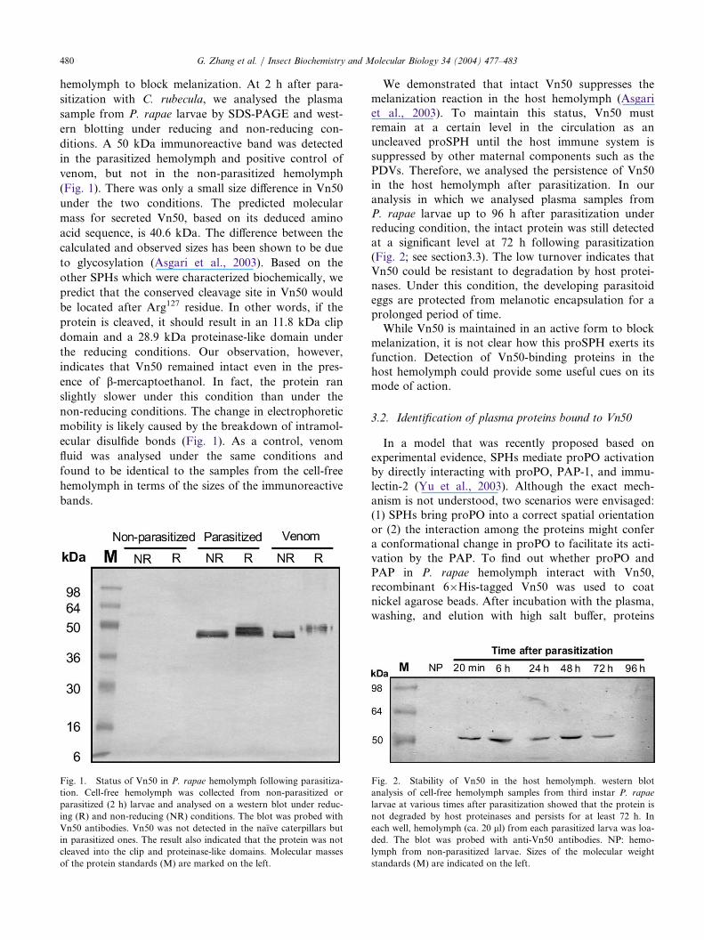
3.1. Status of Vn50 in the host hemolymph followingparasitization
The clip-domain serine proteinases are produced aszymogens and cleaved at a specific location betweenthe clip domain and the proteolytic domain (Jiang andKanost, 2000). The cleavage is necessary for activationof these enzymes. Clip-domain SPHs, with a similardomain structure, were also cleaved (Yu et al., 2003;Lee et al., 2002). Assuming that the cleavage is alsonecessary for SPHs to exert their function as enhan-cers/mediators for proPO activation, we investigatedthe status of Vn50 after it was injected into the host
480 G. Zhang et al. / Insect Biochemistry and Molecular Biology 34 (2004) 477–483
hemolymph to block melanization. At 2 h after para-
sitization with C. rubecula, we analysed the plasma
sample from P. rapae larvae by SDS-PAGE and west-
ern blotting under reducing and non-reducing con-
ditions. A 50 kDa immunoreactive band was detected
in the parasitized hemolymph and positive control of
venom, but not in the non-parasitized hemolymph
(Fig. 1). There was only a small size difference in Vn50
under the two conditions. The predicted molecular
mass for secreted Vn50, based on its deduced amino
acid sequence, is 40.6 kDa. The difference between the
calculated and observed sizes has been shown to be due
to glycosylation (Asgari et al., 2003). Based on the
other SPHs which were characterized biochemically, we
predict that the conserved cleavage site in Vn50 would
be located after Arg127 residue. In other words, if the
protein is cleaved, it should result in an 11.8 kDa clip
domain and a 28.9 kDa proteinase-like domain under
the reducing conditions. Our observation, however,
indicates that Vn50 remained intact even in the pres-
ence of b-mercaptoethanol. In fact, the protein ran
slightly slower under this condition than under the
non-reducing conditions. The change in electrophoretic
mobility is likely caused by the breakdown of intramol-
ecular disulfide bonds (Fig. 1). As a control, venom
fluid was analysed under the same conditions and
found to be identical to the samples from the cell-free
hemolymph in terms of the sizes of the immunoreactive
bands.
We demonstrated that intact Vn50 suppresses themelanization reaction in the host hemolymph (Asgariet al., 2003). To maintain this status, Vn50 mustremain at a certain level in the circulation as anuncleaved proSPH until the host immune system issuppressed by other maternal components such as thePDVs. Therefore, we analysed the persistence of Vn50in the host hemolymph after parasitization. In ouranalysis in which we analysed plasma samples fromP. rapae larvae up to 96 h after parasitization underreducing condition, the intact protein was still detectedat a significant level at 72 h following parasitization(Fig. 2; see section3.3). The low turnover indicates thatVn50 could be resistant to degradation by host protei-nases. Under this condition, the developing parasitoideggs are protected from melanotic encapsulation for aprolonged period of time.While Vn50 is maintained in an active form to block
melanization, it is not clear how this proSPH exerts itsfunction. Detection of Vn50-binding proteins in thehost hemolymph could provide some useful cues on itsmode of action.
3.2. Identification of plasma proteins bound to Vn50
In a model that was recently proposed based onexperimental evidence, SPHs mediate proPO activationby directly interacting with proPO, PAP-1, and immu-lectin-2 (Yu et al., 2003). Although the exact mech-anism is not understood, two scenarios were envisaged:(1) SPHs bring proPO into a correct spatial orientationor (2) the interaction among the proteins might confera conformational change in proPO to facilitate its acti-vation by the PAP. To find out whether proPO andPAP in P. rapae hemolymph interact with Vn50,recombinant 6�His-tagged Vn50 was used to coatnickel agarose beads. After incubation with the plasma,washing, and elution with high salt buffer, proteins
Fig. 1. Status of Vn50 in P. rapae hemolymph following parasitiza-
tion. Cell-free hemolymph was collected from non-parasitized or
parasitized (2 h) larvae and analysed on a western blot under reduc-
ing (R) and non-reducing (NR) conditions. The blot was probed with
Vn50 antibodies. Vn50 was not detected in the naı̈ve caterpillars but
in parasitized ones. The result also indicated that the protein was not
cleaved into the clip and proteinase-like domains. Molecular masses
of the protein standards (M) are marked on the left.
Fig. 2. Stability of Vn50 in the host hemolymph. western blot
analysis of cell-free hemolymph samples from third instar P. rapae
larvae at various times after parasitization showed that the protein is
not degraded by host proteinases and persists for at least 72 h. In
each well, hemolymph (ca. 20 ll) from each parasitized larva was loa-
ded. The blot was probed with anti-Vn50 antibodies. NP: hemo-
lymph from non-parasitized larvae. Sizes of the molecular weight
standards (M) are indicated on the left.
G. Zhang et al. / Insect Biochemistry and Molecular Biology 34 (2004) 477–483 481
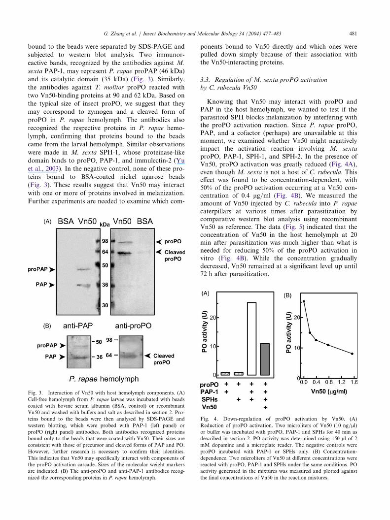
bound to the beads were separated by SDS-PAGE and
subjected to western blot analysis. Two immunor-
eactive bands, recognized by the antibodies against M.
sexta PAP-1, may represent P. rapae proPAP (46 kDa)
and its catalytic domain (35 kDa) (Fig. 3). Similarly,
the antibodies against T. molitor proPO reacted with
two Vn50-binding proteins at 90 and 62 kDa. Based on
the typical size of insect proPO, we suggest that they
may correspond to zymogen and a cleaved form of
proPO in P. rapae hemolymph. The antibodies also
recognized the respective proteins in P. rapae hemo-
lymph, confirming that proteins bound to the beads
came from the larval hemolymph. Similar observations
were made in M. sexta SPH-1, whose proteinase-like
domain binds to proPO, PAP-1, and immulectin-2 (Yu
et al., 2003). In the negative control, none of these pro-
teins bound to BSA-coated nickel agarose beads
(Fig. 3). These results suggest that Vn50 may interact
with one or more of proteins involved in melanization.
Further experiments are needed to examine which com-
ponents bound to Vn50 directly and which ones werepulled down simply because of their association withthe Vn50-interacting proteins.
3.3. Regulation of M. sexta proPO activationby C. rubecula Vn50
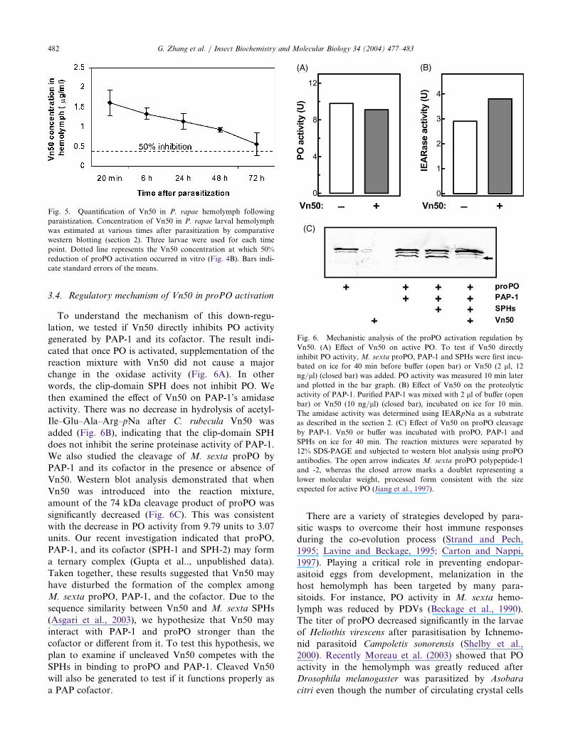
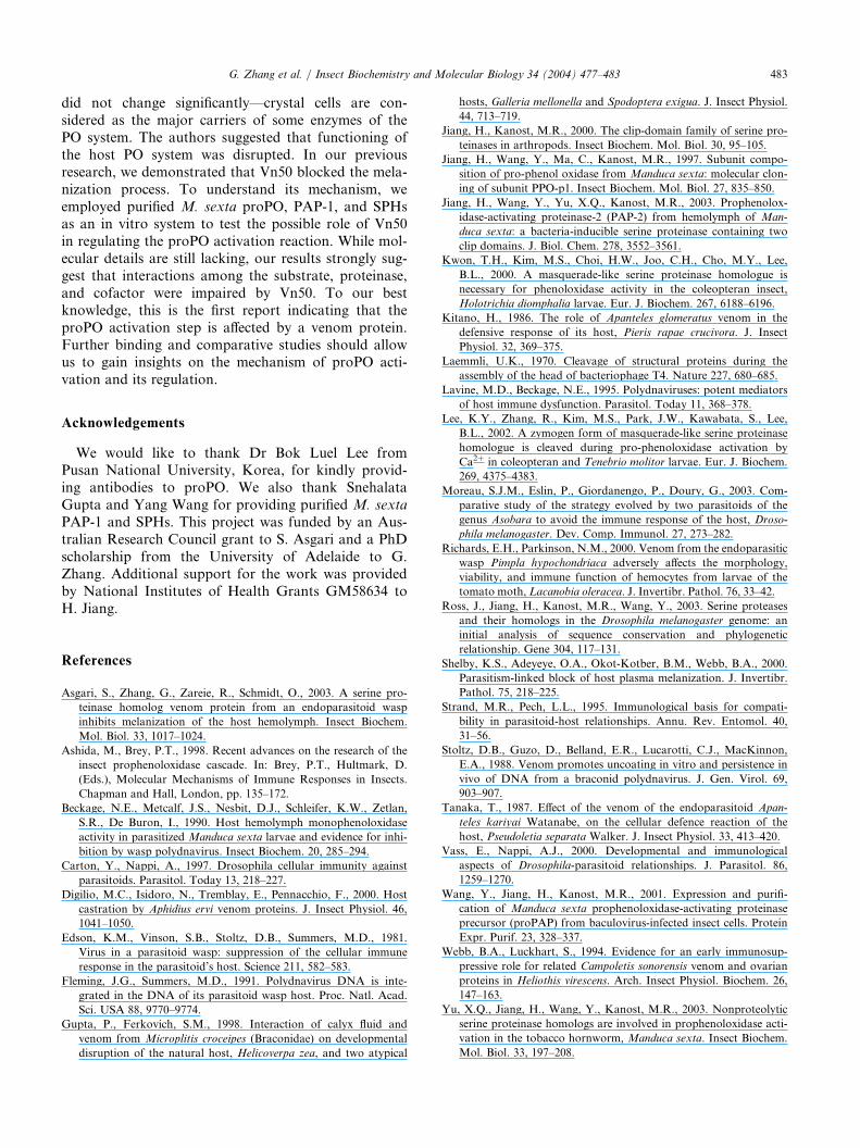
Knowing that Vn50 may interact with proPO andPAP in the host hemolymph, we wanted to test if theparasitoid SPH blocks melanization by interfering withthe proPO activation reaction. Since P. rapae proPO,PAP, and a cofactor (perhaps) are unavailable at thismoment, we examined whether Vn50 might negativelyimpact the activation reaction involving M. sextaproPO, PAP-1, SPH-1, and SPH-2. In the presence ofVn50, proPO activation was greatly reduced (Fig. 4A),even though M. sexta is not a host of C. rubecula. Thiseffect was found to be concentration-dependent, with50% of the proPO activation occurring at a Vn50 con-centration of 0.4 lg/ml (Fig. 4B). We measured theamount of Vn50 injected by C. rubecula into P. rapaecaterpillars at various times after parasitization bycomparative western blot analysis using recombinantVn50 as reference. The data (Fig. 5) indicated that theconcentration of Vn50 in the host hemolymph at 20min after parasitization was much higher than what isneeded for reducing 50% of the proPO activation invitro (Fig. 4B). While the concentration graduallydecreased, Vn50 remained at a significant level up until72 h after parasitization.
Fig. 3. Interaction of Vn50 with host hemolymph components. (A)
Cell-free hemolymph from P. rapae larvae was incubated with beads
coated with bovine serum albumin (BSA, control) or recombinant
Vn50 and washed with buffers and salt as described in section 2. Pro-
teins bound to the beads were then analysed by SDS-PAGE and
western blotting, which were probed with PAP-1 (left panel) or
proPO (right panel) antibodies. Both antibodies recognized proteins
bound only to the beads that were coated with Vn50. Their sizes are
consistent with those of precursor and cleaved forms of PAP and PO.
However, further research is necessary to confirm their identities.
This indicates that Vn50 may specifically interact with components of
the proPO activation cascade. Sizes of the molecular weight markers
are indicated. (B) The anti-proPO and anti-PAP-1 antibodies recog-
nized the corresponding proteins in P. rapae hemolymph.
Fig. 4. Down-regulation of proPO activation by Vn50. (A)
Reduction of proPO activation. Two microliters of Vn50 (10 ng/ll)or buffer was incubated with proPO, PAP-1 and SPHs for 40 min as
described in section 2. PO activity was determined using 150 ll of 2mM dopamine and a microplate reader. The negative controls were
proPO incubated with PAP-1 or SPHs only. (B) Concentration-
dependence. Two microliters of Vn50 at different concentrations were
reacted with proPO, PAP-1 and SPHs under the same conditions. PO
activity generated in the mixtures was measured and plotted against
the final concentrations of Vn50 in the reaction mixtures.
482 G. Zhang et al. / Insect Biochemistry and Molecular Biology 34 (2004) 477–483
3.4. Regulatory mechanism of Vn50 in proPO activation
To understand the mechanism of this down-regu-
lation, we tested if Vn50 directly inhibits PO activity
generated by PAP-1 and its cofactor. The result indi-
cated that once PO is activated, supplementation of thereaction mixture with Vn50 did not cause a major
change in the oxidase activity (Fig. 6A). In other
words, the clip-domain SPH does not inhibit PO. We
then examined the effect of Vn50 on PAP-1’s amidase
activity. There was no decrease in hydrolysis of acetyl-
Ile–Glu–Ala–Arg–pNa after C. rubecula Vn50 was
added (Fig. 6B), indicating that the clip-domain SPH
does not inhibit the serine proteinase activity of PAP-1.We also studied the cleavage of M. sexta proPO by
PAP-1 and its cofactor in the presence or absence of
Vn50. Western blot analysis demonstrated that when
Vn50 was introduced into the reaction mixture,
amount of the 74 kDa cleavage product of proPO was
significantly decreased (Fig. 6C). This was consistent
with the decrease in PO activity from 9.79 units to 3.07units. Our recent investigation indicated that proPO,
PAP-1, and its cofactor (SPH-1 and SPH-2) may form
a ternary complex (Gupta et al.., unpublished data).
Taken together, these results suggested that Vn50 may
have disturbed the formation of the complex among
M. sexta proPO, PAP-1, and the cofactor. Due to the
sequence similarity between Vn50 and M. sexta SPHs
(Asgari et al., 2003), we hypothesize that Vn50 mayinteract with PAP-1 and proPO stronger than the
cofactor or different from it. To test this hypothesis, we
plan to examine if uncleaved Vn50 competes with the
SPHs in binding to proPO and PAP-1. Cleaved Vn50
will also be generated to test if it functions properly as
a PAP cofactor.
There are a variety of strategies developed by para-sitic wasps to overcome their host immune responsesduring the co-evolution process (Strand and Pech,1995; Lavine and Beckage, 1995; Carton and Nappi,1997). Playing a critical role in preventing endopar-asitoid eggs from development, melanization in thehost hemolymph has been targeted by many para-sitoids. For instance, PO activity in M. sexta hemo-lymph was reduced by PDVs (Beckage et al., 1990).The titer of proPO decreased significantly in the larvaeof Heliothis virescens after parasitisation by Ichnemo-nid parasitoid Campoletis sonorensis (Shelby et al.,2000). Recently Moreau et al. (2003) showed that POactivity in the hemolymph was greatly reduced afterDrosophila melanogaster was parasitized by Asobaracitri even though the number of circulating crystal cells
Fig. 5. Quantification of Vn50 in P. rapae hemolymph following
paraistization. Concentration of Vn50 in P. rapae larval hemolymph
was estimated at various times after parasitization by comparative
western blotting (section 2). Three larvae were used for each time
point. Dotted line represents the Vn50 concentration at which 50%
reduction of proPO activation occurred in vitro (Fig. 4B). Bars indi-
cate standard errors of the means.
Fig. 6. Mechanistic analysis of the proPO activation regulation by
Vn50. (A) Effect of Vn50 on active PO. To test if Vn50 directly
inhibit PO activity,M. sexta proPO, PAP-1 and SPHs were first incu-
bated on ice for 40 min before buffer (open bar) or Vn50 (2 ll, 12ng/ll) (closed bar) was added. PO activity was measured 10 min later
and plotted in the bar graph. (B) Effect of Vn50 on the proteolytic
activity of PAP-1. Purified PAP-1 was mixed with 2 ll of buffer (openbar) or Vn50 (10 ng/ll) (closed bar), incubated on ice for 10 min.
The amidase activity was determined using IEARpNa as a substrate
as described in the section 2. (C) Effect of Vn50 on proPO cleavage
by PAP-1. Vn50 or buffer was incubated with proPO, PAP-1 and
SPHs on ice for 40 min. The reaction mixtures were separated by
12% SDS-PAGE and subjected to western blot analysis using proPO
antibodies. The open arrow indicates M. sexta proPO polypeptide-1
and -2, whereas the closed arrow marks a doublet representing a
lower molecular weight, processed form consistent with the size
expected for active PO (Jiang et al., 1997).
G. Zhang et al. / Insect Biochemistry and Molecular Biology 34 (2004) 477–483 483
did not change significantly—crystal cells are con-sidered as the major carriers of some enzymes of thePO system. The authors suggested that functioning ofthe host PO system was disrupted. In our previousresearch, we demonstrated that Vn50 blocked the mela-nization process. To understand its mechanism, weemployed purified M. sexta proPO, PAP-1, and SPHsas an in vitro system to test the possible role of Vn50in regulating the proPO activation reaction. While mol-ecular details are still lacking, our results strongly sug-gest that interactions among the substrate, proteinase,and cofactor were impaired by Vn50. To our bestknowledge, this is the first report indicating that theproPO activation step is affected by a venom protein.Further binding and comparative studies should allowus to gain insights on the mechanism of proPO acti-vation and its regulation.
Acknowledgements
We would like to thank Dr Bok Luel Lee fromPusan National University, Korea, for kindly provid-ing antibodies to proPO. We also thank SnehalataGupta and Yang Wang for providing purified M. sextaPAP-1 and SPHs. This project was funded by an Aus-tralian Research Council grant to S. Asgari and a PhDscholarship from the University of Adelaide to G.Zhang. Additional support for the work was providedby National Institutes of Health Grants GM58634 toH. Jiang.
References
Asgari, S., Zhang, G., Zareie, R., Schmidt, O., 2003. A serine pro-
teinase homolog venom protein from an endoparasitoid wasp
inhibits melanization of the host hemolymph. Insect Biochem.
Mol. Biol. 33, 1017–1024.
Ashida, M., Brey, P.T., 1998. Recent advances on the research of the
insect prophenoloxidase cascade. In: Brey, P.T., Hultmark, D.
(Eds.), Molecular Mechanisms of Immune Responses in Insects.
Chapman and Hall, London, pp. 135–172.
Beckage, N.E., Metcalf, J.S., Nesbit, D.J., Schleifer, K.W., Zetlan,
S.R., De Buron, I., 1990. Host hemolymph monophenoloxidase
activity in parasitized Manduca sexta larvae and evidence for inhi-
bition by wasp polydnavirus. Insect Biochem. 20, 285–294.
Carton, Y., Nappi, A., 1997. Drosophila cellular immunity against
parasitoids. Parasitol. Today 13, 218–227.
Digilio, M.C., Isidoro, N., Tremblay, E., Pennacchio, F., 2000. Host
castration by Aphidius ervi venom proteins. J. Insect Physiol. 46,
1041–1050.
Edson, K.M., Vinson, S.B., Stoltz, D.B., Summers, M.D., 1981.
Virus in a parasitoid wasp: suppression of the cellular immune
response in the parasitoid’s host. Science 211, 582–583.
Fleming, J.G., Summers, M.D., 1991. Polydnavirus DNA is inte-
grated in the DNA of its parasitoid wasp host. Proc. Natl. Acad.
Sci. USA 88, 9770–9774.
Gupta, P., Ferkovich, S.M., 1998. Interaction of calyx fluid and
venom from Microplitis croceipes (Braconidae) on developmental
disruption of the natural host, Helicoverpa zea, and two atypical
hosts, Galleria mellonella and Spodoptera exigua. J. Insect Physiol.
44, 713–719.
Jiang, H., Kanost, M.R., 2000. The clip-domain family of serine pro-
teinases in arthropods. Insect Biochem. Mol. Biol. 30, 95–105.
Jiang, H., Wang, Y., Ma, C., Kanost, M.R., 1997. Subunit compo-
sition of pro-phenol oxidase from Manduca sexta: molecular clon-
ing of subunit PPO-p1. Insect Biochem. Mol. Biol. 27, 835–850.
Jiang, H., Wang, Y., Yu, X.Q., Kanost, M.R., 2003. Prophenolox-
idase-activating proteinase-2 (PAP-2) from hemolymph of Man-
duca sexta: a bacteria-inducible serine proteinase containing two
clip domains. J. Biol. Chem. 278, 3552–3561.
Kwon, T.H., Kim, M.S., Choi, H.W., Joo, C.H., Cho, M.Y., Lee,
B.L., 2000. A masquerade-like serine proteinase homologue is
necessary for phenoloxidase activity in the coleopteran insect,
Holotrichia diomphalia larvae. Eur. J. Biochem. 267, 6188–6196.
Kitano, H., 1986. The role of Apanteles glomeratus venom in the
defensive response of its host, Pieris rapae crucivora. J. Insect
Physiol. 32, 369–375.
Laemmli, U.K., 1970. Cleavage of structural proteins during the
assembly of the head of bacteriophage T4. Nature 227, 680–685.
Lavine, M.D., Beckage, N.E., 1995. Polydnaviruses: potent mediators
of host immune dysfunction. Parasitol. Today 11, 368–378.
Lee, K.Y., Zhang, R., Kim, M.S., Park, J.W., Kawabata, S., Lee,
B.L., 2002. A zymogen form of masquerade-like serine proteinase
homologue is cleaved during pro-phenoloxidase activation by
Ca2+ in coleopteran and Tenebrio molitor larvae. Eur. J. Biochem.
269, 4375–4383.
Moreau, S.J.M., Eslin, P., Giordanengo, P., Doury, G., 2003. Com-
parative study of the strategy evolved by two parasitoids of the
genus Asobara to avoid the immune response of the host, Droso-
phila melanogaster. Dev. Comp. Immunol. 27, 273–282.
Richards, E.H., Parkinson, N.M., 2000. Venom from the endoparasitic
wasp Pimpla hypochondriaca adversely affects the morphology,
viability, and immune function of hemocytes from larvae of the
tomato moth, Lacanobia oleracea. J. Invertibr. Pathol. 76, 33–42.
Ross, J., Jiang, H., Kanost, M.R., Wang, Y., 2003. Serine proteases
and their homologs in the Drosophila melanogaster genome: an
initial analysis of sequence conservation and phylogenetic
relationship. Gene 304, 117–131.
Shelby, K.S., Adeyeye, O.A., Okot-Kotber, B.M., Webb, B.A., 2000.
Parasitism-linked block of host plasma melanization. J. Invertibr.
Pathol. 75, 218–225.
Strand, M.R., Pech, L.L., 1995. Immunological basis for compati-
bility in parasitoid-host relationships. Annu. Rev. Entomol. 40,
31–56.
Stoltz, D.B., Guzo, D., Belland, E.R., Lucarotti, C.J., MacKinnon,
E.A., 1988. Venom promotes uncoating in vitro and persistence in
vivo of DNA from a braconid polydnavirus. J. Gen. Virol. 69,
903–907.
Tanaka, T., 1987. Effect of the venom of the endoparasitoid Apan-
teles kariyai Watanabe, on the cellular defence reaction of the
host, Pseudoletia separataWalker. J. Insect Physiol. 33, 413–420.
Vass, E., Nappi, A.J., 2000. Developmental and immunological
aspects of Drosophila-parasitoid relationships. J. Parasitol. 86,
1259–1270.
Wang, Y., Jiang, H., Kanost, M.R., 2001. Expression and purifi-
cation of Manduca sexta prophenoloxidase-activating proteinase
precursor (proPAP) from baculovirus-infected insect cells. Protein
Expr. Purif. 23, 328–337.
Webb, B.A., Luckhart, S., 1994. Evidence for an early immunosup-
pressive role for related Campoletis sonorensis venom and ovarian
proteins in Heliothis virescens. Arch. Insect Physiol. Biochem. 26,
147–163.
Yu, X.Q., Jiang, H., Wang, Y., Kanost, M.R., 2003. Nonproteolytic
serine proteinase homologs are involved in prophenoloxidase acti-
vation in the tobacco hornworm, Manduca sexta. Insect Biochem.
Mol. Biol. 33, 197–208.
Related Documents