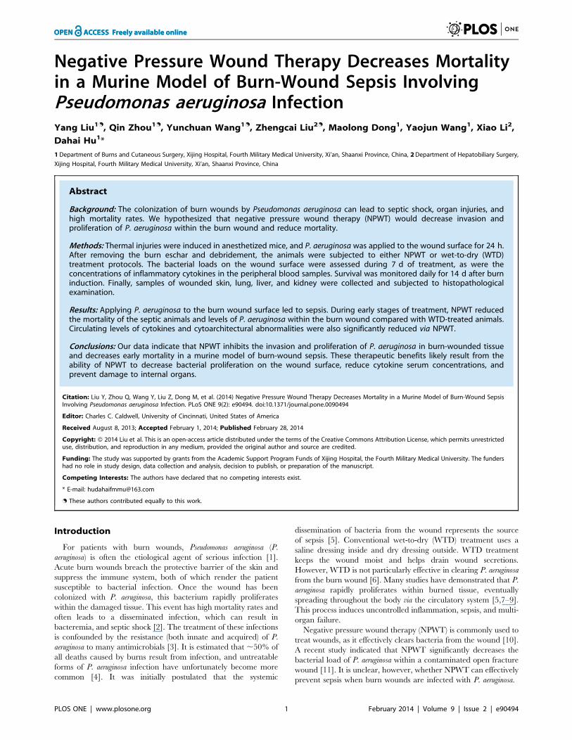
Negative Pressure Wound Therapy Decreases Mortality in a Murine Model of Burn-Wound Sepsis Involving Pseudomonas aeruginosa Infection Yang Liu 1. , Qin Zhou 1. , Yunchuan Wang 1. , Zhengcai Liu 2. , Maolong Dong 1 , Yaojun Wang 1 , Xiao Li 2 , Dahai Hu 1 * 1 Department of Burns and Cutaneous Surgery, Xijing Hospital, Fourth Military Medical University, Xi’an, Shaanxi Province, China, 2 Department of Hepatobiliary Surgery, Xijing Hospital, Fourth Military Medical University, Xi’an, Shaanxi Province, China Abstract Background: The colonization of burn wounds by Pseudomonas aeruginosa can lead to septic shock, organ injuries, and high mortality rates. We hypothesized that negative pressure wound therapy (NPWT) would decrease invasion and proliferation of P. aeruginosa within the burn wound and reduce mortality. Methods: Thermal injuries were induced in anesthetized mice, and P. aeruginosa was applied to the wound surface for 24 h. After removing the burn eschar and debridement, the animals were subjected to either NPWT or wet-to-dry (WTD) treatment protocols. The bacterial loads on the wound surface were assessed during 7 d of treatment, as were the concentrations of inflammatory cytokines in the peripheral blood samples. Survival was monitored daily for 14 d after burn induction. Finally, samples of wounded skin, lung, liver, and kidney were collected and subjected to histopathological examination. Results: Applying P. aeruginosa to the burn wound surface led to sepsis. During early stages of treatment, NPWT reduced the mortality of the septic animals and levels of P. aeruginosa within the burn wound compared with WTD-treated animals. Circulating levels of cytokines and cytoarchitectural abnormalities were also significantly reduced via NPWT. Conclusions: Our data indicate that NPWT inhibits the invasion and proliferation of P. aeruginosa in burn-wounded tissue and decreases early mortality in a murine model of burn-wound sepsis. These therapeutic benefits likely result from the ability of NPWT to decrease bacterial proliferation on the wound surface, reduce cytokine serum concentrations, and prevent damage to internal organs. Citation: Liu Y, Zhou Q, Wang Y, Liu Z, Dong M, et al. (2014) Negative Pressure Wound Therapy Decreases Mortality in a Murine Model of Burn-Wound Sepsis Involving Pseudomonas aeruginosa Infection. PLoS ONE 9(2): e90494. doi:10.1371/journal.pone.0090494 Editor: Charles C. Caldwell, University of Cincinnati, United States of America Received August 8, 2013; Accepted February 1, 2014; Published February 28, 2014 Copyright: ß 2014 Liu et al. This is an open-access article distributed under the terms of the Creative Commons Attribution License, which permits unrestricted use, distribution, and reproduction in any medium, provided the original author and source are credited. Funding: The study was supported by grants from the Academic Support Program Funds of Xijing Hospital, the Fourth Military Medical University. The funders had no role in study design, data collection and analysis, decision to publish, or preparation of the manuscript. Competing Interests: The authors have declared that no competing interests exist. * E-mail: [email protected] . These authors contributed equally to this work. Introduction For patients with burn wounds, Pseudomonas aeruginosa (P. aeruginosa) is often the etiological agent of serious infection [1]. Acute burn wounds breach the protective barrier of the skin and suppress the immune system, both of which render the patient susceptible to bacterial infection. Once the wound has been colonized with P. aeruginosa, this bacterium rapidly proliferates within the damaged tissue. This event has high mortality rates and often leads to a disseminated infection, which can result in bacteremia, and septic shock [2]. The treatment of these infections is confounded by the resistance (both innate and acquired) of P. aeruginosa to many antimicrobials [3]. It is estimated that ,50% of all deaths caused by burns result from infection, and untreatable forms of P. aeruginosa infection have unfortunately become more common [4]. It was initially postulated that the systemic dissemination of bacteria from the wound represents the source of sepsis [5]. Conventional wet-to-dry (WTD) treatment uses a saline dressing inside and dry dressing outside. WTD treatment keeps the wound moist and helps drain wound secretions. However, WTD is not particularly effective in clearing P. aeruginosa from the burn wound [6]. Many studies have demonstrated that P. aeruginosa rapidly proliferates within burned tissue, eventually spreading throughout the body via the circulatory system [5,7–9]. This process induces uncontrolled inflammation, sepsis, and multi- organ failure. Negative pressure wound therapy (NPWT) is commonly used to treat wounds, as it effectively clears bacteria from the wound [10]. A recent study indicated that NPWT significantly decreases the bacterial load of P. aeruginosa within a contaminated open fracture wound [11]. It is unclear, however, whether NPWT can effectively prevent sepsis when burn wounds are infected with P. aeruginosa. PLOS ONE | www.plosone.org 1 February 2014 | Volume 9 | Issue 2 | e90494

Welcome message from author
This document is posted to help you gain knowledge. Please leave a comment to let me know what you think about it! Share it to your friends and learn new things together.
Transcript
Negative Pressure Wound Therapy Decreases Mortalityin a Murine Model of Burn-Wound Sepsis InvolvingPseudomonas aeruginosa InfectionYang Liu1., Qin Zhou1., Yunchuan Wang1., Zhengcai Liu2., Maolong Dong1, Yaojun Wang1, Xiao Li2,
Dahai Hu1*
1Department of Burns and Cutaneous Surgery, Xijing Hospital, Fourth Military Medical University, Xi’an, Shaanxi Province, China, 2Department of Hepatobiliary Surgery,
Xijing Hospital, Fourth Military Medical University, Xi’an, Shaanxi Province, China
Abstract
Background: The colonization of burn wounds by Pseudomonas aeruginosa can lead to septic shock, organ injuries, andhigh mortality rates. We hypothesized that negative pressure wound therapy (NPWT) would decrease invasion andproliferation of P. aeruginosa within the burn wound and reduce mortality.
Methods: Thermal injuries were induced in anesthetized mice, and P. aeruginosa was applied to the wound surface for 24 h.After removing the burn eschar and debridement, the animals were subjected to either NPWT or wet-to-dry (WTD)treatment protocols. The bacterial loads on the wound surface were assessed during 7 d of treatment, as were theconcentrations of inflammatory cytokines in the peripheral blood samples. Survival was monitored daily for 14 d after burninduction. Finally, samples of wounded skin, lung, liver, and kidney were collected and subjected to histopathologicalexamination.
Results: Applying P. aeruginosa to the burn wound surface led to sepsis. During early stages of treatment, NPWT reducedthe mortality of the septic animals and levels of P. aeruginosa within the burn wound compared with WTD-treated animals.Circulating levels of cytokines and cytoarchitectural abnormalities were also significantly reduced via NPWT.
Conclusions: Our data indicate that NPWT inhibits the invasion and proliferation of P. aeruginosa in burn-wounded tissueand decreases early mortality in a murine model of burn-wound sepsis. These therapeutic benefits likely result from theability of NPWT to decrease bacterial proliferation on the wound surface, reduce cytokine serum concentrations, andprevent damage to internal organs.
Citation: Liu Y, Zhou Q, Wang Y, Liu Z, Dong M, et al. (2014) Negative Pressure Wound Therapy Decreases Mortality in a Murine Model of Burn-Wound SepsisInvolving Pseudomonas aeruginosa Infection. PLoS ONE 9(2): e90494. doi:10.1371/journal.pone.0090494
Editor: Charles C. Caldwell, University of Cincinnati, United States of America
Received August 8, 2013; Accepted February 1, 2014; Published February 28, 2014
Copyright: � 2014 Liu et al. This is an open-access article distributed under the terms of the Creative Commons Attribution License, which permits unrestricteduse, distribution, and reproduction in any medium, provided the original author and source are credited.
Funding: The study was supported by grants from the Academic Support Program Funds of Xijing Hospital, the Fourth Military Medical University. The fundershad no role in study design, data collection and analysis, decision to publish, or preparation of the manuscript.
Competing Interests: The authors have declared that no competing interests exist.
* E-mail: [email protected]
. These authors contributed equally to this work.
Introduction
For patients with burn wounds, Pseudomonas aeruginosa (P.
aeruginosa) is often the etiological agent of serious infection [1].
Acute burn wounds breach the protective barrier of the skin and
suppress the immune system, both of which render the patient
susceptible to bacterial infection. Once the wound has been
colonized with P. aeruginosa, this bacterium rapidly proliferates
within the damaged tissue. This event has high mortality rates and
often leads to a disseminated infection, which can result in
bacteremia, and septic shock [2]. The treatment of these infections
is confounded by the resistance (both innate and acquired) of P.
aeruginosa to many antimicrobials [3]. It is estimated that ,50% of
all deaths caused by burns result from infection, and untreatable
forms of P. aeruginosa infection have unfortunately become more
common [4]. It was initially postulated that the systemic
dissemination of bacteria from the wound represents the source
of sepsis [5]. Conventional wet-to-dry (WTD) treatment uses a
saline dressing inside and dry dressing outside. WTD treatment
keeps the wound moist and helps drain wound secretions.
However, WTD is not particularly effective in clearing P. aeruginosa
from the burn wound [6]. Many studies have demonstrated that P.
aeruginosa rapidly proliferates within burned tissue, eventually
spreading throughout the body via the circulatory system [5,7–9].
This process induces uncontrolled inflammation, sepsis, and multi-
organ failure.
Negative pressure wound therapy (NPWT) is commonly used to
treat wounds, as it effectively clears bacteria from the wound [10].
A recent study indicated that NPWT significantly decreases the
bacterial load of P. aeruginosa within a contaminated open fracture
wound [11]. It is unclear, however, whether NPWT can effectively
prevent sepsis when burn wounds are infected with P. aeruginosa.
PLOS ONE | www.plosone.org 1 February 2014 | Volume 9 | Issue 2 | e90494
We hypothesized that NPWT would decrease the levels of P.
aeruginosa within a burn wound, prevent sepsis, and reduce the
mortality rate. To test this hypothesis, a murine model of P.
aeruginosa–induced burn wound sepsis was established according to
our previous report [12]. The effect of NPWT on the bacterial
load on the wound surface was determined using an in vivo imaging
system and direct culturing of the wound tissue. Mortality rates
were also examined. We further speculated that NPWT would
reduce the levels of Th1-type cytokines in the bloodstream and
thereby protect the internal organs against infection.
Materials and Methods
Ethics StatementsAll animal experiments were performed in accordance with the
guidelines from the Administration of Animal Experiments for
Medical Research Purposes issued by the Ministry of Health of
China. The protocol was approved by the Animal Experiment
Administration Committee of Fourth Military Medical University.
All surgical procedures were performed under sodium pentobar-
bital anesthesia and in a clean surgical room with sterilized
instruments. All efforts were made to minimize the suffering of the
mice during the experiments.
MiceEight- to 12-week-old male C57BL/6 mice were obtained from
the Experimental Animal Center of The Fourth Military Medical
University and housed in specific pathogen-free conditions. A total
of 342 mice were involved in our experiments. Because each test
was independently repeated three times, 6 mice were used for the
detection of bioluminescent bacteria in the wound, 36 mice were
used for the bacterial cultures, and 180 mice were used for the
cytokine analysis. In addition, 120 mice were used for the
mortality analysis and were randomly divided into a NPWT
group, WTD group, scald with infection group, or scald without
infection group (30 mice per group).
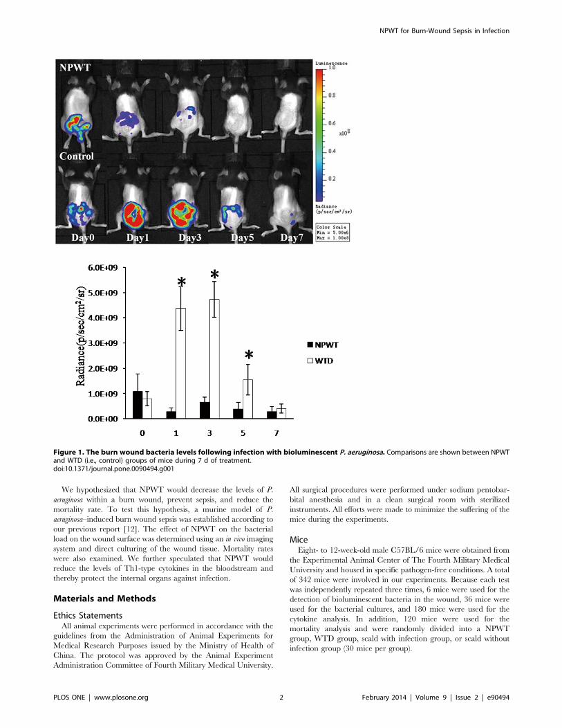
Figure 1. The burn wound bacteria levels following infection with bioluminescent P. aeruginosa. Comparisons are shown between NPWTand WTD (i.e., control) groups of mice during 7 d of treatment.doi:10.1371/journal.pone.0090494.g001
NPWT for Burn-Wound Sepsis in Infection
PLOS ONE | www.plosone.org 2 February 2014 | Volume 9 | Issue 2 | e90494
Bacterial Strain and Culture ConditionsThe P. aeruginosa strain was derived from the PAO1 parental
strain. This bacterium was genetically engineered to fluoresce by
random chromosomal insertion of the modified Photorhabdus
luminescens luxCDABE operon [13]. The bacteria were grown
overnight in Luria-Bertani medium at 37uC with shaking
(120 rpm), which produced cultures with an optical density
(600 nm) of 0.8. This corresponded to a bacterial density of
16109 colony-forming units (CFU)/ml.
Animal Model of Thermal Injury and InfectionThe murine model of sepsis that was originally developed by
Yoav Barnea and Yehuda Barnea [14] was adopted for this study
with slight modifications. All procedures were performed in a
laboratory accredited by the Association for Assessment and
Accreditation of Laboratory Animal Care, and the protocol was
approved by the Institutional Animal Care and Use Committee.
The mice were securely placed into a template with a round
opening (2 cm in diameter) that exposed their shaved backs. A
thermal injury was induced by exposing the shaved area of the skin
to water vapor (100uC) for 8 s. This injury is nonlethal but results
in a third-degree (full-thickness) burn to ,6% of the animal’s body
surface. Fluid replacement therapy, which consisted of an
intraperitoneal injection of 30 ml/kg of 0.9% physiological saline,
was administered immediately following the burn. The mice in the
infection groups had 50 ml of the bacterial inoculums
(1.06109 cfu/ml) directly applied to the eschar. During recovery,
the mice were housed under warming lights and observed
frequently.
Wound Treatment and Bacterial MeasurementsThe bioluminescence of the bacteria in the wound was detected
using the IVIS 100 system (Xenogen Corporation/Caliper Life
Sciences, Alameda, CA, USA). The animals were anesthetized
24 h after bacterial inoculation, and the burn eschar was removed
under aseptic conditions. The wound was then washed twice with
sterile 0.9% physiological saline to clear the wound secretions; the
wound was then imaged. This process determined the baseline
quantity of bacteria. Dressings were then applied to the wounds.
The wounds were dressed using either conventional WTD or the
V.A.C. System (KCI, San Antonio, TX, USA). The V.A.C.
System used GranuFoam Dressing (KCI) and a continuous
negative pressure of –125 mmHg. The wounds were debrided
and irrigated every 24 h. The wounds were imaged after 0, 1, 3, 5,
and 7 d. After the images were collected on day 7, the animals
were euthanized, and tissue specimens were collected. Measure-
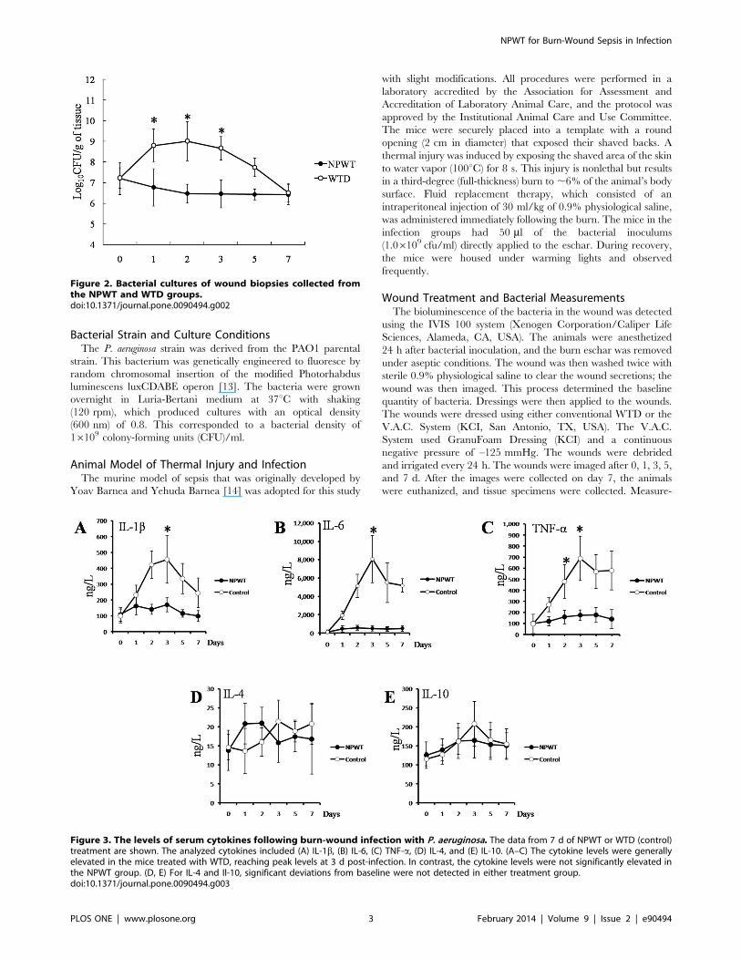
Figure 2. Bacterial cultures of wound biopsies collected fromthe NPWT and WTD groups.doi:10.1371/journal.pone.0090494.g002
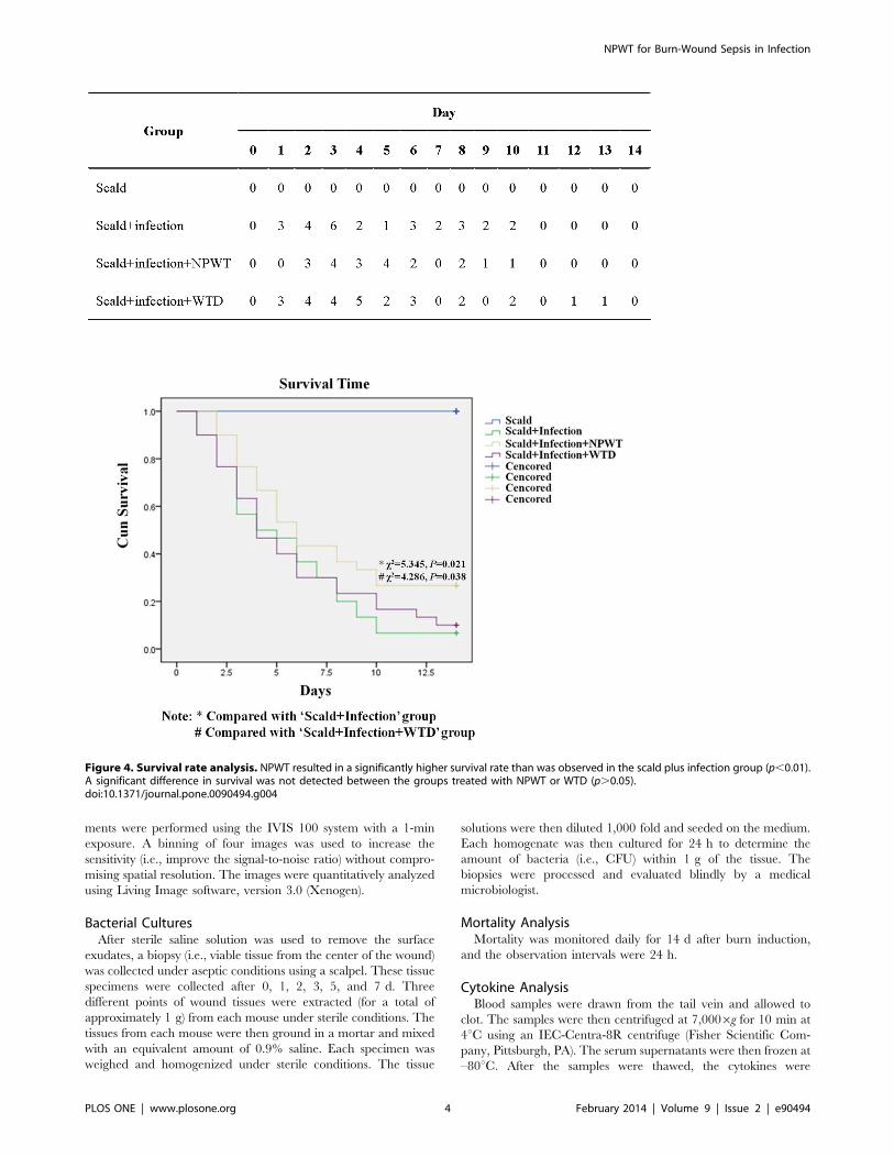
Figure 3. The levels of serum cytokines following burn-wound infection with P. aeruginosa. The data from 7 d of NPWT or WTD (control)treatment are shown. The analyzed cytokines included (A) IL-1b, (B) IL-6, (C) TNF-a, (D) IL-4, and (E) IL-10. (A–C) The cytokine levels were generallyelevated in the mice treated with WTD, reaching peak levels at 3 d post-infection. In contrast, the cytokine levels were not significantly elevated inthe NPWT group. (D, E) For IL-4 and Il-10, significant deviations from baseline were not detected in either treatment group.doi:10.1371/journal.pone.0090494.g003
NPWT for Burn-Wound Sepsis in Infection
PLOS ONE | www.plosone.org 3 February 2014 | Volume 9 | Issue 2 | e90494
ments were performed using the IVIS 100 system with a 1-min
exposure. A binning of four images was used to increase the
sensitivity (i.e., improve the signal-to-noise ratio) without compro-
mising spatial resolution. The images were quantitatively analyzed
using Living Image software, version 3.0 (Xenogen).
Bacterial CulturesAfter sterile saline solution was used to remove the surface
exudates, a biopsy (i.e., viable tissue from the center of the wound)
was collected under aseptic conditions using a scalpel. These tissue
specimens were collected after 0, 1, 2, 3, 5, and 7 d. Three
different points of wound tissues were extracted (for a total of
approximately 1 g) from each mouse under sterile conditions. The
tissues from each mouse were then ground in a mortar and mixed
with an equivalent amount of 0.9% saline. Each specimen was
weighed and homogenized under sterile conditions. The tissue
solutions were then diluted 1,000 fold and seeded on the medium.
Each homogenate was then cultured for 24 h to determine the
amount of bacteria (i.e., CFU) within 1 g of the tissue. The
biopsies were processed and evaluated blindly by a medical
microbiologist.
Mortality AnalysisMortality was monitored daily for 14 d after burn induction,
and the observation intervals were 24 h.
Cytokine AnalysisBlood samples were drawn from the tail vein and allowed to
clot. The samples were then centrifuged at 7,0006g for 10 min at
4uC using an IEC-Centra-8R centrifuge (Fisher Scientific Com-
pany, Pittsburgh, PA). The serum supernatants were then frozen at
–80uC. After the samples were thawed, the cytokines were
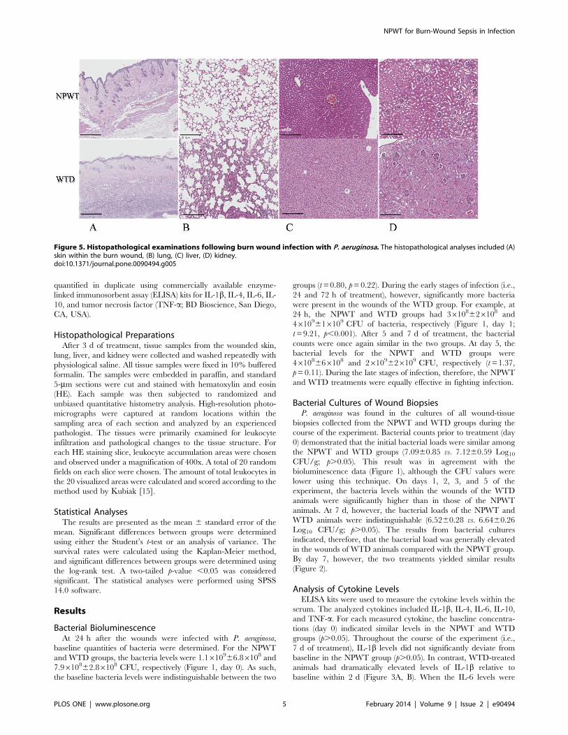
Figure 4. Survival rate analysis. NPWT resulted in a significantly higher survival rate than was observed in the scald plus infection group (p,0.01).A significant difference in survival was not detected between the groups treated with NPWT or WTD (p.0.05).doi:10.1371/journal.pone.0090494.g004
NPWT for Burn-Wound Sepsis in Infection
PLOS ONE | www.plosone.org 4 February 2014 | Volume 9 | Issue 2 | e90494
quantified in duplicate using commercially available enzyme-
linked immunosorbent assay (ELISA) kits for IL-1b, IL-4, IL-6, IL-10, and tumor necrosis factor (TNF-a; BD Bioscience, San Diego,
CA, USA).
Histopathological PreparationsAfter 3 d of treatment, tissue samples from the wounded skin,
lung, liver, and kidney were collected and washed repeatedly with
physiological saline. All tissue samples were fixed in 10% buffered
formalin. The samples were embedded in paraffin, and standard
5-mm sections were cut and stained with hematoxylin and eosin
(HE). Each sample was then subjected to randomized and
unbiased quantitative histometry analysis. High-resolution photo-
micrographs were captured at random locations within the
sampling area of each section and analyzed by an experienced
pathologist. The tissues were primarily examined for leukocyte
infiltration and pathological changes to the tissue structure. For
each HE staining slice, leukocyte accumulation areas were chosen
and observed under a magnification of 400x. A total of 20 random
fields on each slice were chosen. The amount of total leukocytes in
the 20 visualized areas were calculated and scored according to the
method used by Kubiak [15].
Statistical AnalysesThe results are presented as the mean 6 standard error of the
mean. Significant differences between groups were determined
using either the Student’s t-test or an analysis of variance. The
survival rates were calculated using the Kaplan-Meier method,
and significant differences between groups were determined using
the log-rank test. A two-tailed p-value ,0.05 was considered
significant. The statistical analyses were performed using SPSS
14.0 software.
Results
Bacterial BioluminescenceAt 24 h after the wounds were infected with P. aeruginosa,
baseline quantities of bacteria were determined. For the NPWT
and WTD groups, the bacteria levels were 1.1610966.86108 and
7.9610862.86108 CFU, respectively (Figure 1, day 0). As such,
the baseline bacteria levels were indistinguishable between the two
groups (t = 0.80, p=0.22). During the early stages of infection (i.e.,
24 and 72 h of treatment), however, significantly more bacteria
were present in the wounds of the WTD group. For example, at
24 h, the NPWT and WTD groups had 36108626108 and
46109616109 CFU of bacteria, respectively (Figure 1, day 1;
t = 9.21, p,0.001). After 5 and 7 d of treatment, the bacterial
counts were once again similar in the two groups. At day 5, the
bacterial levels for the NPWT and WTD groups were
46108666108 and 26109626109 CFU, respectively (t = 1.37,
p=0.11). During the late stages of infection, therefore, the NPWT
and WTD treatments were equally effective in fighting infection.
Bacterial Cultures of Wound BiopsiesP. aeruginosa was found in the cultures of all wound-tissue
biopsies collected from the NPWT and WTD groups during the
course of the experiment. Bacterial counts prior to treatment (day
0) demonstrated that the initial bacterial loads were similar among
the NPWT and WTD groups (7.0960.85 vs. 7.1260.59 Log10CFU/g; p.0.05). This result was in agreement with the
bioluminescence data (Figure 1), although the CFU values were
lower using this technique. On days 1, 2, 3, and 5 of the
experiment, the bacteria levels within the wounds of the WTD
animals were significantly higher than in those of the NPWT
animals. At 7 d, however, the bacterial loads of the NPWT and
WTD animals were indistinguishable (6.5260.28 vs. 6.6460.26
Log10 CFU/g; p.0.05). The results from bacterial cultures
indicated, therefore, that the bacterial load was generally elevated
in the wounds of WTD animals compared with the NPWT group.
By day 7, however, the two treatments yielded similar results
(Figure 2).
Analysis of Cytokine LevelsELISA kits were used to measure the cytokine levels within the
serum. The analyzed cytokines included IL-1b, IL-4, IL-6, IL-10,and TNF-a. For each measured cytokine, the baseline concentra-
tions (day 0) indicated similar levels in the NPWT and WTD
groups (p.0.05). Throughout the course of the experiment (i.e.,
7 d of treatment), IL-1b levels did not significantly deviate from
baseline in the NPWT group (p.0.05). In contrast, WTD-treated
animals had dramatically elevated levels of IL-1b relative to
baseline within 2 d (Figure 3A, B). When the IL-6 levels were
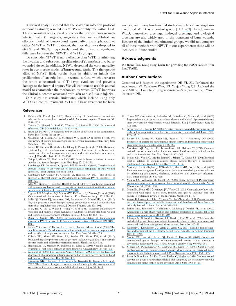
Figure 5. Histopathological examinations following burn wound infection with P. aeruginosa. The histopathological analyses included (A)skin within the burn wound, (B) lung, (C) liver, (D) kidney.doi:10.1371/journal.pone.0090494.g005
NPWT for Burn-Wound Sepsis in Infection
PLOS ONE | www.plosone.org 5 February 2014 | Volume 9 | Issue 2 | e90494
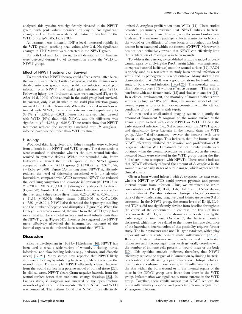
analyzed, this cytokine was significantly elevated in the NPWT
group, with peak values measured on day 3. No significant
changes in IL-6 levels were detected relative to baseline for the
WTD group (p.0.05; Figure 3C).
As treatment was initiated, TNF-a levels increased rapidly in
the WTD group, reaching peak values after 3 d. No significant
changes in TNF-a levels were detected in the NPWT group.
For both IL-4 and IL-10, no significant deviations from baseline
were detected during 7 d of treatment in either the WTD or
NPWT groups.
Effect of NPWT Treatment on SurvivalTo test whether NPWT therapy could affect survival after burn,
the wounds were infected with P. aeruginosa, and the animals were
divided into four groups: scald, scald plus infection, scald plus
infection plus NPWT, and scald plus infection plus WTD.
Following injury, the 14-d survival rates were analyzed (Figure 4).
After 14 d, 100% of the animals in the scald group had survived.
In contrast, only 2 of 30 mice in the scald plus infection group
survived for 14 d (6.7% survival). When the infected wounds were
treated with NPWT, the survival rate increased significantly to
33.3% (x2 = 5.345, p=0.021). Fewer mice survived when treated
with WTD (10%) than with NPWT, and this difference was
significant (x2 = 4.286, p=0.038). These results show that NPWT
treatment reduced the mortality associated with P. aeruginosa–
infected burn wounds more than WTD treatment.
HistologyWounded skin, lung, liver, and kidney samples were collected
from animals in the NPWT and WTD groups. The tissue sections
were examined to determine whether the burn wound infection
resulted in systemic defects. Within the wounded skin, fewer
leukocytes infiltrated the muscle space in the NPWT group
compared with the WTD group (1.4160.22 vs. 3.3760.54;
t = 15.08, p,0.001, Figure 5A). In lung tissue, NPWT significantly
reduced the level of thickening associated with the alveolar
interstitium, compared with WTD treatment. NPWT also reduced
the local lung congestion and leukocyte infiltration (0.9460.25 vs.
2.6660.49; t = 13.98, p,0.001) during early stages of treatment
(Figure 5B). Similar leukocyte infiltration levels were observed in
the liver and kidney tissue (liver tissue: 0.2060.07 vs. 0.5460.11;
t = 11.33, p,0.001; kidney tissue: 0.2860.06 vs. 0.4760.09;
t = 7.92, p,0.001). NPWT also decreased the hepatocyte swelling
and the number of hepatic cord disruptions (Figure 5C). When the
kidney tissues were examined, the mice from the WTD group had
more renal tubular epithelial necrosis and renal tubular casts than
the NPWT group (Figure 5D). These results suggested that NPWT
more effectively alleviated the inflammatory response of the
internal organs to the infected burn wound than WTD.
Discussion
Since its development in 1993 by Fleischman [16], NPWT has
been used to treat a wide variety of wounds, including burns,
infections, and slow-healing lesions (e.g., bedsores, and diabetic
ulcers) [17–21]. Many studies have reported that NPWT likely
aids wound healing by inhibiting bacterial proliferation within the
wound tissue. For example, NPWT effectively cleared bacteria
from the wound surface in a porcine model of burned tissue [22].
In clinical cases, NPWT clears Gram-negative bacteria from the
wound surface better than traditional change dressings [23]. In
Lalliss’s study, P. aeruginosa was smeared on the open fracture
wounds of goats and the therapeutic effect of NPWT and WTD
was compared. The authors found that NPWT more effectively
limited P. aeruginosa proliferation than WTD [11]. These studies
provided preliminary evidence that NPWT inhibits bacterial
proliferation. In each case, however, only the wound surface was
analyzed. The invasion of pathogenic bacteria into deeper levels of
the wound or the diffusion of these bacteria throughout the body
has not been examined within the context of NPWT. Moreover, it
has not been definitively proven that NPWT can effectively limit
the proliferation of P. aeruginosa in burn wounds.
To address these issues, we established a murine model of burn-
wound sepsis by applying the PAO1 strain (which was engineered
to express bacterial luciferase) onto the wound surface [12]. PAO1
is widely used as a test strain to study burn wound infection or
sepsis, and its pathogenicity is representative. Many studies have
demonstrated that PAO1 was a good test strain for fundamental
study in burn wound infection [14,24,25]. The mortality rate in
this model was over 90% without effective treatment. This result is
consistent with our former study [12] and similar to another [14].
In a clinical environment, the mortality of trauma patients with
sepsis is as high as 90% [26]; thus, this murine model of burn
wound sepsis is to a certain extent consistent with the clinical
conditions of burn patients with sepsis.
We then used a small animal imaging system to quantify the
amount of fluorescent P. aeruginosa on the wound surface as the
animals were treated with either NPWT or WTD. During the
early stages of infection (i.e., 3 d of treatment), the NPWT group
had significantly fewer bacteria in the wound than the WTD
group. After 7 d of treatment, however, the bacteria levels were
similar in the two groups. This indicates that, for burned tissue,
NPWT effectively inhibited the invasion and proliferation of P.
aeruginosa, whereas WTD treatment did not. Similar results were
obtained when the wound secretions were cultured, as the wound
bacterial loads were elevated in the WTD group during the first
3 d of treatment (compared with NPWT). These results indicate
that NPWT effectively reduced the amount of P. aeruginosa in the
wound tissue at early stages of burn damage, which agrees with its
clinical effects.
Given a burn wound infected with P. aeruginosa, we next tested
whether NPWT or WTD could prevent pyaemia and protect
internal organs from infection. Thus, we examined the serum
concentrations of IL-1b, IL-4, IL-6, IL-10, and TNF-a during
burn treatment. We also performed histopathological examina-
tions of the wounded skin, lung, liver, and kidney tissue after 3 d of
treatment. In the NPWT group, the serum levels of IL-1b, IL-6,and TNF-a did not significantly deviate from baseline throughout
the course of the experiment. In contrast, the levels of these
proteins in the WTD group were dramatically elevated during the
early stages of treatment. On day 7, the bacterial content
decreased, which may be related to the mouse immune clearance
of the bacteria; a determination of this possibility requires further
study. The four cytokines used are Th1-type cytokines, which play
important roles in acute post-traumatic inflammation [27–29].
Because Th1-type cytokines are primarily secreted by activated
monocytes and macrophages, their levels generally correlate with
the number of immune cells present in wound tissue or the body
[30]. This cytokine analysis indicates, therefore, that NPWT
effectively reduces the degree of inflammation by limiting bacterial
proliferation and alleviating sepsis progression. Histopathological
examinations confirmed these results, as the inflammatory cells in
the skin within the burn wound or in the internal organs of the
mice in the NPWT group were fewer than those in the WTD
group. Inflammation was significantly more extreme in the WTD
group. Together, these results suggest that NPWT reduced the
in vivo inflammatory response and protected internal organs from
P. aeruginosa infection.
NPWT for Burn-Wound Sepsis in Infection
PLOS ONE | www.plosone.org 6 February 2014 | Volume 9 | Issue 2 | e90494
A survival analysis showed that the scald plus infection protocol
(without treatment) resulted in a 93.3% mortality rate within 14 d.
This is consistent with clinical outcomes that involve burn wounds
infected with P. aeruginosa, suggesting that we established an
effective model of burn-wound sepsis. After the application of
either NPWT or WTD treatment, the mortality rates dropped to
66.7% and 90.0%, respectively, and there was a significant
difference between the NPWT and WTD groups.
To conclude, NPWT is more effective than WTD in inhibiting
the invasion and subsequent proliferation of P. aeruginosa into burn-
wounded tissue. In addition, NPWT decreased the early mortality
rates in our murine model of burn-wound sepsis. The therapeutic
effect of NPWT likely results from its ability to inhibit the
proliferation of bacteria from the wound surface, which decreases
the serum concentrations of Th1-type cytokines and prevents
damage to the internal organs. We will continue to use this animal
model to characterize the mechanism by which NPWT improves
the clinical outcomes associated with skin and soft tissue injuries.
Our study has certain limitations, which include using only
WTD as a control treatment. WTD is a basic treatment for burn
wounds, and many fundamental studies and clinical investigations
have used WTD as a control group [11,31–33]. In addition to
WTD, nano-silver dressings, hydrogel dressings, and biological
dressings are also widely used in the treatment of burn wounds.
Because of the limited experimental groups, we did not compare
all of these methods with NPWT in our experiments; these will be
included in future studies.
Acknowledgments
We thank Pro. Kang-Ming Duan for providing the PAO1 labeled with
LuxCDABE.
Author Contributions
Conceived and designed the experiments: DH YL ZL. Performed the
experiments: YL Yunchuan Wang XL Yaojun Wang QZ. Analyzed the
data: MD YL. Contributed reagents/materials/analysis tools: YL. Wrote
the paper: DH.
References
1. McVay CS, Fralick JA (2007) Phage therapy of Pseudomonas aeruginosa
infection in a mouse burn wound model. Antimicrob Agents Chemother 51:
1934–1938.2. Church D, Elsayed S, Reid O, Winston B, Lindsay R (2006) Burn wound
infections. Clin Microbiol Rev.; 19: 403–434.3. Pruitt BA Jr (1984) The diagnosis and treatment of infection in the burn patient.
Burns Incl Therm Inj 11: 79–91.4. McManus AT, Mason AD Jr, McManus WF, Pruitt BA Jr (1985) Twenty-five
year review of Pseudomonas aeruginosa bacteremia in a burn center. Eur J Clin
Microbiol 4: 219–223.5. Pirnay JP, De Vos D, Cochez C, Bilocq F, Pirson J, et al. (2003) Molecular
epidemiology of Pseudomonas aeruginosa colonization in a burn unit:persistence of a multi-drug-resistance clone and a silver sulfadiazine-resistant
clone. J Clin Microbiol. 41: 1192–1202.
6. Chipp E, Milner CS, Blackburn AV (2010) Sepsis in burns: a review of currentpractice and future therapies. Ann Plast Surg 65: 228–236.
7. Rumbaugh KP, Griswold JA, Iglewski BH, Hamood AN (1999) Contribution ofquorum sensing to the virulence of Pseudomonas aeruginosa in burn wound
infections. Infect Immun. 67: 5854–5862.8. Rumbaugh KP, Colmer JA, Griswold JA, Hamood AN (2001) The effects of
infection of thermal injury by Pseudomonas aeruginosa PAO1. Cytokine. 16:
160–168.9. Felts AG, Grainger DW, Slunt JB (2000) Locally delivered antibodies combined
with systemic antibiotics confer synergistic protection against antibiotic-resistantburn wound infection. J Trauma 49: 873–878.
10. Argenta LC, Morykwas MJ, Marks MW, DeFranzo AJ, Molnar JA, et al. (2006)
Vacuum-assisted closure:state of clinic art. Plast Reconstr Surg. 117: 127–142.11. Lalliss SJ, Stinner DJ, Waterman SM, Branstetter JG, Masini BD, et al. (2010)
Negative pressure wound therapy reduces pseudomonas wound contaminationmore than staphylococcus aureus. J Orthop Trauma. 24: 598–602.
12. Li N, Hu X, Liu Y, Wang Y, Wang Y, et al. (2013) Systemic inflammatory
responses and multiple organ dysfunction syndrome following skin burn woundand Pseudomonas aeruginosa infection in mice. Shock 40: 152–159.
13. Duan K, Surette MG (2007) Environmental Regulation of Pseudomonasaeruginosa PAO1 Las and Rhl Quorum-Sensing Systems. J Bacteriol 189: 4827–
4836.14. Barnea Y, Carmeli Y, Kuzmenko B, Gur E, Hammer-Munz O, et al. (2006) The
establishment of a Pseudomonas aeruginosa- infected burn-wound sepsis model
and the effect of imipenem treatment. Ann Plast Surg 56: 674–679.15. Kubiak BD, Albert SP, Gatto LA, Snyder KP, Maier KG, et al. (2010)
Peritoneal negative pressure therapy prevents multiple organ injury in a chronicporcine sepsis and ischemia/reperfusion model. Shock 34: 525–534.
16. Fleischmann W, Strecker W, Bombelli M, Kinzl L (1993) Vacuum sealing as
treatment of soft tissue damage in open fractures. Unfallchirurg 96: 488–492.17. Weinand C (2009) The Vacuum-Assisted Closure (VAC) device for hastened
attachment of a superficial inferior-epigastric flap to third-degree burns on handand fingers. J Burn Care Res 30: 362–365.
18. Kanakaris NK, Thanasas C, Keramaris N, Kontakis G, Granick MS, et al.(2007) The efficacy of negative pressure wound therapy in the management of
lower extremity trauma: review of clinical evidence. Injury 38: 9–18.
19. Tocco MP, Costantino A, Ballardini M, D’Andrea C, Masala M, et al. (2009)
Improved results of the vacuum assisted closure and Nitinol clips sternal closure
after postoperative deep sternal wound infection. Eur J Cardiothorac Surg 35:
833–838.
20. Armstrong DG, Lavery LA (2005) Negative pressure wound therapy after partial
diabetic foot amputation: a multicentre, randomised controlled trial. Lancet 366:
1704–1710.
21. Lavery LA, Barnes SA, Keith MS, Seaman JW Jr, Armstrong DG (2008)
Prediction of healing for postoperative diaetic foot wounds based on early wound
area progression. Diabetes Care 31: 26–29.
22. Morykwas MJ, Argenta LC, Shelton-Brown EI, McGuirt W (1997) Vacuum-
assisted closure: a new method for wound control and treatment: animal studies
and basic foundation. Ann Plast Surg 38: 553–562.
23. Moues CM, Vos MC, van den Bemd GJ, Stijnen T, Hovius SE (2004) Bacterial
load in relation to vacuum-assisted closure wound therapy: a prospective
randomized trial. Wound Repair Regen 12: 11–17.
24. Mulcahy H, O’Callaghan J, O’Grady EP, Macia MD, Borrell N, et al. (2008)
Pseudomonas aeruginosa RsmA plays an important role during murine infection
by influencing colonization, virulence, persistence, and pulmonary inflamma-
tion. Infect Immun 76: 632–638.
25. McVay CS, Velasquez M, Fralick JA. (2007) Phage therapy of Pseudomonas
aeruginosa infection in a mouse burn wound model. Antimicrob Agents
Chemother 51: 1934–1938.
26. Mann EA, Baun MM, Meininger JC, Wade CE (2012) Comparison of mortality
associated with sepsis in the burn, trauma, and general intensive care unit
patient: a systematic review of the literature. Shock 37: 4–16.
27. Zhang B, Huang YH, Chen Y, Yang Y, Hao ZL, et al. (1998) Plasma tumour
necrosis factor-alpha, its soluble receptors and interleukin-1 beta levels in
critically burned patients. Burns 24: 599–603.
28. Dehne MG, Sablotzki A, Hoffmann A, Muhling J, Dietrich FE, et al. (2002)
Alterations of acute phase reaction and cytokine production in patients following
severe burn injury. Burns 28: 535–542.
29. Infanger M, Schmidt O, Kossmehl P, Grad S, Ertel W, et al. (2004) Vascular
endothelial growth factor serum level is strongly enhanced after burn injury and
correlated with local and general tissue edema. Burns 30: 305–311.
30. Ozdemir C, Kucuksezer UC, Akdis M, Akdis CA (2011) Specific immunother-
apy and turning off the T cell: how does it work? Ann Allergy Asthma Immunol
107: 381–392.
31. Moues CM, van den Bemd GJ, Heule F, Hovius SE (2007) Comparing
conventional gauze therapy to vacuum-assisted closure wound therapy: a
prospective randomised trial. J Plast Reconstr Aesthet Surg 60: 672–681.
32. Scherer SS, Pietramaggiori G, Mathews JC, Orgill DP (2009) Short periodic
applications of the vacuum-assisted closure device cause an extended tissue
response in thediabetic mouse model. Plast Reconstr Surg 124: 1458–1465.
33. Perez D, Bramkamp M, Exe C, von Ruden C, Ziegler A (2010) Modern wound
care for the poor: a randomized clinical trial comparing the vacuum system with
conventional saline-soaked gauze dressings. Am J Surg 199: 14–20.
NPWT for Burn-Wound Sepsis in Infection
PLOS ONE | www.plosone.org 7 February 2014 | Volume 9 | Issue 2 | e90494
Related Documents