May 18, 2008

Neck
Oct 21, 2015
Lectures from International School of Capital Medical University
Welcome message from author
This document is posted to help you gain knowledge. Please leave a comment to let me know what you think about it! Share it to your friends and learn new things together.
Transcript
May 18, 2008
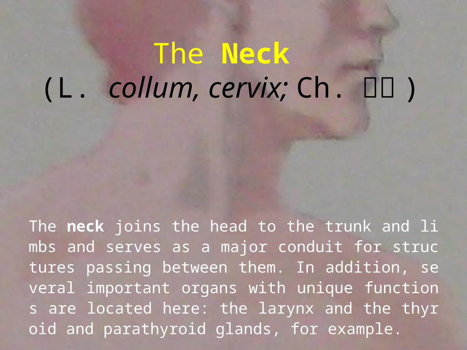
The Neck (L. collum, cervix; Ch. 颈部 )
The neck joins the head to the trunk and limbs and serves as a major conduit for structures passing between them. In addition, several important organs with unique functions are located here: the larynx and the thyroid and parathyroid glands, for example.
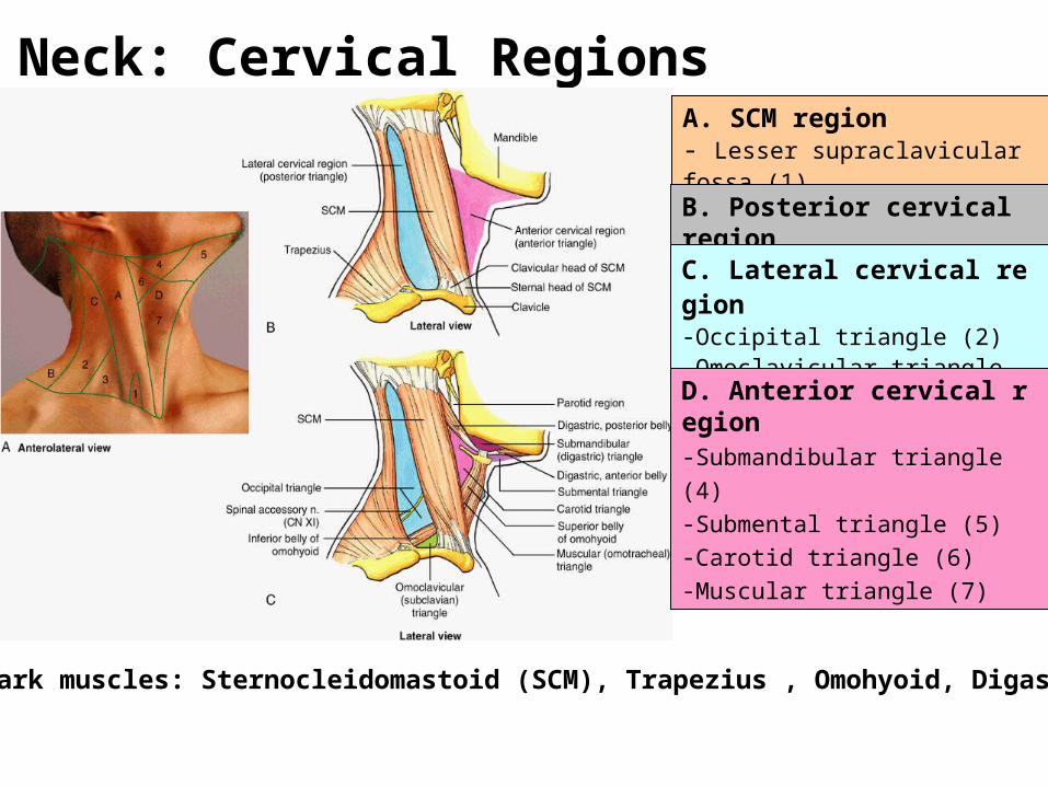
Neck: Cervical Regions A. SCM region- Lesser supraclavicular fossa (1)
B. Posterior cervical region
C. Lateral cervical region-Occipital triangle (2) -Omoclavicular triangle (3)
D. Anterior cervical region-Submandibular triangle (4) -Submental triangle (5) -Carotid triangle (6) -Muscular triangle (7)
Landmark muscles: Sternocleidomastoid (SCM), Trapezius , Omohyoid, Digastric
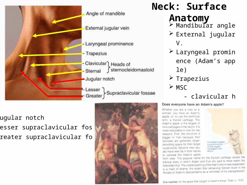
Neck: Surface Anatomy Mandibular angle External jugular V. Laryngeal prominence
(Adam’s apple) Trapezius MSC
- clavicular head
- sternal head
Jugular notch
Lesser supraclavicular fossa
Greater supraclavicular fossa
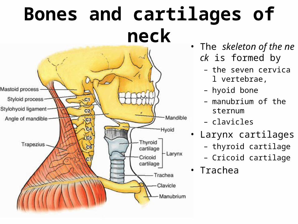
Bones and cartilages of neck• The skeleton of the n
eck is formed by – the seven cervical ver
tebrae, – hyoid bone – manubrium of the ster
num– clavicles
• Larynx cartilages– thyroid cartilage– Cricoid cartilage
• Trachea
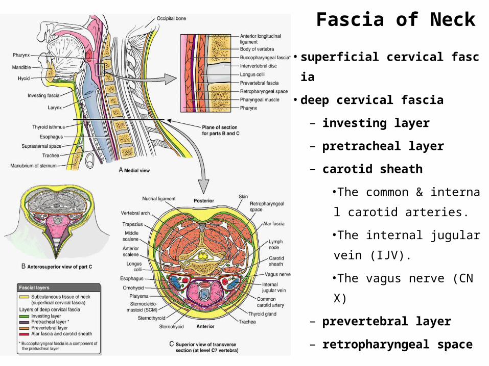
Fascia of Neck
• superficial cervical fascia
• deep cervical fascia
– investing layer
– pretracheal layer
– carotid sheath
•The common & internal
carotid arteries.
•The internal jugular vein
(IJV).
•The vagus nerve (CN X)
– prevertebral layer
– retropharyngeal space
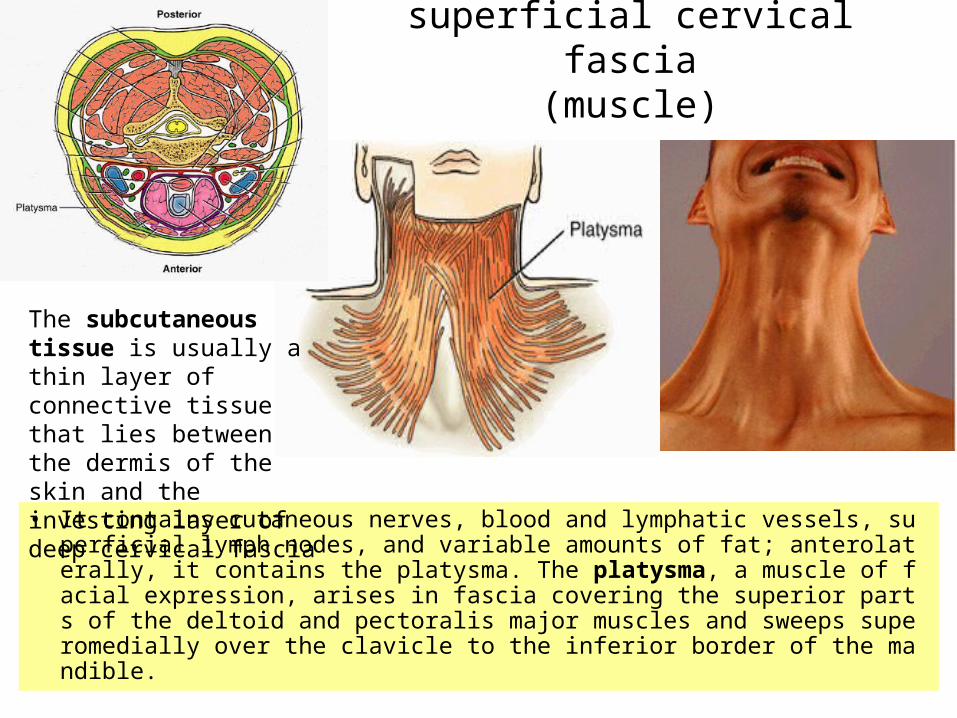
superficial cervical fascia(muscle)
• It contains cutaneous nerves, blood and lymphatic vessels, superficial lymph nodes, and variable amounts of fat; anterolaterally, it contains the platysma. The platysma, a muscle of facial expression, arises in fascia covering the superior parts of the deltoid and pectoralis major muscles and sweeps superomedially over the clavicle to the inferior border of the mandible.
The subcutaneous tissue is usually a thin layer of connective tissue that lies between the dermis of the skin and the investing layer of deep cervical fascia

Superficial veins, nerves, and lymph nodes
external jugular vein (EJV) subclavian vein cutaneous branches of the cervical plexus emerge around the middle of the
posterior border of the SCM, often called the nerve point of the neck, and supply the skin of the neck
superficial cervical lymph nodes inferior deep cervical lymph nodes supraclavicular lymph nodes
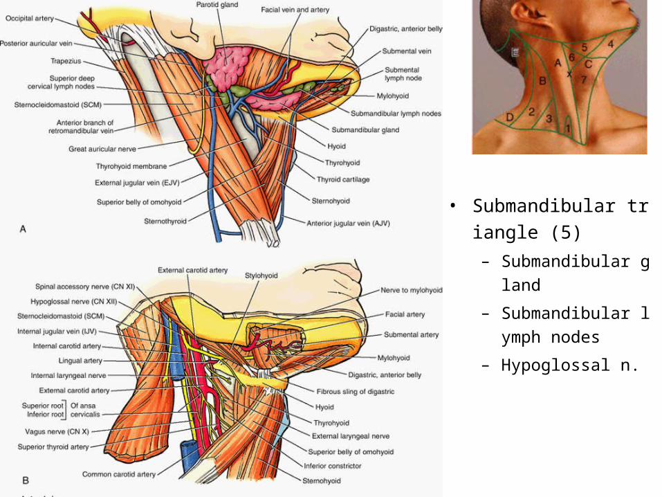
• Submandibular triangl
e (5)
– Submandibular glan
d
– Submandibular lymp
h nodes
– Hypoglossal n.
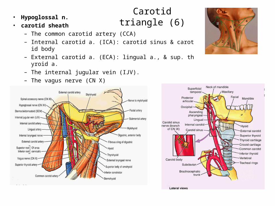
Carotid triangle (6)• Hypoglossal n.
• carotid sheath
– The common carotid artery (CCA)
– Internal carotid a. (ICA): carotid sinus & carotid body
– External carotid a. (ECA): lingual a., & sup. thyroid a.
– The internal jugular vein (IJV).
– The vagus nerve (CN X)
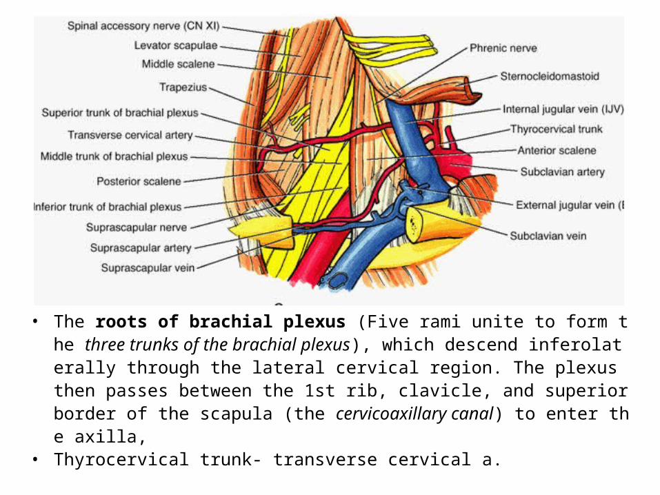
• The roots of brachial plexus (Five rami unite to form the three trunks of the brachial plexus), which descend inferolaterally through the lateral cervical region. The plexus then passes between the 1st rib, clavicle, and superior border of the scapula (the cervicoaxillary canal) to enter the axilla,
• Thyrocervical trunk- transverse cervical a.
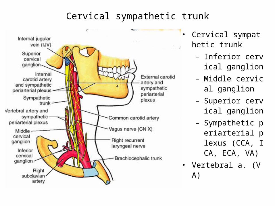
Cervical sympathetic trunk
• Cervical sympathetic trunk
– Inferior cervical ganglion
– Middle cervical ganglion
– Superior cervical ganglion
– Sympathetic periarterial plexus (CCA, ICA, ECA, VA)
• Vertebral a. (VA)
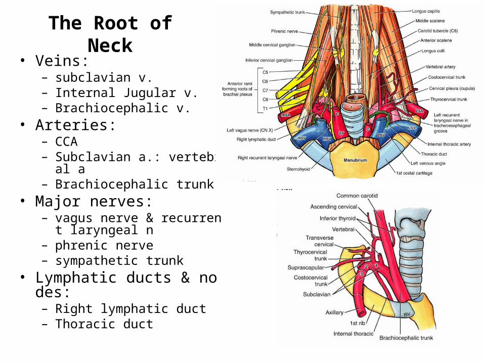
The Root of Neck• Veins:
– subclavian v.– Internal Jugular v.– Brachiocephalic v.
• Arteries:– CCA– Subclavian a.: vertebral a– Brachiocephalic trunk
• Major nerves: – vagus nerve & recurrent lar
yngeal n– phrenic nerve– sympathetic trunk
• Lymphatic ducts & nodes:– Right lymphatic duct– Thoracic duct
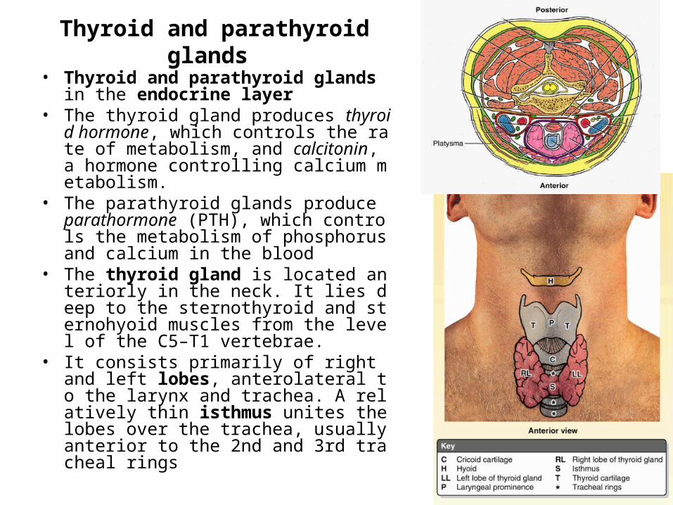
Thyroid and parathyroid glands
• Thyroid and parathyroid glands in the endocrine layer
• The thyroid gland produces thyroid hormone, which controls the rate of metabolism, and calcitonin, a hormone controlling calcium metabolism.
• The parathyroid glands produce parathormone (PTH), which controls the metabolism of phosphorus and calcium in the blood
• The thyroid gland is located anteriorly in the neck. It lies deep to the sternothyroid and sternohyoid muscles from the level of the C5–T1 vertebrae.
• It consists primarily of right and left lobes, anterolateral to the larynx and trachea. A relatively thin isthmus unites the lobes over the trachea, usually anterior to the 2nd and 3rd tracheal rings
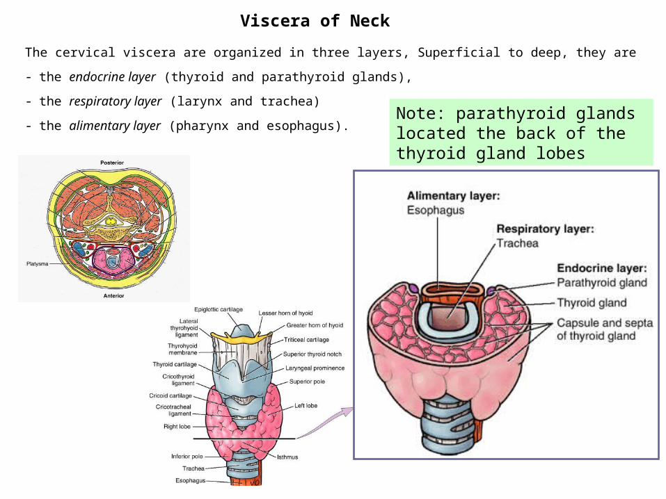
The cervical viscera are organized in three layers, Superficial to deep, they are
- the endocrine layer (thyroid and parathyroid glands),
- the respiratory layer (larynx and trachea)
- the alimentary layer (pharynx and esophagus).
Viscera of Neck
Note: parathyroid glands located the back of the thyroid gland lobes
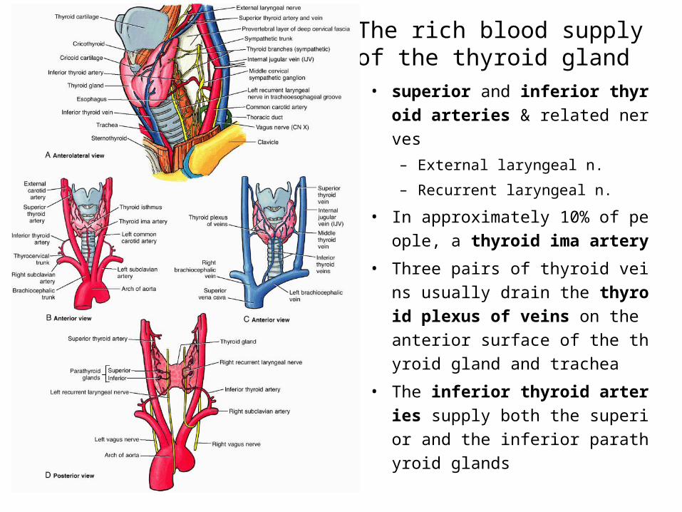
The rich blood supply of the thyroid gland
• superior and inferior thyroid
arteries & related nerves
– External laryngeal n.
– Recurrent laryngeal n.
• In approximately 10% of peopl
e, a thyroid ima artery
• Three pairs of thyroid veins us
ually drain the thyroid plexus
of veins on the anterior surfac
e of the thyroid gland and trac
hea
• The inferior thyroid arteries
supply both the superior and t
he inferior parathyroid glands
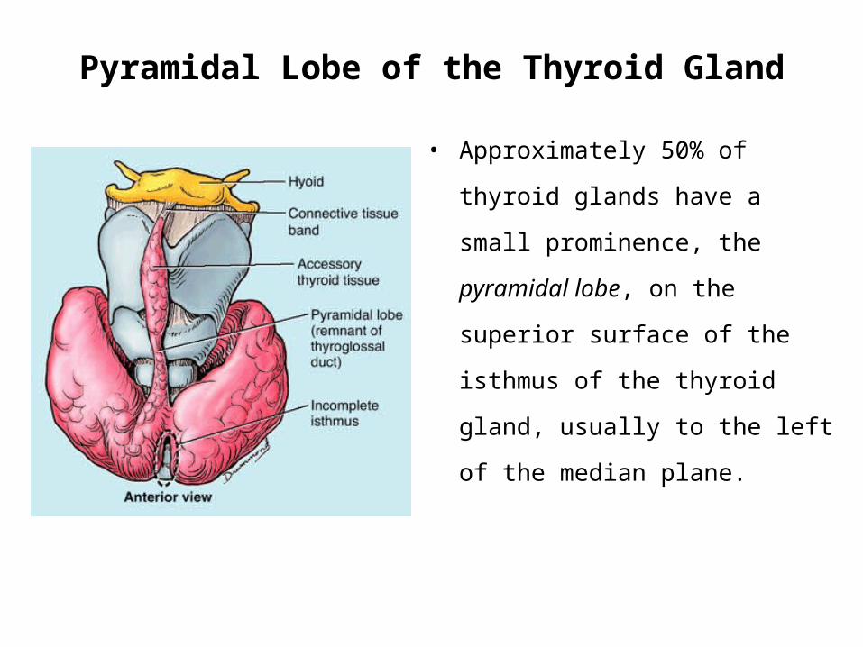
Pyramidal Lobe of the Thyroid Gland
• Approximately 50% of thyroid
glands have a small prominence,
the pyramidal lobe, on the superior
surface of the isthmus of the
thyroid gland, usually to the left of
the median plane.

Review & Questions
Please think and give your answers
during the experimental class
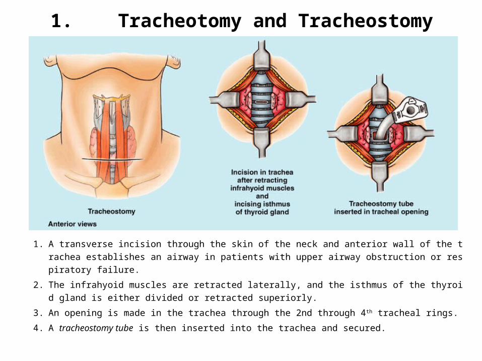
1. Tracheotomy and Tracheostomy
1. A transverse incision through the skin of the neck and anterior wall of the trachea estab
lishes an airway in patients with upper airway obstruction or respiratory failure.
2. The infrahyoid muscles are retracted laterally, and the isthmus of the thyroid gland is ei
ther divided or retracted superiorly.
3. An opening is made in the trachea through the 2nd through 4th tracheal rings.
4. A tracheostomy tube is then inserted into the trachea and secured.
1. Tracheotomy and Tracheostomy
Question
• What layers were passed through to reach the trac
hea during the surgery?
• To avoid complications during a tracheostomy, wh
at anatomical relationships are important?
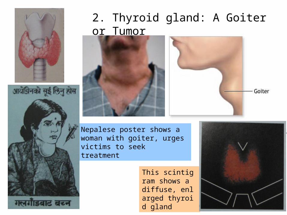
2. Thyroid gland: A Goiter or Tumor
Nepalese poster shows a woman with goiter, urges victims to seek treatment
This scintigram shows a diffuse, enlarged thyroid gland
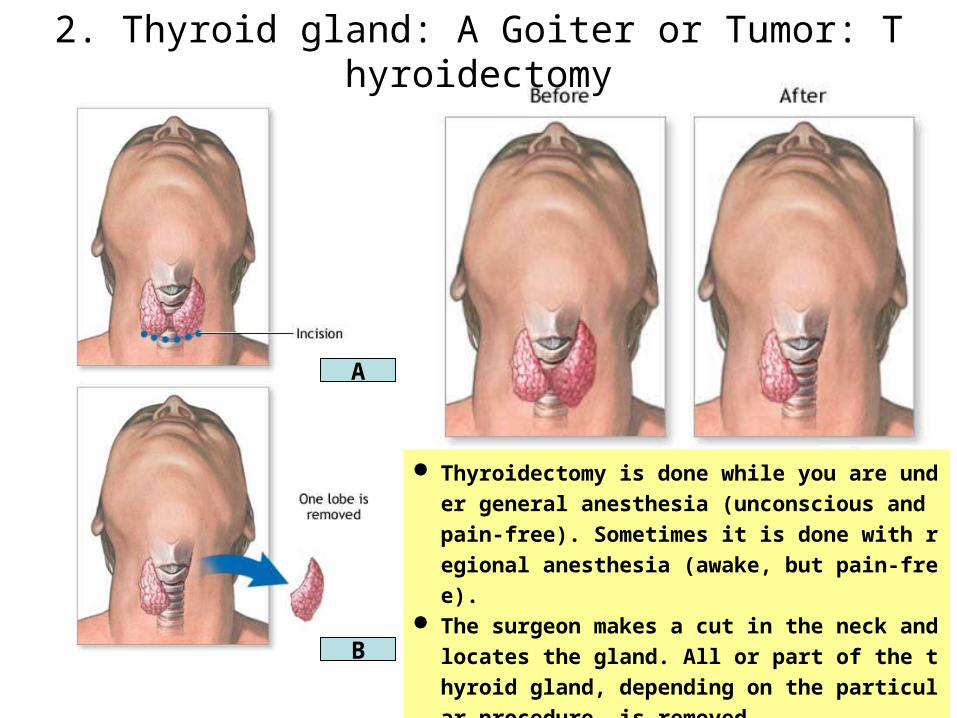
2. Thyroid gland: A Goiter or Tumor: Thyroidectomy
A
B
Thyroidectomy is done while you are under gene
ral anesthesia (unconscious and pain-free). Som
etimes it is done with regional anesthesia (awake,
but pain-free). The surgeon makes a cut in the neck and locates
the gland. All or part of the thyroid gland, depend
ing on the particular procedure, is removed.
2. Thyroidectomy: Questions
• As the figure showed, the left lobe of the thyroid gland was r
emoved. What layers and anatomical structures were passe
d through to reach the lobe during the surgery?
• To avoid bleeding, which vessels were ligated during the su
rgery?
• To avoid hoarseness or aphonia, which nerves should be ca
refully protected during surgery?
• A patient showed tetany, a severe neurologic syndrome cha
racterized by muscle twitches and cramps, days after he rec
eived a thyroidectmy. His serum calcium level decreased.
What organs were damaged during the surgery on him?
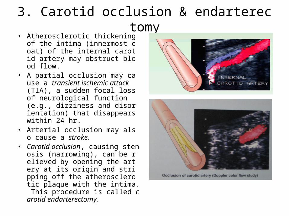
3. Carotid occlusion & endarterectomy• Atherosclerotic thickening of th
e intima (innermost coat) of the internal carotid artery may obstruct blood flow.
• A partial occlusion may cause a transient ischemic attack (TIA), a sudden focal loss of neurological function (e.g., dizziness and disorientation) that disappears within 24 hr.
• Arterial occlusion may also cause a stroke.
• Carotid occlusion, causing stenosis (narrowing), can be relieved by opening the artery at its origin and stripping off the atherosclerotic plaque with the intima. This procedure is called carotid endarterectomy.
3. Carotid occlusion & endarterectomyQuestions
• Where do you make the incision in order to fully explore the right ICA?
• What layers and anatomical structures should be passed through to reach the right ICA during the surgery?
• Because of the relations of the internal carotid artery, which cranial nerves should be carefully protected from the injury during the surgery procedure?
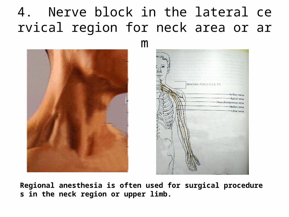
4. Nerve block in the lateral cervical region for neck area or arm
Regional anesthesia is often used for surgical procedures in the neck region or upper limb.
4. Nerve block in the lateral cervical region
• Regional anesthesia is often used for surgical procedures in the neck region or upper limb.
• In a cervical plexus block, which point should be selected for injection of an anesthetic agent to cover the most part of the lateral cervical region?
• For anesthesia of the upper limb, the anesthetic agent in a supraclavicular brachial plexus block is injected around the supraclavicular part of the brachial plexus. Where is the main injection site?
Related Documents