BY- GULAB CH. SONI Nerve conduction studies-Basics

Welcome message from author
This document is posted to help you gain knowledge. Please leave a comment to let me know what you think about it! Share it to your friends and learn new things together.
Transcript
BY- GULAB CH. SONI
Nerve conduction studies-Basics
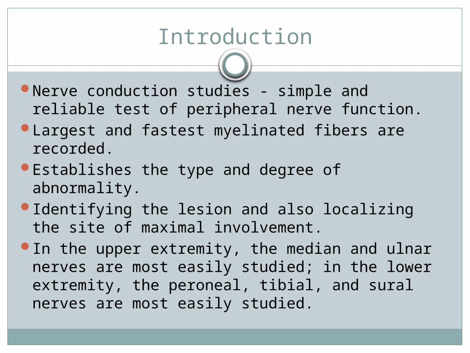
Introduction
Nerve conduction studies - simple and reliable test of peripheral nerve function.
Largest and fastest myelinated fibers are recorded.
Establishes the type and degree of abnormality.Identifying the lesion and also localizing the site
of maximal involvement.In the upper extremity, the median and ulnar
nerves are most easily studied; in the lower extremity, the peroneal, tibial, and sural nerves are most easily studied.

Instruments
OscilloscopeAmplifierSweep- Sensitivity controllerElectrodes


Oscilloscope

Amplifier
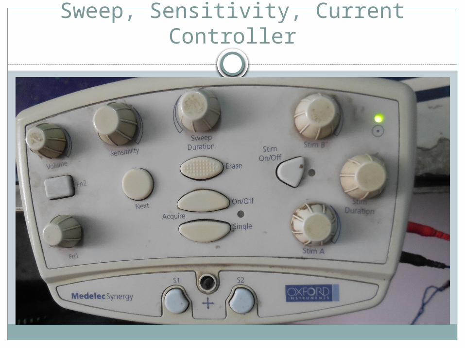
Sweep, Sensitivity, Current Controller

Various Electrode

Stimulator
Cathode( Negative pole ) – depolarize underlying nerve segment
Anode ( Positive pole )– hyperpolarize underlying nerve segment
Placing the cathode closer to the recording site avoids anodal conduction block
Cathode & anode - 2-3 cm apart
Surface electrode
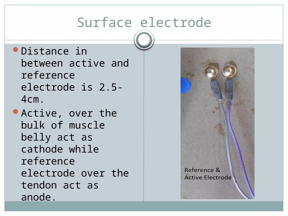
Distance in between active and reference electrode is 2.5-4cm.
Active, over the bulk of muscle belly act as cathode while reference electrode over the tendon act as anode.

Nerve conduction studies
Commonly deal with-Motor conduction studies (MCS)Sensory conduction studies Late responses

Motor Conduction Studies
Motor conduction studies are technically less demanding than sensory and mixed nerve studies; thus; they usually are performed first.
Motor responses typically are in the range of several millivolts (mV), as opposed to sensory and mixed nerve responses, which are in the microvolt (mcV) range.
Thus, motor responses are less affected by electrical noise and other technical factors.

MCS-belly-tendon montage-
Active recording electrode (GI) is placed on the center of the muscle belly (over the motor endplate), and the reference electrode (G2) is placed distally, over the tendon to the muscle
Stimulator- placed over the nerve that supplies the muscle, with the cathode placed closest to the recording electrode.(Black to black)
Ground electrode- In between stimulating and recording electrode
Concept of anodal block
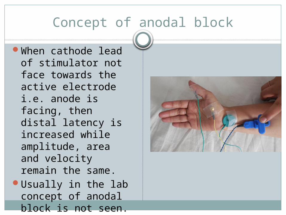
When cathode lead of stimulator not face towards the active electrode i.e. anode is facing, then distal latency is increased while amplitude, area and velocity remain the same.
Usually in the lab concept of anodal block is not seen.

Motor Conduction Studies
Sensitivity- 2 to 5 mV per division Sweep -30ms Current-20 to 50 mA Duration of current: 200mcs As current is slowly increased from a baseline 0 mA, usually by
5- to 10-mA increments, more of the underlying nerve fibers are brought to action potential and subsequently more muscle fiber action potentials i.e. compound muscle action potential (CMAP)
When the current is increased to the point that the CMAP no longer increases in size, one presumes that all nerve fibers have been excited and that supramaximal stimulation has been achieved.
The current then is increased by another 20% to ensure supramaximal stimulation.
Motor Conduction Studies
Measured - 1) CMAP 2) Motor nerve conduction velocity

Compound muscle action potential (CMAP)
Compound term represents the summation of all underlying individual muscle fiber action potentials.
Biphasic potential with an initial negativity, or upward deflection from the baseline.

Compound muscle action potential (CMAP)
Comprises of-Latency, Amplitude,Duration, and Area of the CMAP
Compound muscle action potential (CMAP)
Latency (ms): The latency is the time from the stimulus to the initial CMAP deflection from baseline.
Latency represents three separate processes: (1) the nerve conduction time from thestimulus site to the neuromuscular junction (NMJ), (2) the time delay across the NMJ, and (3) the depolarization time across the muscle.
Compound muscle action potential (CMAP)
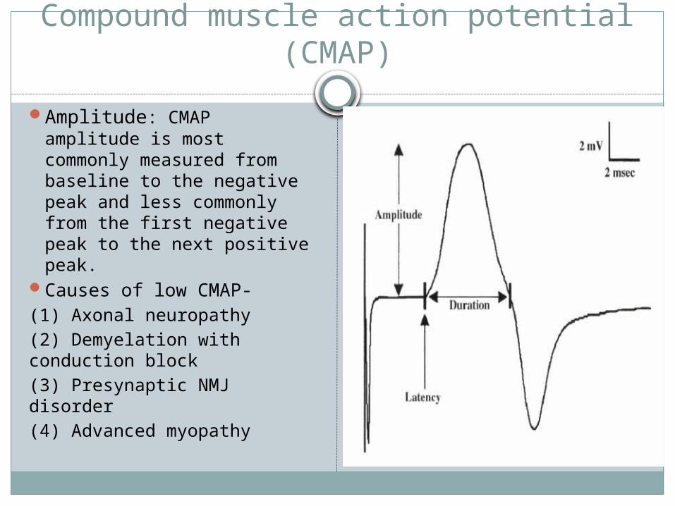
Amplitude: CMAP amplitude is most commonly measured from baseline to the negative peak and less commonly from the first negative peak to the next positive peak.
Causes of low CMAP-(1) Axonal neuropathy(2) Demyelation with conduction block(3) Presynaptic NMJ disorder(4) Advanced myopathy
Compound muscle action potential (CMAP)
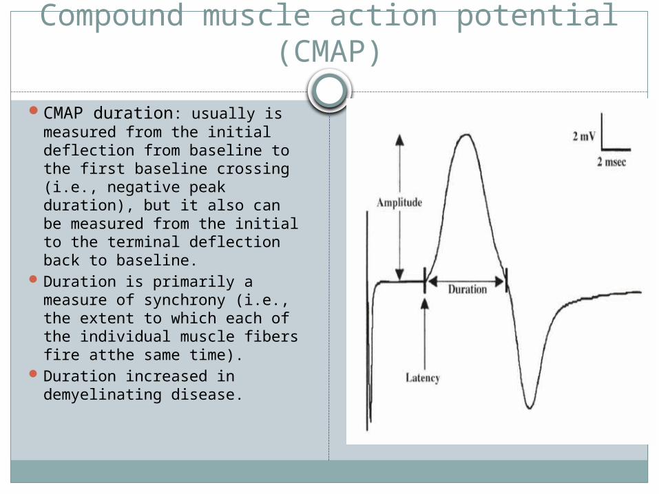
CMAP duration: usually is measured from the initial deflection from baseline to the first baseline crossing (i.e., negative peak duration), but it also can be measured from the initial to the terminal deflection back to baseline.
Duration is primarily a measure of synchrony (i.e., the extent to which each of the individual muscle fibers fire atthe same time).
Duration increased in demyelinating disease.

Compound muscle action potential (CMAP)
CMAP area: also is conventionally measured between the baseline and the negative peak.
Differences in CMAP area between distal and proximal stimulation sites take on special significance in the determination of conduction block from a demyelinating lesion.
Motor nerve conduction velocity
Measure of the speed of the fastest conducting motor axons.
Conduction velocity (m/s) calculated as: distance between the proximal and distal stimulation sites divided by proximal latency - distal latency.

Sensory conduction studies
Sensitivity: 10-20mcv/divisionSweep: 20msCurrent:5-30mA(50-300V)Sensory fibers usually have a lower threshold
to stimulation than do motor ftbers. As in motor studies, the current is slowly increased from a baseline of 0 mA, usually in 3- to 5-mA increments, until the recorded sensory potential is maximized.

Sensory conduction studies
Comprises of-Sensory nerve action potential (SNAP)Sensory nerve conduction velocity
Sensory nerve action potential (SNAP)
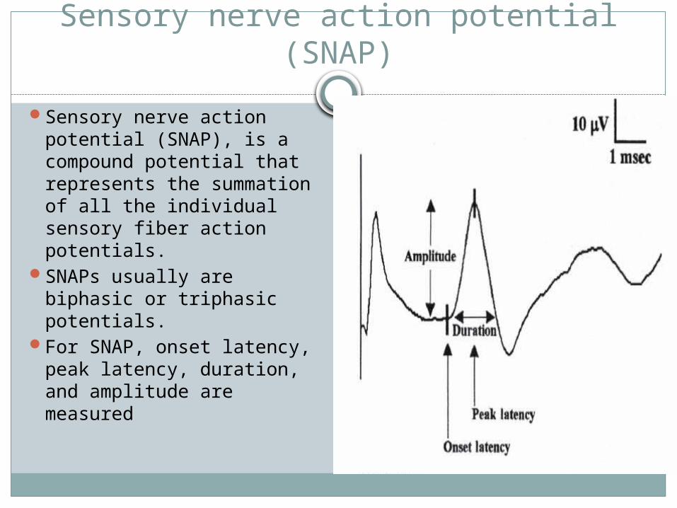
Sensory nerve action potential (SNAP), is a compound potential that represents the summation of all the individual sensory fiber action potentials.
SNAPs usually are biphasic or triphasic potentials.
For SNAP, onset latency, peak latency, duration, and amplitude are measured

Sensory nerve action potential (SNAP)
Onset Latency: is the time from the stimulus to the first deflection from baseline and represents nerve conduction time from the stimulus site to the recording electrodes for the largest cutaneous sensory fibers, so used to calculate conduction velocity.
Peak Latency: The peak latency is measured at the midpoint of the first negative peak. Interobserver variation is less.

Sensory nerve action potential (SNAP)
Duration:SNAP duration usually is measured from the onset of the potential to the firstbaseline crossing (i.e., negative peak duration).The SNAP duration typically is much shorter than the CMAP duration (typically 1.5 ms vs 5-6 ms).
Amplitude: The SNAP amplitude is most commonly measured from baseline to negative peak. Low SNAP amplitudes indicate a definite disorder of peripheral nerve.
Sensory nerve conduction velocity
Sensory conduction velocity represents the speed of the fastest, myelinated cutaneous sensory fibers.
Sensory conduction velocity can be determined with one stimulation, simply by dividing the distance traveled by the onset latency
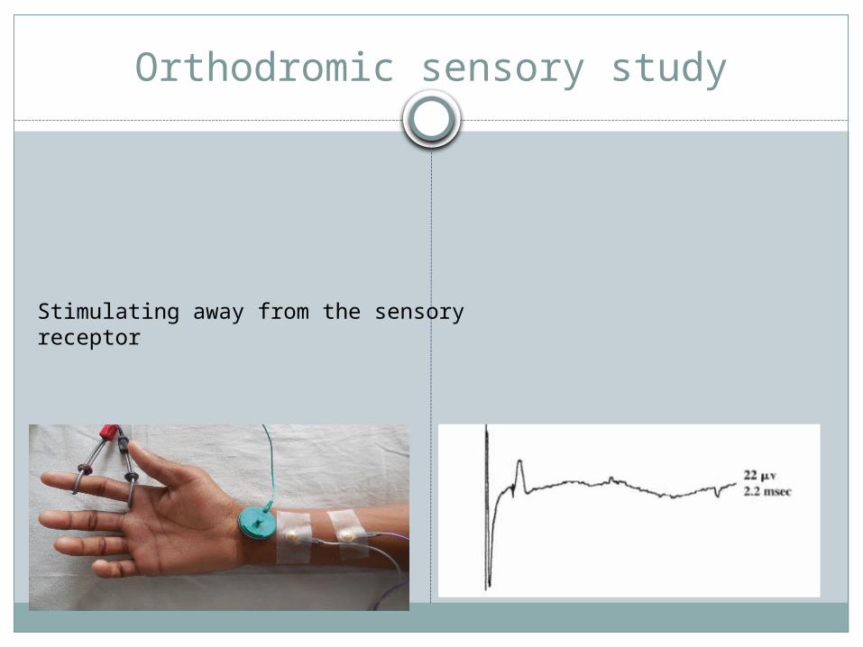
Orthodromic sensory study
Stimulating away from the sensory receptor
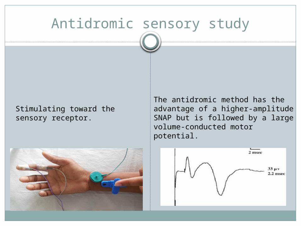
Antidromic sensory study
Stimulating toward thesensory receptor.
The antidromic method has the advantage of a higher-amplitude SNAP but is followed by a large volume-conducted motor potential.

Sensory conduction studies
In orthodomic vs antidromic sensory study, latency and conduction velocity remain same except amplitude which is more in antidromic due to proximity of the recording electrode to the underlying nerve.
Sensory conduction studies
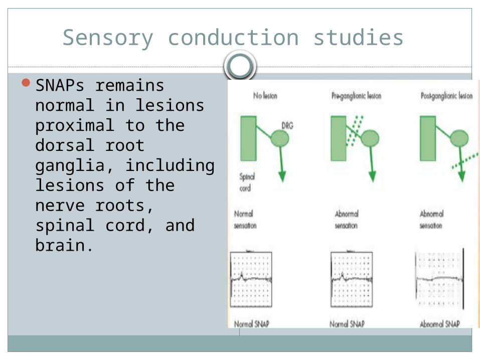
SNAPs remains normal in lesions proximal to the dorsal root ganglia, including lesions of the nerve roots, spinal cord, and brain.


Conduction block & Temporal dispersion
Seen in acquired demyelinating neuropathy.
In tibial nerve conduction block is not commented because distal CMAP reduction up to 50% normally seen in this nerve.

Conduction block
Late response
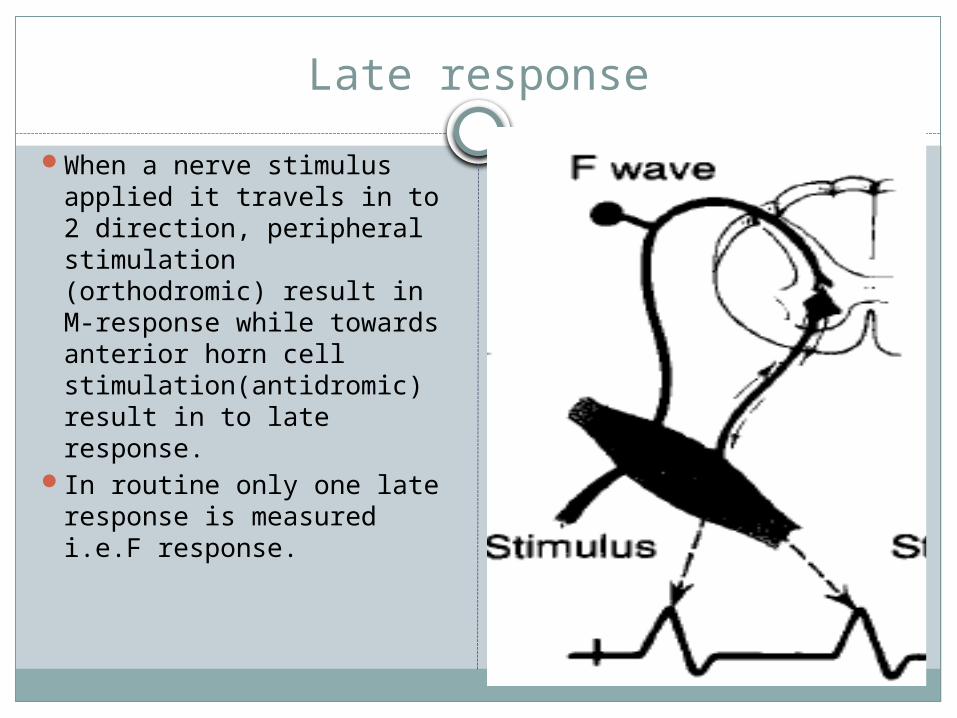
When a nerve stimulus applied it travels in to 2 direction, peripheral stimulation (orthodromic) result in M-response while towards anterior horn cell stimulation(antidromic) result in to late response.
In routine only one late response is measured i.e.F response.

Late response
For eliciting the late response, some author change the direction of stimulator (cathode end proximally) so that maximum number of nerve stimulated.
In lab usually no change seen by doing this.

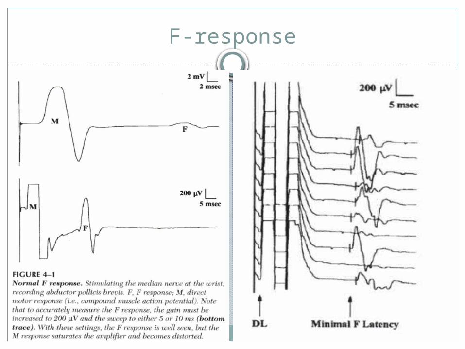
F-response
First described by Magaladery and McDougal.
F response derives its name from foot because it was first recorded from the intrinsic foot muscles.
Sensitivity-500mcv/div
Sweep-100ms
F-response


Factors affecting Nerve conduction study

Factors Influencing Nerve Conduction Studies
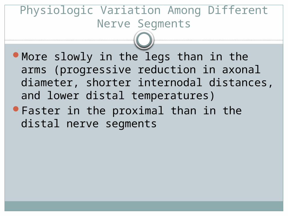
Physiologic Variation Among Different Nerve Segments
More slowly in the legs than in the arms (progressive reduction in axonal diameter, shorter internodal distances, and lower distal temperatures)
Faster in the proximal than in the distal nerve segments
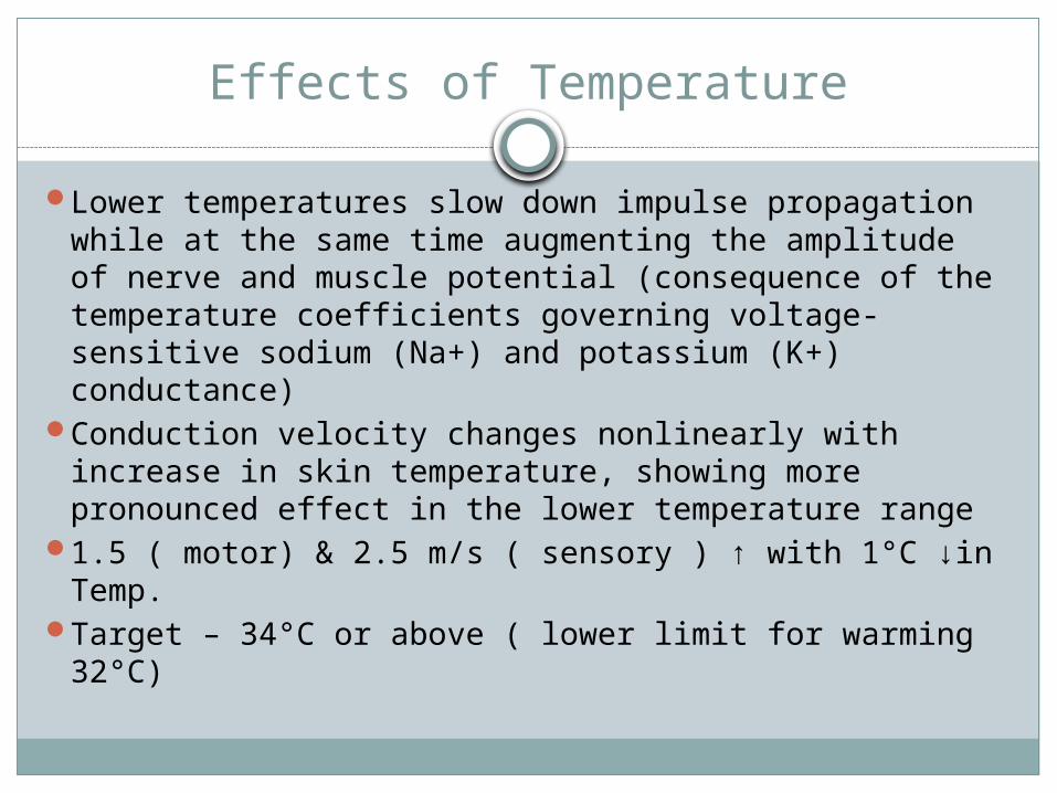
Effects of Temperature
Lower temperatures slow down impulse propagation while at the same time augmenting the amplitude of nerve and muscle potential (consequence of the temperature coefficients governing voltage-sensitive sodium (Na+) and potassium (K+) conductance)
Conduction velocity changes nonlinearly with increase in skin temperature, showing more pronounced effect in the lower temperature range
1.5 ( motor) & 2.5 m/s ( sensory ) ↑ with 1°C ↓in Temp.
Target – 34°C or above ( lower limit for warming 32°C)
Maturation and Aging
Conduction velocity - half the adult value in full-term infants to the adult range at age 3–5 years ( rate of myelination)
In children and adolescents, from age 3 to 19 years, both motor and sensory conduction velocities tend to increase slightly in the upper limb and decrease in the lower limb as a function of age and growth in length
begin to decline after 30–40 years of age, but the values normally change by less than 10 m/s by the sixtieth year
The most distal branches, such as the interdigital nerves, may degenerate earlier.

Height
Height shows negative association with sensory amplitude and positive association with distal latencies
Sural, peroneal and tibial nerve conduction velocities all have inverse correlation with height in normals
Relevant in interpretation of late responses ( F response and H reflex) – impulse travel twice the length of limb

THANK-U

H- reflex
Related Documents