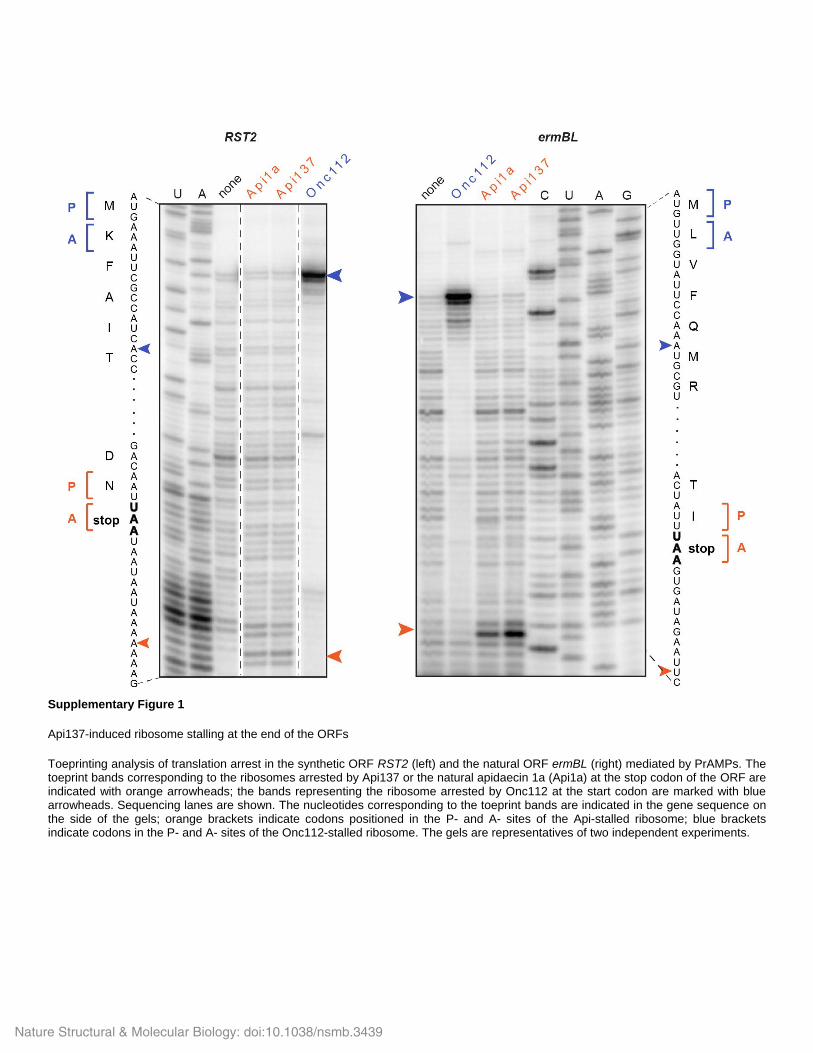
Supplementary Figure 1 Api137-induced ribosome stalling at the end of the ORFs Toeprinting analysis of translation arrest in the synthetic ORF RST2 (left) and the natural ORF ermBL (right) mediated by PrAMPs. The toeprint bands corresponding to the ribosomes arrested by Api137 or the natural apidaecin 1a (Api1a) at the stop codon of the ORF are indicated with orange arrowheads; the bands representing the ribosome arrested by Onc112 at the start codon are marked with blue arrowheads. Sequencing lanes are shown. The nucleotides corresponding to the toeprint bands are indicated in the gene sequence on the side of the gels; orange brackets indicate codons positioned in the P- and A- sites of the Api-stalled ribosome; blue brackets indicate codons in the P- and A- sites of the Onc112-stalled ribosome. The gels are representatives of two independent experiments. Nature Structural & Molecular Biology: doi:10.1038/nsmb.3439

Welcome message from author
This document is posted to help you gain knowledge. Please leave a comment to let me know what you think about it! Share it to your friends and learn new things together.
Transcript
Supplementary Figure 1
Api137-induced ribosome stalling at the end of the ORFs
Toeprinting analysis of translation arrest in the synthetic ORF RST2 (left) and the natural ORF ermBL (right) mediated by PrAMPs. The toeprint bands corresponding to the ribosomes arrested by Api137 or the natural apidaecin 1a (Api1a) at the stop codon of the ORF are indicated with orange arrowheads; the bands representing the ribosome arrested by Onc112 at the start codon are marked with blue arrowheads. Sequencing lanes are shown. The nucleotides corresponding to the toeprint bands are indicated in the gene sequence on the side of the gels; orange brackets indicate codons positioned in the P- and A- sites of the Api-stalled ribosome; blue brackets indicate codons in the P- and A- sites of the Onc112-stalled ribosome. The gels are representatives of two independent experiments.
Nature Structural & Molecular Biology: doi:10.1038/nsmb.3439
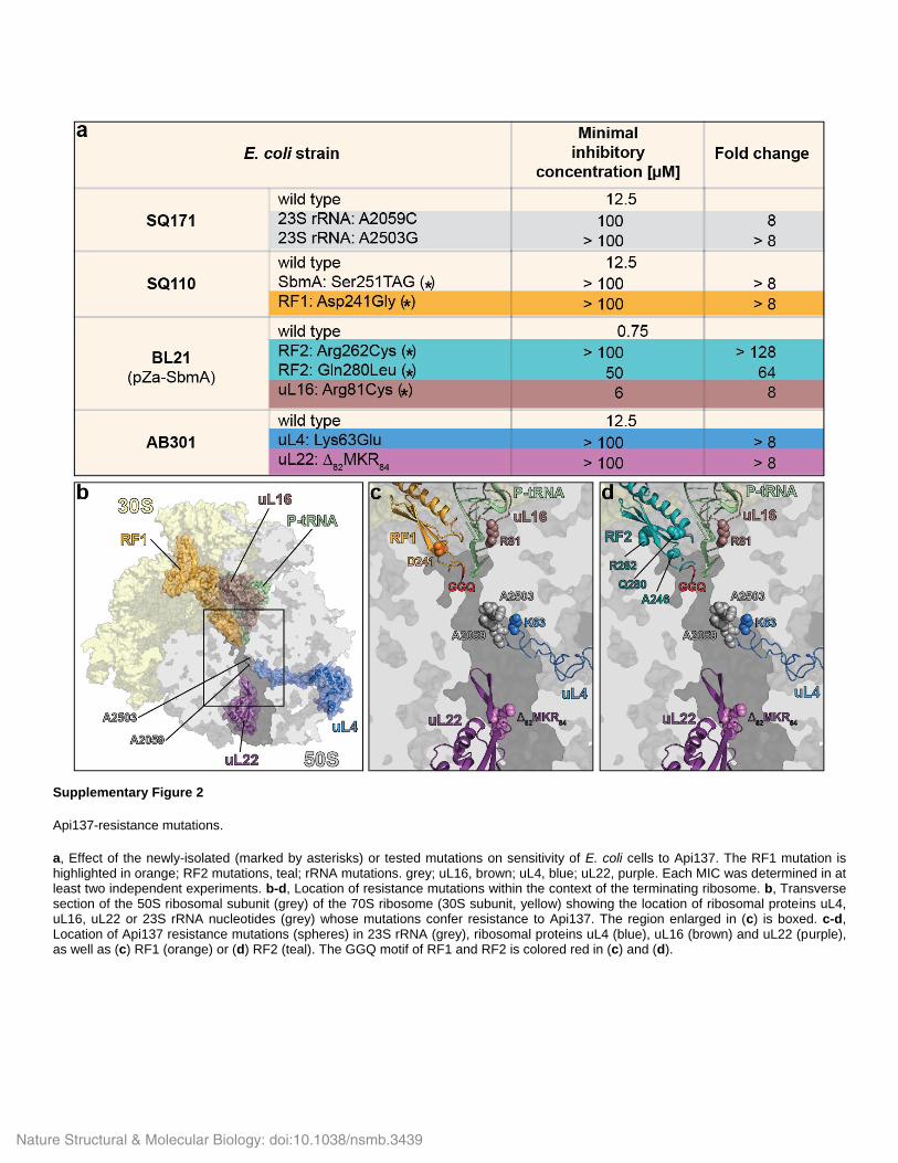
Supplementary Figure 2
Api137-resistance mutations.
a, Effect of the newly-isolated (marked by asterisks) or tested mutations on sensitivity of E. coli cells to Api137. The RF1 mutation is highlighted in orange; RF2 mutations, teal; rRNA mutations. grey; uL16, brown; uL4, blue; uL22, purple. Each MIC was determined in at least two independent experiments. b-d, Location of resistance mutations within the context of the terminating ribosome. b, Transverse section of the 50S ribosomal subunit (grey) of the 70S ribosome (30S subunit, yellow) showing the location of ribosomal proteins uL4, uL16, uL22 or 23S rRNA nucleotides (grey) whose mutations confer resistance to Api137. The region enlarged in (c) is boxed. c-d, Location of Api137 resistance mutations (spheres) in 23S rRNA (grey), ribosomal proteins uL4 (blue), uL16 (brown) and uL22 (purple), as well as (c) RF1 (orange) or (d) RF2 (teal). The GGQ motif of RF1 and RF2 is colored red in (c) and (d).
Nature Structural & Molecular Biology: doi:10.1038/nsmb.3439

Supplementary Figure 3
Mutations allow faster dissociation of RF1 and RF2 from the PostHC.
a, Dissociation of RF1(D241G) from the PostHC in the presence of Api137. RF1(D241G)Qsy was incubated with PreHCFlu (0.05 µM) to generate PostHCFlu and then mixed with a 10-fold excess of unlabeled RF1 and RF3·GTP in the absence (grey) or in the presence (black) of Api137 (1 µM). The traces represent the average of up to eight technical replicates. No dissociation of wt RF1 in the presence of Api137 was observed under the same experimental conditions (Fig. 2f). b, Peptide hydrolysis by K12 strain-specific RF2(Ala246Thr) at turnover conditions in the absence (open circles) or in the presence (closed circles) of Api137 (1 µM). In the presence of Api137, the peptide hydrolysis reaction proceeds faster when it is catalyzed by the K12 strain RF2, compared to the B strain RF2 (Fig. 2c).
Nature Structural & Molecular Biology: doi:10.1038/nsmb.3439

Supplementary Figure 4
In silico sorting and resolution of the Api-RF1-70S complex.
a, In silico sorting was performed with the FreAlign 9.11 software package (as described in Grigorieff, N., J. Struct. Biol. 157, 117-125 (2007)). Initial alignment of 116,212 particles was followed by 3D classification, resulting in six different classes. Class 1 (38,203 particles) was further refined, yielding a (b) final reconstruction consisting of 36,826 particles, with (c) an average resolution of 3.4 Å (based on the Fourier shell correlation (FSC) curve at FSC 0.143). d, Validation of the fit of molecular models to cryo-EM map for the Api137-RF1-70S complex. FSC curves calculated between the refined model and the final map (blue), with the self- and cross-validated correlations in orange and black, respectively. Information beyond 3.4 Å was not used during refinement and preserved for validation. (e) Side view and (f) transverse section of the cryo-EM map of Api137-RF1-70S complex colored according to local resolution as shown previously (Kucukelbir, A., Sigworth, F. J. & Tagare, H. D., Nat. Methods 11, 63-65 (2014)). g-h, Cryo-EM density for Api137 (g) colored according to local resolution and (h) shown as grey mesh with molecular model for residues 5-18.
Nature Structural & Molecular Biology: doi:10.1038/nsmb.3439

Supplementary Figure 5
Features of the Api137-RF1-70S complex.
a, RF1 (orange), deacylated P-site tRNA (green) and Api137 (salmon) in the Api137-RF1-70S complex. The position of RF1 during canonical termination is shown in blue (PDBID 5J30; Pierson, W. E. et al., Cell Rep. 17, 11-18 (2016)). Boxed regions are zoomed in the panels (b) and (c). b, Interaction of the PAT motif of RF1 (orange) with the UAG stop codon of the mRNA (cyan) in the Api137-RF1-70S complex. c, A2602 of the 23S rRNA is in the rotated conformation as observed in previous RF1-70S structures (Korostelev, A. et al., EMBO J. 29, 2577-2585 (2010); Pierson, W. E. et al., Cell Rep. 17, 11-18 (2016); Laurberg, M. et al., Nature 454, 852-857 (2008); Svidritskiy, E. & Korostelev, A. A., Structure 23, 2155-2161 (2015)). Conformation of A2602 (grey) in Api137-RF1-70S complex compared to A2602 (blue) during canonical termination (PDBID 5J30; Pierson, W. E. et al., Cell Rep. 17, 11-18 (2016)) and A2602 (slate) from the pre-attack state (PDBID 1VY4; Polikanov, Y. S., Steitz, T. A. & Innis, C. A., Nat. Struct. Mol. Biol. 21, 787-793 (2014)). Api137 (salmon) and P-site tRNA (green) are shown for reference. d, e, The binding position of Api137 (salmon) relative to the (d) MifM nascent chain (dark green; Sohmen, D. et al., Nat. Commun. 6, 6941 (2015)) or (e) antimicrobial peptide Onc112 (slate; Seefeldt, A. C. et al., Nat. Struct. Mol. Biol. 22, 470-475 (2015)). In (d) and (e) the orientations of the peptides are indicated.
Nature Structural & Molecular Biology: doi:10.1038/nsmb.3439
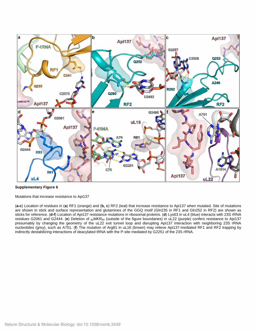
Supplementary Figure 6
Mutations that increase resistance to Api137
(a-c) Location of residues in (a) RF1 (orange) and (b, c) RF2 (teal) that increase resistance to Api137 when mutated. Site of mutations are shown in stick and surface representation and glutamines of the GGQ motif (Gln235 in RF1 and Gln252 in RF2) are shown as sticks for reference. (d-f) Location of Api137 resistance mutations in ribosomal proteins. (d) Lys63 in uL4 (blue) interacts with 23S rRNA residues G2061 and G2444. (e) Deletion of 82MKR84 (outside of the figure boundaries) in uL22 (purple) confers resistance to Api137 presumably by changing the geometry of the uL22 exit tunnel loop and disrupting Api137 interaction with neighboring 23S rRNA nucleotides (grey), such as A751. (f) The mutation of Arg81 in uL16 (brown) may relieve Api137-mediated RF1 and RF2 trapping by indirectly destabilizing interactions of deacylated tRNA with the P-site mediated by G2251 of the 23S rRNA.
Nature Structural & Molecular Biology: doi:10.1038/nsmb.3439

Supplementary Table 1. Deletion of the RF3-encoding prfC gene affects minimal inhibitory concentration (MIC) of Api137 E. coli strain MIC [µM] wild type (BW25113)
a) 6.25
∆xylA b) 6.25
∆prfC c)
0.75
a)
Parental E. coli K-type strain b)
The xylA::kan strain, where an unrelated gene was inactivated, was used as an additional
negative control c) The prfC::kan strain
Nature Structural & Molecular Biology: doi:10.1038/nsmb.3439

Supplementary Table 2: DNA & RNA templates
Promoter – blue, ORF – red, annealing site for toeprinting primer – purple, annealing site for
oligonucleotide for RNase H treatment of disomes – green. Start codons of the ORFs are shown
in bold, stop codons are underlined.
Name DNA Sequence (5’ – 3’)
yrbA-fs TAATACGACTCACTATAGGGCTTAAGTATAAGGAGGAAAACATATGAT
ATACCCCTGCGGAGTGGGCGCGCGATCGCAAACTGAACGGCTTTAG
GCCGACCTCGACAGTTGGATTCACGTGCTGAATCCTGATGCGATGTC
GAGTTAATAAGCAAAATTCATTATAACC
ermCL-UAG TTAATACGACTCACTATAGGGAATTGTGAGCGGATAACAATTGCTAGT
CTTAAGTTTTATAAGGAGGAAAAAATATGGGCATTTTTAGTATTTTTGT
AATCAGCACAGTTCATTATCAACCAAACAAAAAATAGGTGGTTATAATG
AATCGTTAATAAGCAAAATTCATTATAACCAAATTAAAGAGGGTTATAA
RST2 TAATACGACTCACTATAGGGCTTAAGTATAAGGAGGAAAACATATGTA
TTGGGTAACCTCACGTCAGCCGAATATGCTGAAAATCCATGGCTTCGA
AGACTGCGCCTAATAATAATAAAAAAAGTGATAGAATTCTATCGTTAAT
AAGCAAAATTCATTATAACC
ermBL TAATACGACTCACTATAGGGCTTAAGTATAAGGAGGAAAAAATATGTT
GGTATTCCAAATGCGTAATGTAGATAAAACATCTACTATTTAAGTGATA
GAATTCTATCGTTAATAAGCAAAATTCATTATAACC
Start-Stop GGCAAGGAGGUAAAUAAUGUAAACGAUU
tnaC-UAG ACATGGATTCTTGACAATTAATCATCGGCTCGTATAATGTGTGGAAGTT
TTATAAGGAGGAAAACATATGAATATCTTACATATATGTGTGACCTCAA
AATGGTTCAATATTGACAACAAAATTGTCGATCACCGCCCTTAG tnaC-UGA ACATGGATTCTTGACAATTAATCATCGGCTCGTATAATGTGTGGAAGTT
TTATAAGGAGGAAAACATATGAATATCTTACATATATGTGTGACCTCAA
AATGGTTCAATATTGACAACAAAATTGTCGATCACCGCCCTTGA
2XermCL_S10_
UAG
UAAUACGACUCACUAUAGGGAGUUUUAUAAGGAGGAAAAAAUAUGG
GCAUUUUUAGUAUUUUUGUAAUCUAGACAGUUCAUUAUCAACCAAA
CAAAAAAUAAAGUUUUAUAAGGAGGAAAAAAUAUGGGCAUUUUUAGU
AUUUUUGUAAUCUAGACAGUUCAUUAUCAACCAAACAAAAAAUAA
Nature Structural & Molecular Biology: doi:10.1038/nsmb.3439

Supplementary Table 3: DNA primers used in this study
Name Sequence T7-IR-AUG TAATACGACTCACTATAGGGCTTAAGTATAAGGAGGAAAACATATG
IR-yrbA-fs15-
RF1
GTATAAGGAGGAAAACATATGATATACCCCTGCGGAGTGGGCGCGCGAT
CGCAAACTGAACGGCTTTAGGCCGACCTCGACAGTTGGAT
posT-NV1 GGTTATAATGAATTTTGCTTATTAACTCGACATCGCATCAGGATTCAGCAC
GTGAATCCAACTGTCGAGGTCG
T7 TAATACGACTCACTATAGGG
ermCL-UAG TTATAACCCTCTTTAATTTGGTTATAATGAATTTTGCTTATTAACGATTCAT
TATAACCACCTATT
ermCL-TP-term TTATAACCCTCTTTAATTTGGTT
SbmA-seq-fwd CATTTGGCTGACGCTTTGTA
SbmA-seq-rev TACTACACCCCGCTAAAACC
SbmA-EcoRI-
rev
TGACGCGCGGAATTCCTTCT
PrfA-seq-fwd CTGAATATTCTGCGCGACAG
PrfA-seq-rev CAGGATTTCAGCATCACGC
PrfB-seq-fwd GCTCTTATCACCGCATTTTG
PrfB-seq-rev GTTCATTGTTAAGATCGACTACC
PrfC-seq-fwd GAAGGTAAGCTGGATATGCTG
PrfC-seq-rev GCTTCTGATAACGTAGCCAG
rplP-seq-fwd CGTTAAAGTGTGGATCTTCAAAGG
rplP-seq-rev CACTTGCTTCAACAGGTGAG
L2667 GGTCCTCTCGTACTAGGAGCAG
L2180 GGGTGGTATTTCAAGGTCGG
Ptrc-tnaC ACATGGATTCTTGACAATTAATCATCGGCTCGTATAATGTGTGGA
AGTTTTATAAGGAGGAAAACATATG
tnaC-UAG-rev GCAAACTAAGGGCGGTGATCGAC
tnaC-UGA-rev GCAAATCAAGGGCGGTGATCGAC
Nature Structural & Molecular Biology: doi:10.1038/nsmb.3439

Supplementary Table 4: Bacterial strains used in this study
E. coli strain Type Source SQ171 K-strain; F-, Δ(rrsH-aspU)794(::FRT), λ-
, Δ(rrfG-rrsG)791(::FRT), Δ(rrfF-rrsD)793(::FRT), rph-1,
Δ(rrsC-trpT)795(::FRT), Δ(rrsA-rrfA)792(::FRT), Δ(rrsB-rrfB)790(::FRT), Δ(rrsE-rrfE)789(::FRT), ptRNA67, pKK3535
Quan, S. et al., G3 5,
2555-2557 (2015)
SQ110 K-strain, F-, Δ(rrsH-aspU)794(::FRT), λ-, Δ(rrfG-
rrsG)791(::FRT), Δ(rrfF-rrsD)793(::FRT), rph-1,
Δ(rrsC-trpT)795(::FRT), Δ(rrsA-rrfA)792(::FRT), Δ(rrsB-rrfB)790(::FRT), ptRNA67
Quan, S. et al., G3 5,
2555-2557 (2015)
SQ110 ApiR2 derived from SQ110, sbmA(C752A) this study
SQ110 ApiR21 derived from SQ110, prfA(A722G) this study BL21 (DE3) B-strain; F-, lon-11, Δ(ompT-nfrA)885, Δ(galM-
ybhJ)884, λDE3 [lacI, lacUV5-T7 gene 1, ind1, sam7, nin5], Δ46, [mal+]K-12(λ
S), hsdS10
Wood, W.B., J Mol Biol 16, 118-133 (1966);
Studier, F.W. & Moffatt,
B.A., J Mol Biol 189, 113-
30 (1986)
BL21 ApiR10 derived from BL21 (DE3), rplP(G241A), pZa-SbmA this study BL21 ApiR11 derived from BL21 (DE3), prfB(C784T), pZa-SbmA this study BL21 ApiR12 derived from BL21 (DE3), prfB(A839T), pZa-SbmA this study AB301 K-strain; Hfr(PO21), relA1, spoT1, metB1 Bouck, N. & Adelberg,
E.A., J Bacteriol 102,
688-701 (1970)
N281 K-strain; Hfr(PO21), relA1, rplV281, spoT1, metB1 Wittmann, H.G. et al., Mol Gen Genet 127, 175-89.
(1973); Chittum, H.S. &
Champney, W.S., J Bacteriol 176, 6192-8
(1994).
N282 K-strain; Hfr(PO21), relA1, rplD282, spoT1, metB1 Wittmann, H.G. et al., Mol Gen Genet 127, 175-89.
(1973); Chittum, H.S. &
Champney, W.S., J Bacteriol 176, 6192-8
(1994).
SQ171-∆tolC derived from SQ171; ∆tolC, pCSacB Kannan, K., Vázquez-
Laslop, N. & Mankin,
A.S., Cell 151, 508-520
(2012)
SQ171-
∆tolC/W3
derived from SQ171-∆tolC, lacZ(C2035T) pCSacB this study
BW25113 F-, Δ(araD-araB)567, ΔlacZ4787(::rrnB-3), λ-, rph-
1, Δ(rhaD-rhaB)568, hsdR514
Baba, T. et al., Mol Syst Biol 2, 2006 0008 (2006)
BW25113 ∆xylA
(JW3537)
F-, Δ(araD-araB)567, ΔlacZ4787(::rrnB-3), λ-
, ΔxylA748::kan, rph-1, Δ(rhaD-rhaB)568, hsdR514
Baba, T. et al., Mol Syst Biol 2, 2006 0008 (2006)
BW25113 ∆prfC
(JW5873)
F-, Δ(araD-araB)567, ΔlacZ4787(::rrnB-3), λ-, rph-
1, Δ(rhaD-rhaB)568, hsdR514, ΔprfC770::kan
Baba, T. et al., Mol Syst Biol 2, 2006 0008 (2006)
Nature Structural & Molecular Biology: doi:10.1038/nsmb.3439
Related Documents