The past decade has seen an increase in the number of new drugs approved by the US Food and Drug Admin- istration (FDA), leading to an all-time record number of 59 novel drug approvals in 2018. Drugs for oral use continue to dominate the therapeutic landscape, encom- passing over 50% of these approvals 1 . Over one-third of the remaining approvals relate to biologic agents admin- istered via parenteral routes, such as subcutaneous, intravenous or intramuscular injections 1 . Parenteral drug delivery achieves high bioavailability (~100% for infusions) 2,3 . Nonetheless, parenteral admin- istration is associated with considerable disadvantages, including pain or discomfort 4 , severe reactions at the injection site 5,6 , scarring 7 , local allergic reactions 8 and cutaneous infections 9 . Furthermore, intravenous injec- tions require administration by a skilled health-care pro- fessional, and self-administered injections are associated with social stigmatization of patients 10 . Taken together, these deleterious effects can result in poor compliance of patients with their prescribed treatments (BOX 1), especially among individuals with chronic diseases that require long-term monitoring and repeat dosing 11,12 . Historically, oral administration has offered a con- venient, familiar and painless alternative to injections. Alchemists and researchers alike have been delivering herbal remedies and drugs by the oral route dating as far back as 1550 BCE (FIG. 1). Throughout history, techno- logical advances, including the mass manufacture of tablets and capsules, have continued to drive the field of oral drug delivery forward. Today, an estimated 70% of Americans (~230 million individuals) take at least one prescription drug each day, regardless of administration route 13 . Unfortunately, barring some very small pep- tides such as ciclosporin, oral delivery is not a currently available option for protein and antibody drugs 14 . These macromolecular agents have prohibitively low oral bio- availability due to several features of the gastrointestinal tract, including the proteolytic environment of the stom- ach and limited absorption in the intestine 15,16 . A clear, unmet need exists for the design of new materials to enable oral protein delivery, beyond commonly used excipients or already FDA-approved inactive ingredients. In this Review, we discuss key physiological barriers to the oral delivery of biologic therapies and describe state-of-the-art, materials-based approaches for improv- ing their bioavailability. This Review also details the current clinical translational landscape of materials for oral protein delivery, arranged by the characteristic length scale — from small molecules to macromolecular devices. Our selection of this organizational structure reflects the fact that some materials possess multiple mechanisms of action, whereas others do not have an established mechanism of action. Barriers to oral delivery The gastrointestinal tract is designed to digest carbo- hydrates, proteins and other nutrients into their con- stitutive subunits of amino acids and simple sugars. Simultaneously, it also prevents the entry of pathogens. We should not be surprised, therefore, that the oral bio- availability of intact peptides and proteins is <1% and sometimes even <0.1% 17,18 . Indeed, orally administered drugs must overcome numerous biological hurdles prior to their absorption, as detailed in the next sections (FIG. 2). Materials for oral delivery of proteins and peptides Tyler D. Brown 1,2 , Kathryn A. Whitehead 3,4 and Samir Mitragotri 1,2 * Abstract | Throughout history, oral administration has been regarded as the most convenient mode of drug delivery, as it requires minimal expertise and invasiveness. Although oral delivery works well for small-molecule drugs, oral delivery of macromolecules (particularly proteins and peptides) has been limited by acidic conditions in the stomach and low permeability across the intestinal epithelium. Accordingly, the large numbers of biologic drugs that have become available in the past 10 years typically require administration by injection or infusion. As such, a renewed emphasis has been placed on the development of novel materials that overcome the physiological challenges of oral delivery for macromolecular agents. This Review provides an overview of physiological barriers to the oral delivery of biologics and highlights the advances made in materials across various length scales, from small molecules to macroscopic devices. This Review also describes the current status of materials for oral delivery of protein and peptide drugs. 1 John A. Paulson School of Engineering and Applied Sciences, Harvard University, Cambridge, MA, USA. 2 Wyss Institute for Biologically Inspired Engineering, Harvard University, Boston, MA, USA. 3 Department of Chemical Engineering, Carnegie Mellon University, Pittsburgh, PA, USA. 4 Department of Biomedical Engineering, Carnegie Mellon University, Pittsburgh, PA, USA. *e-mail: mitragotri@ seas.harvard.edu https://doi.org/10.1038/ s41578-019-0156-6 REVIEWS NATURE REVIEWS | MATERIALS

Welcome message from author
This document is posted to help you gain knowledge. Please leave a comment to let me know what you think about it! Share it to your friends and learn new things together.
Transcript

The past decade has seen an increase in the number of new drugs approved by the US Food and Drug Administration (FDA), leading to an all time record number of 59 novel drug approvals in 2018. Drugs for oral use continue to dominate the therapeutic landscape, encompassing over 50% of these approvals1. Over one third of the remaining approvals relate to biologic agents administered via parenteral routes, such as subcutaneous, intravenous or intramuscular injections1.
Parenteral drug delivery achieves high bioavailability (~100% for infusions)2,3. Nonetheless, parenteral administration is associated with considerable disadvantages, including pain or discomfort4, severe reactions at the injection site5,6, scarring7, local allergic reactions8 and cutaneous infections9. Furthermore, intravenous injections require administration by a skilled health care professional, and self administered injections are associated with social stigmatization of patients10. Taken together, these deleterious effects can result in poor compliance of patients with their prescribed treatments (Box 1), especially among individuals with chronic diseases that require long term monitoring and repeat dosing11,12.
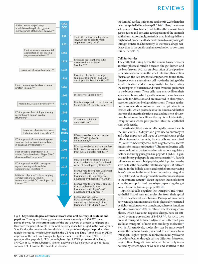
Historically, oral administration has offered a convenient, familiar and painless alternative to injections. Alchemists and researchers alike have been delivering herbal remedies and drugs by the oral route dating as far back as 1550 BCE (Fig. 1). Throughout history, technological advances, including the mass manufacture of tablets and capsules, have continued to drive the field of oral drug delivery forward. Today, an estimated 70% of Americans (~230 million individuals) take at least one prescription drug each day, regardless of administration
route13. Unfortunately, barring some very small peptides such as ciclosporin, oral delivery is not a currently available option for protein and antibody drugs14. These macro molecular agents have prohibitively low oral bioavailability due to several features of the gastrointestinal tract, including the proteolytic environment of the stomach and limited absorption in the intestine15,16. A clear, unmet need exists for the design of new materials to enable oral protein delivery, beyond commonly used excipients or already FDAapproved inactive ingredients.
In this Review, we discuss key physiological barriers to the oral delivery of biologic therapies and describe state ofthe art, materials based approaches for improving their bioavailability. This Review also details the current clinical translational landscape of materials for oral protein delivery, arranged by the characteristic length scale — from small molecules to macromolecular devices. Our selection of this organizational structure reflects the fact that some materials possess multiple mechanisms of action, whereas others do not have an established mechanism of action.
Barriers to oral deliveryThe gastrointestinal tract is designed to digest carbohydrates, proteins and other nutrients into their constitutive subunits of amino acids and simple sugars. Simultaneously, it also prevents the entry of pathogens. We should not be surprised, therefore, that the oral bioavailability of intact peptides and proteins is <1% and sometimes even <0.1%17,18. Indeed, orally administered drugs must overcome numerous biological hurdles prior to their absorption, as detailed in the next sections (Fig. 2).
Materials for oral delivery of proteins and peptidesTyler D. Brown 1,2, Kathryn A. Whitehead 3,4 and Samir Mitragotri 1,2*
Abstract | Throughout history , oral administration has been regarded as the most convenient mode of drug delivery , as it requires minimal expertise and invasiveness. Although oral delivery works well for small- molecule drugs, oral delivery of macromolecules (particularly proteins and peptides) has been limited by acidic conditions in the stomach and low permeability across the intestinal epithelium. Accordingly , the large numbers of biologic drugs that have become available in the past 10 years typically require administration by injection or infusion. As such, a renewed emphasis has been placed on the development of novel materials that overcome the physiological challenges of oral delivery for macromolecular agents. This Review provides an overview of physiological barriers to the oral delivery of biologics and highlights the advances made in materials across various length scales, from small molecules to macroscopic devices. This Review also describes the current status of materials for oral delivery of protein and peptide drugs.
1John A. Paulson School of Engineering and Applied Sciences, Harvard University, Cambridge, MA, USA.2Wyss Institute for Biologically Inspired Engineering, Harvard University, Boston, MA, USA.3Department of Chemical Engineering, Carnegie Mellon University, Pittsburgh, PA, USA.4Department of Biomedical Engineering, Carnegie Mellon University, Pittsburgh, PA, USA.
*e- mail: mitragotri@ seas.harvard.edu
https://doi.org/10.1038/ s41578-019-0156-6
REVIEwS
Nature reviews | Materials
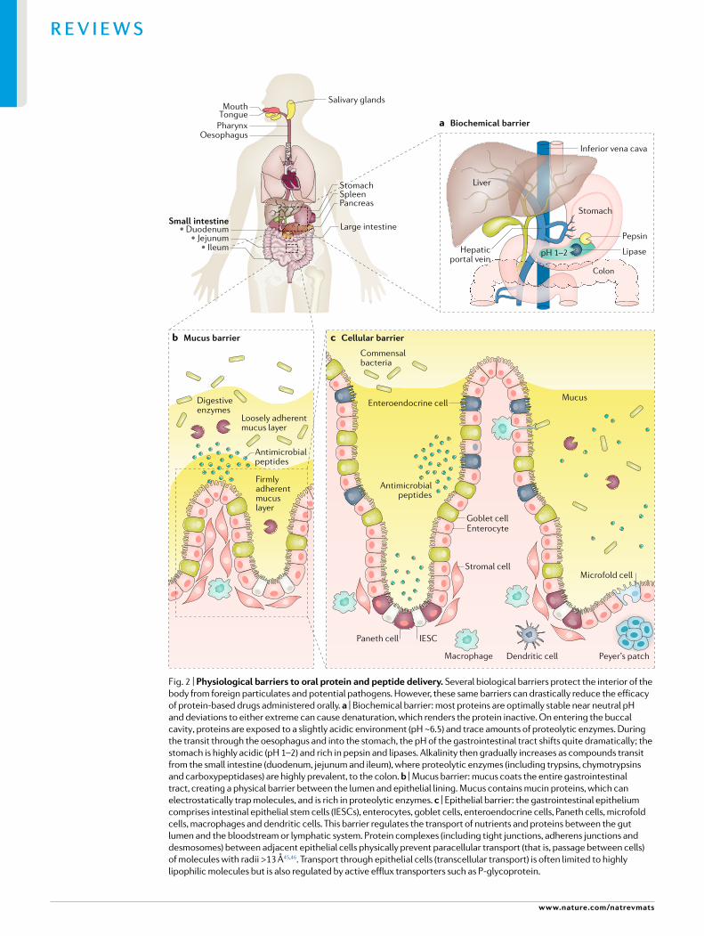
Biochemical barrierTwo major categories of biochemical barriers exist for proteins: enzymatic and pH. Proteases and other enzymes readily cleave proteins at specific cleavage sites and are located throughout the gastrointestinal tract. Likewise, drastic deviations from neutral pH can readily denature (unfold) proteins, rendering them inactive. Digestion first begins in the mouth, where the slightly acidic (pH ~6.5) conditions and saliva rich in salivary amylases and lysozymes initiate degradation of carbohydrates and peptidoglycans, respectively19. However, the buccal cavity is not considered a prominent barrier to oral drug delivery as the residence time of a pill or capsule is minimal, and the drug exposure is correspondingly minimal.
The stomach and intestine possess the most active biochemical barriers to bioavailability of orally ingested proteins20 (Fig. 2a). Digestive fluids of the stomach, secreted by gastric glands, are composed of hydrochloric acid, the protein digesting enzyme pepsin and mucus. Hydrochloric acid renders the stomach the most acidic environment in the body (pH 1–2). In such highly acidic conditions, pepsin performs optimally. Found at high concentrations in the stomach, pepsin acts as a broad endopeptidase, hydrolysing peptide linkages of aromatic residues such as phenylalanine, tryptophan and tyro sine21. Hydrolysis of fats, oils and triglycerides also occurs in the stomach and is catalysed by lipase enzymes. Digestion continues in the small
intestine, which is brimming with digestive enzymes secreted by the pancreas. Common enzymes found here include trypsins, chymotrypsins, carboxypeptidases and elastases. Enterocytes of the small intestine also produce several aminopeptidases, carboxypeptidases, endopeptidases and γ glutamyl transpeptidases22.
The small intestine is divided into three distinct regions: duodenum, jejunum and ileum, each of which possesses unique features that affect nutrient absorption23. As partly digested food and other particulates transit through the gastrointestinal tract, they are subjected to a luminal pH that steadily increases from the stomach (pH 1.0–2.0) through the duodenum (pH 4–5.5.0), jejunum (pH 5.5–7.0) and ileum (pH 7.0–7.5), before transiting to the colon and rectum (pH 7.0–7.5)24,25. This pH gradient, along with varying gastric emptying rates and gastrointestinal motility, strongly influence the pharmacokinetics of orally administered drugs26. Inactivation and/or protection from these enzymes and pH changes are essential for the effective oral delivery of protein based drugs.
Mucus barrier. Mucus is a viscoelastic, hydrogel like substance lining the gastrointestinal tract that is secreted by goblet cells (Fig. 2b). Mucus is predominately composed of mucins, which are a heavily glycosylated class of glycoproteins with a propensity to form gels due to their charged, bottlebrushlike architecture27. Mucus also contains water, lipids, electrolytes, immunoglobulins, antimicrobial peptides, protease inhibitors and various other active proteins28. Mucus, therefore, provides a nutrient rich niche for commensal bacterial colonization throughout the gastrointestinal tract, while serving as a barrier to pathogenic bacteria29.
One of the main functions of mucus is to facilitate the passage of food, chyme and faeces through the body. Mucus also acts as a physical barrier that limits the diffusion of drugs and other molecules from the lumen to the underlying epithelium28,30. The pore size of mucus has been estimated to be approximately 0.2 μm on average31,32. Pore size varies, however, depending on the location, dynamic responses to the presence of endogenous and exogenous stimuli and the patient’s state of health33.
Gastrointestinal mucus also has unique dynamic, physiochemical characteristics. The gastrointestinal tract is lined by two mucus layers: a loosely adherent layer and a firmly adherent layer. The firmly adherent layer lies immediately adjacent to the epithelial lining and includes cellbound mucins, as well as glycolipids and glycoproteins of the glycocalyx. The loosely adherent mucus layer undergoes constant turnover, which aids in the elimination of potentially harmful compounds. In early mucoadhesive studies, researchers estimated the turnover of mucus to be similar to the gut transit time, approximately 24–48 h34,35. The rapid cycle of mucin synthesis, degradation and removal contributes to considerable variability in the thickness of the mucus layer29. Disease states, exposure to environmental factors and increased age can further exacerbate this variability36,37. The pH of mucus can also vary based on its location. For example, the gastric mucus layer exhibits a pH gradient such that
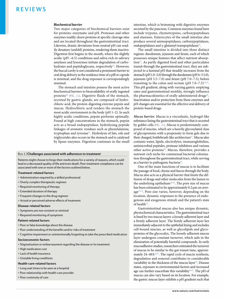
Box 1 | Challenges associated with adherence to treatment
Patients might choose to forgo their medications for a variety of reasons, which could lead to a decreased quality of life and even death. Poor treatment compliance can be associated with one or more of the factors outlined below.
treatment- related factors•administration required by a skilled professional
•Overly complex therapeutic regimen
•required monitoring of therapy
•extended duration of therapy
•Frequent changes to the drug regimen
•actual or perceived adverse effects of treatment
Disease- related factors•symptoms are non- existent or minimal
•required monitoring of symptoms
Patient- related factors•Poor or false knowledge about the disease
•Poor understanding of the benefits and/or risks of treatment
•Cognitive impairment or unintentionally forgetting to take the prescribed medication
socioeconomic factors•stigmatization or embarrassment regarding the disease or its treatment
•High medication cost
•Lack of health insurance
•unstable living conditions
Health- care-related factors•Long wait times to be seen at a hospital
•Poor relationship with health- care provider
•Poor continuity of care
www.nature.com/natrevmats
R e v i e w s
the luminal surface is far more acidic (pH 2.25) than that near the epithelial interface (pH 6.96)38. Here, the mucus acts as a selective barrier that buffers the mucosa from gastric juices and prevents autodigestion of the stomach epithelium. Accordingly, materials used in drug delivery might need properties that enable them to easily navigate through mucus or, alternatively, to increase a drug’s residence time in the gut through mucoadhesion to overcome this barrier (Fig. 3).
Cellular barrierThe epithelial lining below the mucus barrier creates another physical hurdle between the gut lumen and the bloodstream (Fig. 2c). As absorption of oral particulates primarily occurs in the small intestine, this section focuses on the key structural components found there. Enterocytes are a prominent cell type in the lining of the small intestine and are responsible for facilitating the transport of nutrients and water from the gut lumen to the bloodstream. These cells have microvilli on their apical membrane, which greatly increase the surface area available for diffusion and are involved in absorption, secretion and other biological functions. The gut epithelium also extends as columnar macroscopic structures termed villi, which protrude into the lumen and further increase the intestinal surface area and nutrient absorption. In between the villi are the crypts of Lieberkühn, invaginations where pluripotent intestinal epithelial stem cells reside.
Intestinal epithelial stem cells rapidly renew the epithelium every 2–6 days39 and give rise to enterocytes and other important cell types of the epithelium: goblet cells, enteroendocrine cells, Paneth cells and microfold (M) cells40,41. Secretory cells, such as goblet cells, secrete mucins for mucus production42. Enteroendocrine cells can sense luminal contents and secrete various regulatory factors, including glucagon like peptide 1 (GLP1), gastric inhibitory polypeptide and somatostatin42,43. Paneth cells release antimicrobial peptides, which protect nearby stem cells at the base of the intestinal crypts42. M cells are located in the follicle associated epithelium overlaying Peyer’s patches in the small intestine and are integral to the uptake and eventual presentation of luminal antigens to the immune system42. Taken together, these cells form a continuous, polarized monolayer separating the gut lumen from the lamina propria (Fig. 2b).
Epithelial cells regulate the transport and transepithelial flux of ions and molecules from their apical to their basolateral membranes. Passage of molecules between adjacent intestinal cells is physically restricted by tight junction protein complexes, adherens junctions and desmosomes44 (Fig. 3). These interlocking complexes, which have a net negative charge, have an estimated average pore radius of 8–13 Å45,46. As such, they prevent transport between adjacent cells (termed paracellular transport) of most ions and large molecules44 (Fig. 3). Alternatively, molecules can be transported across the cellular barrier, referred to as transcellular transport. Highly lipophilic molecules readily traverse the cellular barrier through passive diffusion. Moreover, large (often charged) molecules can be actively internalized by enterocytes or M cells and shuttled to the
First pure protein therapeutic discovered and isolated (insulin)295,296
FDA approval of exenatide, the first GLP-1 receptor agnoist used to treat type 2 diabetes mellitus309
FDA approval of a desmopressin tablet308 with 0.1% oral bioavailability18
Invention of enteric coatings soluble at alkaline pH (Eudragit) developed by Röhm and Haas298
Positive results for phase 3 clinical trial of oral semaglutide, formulated with Eligen SNAC developed by Emisphere315
PLease change text toFDA approval of first oral GLP-1 receptor agonist semaglutide formulated with Eligen SNAC58
Creation of solid lipid nanoparticles305
Discovery of liposomes300
First human protein to be cloned in Escherichia coli (somatostatin)303
First pill coating: mucilage from psyllium seeds used to mask unpleasant drug taste293
Earliest recording of drugs administered as pills in Egyptian hieroglyphics of the Ebers Papyrus292
First successful commercial application of pill coating: sugar-coated tablets294
FDA approval of an oral ciclosporin solution that forms microemulsions in aqueous environments307
Initiation of third phase 3 clinical trial of oral octreotide, formulated with TPE developed by Chiasma312FDA approval for GLP-1 receptor
agonist semaglutide, only for subcutaneous injection311
Positive results for phase 2a clinical trial of oral leuprolide tablet, formulated with Peptelligence developed by Enteris BioPharma314
Initiation of phase 2b dose-ranging clinical trial of oral insulin, formulated with POD developed by Oramed313
First effective oral vitamin B12 tablet, formulated with Eligen SNAC developed by Emisphere310
Invention of microfabrication techniques (microneedles)306
FDA approves first biologic therapy: recombinant human insulin (Humulin)304
Protein PEGylation invented301,302
First chemical synthesis of a human protein (insulin)299
297
1550BCE
865
925
1866
1922
1934
1953
1963
1965
1977
1982
1991
Mid1990s
1995
2005
2015
2017
2018
2019
Fig. 1 | Key technological advances towards the oral delivery of proteins and peptides. Throughout history , paramount events as early as 1550 BCE have paved the way for the current status of the oral delivery of proteins and peptides. However, the pace of events in the oral- delivery space has surged in the past 5 years. Specifically , the number of clinical trials of oral protein and peptide products has rapidly increased, which culminated in the US Food and Drug Administration (FDA) approval of the first oral biologic for type 2 diabetes mellitus in late 2019. GLP-1, glucagon- like peptide 1; PEG, polyethylene glycol; POD, protein oral delivery ; SNAC, N-(8-[2-hydroxybenzoyl]-amino) caprylic acid, also known as salcaprozate sodium; TPE, Transient Permeability Enhancer.
Nature reviews | Materials
R e v i e w s
Hepaticportal vein
Inferior vena cava
MouthSalivary glands
PharynxOesophagus
Tongue
StomachSpleenPancreas
Large intestineSmall intestine
• Duodenum• Jejunum
• Ileum
b Mucus barrier
Loosely adherentmucus layer
Firmlyadherentmucuslayer
Digestiveenzymes
c Cellular barrier
Dendritic cell Peyer’s patchMacrophage
Mucus
Antimicrobialpeptides
Antimicrobialpeptides
Commensalbacteria
Goblet cell
Enteroendocrine cell
Enterocyte
Stromal cell
Paneth cell IESC
Stomach
Colon
pH 1–2
Pepsin
Lipase
a Biochemical barrier
Microfold cell
Liver
Fig. 2 | Physiological barriers to oral protein and peptide delivery. Several biological barriers protect the interior of the body from foreign particulates and potential pathogens. However, these same barriers can drastically reduce the efficacy of protein- based drugs administered orally. a | Biochemical barrier: most proteins are optimally stable near neutral pH and deviations to either extreme can cause denaturation, which renders the protein inactive. On entering the buccal cavity , proteins are exposed to a slightly acidic environment (pH ~6.5) and trace amounts of proteolytic enzymes. During the transit through the oesophagus and into the stomach, the pH of the gastrointestinal tract shifts quite dramatically ; the stomach is highly acidic (pH 1–2) and rich in pepsin and lipases. Alkalinity then gradually increases as compounds transit from the small intestine (duodenum, jejunum and ileum), where proteolytic enzymes (including trypsins, chymotrypsins and carboxypeptidases) are highly prevalent, to the colon. b | Mucus barrier: mucus coats the entire gastrointestinal tract, creating a physical barrier between the lumen and epithelial lining. Mucus contains mucin proteins, which can electrostatically trap molecules, and is rich in proteolytic enzymes. c | Epithelial barrier: the gastrointestinal epithelium comprises intestinal epithelial stem cells (IESCs), enterocytes, goblet cells, enteroendocrine cells, Paneth cells, microfold cells, macrophages and dendritic cells. This barrier regulates the transport of nutrients and proteins between the gut lumen and the bloodstream or lymphatic system. Protein complexes (including tight junctions, adherens junctions and desmosomes) between adjacent epithelial cells physically prevent paracellular transport (that is, passage between cells) of molecules with radii >13 Å45,46. Transport through epithelial cells (transcellular transport) is often limited to highly lipophilic molecules but is also regulated by active efflux transporters such as P- glycoprotein.
www.nature.com/natrevmats
R e v i e w s
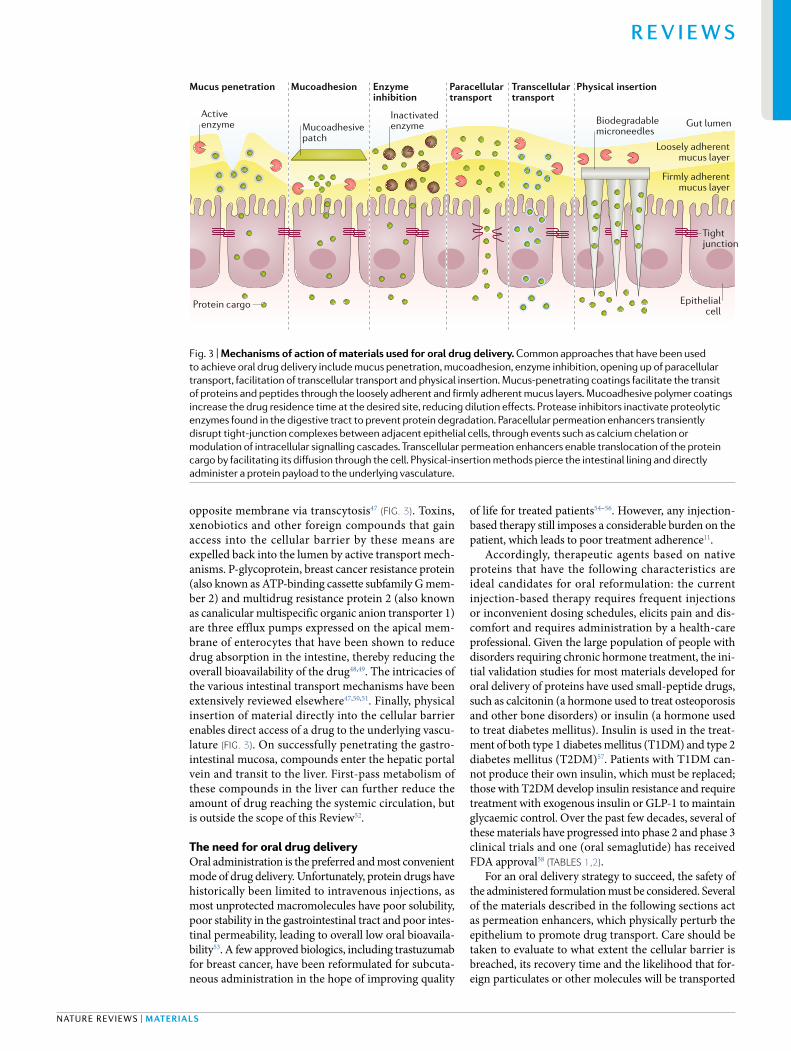
opposite membrane via transcytosis47 (Fig. 3). Toxins, xenobiotics and other foreign compounds that gain access into the cellular barrier by these means are expelled back into the lumen by active transport mechanisms. P glycoprotein, breast cancer resistance protein (also known as ATP binding cassette subfamily G member 2) and multidrug resistance protein 2 (also known as canalicular multispecific organic anion transporter 1) are three efflux pumps expressed on the apical membrane of enterocytes that have been shown to reduce drug absorption in the intestine, thereby reducing the overall bioavailability of the drug48,49. The intricacies of the various intestinal transport mechanisms have been extensively reviewed elsewhere47,50,51. Finally, physical insertion of material directly into the cellular barrier enables direct access of a drug to the underlying vasculature (Fig. 3). On successfully penetrating the gastrointestinal mucosa, compounds enter the hepatic portal vein and transit to the liver. First pass metabolism of these compounds in the liver can further reduce the amount of drug reaching the systemic circulation, but is outside the scope of this Review52.
The need for oral drug deliveryOral administration is the preferred and most convenient mode of drug delivery. Unfortunately, protein drugs have historically been limited to intravenous injections, as most unprotected macromolecules have poor solubility, poor stability in the gastrointestinal tract and poor intestinal permeability, leading to overall low oral bioavailability53. A few approved biologics, including trastuzumab for breast cancer, have been reformulated for subcutaneous administration in the hope of improving quality
of life for treated patients54–56. However, any injection based therapy still imposes a considerable burden on the patient, which leads to poor treatment adherence11.
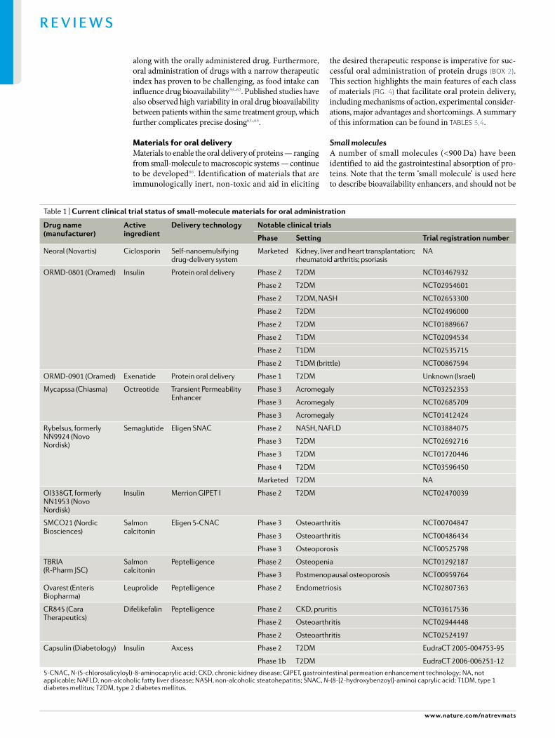
Accordingly, therapeutic agents based on native proteins that have the following characteristics are ideal candidates for oral reformulation: the current injection based therapy requires frequent injections or inconvenient dosing schedules, elicits pain and discomfort and requires administration by a health care professional. Given the large population of people with disorders requiring chronic hormone treatment, the initial validation studies for most materials developed for oral delivery of proteins have used small peptide drugs, such as calcitonin (a hormone used to treat osteoporosis and other bone disorders) or insulin (a hormone used to treat diabetes mellitus). Insulin is used in the treatment of both type 1 diabetes mellitus (T1DM) and type 2 diabetes mellitus (T2DM)57. Patients with T1DM cannot produce their own insulin, which must be replaced; those with T2DM develop insulin resistance and require treatment with exogenous insulin or GLP1 to maintain glycaemic control. Over the past few decades, several of these materials have progressed into phase 2 and phase 3 clinical trials and one (oral semaglutide) has received FDA approval58 (TaBles 1,2).
For an oral delivery strategy to succeed, the safety of the administered formulation must be considered. Several of the materials described in the following sections act as permeation enhancers, which physically perturb the epithelium to promote drug transport. Care should be taken to evaluate to what extent the cellular barrier is breached, its recovery time and the likelihood that foreign particulates or other molecules will be transported
Epithelialcell
Loosely adherentmucus layer
Firmly adherentmucus layer
Mucoadhesivepatch
Biodegradable microneedles
Gut lumen
Tightjunction
Activeenzyme
Inactivatedenzyme
Protein cargo
Mucus penetration Enzymeinhibition
Paracellulartransport
Transcellulartransport
Physical insertionMucoadhesion
Fig. 3 | Mechanisms of action of materials used for oral drug delivery. Common approaches that have been used to achieve oral drug delivery include mucus penetration, mucoadhesion, enzyme inhibition, opening up of paracellular transport, facilitation of transcellular transport and physical insertion. Mucus- penetrating coatings facilitate the transit of proteins and peptides through the loosely adherent and firmly adherent mucus layers. Mucoadhesive polymer coatings increase the drug residence time at the desired site, reducing dilution effects. Protease inhibitors inactivate proteolytic enzymes found in the digestive tract to prevent protein degradation. Paracellular permeation enhancers transiently disrupt tight- junction complexes between adjacent epithelial cells, through events such as calcium chelation or modulation of intracellular signalling cascades. Transcellular permeation enhancers enable translocation of the protein cargo by facilitating its diffusion through the cell. Physical- insertion methods pierce the intestinal lining and directly administer a protein payload to the underlying vasculature.
Nature reviews | Materials
R e v i e w s
along with the orally administered drug. Furthermore, oral administration of drugs with a narrow therapeutic index has proven to be challenging, as food intake can influence drug bioavailability59–62. Published studies have also observed high variability in oral drug bioavailability between patients within the same treatment group, which further complicates precise dosing63–65.
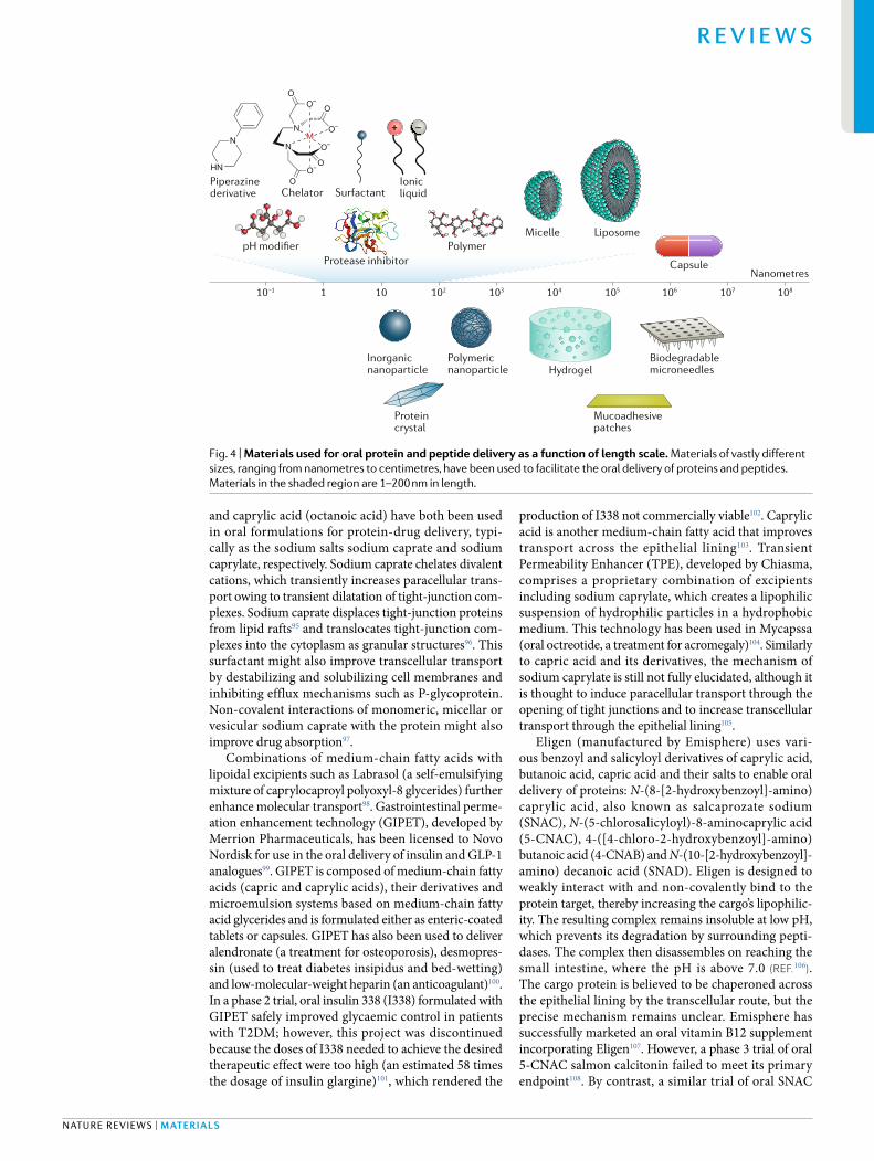
Materials for oral deliveryMaterials to enable the oral delivery of proteins — ranging from smallmolecule to macroscopic systems — continue to be developed66. Identification of materials that are immunologically inert, non toxic and aid in eliciting
the desired therapeutic response is imperative for successful oral administration of protein drugs (Box 2). This section highlights the main features of each class of materials (Fig. 4) that facilitate oral protein delivery, including mechanisms of action, experimental considerations, major advantages and shortcomings. A summary of this information can be found in TaBles 3,4.
Small moleculesA number of small molecules (<900 Da) have been identified to aid the gastrointestinal absorption of proteins. Note that the term ‘small molecule’ is used here to describe bioavailability enhancers, and should not be
Table 1 | Current clinical trial status of small- molecule materials for oral administration
Drug name (manufacturer)
active ingredient
Delivery technology Notable clinical trials
Phase setting trial registration number
Neoral (Novartis) Ciclosporin Self- nanoemulsifying drug- delivery system
Marketed Kidney , liver and heart transplantation; rheumatoid arthritis; psoriasis
NA
ORMD-0801 (Oramed) Insulin Protein oral delivery Phase 2 T2DM NCT03467932
Phase 2 T2DM NCT02954601
Phase 2 T2DM, NASH NCT02653300
Phase 2 T2DM NCT02496000
Phase 2 T2DM NCT01889667
Phase 2 T1DM NCT02094534
Phase 2 T1DM NCT02535715
Phase 2 T1DM (brittle) NCT00867594
ORMD-0901 (Oramed) Exenatide Protein oral delivery Phase 1 T2DM Unknown (Israel)
Mycapssa (Chiasma) Octreotide Transient Permeability Enhancer
Phase 3 Acromegaly NCT03252353
Phase 3 Acromegaly NCT02685709
Phase 3 Acromegaly NCT01412424
Rybelsus, formerly NN9924 (Novo Nordisk)
Semaglutide Eligen SNAC Phase 2 NASH, NAFLD NCT03884075
Phase 3 T2DM NCT02692716
Phase 3 T2DM NCT01720446
Phase 4 T2DM NCT03596450
Marketed T2DM NA
OI338GT, formerly NN1953 (Novo Nordisk)
Insulin Merrion GIPET I Phase 2 T2DM NCT02470039
SMCO21 (Nordic Biosciences)
Salmon calcitonin
Eligen 5-CNAC Phase 3 Osteoarthritis NCT00704847
Phase 3 Osteoarthritis NCT00486434
Phase 3 Osteoporosis NCT00525798
TBRIA (R-Pharm JSC)
Salmon calcitonin
Peptelligence Phase 2 Osteopenia NCT01292187
Phase 3 Postmenopausal osteoporosis NCT00959764
Ovarest (Enteris Biopharma)
Leuprolide Peptelligence Phase 2 Endometriosis NCT02807363
CR845 (Cara Therapeutics)
Difelikefalin Peptelligence Phase 2 CKD, pruritis NCT03617536
Phase 2 Osteoarthritis NCT02944448
Phase 2 Osteoarthritis NCT02524197
Capsulin (Diabetology) Insulin Axcess Phase 2 T2DM EudraCT 2005-004753-95
Phase 1b T2DM EudraCT 2006-006251-12
5-CNAC, N-(5-chlorosalicyloyl)-8-aminocaprylic acid; CKD, chronic kidney disease; GIPET, gastrointestinal permeation enhancement technology ; NA , not applicable; NAFLD, non- alcoholic fatty liver disease; NASH, non- alcoholic steatohepatitis; SNAC, N-(8-[2-hydroxybenzoyl]-amino) caprylic acid; T1DM, type 1 diabetes mellitus; T2DM, type 2 diabetes mellitus.
www.nature.com/natrevmats
R e v i e w s
confused with similar terminology referring to the active pharmaceutical ingredients. Here, we discuss enzyme inhibitors, buffering agents, chelating agents, surfactants, bile salts, aromatic alcohols and ionic liquids, which are often physically mixed with the protein to form a tablet or solubilized with the protein inside a capsule. The mechanisms of action of these compounds include improved transport across the mucus and cellu lar barriers, inactivation of proteases or other gastro intestinal enzymes and stabilization of the cargo protein’s structure.
Protease inhibitors. Ingested proteins are rapidly subjected to proteolytic degradation by proteases in the gastrointestinal tract. These enzymes recognize specific sequences of amino acids and hydrolyse (cleave) the peptide bonds between them, thereby reducing the amount of active protein available to be absorbed67. As a result, an enteric coating alone will not be sufficient to protect the drug cargo once it is released in the gut lumen. Protease inhibitors can be mixed with protein drugs to dampen this enzymatic activity. The choice of protease inhibitor depends on the amino acid sequence of the drug being delivered, as proteases are sequence specific68. Small molecule enzyme inhibitors bind reversibly or irreversibly to the target enzyme, inactivating it and, therefore, promoting survival of the protein cargo.
Several groups have used protease inhibitors to improve the bioavailability of orally administered
proteins. For instance, coadministration of calcitonin and the protease inhibitor aprotinin resulted in reduced calcitonin degradation in the colon. However, the addition of this protease inhibitor did not increase the plasma concentration of calcitonin in treated patients69. Several small molecule inhibitors, including camostat mesylate, bacitracin, soybean trypsin inhibitor and aprotinin, have been investigated to determine whether they might influence the intestinal metabolism of insulin. Of note, camostat mesylate and bacitracin improved the bioavailability of insulin only in the large intestine, and none of the molecules tested improved the absorption of insulin in the small intestine70. The activity of these inhibitors might be impaired by rapid dilution, low potency, digestion and absorption. Although these factors might be overcome by the use of high inhibitor doses to elicit a therapeutic effect, such high doses also pose substantial safety concerns, including pancreatic hypertrophy and hyperplasia71,72. Some small molecule inhibitors, including bacitracin, have also been linked to nephrotoxicity73. One explanation for the observed lack of benefit is that the pancreas readily compensates for the presence of inhibitors by activating a feedback loop that increases the secretion of proteases74. As these inhibitors also prevent the degradation and absorption of other proteins besides the drug cargo, thereby altering the metabolism within the gastrointestinal tract, the use of protease inhibitors should be localized to the specific
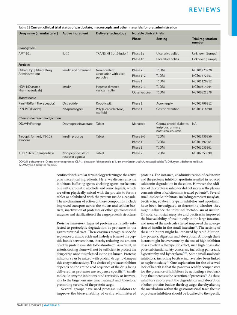
Table 2 | Current clinical trial status of particulate, macroscopic and other materials for oral administration
Drug name (manufacturer) active ingredient Delivery technology Notable clinical trials
Phase setting trial registration number
Biopolymers
AMT-101 IL-10 TRANSINT (IL-10 fusion) Phase 1a Ulcerative colitis Unknown (Europe)
Phase 1b Ulcerative colitis Unknown (Europe)
Particles
Oshadi Icp (Oshadi Drug Administration)
Insulin and proinsulin Non- covalent association with silica particles
Phase 2 T1DM NCT01973920
Phase 1–2 T1DM NCT01772251
Phase 1 T1DM NCT01120912
HDV- I (Diasome Pharmaceuticals)
Insulin Hepatic- directed vesicle insulin
Phase 2–3 T1DM NCT00814294
Observational T1DM NCT00521378
Macroscopic
RaniPill (Rani Therapeutics) Octreotide Robotic pill Phase 1 Acromegaly NCT03798912
LYN- PLT (Lyndra) NA (prototype) Poly (ε- caprolactone) scaffold
Phase 1 Gastric retention NCT03718390
Chemical or other modification
DDAVP (Ferring) Desmopressin acetate Tablet Marketed Central cranial diabetes insipidus; primary nocturnal enuresis
NA
Tregopil, formerly IN-105 (Biocon)
Insulin prodrug Tablet Phase 2–3 T2DM NCT03430856
Phase 1 T2DM NCT03392961
Phase 1 T1DM NCT01035801
TTP273 (vTv Therapeutics) Non- peptide GLP-1 receptor agonist
Tablet Phase 2 T2DM NCT02653599
DDAVP, 1-deamino-8-D- arginine vasopressin; GLP-1, glucagon- like peptide 1; IL-10, interleukin 10; NA , not applicable; T1DM, type 1 diabetes mellitus; T2DM, type 2 diabetes mellitus.
Nature reviews | Materials
R e v i e w s
site of action. One possible method of localizing protease inhibitors would be to incorporate them into a muco adhesive system. However, as yet, no commercial products have used this approach.
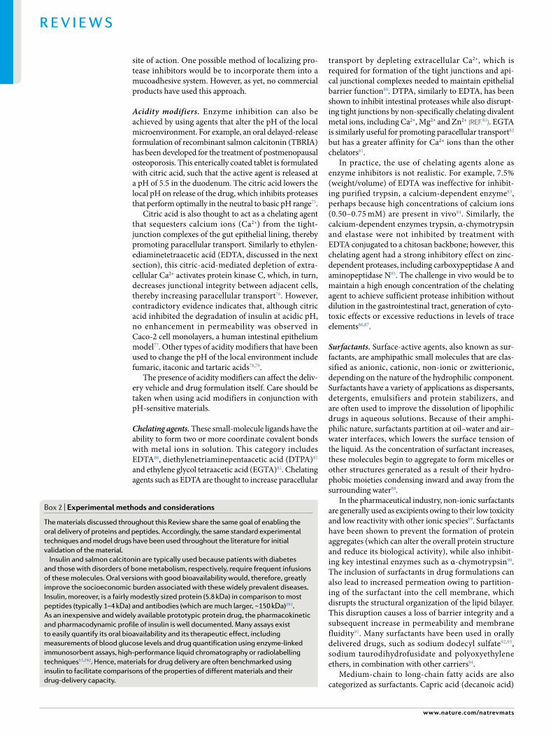
Acidity modifiers. Enzyme inhibition can also be achieved by using agents that alter the pH of the local microenvironment. For example, an oral delayed release formulation of recombinant salmon calcitonin (TBRIA) has been developed for the treatment of postmenopausal osteoporosis. This enterically coated tablet is formulated with citric acid, such that the active agent is released at a pH of 5.5 in the duodenum. The citric acid lowers the local pH on release of the drug, which inhibits proteases that perform optimally in the neutral to basic pH range75.
Citric acid is also thought to act as a chelating agent that sequesters calcium ions (Ca2+) from the tight junction complexes of the gut epithelial lining, thereby promoting paracellular transport. Similarly to ethylenediaminetetraacetic acid (EDTA, discussed in the next section), this citric acidmediated depletion of extracellular Ca2+ activates protein kinase C, which, in turn, decreases junctional integrity between adjacent cells, thereby increasing paracellular transport76. However, contradictory evidence indicates that, although citric acid inhibited the degradation of insulin at acidic pH, no enhancement in permeability was observed in Caco2 cell monolayers, a human intestinal epithelium model77. Other types of acidity modifiers that have been used to change the pH of the local environment include fumaric, itaconic and tartaric acids78,79.
The presence of acidity modifiers can affect the delivery vehicle and drug formulation itself. Care should be taken when using acid modifiers in conjunction with pH sensitive materials.
Chelating agents. These small molecule ligands have the ability to form two or more coordinate covalent bonds with metal ions in solution. This category includes EDTA80, diethylenetriaminepentaacetic acid (DTPA)81 and ethylene glycol tetraacetic acid (EGTA)82. Chelating agents such as EDTA are thought to increase para cellular
transport by depleting extracellular Ca2+, which is required for formation of the tight junctions and apical junctional complexes needed to maintain epithelial barrier function80. DTPA, similarly to EDTA, has been shown to inhibit intestinal proteases while also disrupting tight junctions by non specifically chelating divalent metal ions, including Ca2+, Mg2+ and Zn2+ (reF.81). EGTA is similarly useful for promoting paracellular transport82 but has a greater affinity for Ca2+ ions than the other chelators81.
In practice, the use of chelating agents alone as enzyme inhibitors is not realistic. For example, 7.5% (weight/volume) of EDTA was ineffective for inhibiting purified trypsin, a calcium dependent enzyme83, perhaps because high concentrations of calcium ions (0.50–0.75 mM) are present in vivo84. Similarly, the calcium dependent enzymes trypsin, α chymotrypsin and elastase were not inhibited by treatment with EDTA conjugated to a chitosan backbone; however, this chelating agent had a strong inhibitory effect on zinc dependent proteases, including carboxypeptidase A and aminopeptidase N85. The challenge in vivo would be to maintain a high enough concentration of the chelating agent to achieve sufficient protease inhibition without dilution in the gastrointestinal tract, generation of cytotoxic effects or excessive reductions in levels of trace elements86,87.
Surfactants. Surface active agents, also known as surfactants, are amphipathic small molecules that are classified as anionic, cationic, non ionic or zwitterionic, depending on the nature of the hydrophilic component. Surfactants have a variety of applications as dispersants, detergents, emulsifiers and protein stabilizers, and are often used to improve the dissolution of lipophilic drugs in aqueous solutions. Because of their amphiphilic nature, surfactants partition at oil–water and air–water interfaces, which lowers the surface tension of the liquid. As the concentration of surfactant increases, these molecules begin to aggregate to form micelles or other structures generated as a result of their hydrophobic moieties condensing inward and away from the surrounding water88.
In the pharmaceutical industry, non ionic surfactants are generally used as excipients owing to their low toxicity and low reactivity with other ionic species89. Surfactants have been shown to prevent the formation of protein aggregates (which can alter the overall protein structure and reduce its biological activity), while also inhibiting key intestinal enzymes such as α chymotrypsin90. The inclusion of surfactants in drug formulations can also lead to increased permeation owing to partitioning of the surfactant into the cell membrane, which disrupts the structural organization of the lipid bilayer. This disruption causes a loss of barrier integrity and a subsequent increase in permeability and membrane fluidity91. Many surfactants have been used in orally delivered drugs, such as sodium dodecyl sulfate92,93, sodium taurodihydrofusidate and polyoxyethylene ethers, in combination with other carriers94.
Medium chain to long chain fatty acids are also categorized as surfactants. Capric acid (decanoic acid)
Box 2 | experimental methods and considerations
the materials discussed throughout this review share the same goal of enabling the oral delivery of proteins and peptides. accordingly, the same standard experimental techniques and model drugs have been used throughout the literature for initial validation of the material.
insulin and salmon calcitonin are typically used because patients with diabetes and those with disorders of bone metabolism, respectively, require frequent infusions of these molecules. Oral versions with good bioavailability would, therefore, greatly improve the socioeconomic burden associated with these widely prevalent diseases. insulin, moreover, is a fairly modestly sized protein (5.8 kDa) in comparison to most peptides (typically 1–4 kDa) and antibodies (which are much larger, ~150 kDa)291. as an inexpensive and widely available prototypic protein drug, the pharmacokinetic and pharmacodynamic profile of insulin is well documented. Many assays exist to easily quantify its oral bioavailability and its therapeutic effect, including measurements of blood glucose levels and drug quantification using enzyme- linked immunosorbent assays, high- performance liquid chromatography or radiolabelling techniques57,292. Hence, materials for drug delivery are often benchmarked using insulin to facilitate comparisons of the properties of different materials and their drug- delivery capacity.
www.nature.com/natrevmats
R e v i e w s
and caprylic acid (octanoic acid) have both been used in oral formulations for protein drug delivery, typically as the sodium salts sodium caprate and sodium caprylate, respectively. Sodium caprate chelates divalent cations, which transiently increases paracellular transport owing to transient dilatation of tight junction complexes. Sodium caprate displaces tight junction proteins from lipid rafts95 and translocates tight junction complexes into the cytoplasm as granular structures96. This surfactant might also improve transcellular transport by destabilizing and solubilizing cell membranes and inhibiting efflux mechanisms such as P glycoprotein. Non covalent interactions of monomeric, micellar or vesicular sodium caprate with the protein might also improve drug absorption97.
Combinations of medium chain fatty acids with lipoidal excipients such as Labrasol (a self emulsifying mixture of caprylocaproyl polyoxyl8 glycerides) further enhance molecular transport98. Gastrointestinal permeation enhancement technology (GIPET), developed by Merrion Pharmaceuticals, has been licensed to Novo Nordisk for use in the oral delivery of insulin and GLP1 analogues99. GIPET is composed of medium chain fatty acids (capric and caprylic acids), their derivatives and microemulsion systems based on medium chain fatty acid glycerides and is formulated either as enteric coated tablets or capsules. GIPET has also been used to deliver alendronate (a treatment for osteoporosis), desmopressin (used to treat diabetes insipidus and bed wetting) and low molecularweight heparin (an anticoagulant)100. In a phase 2 trial, oral insulin 338 (I338) formulated with GIPET safely improved glycaemic control in patients with T2DM; however, this project was discontinued because the doses of I338 needed to achieve the desired therapeutic effect were too high (an estimated 58 times the dosage of insulin glargine)101, which rendered the
production of I338 not commercially viable102. Caprylic acid is another medium chain fatty acid that improves transport across the epithelial lining103. Transient Permeability Enhancer (TPE), developed by Chiasma, comprises a proprietary combination of excipients including sodium caprylate, which creates a lipophilic suspension of hydrophilic particles in a hydrophobic medium. This technology has been used in Mycapssa (oral octreotide, a treatment for acromegaly)104. Similarly to capric acid and its derivatives, the mechanism of sodium caprylate is still not fully elucidated, although it is thought to induce paracellular transport through the opening of tight junctions and to increase transcellular transport through the epithelial lining105.
Eligen (manufactured by Emisphere) uses various benzoyl and salicyloyl derivatives of caprylic acid, butanoic acid, capric acid and their salts to enable oral delivery of proteins: N(8[2hydroxybenzoyl]amino) caprylic acid, also known as salcaprozate sodium (SNAC), N(5chlorosalicyloyl)8aminocaprylic acid (5CNAC), 4([4chloro2hydroxybenzoyl]amino) butanoic acid (4CNAB) and N(10[2hydroxybenzoyl]amino) decanoic acid (SNAD). Eligen is designed to weakly interact with and non covalently bind to the protein target, thereby increasing the cargo’s lipophilicity. The resulting complex remains insoluble at low pH, which prevents its degradation by surrounding peptidases. The complex then disassembles on reaching the small intestine, where the pH is above 7.0 (reF.106). The cargo protein is believed to be chaperoned across the epithelial lining by the transcellular route, but the precise mechanism remains unclear. Emisphere has successfully marketed an oral vitamin B12 supplement incorporating Eligen107. However, a phase 3 trial of oral 5CNAC salmon calcitonin failed to meet its primary endpoint108. By contrast, a similar trial of oral SNAC
+ –
108107105 1061041031021010–1 1
Polymericnanoparticle
Proteincrystal
HydrogelBiodegradablemicroneedles
Mucoadhesive patches
Inorganicnanoparticle
Liposome
CapsuleNanometres
Micelle
Protease inhibitor
Ionicliquid
Piperazinederivative SurfactantChelator
pH modifier Polymer
HN
NO–N
N
O–
M
O
OO–
O
O
O–
Fig. 4 | Materials used for oral protein and peptide delivery as a function of length scale. Materials of vastly different sizes, ranging from nanometres to centimetres, have been used to facilitate the oral delivery of proteins and peptides. Materials in the shaded region are 1–200 nm in length.
Nature reviews | Materials
R e v i e w s
semaglutide tablets successfully met its predefined primary endpoints, and bioavailability of the oral treatment was similar to that of the conventional injected drug109,110. Clinical and preclinical studies have demonstrated that absorption of oral semaglutide SNAC takes place in the stomach and is confined to the region where the tablet interfaces with the stomach lining111. SNAC and sodium caprate have been reviewed in depth elsewhere112.
Nausea is a known adverse effect of SNAC when given in the high doses needed for oral protein delivery in humans113. In the PIONEER 4 trial, patients receiving oral semaglutide were slightly more likely than those receiving subcutaneous liraglutide to experience adverse events (56 versus 51 events); the most frequent adverse events were mild tomoderate and transient nausea, followed by diarrhoea114. More patients withdrew prematurely from the trial in the oral semaglutide group (11%) than in the liraglutide group (9%)114. In 2019, this oral SNAC semaglutide formulation (Rybelsus) was the first oral GLP1 treatment for T2DM to be approved by the FDA58.
Zwitterionic small molecules have also been used in a variety of protein oral delivery applications. For example, acylcarnitines are esters of L carnitine and fatty acids containing a quaternary ammonium group and a carboxyl group. These zwitterionic compounds transport activated long chain fatty acids into mitochondria for subsequent β oxidation to create the energy needed for cellular activities115. Lauroylcarnitine and palmitoylcarnitine are two zwitterionic excipients included in the Peptelligence technology developed by Enteris BioPharma116. Here, these molecules act as permeation enhancers — by increasing paracellular transport
through tight junctions while also increasing the solubility of the peptide cargo117. Another zwitterionic compound, palmityl dimethyl ammonio propane sulfonate (PPS, also known as 3[N,N dimethyl(3palmitoylaminopropyl)ammonio]propane sulfonate), contains a quaternary ammonium group and a sulfate group. This molecule has been shown to have good intracellular delivery in vitro118 and to enable the oral delivery of protein compounds such as salmon calcitonin in vivo119.
Bile salts. Bile, a complex fluid secreted by the liver containing bile acids, cholesterol and other components, aids in the digestion of lipids in the small intestine. Bile acids are synthesized in hepatocytes from cholesterol and exist as ionic, amphiphilic molecules with a steroid backbone. The vast majority of bile acids are conjugated to glycine or taurine to form monovalent bile salts, which act as amphipathic, steroidal biosurfactants. Bile salts are the major route of elimination of cholesterol from the body; they solubilize lipids in the gut, increase their proteolytic cleavage and aid in their absorption. Thus, several bile salts (sodium deoxycholate120, sodium tauro cholate121, sodium glycodeoxycholate122 and sodium taurodihydrofusidate123) have been used as permeation enhancers to improve drug absorption across various biological barriers, including the intestine. Bile salts increase paracellular transport by opening up tight junctions; they can also improve drug stability against enzymatic activity and fluidize cell membranes of the intestinal epithelium124. Sodium taurodeoxycholate has been used in the oral delivery of salmon calcitonin122 and sodium glycocholate has been used in the oral delivery of
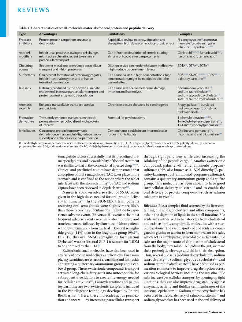
Table 3 | Characteristics of small- molecule materials for oral protein and peptide delivery
type advantages limitations examples
Protease inhibitors
Protect protein cargo from enzymatic degradation
Rapid dilution, low potency, digestion and absorption; high doses can elicit cytotoxic effect
N- acetylcysteine68, camostat mesylate70, soybean trypsin inhibitor317, aprotinin318,319
Acid pH modifiers
Inhibit local proteases owing to pH change, might act as chelating agent to enhance paracellular transport
Can influence dissolution of enteric coating; shifts in pH could alter cargo contents
Citric acid75,78,79, fumaric acid78,79, itaconic acid79, tartaric acid78
Chelating agents
Sequester metal ions to enhance paracellular transport and inhibit proteases
Dilution in vivo can render chelators ineffective; might reduce trace- element levels
EDTA80, DTPA81, EGTA82
Surfactants Can prevent formation of protein aggregates, inhibit intestinal enzymes and enhance intestinal permeation
Can cause nausea in high concentrations; high concentrations might be needed to elicit the desired effect
SDS92,93, SNAC109,110,113, PPS118,119, palmitoylcarnitine116
Bile salts Naturally produced by the body to eliminate cholesterol, increase paracellular transport and protect against enzymatic degradation
Can cause irreversible membrane damage, irritation and haemolysis
Sodium deoxycholate120, sodium taurocholate121, sodium glycodeoxycholate122, sodium taurodihydrofusidate123
Aromatic alcohols
Enhance transcellular transport; used as antioxidants
Chronic exposure shown to be carcinogenic Propyl gallate129, butylated hydroxytoluene129, butylated hydroxyanisole129
Piperazine derivatives
Transiently enhance transport, enhanced permeation when colocalized with protein of interest
Potential for psychoactivity 1-phenylpiperazine134,135, 1-methyl-4-phenylpiperazine137, 1-(4-methylphenyl)piperazine137
Ionic liquids Can protect protein from enzymatic degradation, enhance solubility, reduce mucus viscosity and enhance intestinal permeation
Contaminants could disrupt intermolecular forces in ionic liquids
Choline and gernanate142, nicotinic acid and trigonelline144
DTPA , diethylenetriaminepentaacetic acid; EDTA , ethylenediaminetetraacetic acid; EGTA , ethylene glycol tetraacetic acid; PPS, palmityl-dimethyl ammonio propanesulfonate; SDS, sodium dodecyl sulfate; SNAC, N-(8-[2-hydroxybenzoyl]-amino) caprylic acid, also known as salcaprozate sodium.
www.nature.com/natrevmats
R e v i e w s
insulin125. However, bile salts can cause irreversible damage to cell membranes126, irritation127 and haemolysis, which has limited their clinical applications128.
Aromatic alcohols. Aromatic alcohols are another class of small molecules used as permeation enhancers and solubilizers to enhance transcellular transport of orally delivered proteins. For example, aromatic alcohols are included in the Axcess drug delivery system129 developed by Proxima Concepts and licensed to its subsidiary Diabetology130. Axcess technology is used in several oral antidiabetic drugs, including Capsulin (for T2DM),
Capsulin IR (insulin replacement; for T1DM), Combulin (for T2DM) and an oral GLP1 analogue (for T2DM)131. Other applications for this technology include the oral delivery of anticancer agents, vaccines and treatments for infectious diseases132. In addition to several aromatic alcohols (such as propyl gallate, butylated hydroxytoluene, butylated hydroxyanisole and derivatives thereof), Axcess includes a biguanide to increase the solubility of the aromatic alcohol in aqueous media. These aromatic alcohols are commonly used as antioxidants in both the pharmaceutical and the food industries and do not pose a health hazard at their administered doses133.
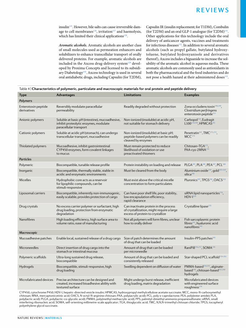
Table 4 | Characteristics of polymeric, particulate and macroscopic materials for oral protein and peptide delivery
type advantages limitations examples
Polymers
Enterotoxin peptide derivatives
Reversibly modulate paracellular permeability
Readily degraded without protection Zona occludens toxin148,149, Clostridium perfringens enterotoxin peptide151
Anionic polymers Soluble at basic pH (intestine), mucoadhesive, inhibit proteolytic enzymes, modulate paracellular transport
Non- ionized (insoluble) at acidic pH, not suitable for stomach delivery
Carbopol154, Eudragit L100159,160, HPMCAS161
Cationic polymers Soluble at acidic pH (stomach), can undergo transcellular transport, mucoadhesive
Non- ionized (insoluble) at basic pH; peptide- based polymers can be readily cleaved by enzymes
Penetratin162, TMC171,172, MCC174
Thiolated polymers Mucoadhesive, inhibit gastrointestinal CYP450 enzymes, form covalent linkages to mucus
Must remain protected to reduce likelihood of oxidation or use preactivated thiomers
Chitosan–TGA175, PAA- cys-2MNA179
Particles
Polymeric Biocompatible, tunable release profile Protein instability on loading and release PLGA185, PL A186, PEA187, PCL188
Inorganic Biocompatible, thermally stable, stable in acidic and enzymatic environments
Must be cleared from the body Aluminium oxide194, gold195–197, silica200
Micelles Hydrophobic core acts as a reservoir for lipophilic compounds, can be stimuli- responsive
Must exist above the critical micelle concentration to form particulates
Pluronics207, TPGS206, OACS210
Liposomal carriers Biocompatible, inherently non- immunogenic, easily scalable, provides protection of cargo
Can have poor shelf life, poor stability , low encapsulation efficiency , rapid clearance
siRNA lipid nanoparticles216, HDV- I320
Drug crystals No excess carrier polymer or surfactant, high drug loading, protection from enzymatic degradation
Can inactivate protein in the process of crystallization, might require a large excess of protein to crystallize
Crystalline lipase235
Nanofibres High loading efficiency , high surface area to volume ratio, ease of manufacturing
Not all polymers will form fibres, unclear how to orally deliver
Fish sarcoplasmic protein fibres241, hyaluronic acid nanofibres242
Macroscopic
Mucoadhesive patches Enable local, sustained release of a drug cargo Size of patch determines the amount of drug that can be loaded
Insulin–PPS patches245
Microneedles Direct insertion of drug cargo into the stomach or intestinal mucosa
Amount of drug that can be loaded per microneedle
RaniPill256,257, SOMA258
Polymeric scaffolds Ultra- long sustained drug release, biocompatible
Amount of drug that can be loaded and consistently released
Star- shaped PCL scaffold259–261
Hydrogels Biocompatible, stimuli- responsive, high drug loading
Swelling dependent on diffusion of water PMMA- based264,265, alginate- based266, chitosan- based267,268 hydrogels
Microfabricated devices Precise architecture can be designed and created, increased bioadhesive ability with textured surface
Might undergo burst release, inefficient drug loading, matrix degradation
Microfabricated devices with engineered surface roughness278
CYP450, cytochrome P450; HDV- I, hepatic- directed vesicle insulin; HPMCAS, hydroxypropyl methylcellulose acetate succinate; MCC, mono- N-carboxymethyl chitosan; MNA , mercaptonicotinic acid; OACS, N- octyl-N- arginine chitosan; PAA , poly(acrylic acid); PCL , poly- ε-caprolactone; PEA , poly(ester amide); PL A , poly(lactic acid); PLGA , poly(lactic- co-glycolic acid); PMMA , poly(methyl methacrylic acid); PPS, palmityl- dimethyl ammonio propanesulfonate; siRNA , small interfering ribonucleic acid; SOMA , self- orienting millimetre- scale applicator ; TGA , thioglycolic acid; TMC, N,N,N- trimethyl chitosan chloride; TPGS, tocopheryl polyethylene glycol succinate.
Nature reviews | Materials
R e v i e w s
However, chronic exposure to elevated levels of these compounds is carcinogenic133.
Piperazine derivatives. Piperazines are molecules with a fully saturated, six membered ring with nitrogen atoms at positions 1 and 4. After screening >50 potential small molecule permeation enhancers, piperazine derivatives were identified as offering an unusual combination of good permeation enhancing ability and low cytotoxi city134. The permeation enhancing efficacy of 1phenyl piperazine was confirmed in an ex vivo study, which suggested that its paracellular permeability increasing effect is mediated by an interaction with serotonin (5hydroxytryptamine) receptor 4, which is present on the apical epithelial surface. This interaction is thought to cause a cascade of events leading to modulation of tight junction complexes134,135. Subsequently, additional piperazine derivatives, including 1methyl4 phenylpiperazine and 1(4methylphenyl)piperazine, were identified as permeation enhancers with lower toxicity than 1phenylpiperazine136,137. With regard to safety considerations, some piperazine derivatives (including 1benzylpiperazine), although not those identified as permeation enhancers, can elicit psychoactive effects138.
Piperazines have also been incorporated into protein– polymer conjugates used for oral delivery of protein drugs. Conjugates synthesized from bovine serum albumin and piperazine containing monomers facilitate colocalization of the permeation enhancer with the protein drug. Use of such conjugates increased transepithelial protein transport by up to 35fold compared with the modest permeation improvements observed for the coadministered, small molecule, calcein. These data suggest that piperazine containing protein–polymer conjugates selectively increase protein permeability, which could mitigate the unwanted transepithelial transport of other molecules in the gut lumen139.
Ionic liquids. Ionic liquids comprise loosely coordinated anions and cations, which provide their unique solvating and permeation enhancing properties. Various cations (such as quaternary ammonium, imidazolium, pyrrolidinium, pyridinium, cholinium and guanidinium) have been used together with various anions (such as carboxylate, alkyl sulfate, dicyanamide and bistriflimide) in ionic liquid formulations140–143. For example, treatment with insulin in a choline and geranate (CAGE) ionic liquid demonstrated considerable lowering of blood glucose when delivered via oral gavage142. This ionic liquid possesses mucolytic activity resulting from decreased mucus viscosity, inhibits intestinal enzymes such as trypsin and directly enhances permeation across the epithelial lining with minimal toxicity. CAGE also offers long term stability of the protein both at room temperature and at 4 °C142. Ionic liquids composed of nicotinic acid and its metabolite, trigonelline (N methylnicotinic acid) have also demonstrated utility in the oral delivery of poorly water soluble drugs144. However, the presence of additional ions, solvents and water molecules in formulated drugs might alter important intermolecular properties (such as viscosity and electrostatic forces) of pure ionic liquids; currently, it is unknown how such alterations
might affect the overall permeation enhancing capacity of the agent145,146.
Natural and synthetic biopolymersNaturally derived and synthetic biopolymers have been extensively used for oral drug delivery, both as individual molecules and as building blocks for use in the macroscopic systems discussed later in this article.
Enterotoxin peptide derivatives. Toxins produced by bacteria and multicellular organisms have been used to develop permeation enhancers derived from specific purified toxin peptides. For instance, Vibrio cholerae is a Gram negative bacterium that induces severe diarrhoea when ingested in contaminated food or water. On attaching to the intestinal lining, this bacterium produces cholera toxin, which enters enterocytes and causes a dramatic dehydrating efflux of ions and water from these cells147. Other virulence factors are also secreted by V. cholerae, such as cholix toxin and zona occludens toxin (Zot)148,149. Zot is a bacterial surface protein that, along with its synthetic mimic AT1002 (reF.150), reversibly increases paracellular permeability by activating intracellular signalling pathways leading to modulation of actin polymerization148,149. Other enterotoxin peptides and their derivatives include the Clostridium perfringens enterotoxin peptide151 and melittin, which is found in the venom of European honey bees152. The TRANSINT permeability enhancer developed by Applied Molecular Transport targets the local intestinal submucosa and gut associated lymphatic tissue153, presumably using cholix toxinderived fusion molecules. This truncated exotoxin based technology enables the successful transcellular transport of protein molecules such as IL10, which is used in the treatment of inflammatory bowel disease. Notably, these peptides and their derivatives require additional protective measures to prevent them from being subjected to proteolytic degradation.
Anionic polymers. Anionic (negatively charged) polymers such as polyacrylic acid or cellulose have frequently been used to deliver small molecule drugs. Anionic poly mers can exhibit mucoadhesive properties, inhibit proteo lytic enzymes and/or modulate intestinal transport by chelating extracellular calcium ions from the surrounding environment154–156. Carbopol polymers, for instance, inhibit the degradation of insulin, calcitonin and insulin like growth factor I by inactivating trypsin and chymo trypsin in the gut lumen154. Enteric coatings157,158 consisting of anionic copolymers, such as methacrylic acid and methyl methacrylate159,160 or hydroxypropyl methylcellulose acetate succinate161, facilitate the release of a drug at a desired pH.
Cationic polymers. Natural and synthetic, positively charged (cationic) polymers are used in oral drug delivery, including as cell penetrating peptides (CPPs), chitosan and chitosan derivatives. CPPs are rich in the two basic amino acid residues arginine and lysine, which are positively charged and, therefore, facilitate electrostatic interactions with negatively charged cell surfaces
www.nature.com/natrevmats
R e v i e w s
and drug molecules. CPPs also contain hydrophobic domains from amino acids such as tryptophan, which promote membrane translocation of the CPP through the lipid bilayer. The amphipathic CPP penetratin and its analogue PenetraMax both increase intestinal permeation of insulin on coadministration162–164. Use of a medium chain fatty acid–CPP hybrid both reduced the cytotoxic effect associated with the medium chain fatty acid and enhanced the transport of insulin glulisine165. However, similarly to enterotoxins, CPPs can be cleaved by intestinal proteases, which inactivates their permeation enhancing activity166. Strategies to reduce this degradation (such as altering the amino acid stereochemistry from L to D) also reduce their efficacy167.
Chitosan improves paracellular transport by opening tight junctions168. This polysaccharide consists of copolymers of glucosamine and N acetylglucosamine, which are insoluble at neutral and alkaline pH, but form salts with inorganic and organic acids. Chitosan is generally regarded as non toxic, biocompatible and biodegradable and is used as a food additive169. Chitosan absorbs water from its local microenvironment and, in its swollen state, has demonstrated excellent mucoadhesive properties, resulting in its capacity for repeated adhesion events, during which positively charged amino groups in the chitosan bind to negatively charged moieties in mucin glycoproteins170. However, drug delivery approaches based solely on mucoadhesion can have several indirect drawbacks, including potential shifting or dislodgment of the material from the mucosal lining owing to mucus turnover or physical disruption.
The quaternized chitosan derivative N,N,N trimethyl chitosan chloride has been used in targeted intestinal delivery. This quaternized chitosan shows higher aqueous solubility than chitosan, in much broader pH and concentration ranges, without affecting its cationic nature. As the primary amine has been substituted with methyl groups, hydrogen bonds cannot form between this amine and the hydroxyl groups of the chitosan backbone, which promotes increased absorption of hydrophilic compounds at pH values similar to those found in the jejunum171,172. Other derivatives, such as acrylated173 and mono carboxymethylated174 chitosan, have also been used as paracellular absorption enhancers to deliver molecules such as low molecularweight heparin174. These chitosan derivatives feature moieties bearing carboxyl groups, which yield polymers with strong mucoadhesive and polyampholytic properties.
Thiolated polymers. Thiolated polymers, or thiomers, have thiol side chains that are responsible for these agents’ mucoadhesive and permeation enhancing properties. Thiomers enable controlled drug release through the inhibition of gastrointestinal enzymes and P glycoprotein efflux pumps.
One major impediment to oral drug administration is the superfamily of haem thiolate cytochrome P450 (CYP450) enzymes, which contribute to oxidative metabolism of administered drugs. Thiolated polymers can inhibit the activity of both CYP450 enzymes and active P glycoprotein efflux pumps. Thiomers exist in both cationic (chitosan derived175) and anionic
(with carboxylic acid side groups) forms176–179; both are suggested to form covalent disulfide bonds with cysteine residues of mucin and CYP450 enzymes, which eliminates the reducing environment and inactivates the enzyme180. As the thiol groups of unstabilized polymers are susceptible to early oxidation at pH 5 or greater, the enzyme inhibiting activity of unprotected thiomers can be severely reduced175. Poly(acrylic acid)cysteine2mercaptonicotinic acid conjugates179 and other preactivated thiomers have been developed, which have enhanced stability and mucoadhesive and cohesive properties because they contain disulfide linkages that cannot oxidize further at high pH181,182.
Particle- based systemsThe advent of the nanomedicine revolution led to the development of an entire host of systems based on nanoparticles or micrometre sized particles to enable the oral delivery of drugs. Owing to limitations of space, we cannot include an extended discussion of every particle system developed to date within this Review29,183; however, we highlight the predominant classes of particulate materials used for oral drug delivery.
Polymeric particles. A widely used class of particles for oral drug delivery is derived from biocompatible and biodegradable polymers. These polymers undergo hydrolysis, driven by pH, temperature and other environmental factors, which cause them to break down at the desired location and release their drug payload. Poly(lactic coglycolic acid) is a commonly used biodegradable polymer that produces lactic and glycolic acids on being hydrolysed. These by products are readily metabolized via the Krebs cycle and consequently yield minimal systemic toxicity184,185. Other biodegradable polymers used in oral delivery include poly(lactic acid)186, poly(ester amide)187 and poly(ε caprolactone)188. Poly(fumaric cosebacic anhydride)189, polyglycerol esters of fatty acids190 and other similar biodegradable polymers possess strong mucoadhesive properties owing to hydrogen bonding, polymer entanglements with mucins, hydrophobic interactions or any combination of these mechanisms, which all increase drug retention time at the epithelial lining30,191,192. Nanoparticles made of these polymers (including those derived from the cationic biopolymer chitosan, discussed above) can also be coated with CPPs to further improve drug delivery across the intestine163. Challenges associated with the use of biodegradable polymeric nanoparticles include protein instability resulting from loading and release, denaturation and aggregation of the cargo protein as a result of the acidic microenvironment created by polymer degradation, and the potential for burst release193.
Inorganic particles. Inorganic nanoparticles have been used for the oral delivery of peptides and proteins. Key advantages of inorganic nanoparticle systems include the wide variety of core materials available, biocompatibility, thermal stability, responsiveness to specific stimuli and the potential for monodisperse production. Unlike their biodegradable counterparts, inorganic particles
Nature reviews | Materials
R e v i e w s
remain stable in acidic and highly enzymatic environments, where they continue to provide protection to their protein cargo. As a result, additional components, such as enteric coatings or protease inhibitors, might not be necessary.
Aluminium oxide194, gold195–197, selenium198,199, silica200 and zirconium phosphate201–203 have been used to deliver proteins orally. Oshadi Drug Administration has developed an oral insulin formulation containing insulin and proinsulin C peptide in Oshadi carrier (Oshadi Icp), in which the cargo protein is non covalently associated with silica nanoparticles. Oshadi Icp has recently completed phase 2 clinical trials204. As inorganic nanoparticles are not biodegradable, care must be taken to ensure these particles are completely cleared and/or excreted without accumulating anywhere within the body or eliciting an immune response.
Micelles. In an aqueous solution, surfactant molecules can aggregate and self assemble into dynamic 20–100 nm particles termed micelles. The hydrophilic moieties form the corona of the particle and the hydrophobic moieties form the core, which acts as a reservoir that protects lipophilic compounds from the aqueous environment. Amphiphilic copolymers are frequently used in drug delivery because they form micelles spontaneously in water and these micelles remain stable before dilution in the gastrointestinal tract205. On PEGylation (that is, conjugation with poly(ethylene glycol), PEG), the lipophilic vitamin α tocopherol forms micelles consisting of tocopheryl polyethylene glycol succinate, which provide water solubility and surfactant properties206.
The use of pH sensitive polymeric micelles might minimize unwanted burst release in the acid conditions of the stomach while also promoting mucoadhesion and increasing the gut residence time of the micelles. Pluronic block copolymers — hydrophilic poly(ethylene oxide) and hydrophobic poly(propylene oxide) blocks arranged in a three block (ABA) configuration — have also been used to create micelles that solubilize and enhance drug transport across the intestinal lining207,208. Glucose responsive micelles have also been developed based on phenylboronic acidcontaining block copolymers, such as poly(ethylene glycol)b poly(aspartic acid coaspartamidophenylboronic acid), and a glycopolymer, such as poly(aspartic acidcoaspartglucosamine)209. N octyl N arginine chitosan also readily forms micelles and has been used for the oral delivery of insulin. These micelles combine the CPP character istics of the arginine residues with the mucoadhesive and permeation enhancing properties of the chitosan210.
Liposomal carriers. Liposomes are spherical vesicles with an aqueous internal core encapsulated by a lipid bilayer. They can range in size from 25 nm up to 2.5 μm, depending on the preparation method211,212. Liposomal carriers protect drugs and proteins from enzymatic degradation and have the advantages of minimal toxicity, biocompatibility, biodegradability, easy scalability, reproducibility and inherent non immunogenicity213–215. Liposomal formulations have been widely used for oral delivery of molecules such as small interfering RNAs216,
insulin, calcitonin, ciclosporin and gonadorelin217. However, liposomal drug delivery systems are limited by poor shelf life, poor stability, low encapsulation efficacy and rapid clearance by the reticuloendothelial system218.
Although some liposomes are broken down in the stomach, a variable proportion (influenced by factors such as size, composition and drug cargo) will transit intact to the small intestine to deliver their cargo. Different mechanisms have been suggested to account for the oral bioavailabilityenhancing property of liposomes, including absorption in the small intestine followed by transit either to the liver (via the hepatic portal vein) or via the lymphatic route, bypassing the liver altogether219. When administered orally, liposomes are broken down by lipases. The presence of lipids in the small intestine also stimulates the secretion of bile salts, phospho lipids and cholesterol, which form a variety of vesicles and micelles that then undergo absorption220. Other proposed mechanisms of liposome related increases in oral bioavailability of the protein cargo include the increased solubility of hydrophobic drugs, enhanced particle stability, shielding of the drug cargo from enzymatic activity, prolonged retention in the gastrointestinal tract, improved mucus penetrating ability, the potential for receptor mediated uptake and improved shuttling via M cells221.
The use of exosomes for oral drug delivery is attracting increased interest. Exosomes are extracellular vesicles 40–100 nm in diameter that are secreted by cells and thought to be vital for intercellular communication and trafficking of molecules, including proteins, lipids and nucleic acids222. Exosomes are thought to be released as a result of fusion events involving organelles and the cell membrane, although exosome biogenesis is a complex process that continues to be heavily investigated223. Depending on their source and structure, exosomes can be immunogenic224 or non immunogenic225. Naturally occurring exosomes isolated from cow milk have been used as vehicles for oral drug delivery226,227 and for oral delivery of microRNA228. The stability, scalability and reproducibility of exosomebased oral delivery systems remain to be characterized.
Drug crystals. Most drugs have poor aqueous solubility, low physiochemical stability, a short half life and low bioavailability. To overcome these hurdles, researchers have created a number of pharmaceutical nanocrystal products containing minimal amounts of surfactants for stabilization. As the drug and the carrier are one and the same, high drug loads can be achieved (>200 mg/ml)229. Crystalline drugs can exhibit reduced physical and chemi cal degradation, including that caused by lysosomal proteases230. Of note, a process to create crosslinked enzyme crystals231 and cross linked protein crystals232 was initially developed by Altus Pharmaceuticals (successively acquired by Althea Technologies233 and Ajinomoto Company234). These methods involve batch crystallization of the protein of interest and cross linking of the resulting crystalline particle, which has been used to create crystalline forms of proteases, lipases and esterases232,235. Although crystallization can improve the overall solubility of a drug, the crystallization process
www.nature.com/natrevmats
R e v i e w s
itself might inactivate the protein232. How much protein is needed to induce crystallization and whether this process would be readily scalable for any protein of interest remain unclear.
Nanofibres. Polymers can be spun, extruded or prepared to form long, continuous nanofibres236,237 with a high drug loading efficiency, a high surface area to volume ratio and various surface functions resulting in rapid dissolution rates238,239. Electrospinning is a common nanofibre manufacturing method in which electric fields are used to draw out charged threads with diameters on the order of a few hundred nanometres from polymer solutions238. Nanofibres can be formed continuously from a single polymer solution or can be co spun from mixtures of polymers237,240. Electrospun nanofibres derived from a solution of water soluble fish sarcoplasmic proteins and insulin have been used for the oral delivery of insulin. Fish sarcoplasmic protein nanofibres are insoluble in aqueous media but are readily degraded in the presence of proteolytic enzymes. Fish sarcoplasmic protein nanofibres also exhibit inhibitory effects on dipeptidyl peptidase 4, and this bioactive material has been used in the development of treatments for T1DM and T2DM241. Hyaluronic acid nanofibres have been loaded with the antidiabetic drug metformin, which has low oral bioavailability due to poor intestinal absorption. In this study, however, the apparent permeability coefficient for a solution of metformin was 2–4 times higher than for metformin loaded nanofibres in Caco2 monolayers242.
Macroscopic systemsMacroscopic materials (>0.1 mm) are often classified as medical devices and have been used extensively in the oral drugdelivery field. Some macroscopic oral drugdelivery systems comprise combinations of the materials previously discussed.
Mucoadhesive patches. Inspired by transdermal patch technology, intestinal mucoadhesive patches aim to overcome the disadvantages of traditional oral delivery of protein drugs. By creating a drug reservoir that releases a drug at a specific location and in only one direction, mucoadhesive patches minimize the dilution effects experienced by other oral delivery strategies.
Intestinal patches consist of several layers: a water impenetrable backing layer (typically ethylcellulose or cellulose acetate) that promotes unidirectional release, a mucoadhesive layer (such as Carbopol and pectin) that promotes adhesion to the intestinal lining and an enteric coating (such as Eudragit L or Eudragit S) that prevents premature drug release and proteolytic degradation in acidic environments243. The drug or protein reservoir can be mixed into the adhesive layer or included as a separate layer. Intestinal patches have been used to deliver insulin244–248, salmon calcitonin249, exenatide244, interferon α250, erythropoietin251 and human granulocyte colony stimulating factor252. Early versions of intestinal patches had a simple layered construction that resulted in drug leakage from the open sides; in later versions, an impenetrable liquid coating on all sides except
for the mucoadhesive region substantially reduced this phenomenon253. The addition of permeation enhancers such as PPS to an oral mucoadhesive patch for oral delivery of insulin resulted in a 30% drop from baseline in blood glucose levels in diabetic rats245. Similar patch architecture has also been used in conjunction with iontophoresis, a procedure extensively used to transport drugs across biological barriers such as the skin254.
As mucoadhesive patches adhere to the intestinal lining, patches that are too thick can either cause disruptions in fluid flow or become dislodged. Patch size could be adjusted to accommodate the required amount of therapeutic protein. However, if the formulation is intended to be released over an extended period of time, patches should be designed to have a minimal propensity to release the entire payload at once.
Microneedles. Microneedle technology is typically implemented as an array of micrometre scale needles attached to a macroscopic substrate. Microneedles were first designed as a minimally invasive procedure to deliver drugs across the outermost layer of the skin. Their applications have since expanded to include drug delivery across a variety of tissue types, including in the gastrointestinal tract255. Microneedles are advantageous for oral delivery as they enable direct injection of the drug cargo into the intestinal wall with minimal perception of pain. The high turnover of mucus and epithelial lining in the gut enables any epithelial disruption caused by microneedles to be readily repaired.
The ‘robotic’ RaniPill developed by Rani Therapeutics sheds its outer cellulose coating on reaching the intestine (pH 6.5–7.0). Dissolution of the outer coating activates chemicals within the capsule that inflate a balloon with carbon dioxide, which forces drug loaded sugar microneedles to pierce the intestinal wall. The company’s selfreported data suggest that the RaniPill achieves >50% oral bioavailability of insulin and adalimumab in pigs. This technology is limited by the small drug payloads it can deliver (3–5 mg of drug per pill)256. Rani Therapeutics’ first inhuman safety study of its RaniPill reported no adverse events257.
An ingestible self orienting millimetre scale applicator (SOMA) inspired by the self righting tortoise shell has been designed that delivers its payload into the stomach lining258. Actuation is triggered by a fluid induced dissolution process, which deploys a stainless steel spring that forces the injection of a pressurized insulin infused tip fused to a biopolymer shaft. As the SOMA delivery vehicles were intragastrically placed in these experiments, a greater degree of variation in payload delivery dose could potentially be expected if the device was placed in an oral capsule. Animals passed the units over a period of a few days and long term toxicity monitoring is ongoing. The researchers used payloads of up to 0.5 mg insulin per device but suggest that payloads >1 mg per device could be attained258.
Polymeric scaffolds. Macroscopic devices made from biodegradable polymeric scaffolds can carry high drug loads and enable a tailored release profile. Such a device was first designed to achieve ultra longlasting delivery
Nature reviews | Materials
R e v i e w s
(up to 14 days) of ivermectin treatment for malaria259. This same approach has been used to deliver oral once weekly HIV antiretroviral therapy260 and oral once weekly memantine for Alzheimer disease261.
The scaffold developed by Lyndra Therapeutics259 fits inside a capsule, which breaks open in the stomach and releases a compressed, drug infused poly(ε caprolactone) polymer structure that self expands radially into a stellate, or star like, architecture. The expanded stellate shape of this biopolymer prevents its passage through the pylorus until the degradation of pH dependent linkers incorporated into the appendages. This polymer scaffold protects the therapeutic agent in the low pH gastric environment for an extended duration, while also retaining its form259. This technology could also be used to deliver protein drugs and other large molecules; however, additional components might be required to offer additional cargo protection or enhanced permeation across the epithelial lining. Applications of this technology are also currently limited with regard to the amount and type of compatible drugs. For example, during device manufacture, the drug cargo is exposed to high temperatures, acidic conditions and high humidity, which pose a considerable challenge for the incorporated biologic agent. The maximum loading capacity of this device is also 10–30% by weight. Thus, this technology is best suited to the delivery of very potent compounds, as a fixed amount of the drug is loaded and gradually released over an extended period, resulting in a low (<50mg) daily dose. Only four of the many anti HIV agents tested were sufficiently potent to be suitable for inclusion in a once weekly formulation using this device260.
Hydrogels. Hydrogels are a class of large molecules consisting of cross linked networks of hydrophilic polymer chains. Hydrogels can possess a high water content yet remain insoluble and retain their three dimensional structure. Physical integrity is maintained by physical or chemical cross links, which yield highly biocompatible materials with a soft consistency and low interfacial tension in aqueous media262, properties resembling those of tissues.
Hydrogel systems are readily tailored for site specific, sustained oral drug delivery. For instance, anionic hydrogels remain in a collapsed, low volume state at the pH of the stomach because this pH is below the acid dissociation constant (pKa) of the hydrogel network. This collapsed state shields the complexed protein cargo from enzymatic degradation and acidic conditions. On reaching the intestine and colon, where the pH is above the hydrogel network’s pKa value, the hydrogel becomes ionized, absorbs water and swells, enabling gradual release of its drug payload at the desired location in a sustained, controlled manner263. Anionic hydrogels consisting of poly(methacrylic acid) grafted with PEG, alginate, polyacrylic acid or hyaluronic acid have been used to deliver insulin264, calcitonin265, interferon β265 and heparin266. Conversely, cationic hydrogels become ionized and swell at pH values below their pKa, such as those found in the stomach. This property makes cationic hydrogels ideal for stomach delivery. The muco adhesive nature of
chitosan based cationic hydrogels have led to their use in the oral delivery of insulin267 and other proteins268 that benefit from prolonged residence in the stomach. One limitation of these pH responsive hydrogels is the timescale over which the hydrogel can fully swell and release the drug cargo. Owing to the slow rate of water diffusion into the gel, swelling could take tens of minutes269.
Other hydrogels used for oral drug delivery include enzymatically degradable scaffolds. A dextran hydrogel has been used to deliver salmon calcitonin to the colon, where dextranase is readily present and can locally degrade the polymeric network270.
Microfabricated devices. Microfabrication techniques have enabled the creation of devices with a precise geometry and programmable systems to overcome barriers to oral delivery on the macroscopic scale. Similar to mucoadhesive patch technology, microfabricated delivery systems can be designed to achieve maximal drug loading, precise drug unloading at the device–cell interface, optimal adhesive ability and to have minimal shear disturbance due to their low profile. Bioadhesive, multilayered patches constructed using photolithography and reactive ion etching can deliver insulin or camptothecin encapsulated in a hydrogel to the intestine in a controlled fashion271. These fabricated devices were designed to be small enough to fit inside a capsule but large enough to resist endocytosis once in contact with the intestinal wall. Microfabrication has also been used to produce microcontainers (reservoirbased, polymeric, cylindrical microdevices having a diameter and height of approximately 300 μm) designed to achieve unidirectional delivery of a volumetrically large payload while protecting the drug cargo from exposure to the surrounding environment272–275. Advances in 3D printing have also simplified the creation of microfabricated devices. For example, the MucoJet is a 3D printed device for oral vaccination that uses a high pressure liquid jet to transport vaccines across the buccal mucosa276.
The nanoscale surface texture of microfabricated devices can dramatically alter the behaviour of cellular barriers. Building on the planar, asymmetric device discussed previously271, the addition of nanostraw structures enabled direct loading of the drug of choice in solution, increased the bioadhesive ability of the device and reduced the influx of molecules from the surrounding environment into the drug reservoir277. In an alternative approach, altering the surface roughness of microfabricated devices by adding nanoscale structures improved the transport of biologics, including bovine serum albumin, immunoglobulin G antibodies and etanercept278.
The presence of nanostructures modulates energy dependent (active) transcellular pathways such as transcytosis and transiently disrupts tight junctions, which improves (passive) paracellular transport279. However, creating devices with microscopic and nanoscopic features can be challenging and cost prohibitive from a manufacturing and scaling perspective. To begin to address this issue, several researchers have proposed alternative, bottom up, layer bylayer fabrication approaches that enable high drug encapsulation efficiency, rapid iteration and optimization and release
www.nature.com/natrevmats
R e v i e w s
kinetics that can readily be tuned simply by changing the capping material280.
Other design considerationsSeveral parameters other than the materials used for oral drug delivery must be considered, such as the device’s drugloading capacity and release kinetics. The functions of the core material, along with those of the protein cargo, can also be modified to further improve oral drug delivery. In this section, we briefly discuss practical limitations and regulatory matters that should be considered when designing a system for oral delivery of protein based therapy.
Drug load, potency and commercial feasibilityMaterials for oral delivery of proteins often have an upper limit on the amount of protein that can be delivered. For sustained release applications, the precise amount of drug needed should be calculated and the formulation optimized accordingly by tuning the material constituents or encapsulation process. The amount of drug required for dosing depends on its potency and should be determined early in the product development process. For example, when comparing semaglutide and liraglutide, the less potent therapeutic (liraglutide) would require over six times the oral dose needed for the more potent therapeutic (semaglutide) to achieve a comparable therapeutic effect when both agents are administered using SNAC281. Similarly, a cost–benefit analysis should be performed for any new oral formulation, as high costs associated with the manufacture of either the drug or the carrier material might render an effective therapy not commercially viable.
Release kineticsThe nature of the disease directs the selection of an optimal drug release profile. For instance, in a controlled release formulation, a drug might need to be continuously released at a predetermined rate. This constant rate of release is independent of the drug concentration. By contrast, a sustained release formulation might immediately deliver an initial dose, followed by the sequential release of additional doses, which might not be maintained at a constant rate. Materials can be tuned to exhibit the desired release profile through the incorporation of coatings, modifying cross linking density or altering the way the protein is encapsulated. Materials can also be combined (for example, nanoparticles can be included within a hydrogel) to further fine tune the desired release profile.
Direct structural modificationThe drug itself or the carrier materials (vehicle) can be chemically or physically modified to improve drug loading or efficacy. Common approaches that have been used include the creation of a prodrug, PEGylation to evade immune clearance, incorporation of mucus penetrating or mucoadhesive polymers, fusion of short chain glycosphingolipids to enhance transcellular transport through lipid sorting and trafficking282, conjugation of trehalose glycopolymers to enhance stability and extend the plasma half life283, chemical conjugation to
and/or physical interaction with bile acids284 and other alterations to the protein backbone, formulation components or vehicle geometry285. For instance, the improved efficacy of semaglutide (a long acting analogue of human GLP1) results from several structural modifications of the native amino acid sequence: replacement of the alanine at position 8 with 2aminoisobutyric acid, which increases its stability against dipeptidyl peptidase 4; substitution of the lysine in position 34 with arginine, which prevents acylation at this site; and acylation of the lysine in position 26 with a spacer consisting of two 8amino3,6dioxaoctanoic acid groups, a glutamic acid and a C18 fatty diacid, which increases the strength of binding to serum albumin. These modifications extend the drug’s halflife to approximately 1 week, rendering it a prime candidate for oral delivery286. Attempts have also been made to render structurally complex molecules such as enzymes amenable to oral delivery through PEGylation287 and other polymerconjugation techniques that provide protection from peptidases and promote permeation288,289.
Practical implicationsThe practical implications of a therapy should be considered at the outset of the development of new oral formulations. For instance, production of materials might need to be scaled up either physically or volumetrically when transitioning from in vitro studies to larger animal models and humans, or when delivering less potent drugs. Care should be taken to determine the upper limit of the amount of material that patients can reasonably ingest, along with any associated safety concerns. Capsules or tablets must be sized appropriately for oral ingestion and able to transit through the pyloric sphincter if drugs are to be delivered to the small intestine or colon. Additionally, the target population of patients must be considered when designing new therapeutics or reformulating pre existing ones to have new modes of administration. For example, young children, unlike adults, might not be able to easily ingest large capsules. In most preclinical studies, the effect of diet is overlooked. Whether the individual is in a fed or fasted state could influence the delivery of oral drugs.
Regulatory considerationsTo expedite the regulatory approval process, companies might decide to pursue ‘generally regarded as safe’ (GRAS) approval from the FDA to reduce the regulatory requirements for drug delivery technologies such as Eligen SNAC112. Materials that attain this designation from the FDA are exempt from premarketing approval and review for use as a food additive, on the grounds that “the substance is generally recognized, among qualified experts, as having been adequately shown to be safe under the conditions of its intended use, or unless the use of the substance is otherwise excepted from the definition of a food additive”290. Similarly, GRAS designation could help to mitigate perceived safety risks associated with these types of materials among investors and the broader community. As GRAS designation does not guarantee either a material’s efficacy or FDA approval of the resulting formulation, companies should
Nature reviews | Materials
R e v i e w s
strive to explore materials beyond those already GRAS designated to be truly innovative and further advance the field of oral protein and peptide delivery.
Conclusions and future directionsIn the past decade, important oral protein delivery milestones have been achieved thanks to innovative technologies and materials. First generation systems addressed only one of the two major barriers to oral protein delivery: protection of the protein cargo or intestinal permeation enhancement. More recently, we have witnessed an influx of potent holistic approaches, including smart hydrogels, ionic liquids, and silica nanoparticle systems. Additionally, renewed emphasis has been placed on the development of cost effective, tunable, biodegradable, biocompatible and easily scalable materials to accommodate the growing pool of biologic therapies. Despite
these advances, oral delivery of protein based drugs persists as a formidable challenge, as indicated by the small number of FDA approved oral biologics and their low oral bioavailability (<2%). Inter patient variation in absorption along the gastrointestinal tract and the effects of everyday diet contribute to these low numbers. Nonetheless, the future of oral protein delivery is bright, given the growing number of materials, combinatorial approaches and late stage clinical trials. Next generation materials must achieve oral bioavailability in the double digit range (that is, >10%), be compatible with a wide range of proteins, deliver precise and reproducible drug doses and cause minimal adverse effects. Such advances will break open this field and put protein therapy control into the hands and mouths of patients.
Published online xx xx xxxx
1. US Food and Drug Administration. Novel drug approvals for 2018. FDA https://www.fda.gov/drugs/new- drugs-fda- cders-new- molecular-entities- and-new- therapeutic-biological- products/novel- drug-approvals-2018 (2018).
2. Toutain, P. L. & Bousquet- Mélou, A. Bioavailability and its assessment. J. Vet. Pharmacol. Ther. 27, 455–466 (2004).
3. Bardal, S. K., Waechter, J. E. & Martin, D. S. Applied Pharmacology 17–34 (Elsevier Saunders, 2011).
4. Shone, A., Burnside, J., Chipchase, S., Game, F. & Jeffcoate, W. Probing the validity of the probe-to-bone test in the diagnosis of osteomyelitis of the foot in diabetes. Diabetes Care 29, 945 (2006).
5. Thomaidou, E. & Ramot, Y. Injection site reactions with the use of biological agents. Dermatol. Ther. 32, e12817 (2019).
6. Hilhorst, N., Spanoudi- Kitrimi, I., Goemans, N. & Morren, M. A. Injection site reactions after long- term subcutaneous delivery of drisapersen: a retrospective study. Eur. J. Pediatr. 178, 253–258 (2018).
7. Messer, L. H., Berget, C., Beatson, C., Polsky, S. & Forlenza, G. P. Preserving skin integrity with chronic device use in diabetes. Diabetes Technol. Ther. 20, S254–S264 (2018).
8. Richardson, T. & Kerr, D. Skin- related complications of insulin therapy: epidemiology and emerging management strategies. Am. J. Clin. Dermatol. 4, 661–667 (2003).
9. Kerbleski, J. F. & Gottlieb, A. B. Dermatological complications and safety of anti- TNF treatments. Gut 58, 1033–1039 (2009).
10. Liu, N. F. et al. Stigma in people with type 1 or type 2 diabetes. Clin. Diabetes 35, 27–34 (2017).
11. Spain, C. V., Wright, J. J., Hahn, R. M., Wivel, A. & Martin, A. A. Self- reported barriers to adherence and persistence to treatment with injectable medications for type 2 diabetes. Clin. Ther. 38, 1653–1664.e1 (2016).
12. Crawford, A., Jewell, S., Mara, H., McCatty, L. & Pelfrey, R. Managing treatment fatigue in patients with multiple sclerosis on long- term therapy: the role of multiple sclerosis nurses. Patient Prefer. Adherence 8, 1093–1099 (2014).
13. Zhong, W. et al. Age and sex patterns of drug prescribing in a defined American population. Mayo Clin. Proc. 88, 697–707 (2013).
14. Zelikin, A. N., Ehrhardt, C. & Healy, A. M. Materials and methods for delivery of biological drugs. Nat. Chem. 8, 997–1007 (2016).
15. Antosova, Z., Mackova, M., Kral, V. & Macek, T. Therapeutic application of peptides and proteins: parenteral forever? Trends Biotechnol. 27, 628–635 (2009).
16. Roger, E., Lagarce, F., Garcion, E. & Benoit, J. P. Biopharmaceutical parameters to consider in order to alter the fate of nanocarriers after oral delivery. Nanomedicine 5, 287–306 (2010).
17. Zhou, X. H. & Po, A. L. W. Peptide and protein drugs: II. Non- parenteral routes of delivery. Int. J. Pharm. 75, 117–130 (1991).
18. Fjellestad- Paulsen, A., Hoglund, P., Lundin, S. & Paulsen, O. Pharmacokinetics of 1-deamino-8-
D-arginine vasopressin after various routes of administration in healthy volunteers. Clin. Endocrinol. 38, 177–182 (1993). Reports the oral bioavailability (0.1%) of one of the first oral formulations of the synthetic hormone desmopressin.
19. Fábián, T. K., Hermann, P., Beck, A., Fejérdy, P. & Fábián, G. Salivary defense proteins: their network and role in innate and acquired oral immunity. Int. J. Mol. Sci. 13, 4295–4320 (2012).
20. Allen, A. & Carroll, N. J. Adherent and soluble mucus in the stomach and duodenum. Dig. Dis. Sci. 30, 55S–62S (1985).
21. Allen, A., Flemstrom, G., Garner, A. & Kivilaakso, E. Gastroduodenal mucosal protection. Physiol. Rev. 73, 823–857 (1993).
22. Whitcomb, D. C. & Lowe, M. E. Human pancreatic digestive enzymes. Dig. Dis. Sci. 52, 1–17 (2007).
23. Masaoka, Y., Tanaka, Y., Kataoka, M., Sakuma, S. & Yamashita, S. Site of drug absorption after oral administration: assessment of membrane permeability and luminal concentration of drugs in each segment of gastrointestinal tract. Eur. J. Pharm. Sci. 29, 240–250 (2006).
24. Kararli, T. T. Comparison of the gastrointestinal anatomy, physiology, and biochemistry of humans and commonly used laboratory animals. Biopharm. Drug Dispos. 16, 351–380 (1995).
25. Daugherty, A. L. & Mrsny, R. J. Transcellular uptake mechanisms of the intestinal epithelial barrier Part one. Pharm. Sci. Technol. Today 2, 144–151 (1999).
26. Golub, A. L., Frost, R. W., Betlach, C. J. & Gonzalez, M. A. Physiologic considerations in drug absorption from the gastrointestinal tract. J. Allergy Clin. Immunol. 78, 689–694 (1986).
27. Perez- Vilar, J. & Hill, R. L. The structure and assembly of secreted mucins. J. Biol. Chem. 274, 31751–31754 (1999).
28. Murgia, X., Loretz, B., Hartwig, O., Hittinger, M. & Lehr, C. M. The role of mucus on drug transport and its potential to affect therapeutic outcomes. Adv. Drug Deliv. Rev. 124, 82–97 (2018).
29. Ensign, L. M., Cone, R. & Hanes, J. Oral drug delivery with polymeric nanoparticles: the gastrointestinal mucus barriers. Adv. Drug Deliv. Rev. 64, 557–570 (2012). Outlines key features of the mucus barriers that can impede oral delivery.
30. Lai, S. K., Wang, Y. Y. & Hanes, J. Mucus- penetrating nanoparticles for drug and gene delivery to mucosal tissues. Adv. Drug Deliv. Rev. 61, 158–171 (2009).
31. Yildiz, H. M., McKelvey, C. A., Marsac, P. J. & Carrier, R. L. Size selectivity of intestinal mucus to diffusing particulates is dependent on surface chemistry and exposure to lipids. J. Drug Target. 23, 768–774 (2015).
32. Maisel, K., Ensign, L., Reddy, M., Cone, R. & Hanes, J. Effect of surface chemistry on nanoparticle interaction with gastrointestinal mucus and distribution in the gastrointestinal tract following oral and rectal administration in the mouse. J. Control. Release 197, 48–57 (2015).
33. Carlson, T. L., Lock, J. Y. & Carrier, R. L. Engineering the mucus barrier. Annu. Rev. Biomed. Eng. 20, 197–220 (2018).
34. Ducheˇne, D., Touchard, F. & Peppas, N. A. Pharmaceutical and medical aspects of bioadhesive systems for drug administration. Drug Dev. Ind. Pharm. 14, 283–318 (1988).
35. Ch’Ng, H. S., Park, H., Kelly, P. & Robinson, J. R. Bioadhesive polymers as platforms for oral controlled drug delivery II: synthesis and evaluation of some swelling, water- insoluble bioadhesive polymers. J. Pharm. Sci. 74, 399–405 (1985).
36. Pullan, R. D. et al. Thickness of adherent mucus gel on colonic mucosa in humans and its relevance to colitis. Gut 35, 353–359 (1994).
37. Elderman, M. et al. The effect of age on the intestinal mucus thickness, microbiota composition and immunity in relation to sex in mice. PLOS One 12, e0184274 (2017).
38. Bahari, H. M., Ross, I. N. & Turnberg, L. A. Demonstration of a pH gradient across the mucus layer on the surface of human gastric mucosa in vitro. Gut 23, 513–516 (1982).
39. Mayhew, T. M., Myklebust, R., Whybrow, A. & Jenkins, R. Epithelial integrity, cell death and cell loss in mammalian small intestine. Histol. Histopathol. 14, 257–267 (1999).
40. Middleton, C. Crypts, villi and microvilli in the small intestine of the rat. A stereological study of their variability within and between animals. J. Anat. 141, 1–17 (1985).
41. Salim, S. Y. & Söderholm, J. D. Importance of disrupted intestinal barrier in inflammatory bowel diseases. Inflamm. Bowel Dis. 17, 362–381 (2011).
42. Allaire, J. M. et al. The intestinal epithelium: central coordinator of mucosal immunity. Trends Immunol. 39, 677–696 (2018). Provides a thorough overview of the intestinal epithelium and surrounding environment.
43. Mace, O. J., Tehan, B. & Marshall, F. Pharmacology and physiology of gastrointestinal enteroendocrine cells. Pharmacol. Res. Perspect. 3, e00155 (2015).
44. Denker, B. M. & Nigam, S. K. Molecular structure and assembly of the tight junction. Am. J. Physiol. 274, F1–F9 (1998).
45. Fine, K. D., Santa Ana, C. A., Porter, J. L. & Fordtran, J. S. Effect of changing intestinal flow rate on a measurement of intestinal permeability. Gastroenterology 108, 983–989 (1995).
46. Linnankoski, J. et al. Paracellular porosity and pore size of the human intestinal epithelium in tissue and cell culture models. J. Pharm. Sci. 99, 2166–2175 (2010).
47. Turner, J. R. Intestinal mucosal barrier function in health and disease. Nat. Rev. Immunol. 9, 799–809 (2009).
48. Amin, M. L. P- glycoprotein inhibition for optimal drug delivery. Drug Target Insights 7, 27–34 (2013).
49. Sjöstedt, N., Holvikari, K., Tammela, P. & Kidron, H. Inhibition of breast cancer resistance protein and multidrug resistance associated protein 2 by natural compounds and their derivatives. Mol. Pharm. 14, 135–146 (2017).
50. Lea, T. in The Impact of Food Bioactives on Health: In Vitro and Ex Vivo Models (eds Verhoeckx, K. et al.) 95–102 (Springer International, 2015).
51. Karasov, W. H. Integrative physiology of transcellular and paracellular intestinal absorption. J. Exp. Biol. 220, 2495–2501(2017).
www.nature.com/natrevmats
R e v i e w s
52. Pereira de Sousa, I. & Bernkop- Schnürch, A. Pre- systemic metabolism of orally administered drugs and strategies to overcome it. J. Control. Release 192, 301–309 (2014).
53. Goldberg, M. & Gomez- Orellana, I. Challenges for the oral delivery of macromolecules. Nat. Rev. Drug Discov. 2, 289–295 (2003).
54. Bittner, B. et al. Erratum: development of a subcutaneous formulation for trastuzumab — nonclinical and clinical bridging approach to the approved intravenous dosing regimen. Drug Res 63, 602 (2013).
55. Pivot, X. et al. Preference for subcutaneous or intravenous administration of trastuzumab in patients with HER2-positive early breast cancer (PrefHer): an open- label randomised study. Lancet Oncol. 14, 962–970 (2013).
56. Hourcade- Potelleret, F. et al. Use of a population pharmacokinetic approach for the clinical development of a fixed- dose subcutaneous formulation of trastuzumab. CPT Pharmacometrics Syst. Pharmacol. 3, e87 (2014).
57. Shah, R. B., Patel, M., Maahs, D. M. & Shah, V. N. Insulin delivery methods: Past, present and future. Int. J. Pharm. Investig. 6, 1–9 (2016). Outlines key ways in which insulin has been attempted to be delivered throughout history.
58. US Food and Drug Administration. FDA approves first oral GLP-1 treatment for type 2 diabetes. FDA https://www.fda.gov/news- events/press- announcements/ fda- approves-first- oral-glp-1-treatment- type-2-diabetes (2019). This is the first FDA- approved oral biologic for type 2 diabetes mellitus.
59. Winstanley, P. A. & Orme, M. L. The effects of food on drug bioavailability. Br. J. Clin. Pharmacol. 28, 621–628 (1989).
60. Melander, A. Influence of food on the bioavailability of drugs. Clin. Pharmacokinet. 3, 337–351 (1978).
61. Karsdal, M. A. et al. Influence of food intake on the bioavailability and efficacy of oral calcitonin. Br. J. Clin. Pharmacol. 67, 413–420 (2009).
62. Yasuji, T., Kondo, H. & Sako, K. The effect of food on the oral bioavailability of drugs: a review of current developments and pharmaceutical technologies for pharmacokinetic control. Ther. Deliv. 3, 81–90 (2012).
63. Hoppu, K. Prehepatic metabolism of drugs — a mechanism for low and variable oral bioavailability. Pediatr. Nephrol. 13, 85–89 (1999).
64. Agrawal, S. & Panchagnula, R. Implication of biopharmaceutics and pharmacokinetics of rifampicin in variable bioavailability from solid oral dosage form. Biopharm. Drug Dispos. 26, 321–334 (2005).
65. El- Kattan, A. & Varma, M. in Topics on Drug Metabolism Ch. 1 (ed Paxton, J.) (IntechOpen, 2012).
66. Lamson, N. L., Berger, A., Fein, K. C., & Whitehead, K. A. Anionic nanoparticles enable the oral delivery of proteins by enhancing intestinal permeability. Nat. Biomed. Eng. https://doi.org/10.1038/s41551- 019-0465-5 (2019).
67. Bernkop- Schnürch, A. The use of inhibitory agents to overcome the enzymatic barrier to perorally administered therapeutic peptides and proteins. J. Control. Release 52, 1–16 (1998).
68. Bernkop- Schnürch, A. & Marschütz, M. K. Development and in vitro evaluation of systems to protect peptide drugs from aminopeptidase N. Pharm. Res. 14, 181–185 (1997).
69. Hastewell, J., Antonin, K. H., Fox, R. & Mackay, M. The colonic absorption of human calcitonin: the effects of increasing local concentration and co- administration with a protease inhibitor. Int. J. Pharm. 126, 245–251 (1995).
70. Yamamoto, A. et al. Effects of various protease inhibitors on the intestinal absorption and degradation of insulin in rats. Pharm. Res. 11, 1496–1500 (1994).
71. Otsuki, M., Ohki, A., Okabayashi, Y., Suehiro, I. & Baba, S. Effect of synthetic protease inhibitor camostate on pancreatic exocrine function in rats. Pancreas 2, 164–169 (1987).
72. Melmed, R. N., El- Aaser, A. A. A. & Holt, S. J. Hypertrophy and hyperplasia of the neonatal rat exocrine pancreas induced by orally administered soybean trypsin inhibitor. Biochim. Biophys. Acta 421, 280–288 (1976).
73. Kunin, C. M. Nephrotoxicity of antibiotics. JAMA 202, 204–208 (1967).
74. Friess, H., Kleeff, J., Isenmann, R., Malfertheiner, P. & Büchler, M. W. Adaptation of the human pancreas to inhibition of luminal proteolytic activity. Gastroenterology 115, 388–396 (1998).
75. Binkley, N. et al. A phase 3 trial of the efficacy and safety of oral recombinant calcitonin: the
Oral Calcitonin in Postmenopausal Osteoporosis (ORACAL) trial. J. Bone Miner. Res. 27, 1821–1829 (2012).
76. Tomita, M., Hayashi, M. & Awazu, S. Absorption- enhancing mechanism of EDTA, caprate, and decanoylcarnitine in Caco-2 cells. J. Pharm. Sci. 85, 608–611 (1996).
77. Welling, S. H. et al. The role of citric acid in oral peptide and protein formulations: relationship between calcium chelation and proteolysis inhibition. Eur. J. Pharm. Biopharm. 86, 544–551 (2014).
78. Bolourchian, N. & Dadashzadeh, S. pH- independent release of propranolol hydrochloride from HPMC- based matrices using organic acids. Daru 16, 136–142 (2008).
79. Dvorˇácˇková, K. et al. The effect of acid pH modifiers on the release characteristics of weakly basic drug from hydrophlilic–lipophilic matrices. AAPS PharmSciTech 14, 1341–1348 (2013).
80. Noach, A. B. J., Kurosaki, Y., Blom- Roosemalen, M. C. M., de Boer, A. G. & Breimer, D. D. Cell- polarity dependent effect of chelation on the paracellular permeability of confluent Caco-2 cell monolayers. Int. J. Pharm. 90, 229–237 (1993).
81. Shen, L., Zhao, H. Y., Du, J. & Wang, F. Anti- tumor activities of four chelating agents against human neuroblastoma cells. In Vivo 19, 233–236 (2005).
82. Collares- Buzato, C. B., McEwan, G. T. A., Jepson, M. A., Simmons, N. L. & Hirst, B. H. Paracellular barrier and junctional protein distribution depend on basolateral extracellular Ca2+ in cultured epithelia. Biochim. Biophys. Acta 1222, 147–158 (1994).
83. Lueßen, H. L. et al. Mucoadhesive polymers in peroral peptide drug delivery. I. Influence of mucoadhesive excipients on the proteolytic activity of intestinal enzymes. Eur. J. Pharm. Sci. 4, 117–128 (1996).
84. Lindahl, A., Ungell, A.-L., Knutson, L. & Lennernäs, H. Characterization of fluids from the stomach and proximal jejunum in men and women. Pharm. Res. 14, 497–502 (1997).
85. Bernkop- Schnürch, A. & Krajicek, M. E. Mucoadhesive polymers as platforms for peroral peptide delivery and absorption: synthesis and evaluation of different chitosan- EDTA conjugates. J. Control. Release 50, 215–223 (1998).
86. Lannigan, R. S. & Yamarik, T. A. Final report on the safety assessment of EDTA, calcium disodium EDTA, diammonium EDTA, dipotassium EDTA, disodium EDTA, TEA- EDTA, tetrasodium EDTA, tripotassium EDTA, trisodium EDTA, HEDTA and trisodium HEDTA. Int. J. Toxicol. 21, 95–142 (2002).
87. Ilbäck, N. G., Stålhandske, T. & Lindh, U. Effects of EDTA on trace elements and cardiovascular function in the anesthetised rabbit. Biol. Trace Elem. Res. 76, 133–148 (2000).
88. Lee, H. J., McAuley, A., Schilke, K. F. & McGuire, J. Molecular origins of surfactant- mediated stabilization of protein drugs. Adv. Drug Deliv. Rev. 63, 1160–1171 (2011).
89. Jones, L. S., Bam, N. B. & Randolph, T. W. in Therapeutic Protein and Peptide Formulation and Delivery Ch. 12 (eds Shahrokh, Z., Sluzky, V., Cleland, J. L., Shire, S. J. & Randolph, T. W.) 206–222 (American Chemical Society, 1997).
90. Shao, Z., Li, Y., Krishnamoorthy, R., Chermak, T. & Mitra, A. K. Differential effects of anionic, cationic, nonionic, and physiologic surfactants on the dissociation, α- chymotryptic degradation, and enteral absorption of insulin hexamers. Pharm. Res. 10, 243–251 (1993).
91. Gupta, S., Kesarla, R. & Omri, A. Formulation strategies to improve the bioavailability of poorly absorbed drugs with special emphasis on self- emulsifying systems. ISRN Pharm. 2013, 848043 (2013).
92. Dahlgren, D. et al. Effect of absorption- modifying excipients, hypotonicity, and enteric neural activity in an in vivo model for small intestinal transport. Int. J. Pharm. 549, 239–248 (2018).
93. Elsayed, A. et al. Chitosan–sodium lauryl sulfate nanoparticles as a carrier system for the in vivo delivery of oral insulin. AAPS PharmSciTech 12, 958–964 (2011).
94. Lo, Y. L. Relationships between the hydrophilic–lipophilic balance values of pharmaceutical excipients and their multidrug resistance modulating effect in Caco-2 cells and rat intestines. J. Control. Release 90, 37–48 (2003).
95. Sugibayashi, K., Onuki, Y. & Takayama, K. Displacement of tight junction proteins from detergent- resistant membrane domains by treatment with sodium caprate. Eur. J. Pharm. Sci. 36, 246–253 (2009).
96. Kurasawa, M. et al. Regulation of tight junction permeability by sodium caprate in human keratinocytes and reconstructed epidermis. Biochem. Biophys. Res. Commun. 381, 171–175 (2009).
97. Maher, S., Leonard, T. W., Jacobsen, J. & Brayden, D. J. Safety and efficacy of sodium caprate in promoting oral drug absorption: from in vitro to the clinic. Adv. Drug Deliv. Rev. 61, 1427–1449 (2009).
98. Heade, J., Maher, S., Bleiel, S. B. & Brayden, D. J. Labrasol and salts of medium- chain fatty acids can be combined in low concentrations to increase the permeability of a macromolecule marker across isolated rat intestinal mucosae. J. Pharm. Sci. 107, 1648–1655 (2018).
99. Keown, A. Merrion Pharma looks to wind up operations, announces liquidation plans. BioSpace https://www.biospace.com/article/merrion-pharma-looks-to-wind- up-operations-announces-liquidation-plans-/ (2016).
100. Leonard, T. W., Lynch, J., McKenna, M. J. & Brayden, D. J. Promoting absorption of drugs in humans using medium- chain fatty acid- based solid dosage forms: GIPET. Expert Opin. Drug Deliv. 3, 685–692 (2006).
101. Tucker, M. E. Oral basal insulin shows promise in type 2 diabetes. Medscape Medical News https://www.medscape.com/viewarticle/882211 (2017).
102. Halberg, I. B. et al. Efficacy and safety of oral basal insulin versus subcutaneous insulin glargine in type 2 diabetes: a randomised, double- blind, phase 2 trial. Lancet Diabetes Endocrinol. 7, 179–188 (2019).
103. Muranushi, N., Mack, E. & Kim, S. W. The effects of fatty acids and their derivatives on the intestinal absorption of insulin in rat. Drug Dev. Ind. Pharm. 19, 929–941 (1993).
104. Chiasma. Chiasma Provides Update On Ongoing Mycapssa Phase 3 Clinical Trials. Chiasma http://ir.chiasmapharma.com/news- releases/news- release-details/chiasma- provides-update- ongoing-mycapssar- phase-3-clinical (2019).
105. Sharma, P., Varma, M. V. S., Chawla, H. P. S. & Panchagnula, R. Absorption enhancement, mechanistic and toxicity studies of medium chain fatty acids, cyclodextrins and bile salts as peroral absorption enhancers. Farmaco 60, 884–893 (2005).
106. Malkov, D. et al. Oral delivery of insulin with the Eligen technology: mechanistic studies. Curr. Drug Deliv. 2, 191–197 (2005).
107. Castelli, M. C. et al. Comparing the efficacy and tolerability of a new daily oral vitamin B12 formulation and intermittent intramuscular vitamin B12 in normalizing low cobalamin levels: a randomized, open-label, parallel- group study. Clin. Ther. 33, 358–371.e2 (2011).
108. Emisphere. Improved oral delivery with Eligen. Emisphere http://www.emisphere.com/improved- oral-delivery- eligen/ (2019).
109. Novo Nordisk. Company announcement: Novo Nordisk files for EU regulatory approval of oral semaglutide for the treatment of type 2 diabetes. Novo Nordisk http://hugin.info/2013/R/2242550/ 885282.pdf (2019).
110. Novo Nordisk. Novo Nordisk files for US FDA approval of oral semaglutide for blood sugar control and cardiovascular risk reduction in adults with type 2 diabetes. Novo Nordisk https:// www.novonordisk- us.com/media/news- releases.html?122958 (2019).
111. Buckley, S. T. et al. Transcellular stomach absorption of a derivatized glucagon- like peptide-1 receptor agonist. Sci. Transl. Med. 10, eaar7047 (2018). A report indicating that oral delivery of semaglutide using SNAC takes place in the stomach and is confined to tablet vicinity.
112. Twarog, C. et al. Intestinal permeation enhancers for oral delivery of macromolecules: a comparison between salcaprozate sodium (SNAC) and sodium caprate (C10). Pharmaceutics 11, E78 (2019). In- depth analysis of SNAC and C10 as permeation enhancers for oral delivery.
113. Gonze, M. D. et al. Orally administered heparin for preventing deep venous thrombosis. Am. J. Surg. 176, 176–178 (1998).
114. Pratley, R. et al. Oral semaglutide versus subcutaneous liraglutide and placebo in type 2 diabetes (PIONEER 4): a randomised, double- blind, phase 3a trial. Lancet 394, 39–50 (2019).
115. Tarasenko, T. N., Cusmano- Ozog, K. & McGuire, P. J. Tissue acylcarnitine status in a mouse model of mitochondrial β- oxidation deficiency during metabolic decompensation due to influenza virus infection. Mol. Genet. Metab. 125, 144–152 (2018).
Nature reviews | Materials
R e v i e w s
116. Gilligan, J. P., Maurer, G. R., Railkar, A. M., Daggs, T. A. & Shields, P. P. Room temperature stable oral calcitonin formulation. Patent application WO2018026993A1 (2018).
117. Doi, N., Tomita, M. & Hayashi, M. Absorption enhancement effect of acylcarnitines through changes in tight junction protein in Caco-2 cell monolayers. Drug Metab. Pharmacokinet. 26, 162–170 (2010).
118. Whitehead, K. & Mitragotri, S. Mechanistic analysis of chemical permeation enhancers for oral drug delivery. Pharm. Res. 25, 1412–1419 (2008).
119. Gupta, V., Hwang, B. H., Doshi, N. & Mitragotri, S. A permeation enhancer for increasing transport of therapeutic macromolecules across the intestine. J. Control. Release 172, 541–549 (2013).
120. Sakai, M., Imai, T., Ohtake, H., Azuma, H. & Otagiri, M. Simultaneous use of sodium deoxycholate and dipotassium glycyrrhizinate enhances the cellular transport of poorly absorbed compounds across Caco-2 cell monolayers. J. Pharm. Pharmacol. 51, 27–33 (2003).
121. Qiao, J. et al. Oral bioavailability and lymphatic transport of pueraria flavone- loaded self- emulsifying drug- delivery systems containing sodium taurocholate in rats. Pharmaceutics 10, 147 (2018).
122. Song, K. H., Chung, S. J. & Shim, C. K. Enhanced intestinal absorption of salmon calcitonin (sCT) from proliposomes containing bile salts. J. Control. Release 106, 298–308 (2005).
123. Lundin, S., Pantzar, N., Hedin, L. & Weström, B. R. Intestinal absorption enhancement by sodium taurodihydrofusidate of a peptide hormone analogue (dDAVP) and a macromolecule (BSA) in vitro and in vivo. Int. J. Pharm. 59, 263–269 (1990).
124. Moghimipour, E., Ameri, A. & Handali, S. Absorption- enhancing effects of bile salts. Molecules 20, 14451–14473 (2015).
125. Moghimipour, E., Jalali, A., Sajjadi Tabassi, S. A. & Löbenberg, R. The enhancing effect of sodium glycocholate and sodium salicylate on rats gastro- intestinal permeability to insulin. Iran. J. Pharm. Res. 3, 87–91 (2004).
126. Gordon, G. S., Moses, A. C., Silver, R. D., Flier, J. S. & Carey, M. C. Nasal absorption of insulin: enhancement by hydrophobic bile salts. Proc. Natl Acad. Sci. USA 82, 7419–7423 (2006).
127. Bowe, C. L. et al. Design of compounds that increase the absorption of polar molecules. Proc. Natl Acad. Sci. USA 94, 12218–12223 (2002).
128. Greenwood, J., Adu, J., Davey, A. J., Abbott, N. J. & Bradbury, M. W. B. The effect of bile salts on the permeability and ultrastructure of the perfused, energy- depleted, rat blood–brain barrier. J. Cereb. Blood Flow Metab. 11, 644–654 (1991).
129. New, R. R. C. Dissolution aids for oral peptide delivery comprising a biguanide. Patent application WO2007093806A1 (2007).
130. Diabetology, Technology. Axcess oral delivery system. Diabetology http://www.diabetology.co.uk/technology/ (2019).
131. Diabetology, Projects. Capsulin, Combulin, Oral GLP-1. Broad product pipeline. Diabetology http://www.diabetology.co.uk/projects/ (2019).
132. Proxima Concepts. Group development & licensee companies. Proxima Concepts http:// www.oralcalcitonin.com/group.htm (2019).
133. Williams, G. M., Iatropoulos, M. J. & Whysner, J. Safety assessment of butylated hydroxyanisole and butylated hydroxytoluene as antioxidant food additives. Food Chem. Toxicol. 37, 1027–1038 (1999).
134. Whitehead, K., Karr, N. & Mitragotri, S. Safe and effective permeation enhancers for oral drug delivery. Pharm. Res. 25, 1782–1788 (2008).
135. Bzik, V. A. & Brayden, D. J. An assessment of the permeation enhancer, 1-phenyl- piperazine (PPZ), on paracellular flux across rat intestinal mucosae in Ussing chambers. Pharm. Res. 33, 2506–2516 (2016).
136. Lamson, N. G., Cusimano, G., Suri, K., Zhang, A. & Whitehead, K. A. The pH of piperazine derivative solutions predicts their utility as transepithelial permeation enhancers. Mol. Pharm. 13, 578–585 (2016).
137. Fein, K. C., Lamson, N. G. & Whitehead, K. A. Structure–function analysis of phenylpiperazine derivatives as intestinal permeation enhancers. Pharm. Res. 34, 1320–1329 (2017).
138. Dickson, A. J., Vorce, S. P., Holler, J. M. & Lyons, T. P. Detection of 1-benzylpiperazine, 1-(3-trifluoromethylphenyl)-piperazine, and 1-(3-chlorophenyl)-piperazine in
3,4-methylenedioxymethamphetamine-positive urine samples. J. Anal. Toxicol. 34, 464–469 (2010).
139. Cummings, C. S. et al. ATRP- grown protein–polymer conjugates containing phenylpiperazine selectively enhance transepithelial protein transport. J. Control. Release 255, 270–278 (2017).
140. Egorova, K. S. & Ananikov, V. P. Toxicity of ionic liquids: eco(cyto)activity as complicated, but unavoidable parameter for task- specific optimization. ChemSusChem 7, 336–360 (2014).
141. Egorova, K. S., Gordeev, E. G. & Ananikov, V. P. Biological activity of ionic liquids and their application in pharmaceutics and medicine. Chem. Rev. 117, 7132–7189 (2017).
142. Banerjee, A. et al. Ionic liquids for oral insulin delivery. Proc. Natl Acad. Sci. USA 115, 7296–7301 (2018).
143. Petkovic, M. et al. Novel biocompatible cholinium- based ionic liquids — toxicity and biodegradability. Green Chem. 12, 643–649 (2010).
144. Williams, H. D. et al. Ionic liquids provide unique opportunities for oral drug delivery: structure optimization and in vivo evidence of utility. Chem. Commun. 50, 1688–1690 (2014).
145. Ma, C., Laaksonen, A., Liu, C., Lu, X. & Ji, X. The peculiar effect of water on ionic liquids and deep eutectic solvents. Chem. Soc. Rev. 47, 8685–8720 (2018).
146. McQueen, L. & Lai, D. Ionic liquid aqueous two- phase systems from a pharmaceutical perspective. Front. Chem. 7, 135 (2019).
147. Kaper, J. B., Morris, J. G. Jr & Levine, M. M. Cholera. Clin. Microbiol. Rev. 8, 48–86 (1995).
148. Uzzau, S., Cappuccinelli, P. & Fasano, A. Expression of Vibrio cholerae zonula occludens toxin and analysis of its subcellular localization. Microb. Pathog. 27, 377–385 (1999).
149. Fasano, A. & Uzzau, S. Modulation of intestinal tight junctions by zonula occludens toxin permits enteral administration of insulin and other macromolecules in an animal model. J. Clin. Invest. 99, 1158–1164 (1997). One of the first reports to use zonula occludens toxin to deliver biologic drugs via enteral administration.
150. Goldblum, S. E. et al. The active Zot domain (aa 288–293) increases ZO-1 and myosin 1C serine/threonine phosphorylation, alters interaction between ZO-1 and its binding partners, and induces tight junction disassembly through proteinase activated receptor 2 activation. FASEB J. 25, 144–158 (2011).
151. Takahashi, A. et al. Mutated C- terminal fragments of Clostridium perfringens enterotoxin have increased affinity to claudin-4 and reversibly modulate tight junctions in vitro. Biochem. Biophys. Res. Commun. 410, 466–470 (2011).
152. Maher, S., Wang, X., Bzik, V., McClean, S. & Brayden, D. J. Evaluation of intestinal absorption and mucosal toxicity using two promoters. II. Rat instillation and perfusion studies. Eur. J. Pharm. Sci. 38, 301–311 (2009).
153. Applied Molecular Transport. Platform technology. Applied Molecular Transport https://www.appliedmt.com/platform- technology/ (2019).
154. Bai, J. P. F., Chang, L. L. & Guo, J. H. Effects of polyacrylic polymers on the degradation of insulin and peptide drugs by chymotrypsin and trypsin. J. Pharm. Pharmacol. 48, 17–21 (1996).
155. Roy, S., Pal, K., Anis, A., Pramanik, K. & Prabhakar, B. Polymers in mucoadhesive drug- delivery systems: a brief note. Des. Monomers Polym. 12, 483–495 (2009).
156. Alexander, A., Ajazuddin, M., Swarna, M., Sharma, M. & Tripathi, D. Polymers and permeation enhancers: specialized components of mucoadhesives. Stamford J. Pharm. Sci. 4, 91–95 (2011).
157. Marvola, M., Nykänen, P., Rautio, S., Isonen, N. & Autere, A. M. Enteric polymers as binders and coating materials in multiple- unit site- specific drug delivery systems. Eur. J. Pharm. Sci. 7, 259–267 (1999).
158. Fang, Y. et al. Eudragit L/HPMCAS blend enteric- coated lansoprazole pellets: enhanced drug stability and oral bioavailability. AAPS PharmSciTech 15, 513–521 (2014).
159. Bando, H. & McGinity, J. W. Relationship between drug dissolution and leaching of plasticizer for pellets coated with an aqueous Eudragit S100:L100 dispersion. Int. J. Pharm. 323, 11–17 (2006).
160. Liu, F., Merchant, H. A., Kulkarni, R. P., Alkademi, M. & Basit, A. W. Evolution of a physiological pH 6.8 bicarbonate buffer system: application to the dissolution testing of enteric coated products. Eur. J. Pharm. Biopharm. 78, 151–157 (2011).
161. Siepmann, F., Siepmann, J., Walther, M., MacRae, R. & Bodmeier, R. Aqueous HPMCAS coatings: effects of formulation and processing parameters on drug release and mass transport mechanisms. Eur. J. Pharm. Biopharm. 63, 262–269 (2006).
162. Kamei, N., Aoyama, Y., Khafagy, E. S., Henmi, M. & Takeda- Morishita, M. Effect of different intestinal conditions on the intermolecular interaction between insulin and cell- penetrating peptide penetratin and on its contribution to stimulation of permeation through intestinal epithelium. Eur. J. Pharm. Biopharm. 94, 42–51 (2015).
163. Kamei, N. et al. Applicability and limitations of cell- penetrating peptides in noncovalent mucosal drug or carrier delivery systems. J. Pharm. Sci. 105, 747–753 (2016).
164. Kamei, N., Shigei, C., Hasegawa, R. & Takeda-Morishita, M. Exploration of the key factors for optimizing the in vivo oral delivery of insulin by using a noncovalent strategy with cell- penetrating peptides. Biol. Pharm. Bull. 41, 239–246 (2018).
165. Garcia, J., Fernández- Blanco, Á., Teixidó, M., Sánchez-Navarro, M. & Giralt, E. D- polyarginine lipopeptides as intestinal permeation enhancers. ChemMedChem 13, 2045–2052 (2018).
166. Zhang, D., Wang, J. & Xu, D. Cell- penetrating peptides as noninvasive transmembrane vectors for the development of novel multifunctional drug-delivery systems. J. Control. Release 229, 130–139 (2016).
167. Khafagy, E. S. et al. Efficiency of cell- penetrating peptides on the nasal and intestinal absorption of therapeutic peptides and proteins. Int. J. Pharm. 381, 49–55 (2009).
168. Ways, T. M. M., Lau, W. M. & Khutoryanskiy, V. V. Chitosan and its derivatives for application in mucoadhesive drug delivery systems. Polymers. 10, E267 (2018).
169. Agulló, E., Rodríguez, M. S., Ramos, V. & Albertengo, L. Present and future role of chitin and chitosan in food. Macromol. Biosci. 3, 521–530 (2003).
170. Sogias, I. A., Williams, A. C. & Khutoryanskiy, V. V. Why is chitosan mucoadhesive? Biomacromolecules 9, 1837–1842 (2008).
171. Thanou, M. M. et al. Effects of N- trimethyl chitosan chloride, a novel absorption enhancer, on Caco-2 intestinal epithelia and the ciliary beat frequency of chicken embryo trachea. Int. J. Pharm. 185, 73–82 (1999).
172. Thanou, M., Verhoef, J. C., Marbach, P. & Junginger, H. E. Intestinal absorption of octreotide N- trimethyl chitosan chloride (TMC) ameliorates the permeability and absorption properties of the somatostatin analogue in vitro and in vivo. J. Pharm. Sci. 89, 951–957 (2000).
173. Shitrit, Y. & Bianco- Peled, H. Acrylated chitosan for mucoadhesive drug delivery systems. Int. J. Pharm. 517, 247–255 (2017).
174. Thanou, M., Nihot, M. T., Jansen, M., Verhoef, J. C. & Junginger, H. E. Mono- N-carboxymethyl chitosan (MCC), a polyampholytic chitosan derivative, enhances the intestinal absorption of low molecular weight heparin across intestinal epithelia in vitro and in vivo. J. Pharm. Sci. 90, 38–46 (2001).
175. Kast, C. E. & Bernkop- Schnürch, A. Thiolated polymers — thiomers: development and in vitro evaluation of chitosan–thioglycolic acid conjugates. Biomaterials 22, 2345–2352 (2001).
176. Bernkop- Schnürch, A. Thiomers: a new generation of mucoadhesive polymers. Adv. Drug Deliv. Rev. 57, 1569–1582 (2005).
177. Bernkop- Schnürch, A., Kast, C. E. & Guggi, D. Permeation enhancing polymers in oral delivery of hydrophilic macromolecules: thiomer/GSH systems. J. Control. Release 93, 95–103 (2003).
178. Hanif, M., Zaman, M. & Qureshi, S. Thiomers: a blessing to evaluating era of pharmaceuticals. Int. J. Polym. Sci. 2015, 146329 (2015).
179. Iqbal, J. et al. Preactivated thiomers as mucoadhesive polymers for drug delivery. Biomaterials 33, 1528–1535 (2012).
180. Iqbal, J., Sakloetsakun, D. & Bernkop- Schnürch, A. Thiomers: inhibition of cytochrome P450 activity. Eur. J. Pharm. Biopharm. 78, 361–365 (2011).
181. Wang, X., Iqbal, J., Rahmat, D. & Bernkop- Schnürch, A. Preactivated thiomers: permeation enhancing properties. Int. J. Pharm. 438, 217–224 (2012).
182. Ijaz, M. & Bernkop- Schnürch, A. Preactivated thiomers: their role in drug delivery. Expert Opin. Drug Deliv. 12, 1269–1281 (2015).
183. Date, A. A., Hanes, J. & Ensign, L. M. Nanoparticles for oral delivery: design, evaluation and state- of-the-art. J. Control. Release 240, 504–526 (2016).
www.nature.com/natrevmats
R e v i e w s
184. Houchin, M. L. & Topp, E. M. Chemical degradation of peptides and proteins in PLGA: a review of reactions and mechanisms. J. Pharm. Sci. 97, 2395–2404 (2008).
185. Vaishya, R. D., Mandal, A., Gokulgandhi, M., Patel, S. & Mitra, A. K. Reversible hydrophobic ion- paring complex strategy to minimize acylation of octreotide during long- term delivery from PLGA microparticles. Int. J. Pharm. 489, 237–245 (2015).
186. Vila, A., Sánchez, A., Tobío, M., Calvo, P. & Alonso, M. J. Design of biodegradable particles for protein delivery. J. Control. Release 78, 15–24 (2002).
187. He, P. et al. Poly(ester amide) blend microspheres for oral insulin delivery. Int. J. Pharm. 455, 259–266 (2013).
188. Damgé, C., Socha, M., Ubrich, N. & Maincent, P. Poly(ε- caprolactone)/Eudragit nanoparticles for oral delivery of aspart- insulin in the treatment of diabetes. J. Pharm. Sci. 99, 879–889 (2010).
189. Mathiowitz, E. et al. Biologically erodable microspheres as potential oral drug delivery systems. Nature 386, 410–414 (1997).
190. Geary, R. S. & Wade Schlameus, H. Vancomycin and insulin used as models for oral delivery of peptides. J. Control. Release 23, 65–74 (1993).
191. Allémann, E., Leroux, J.-C. & Gurny, R. Polymeric nano- and microparticles for the oral delivery of peptides and peptidomimetics. Adv. Drug Deliv. Rev. 34, 171–189 (1998).
192. Kumari, A., Yadav, S. K. & Yadav, S. C. Biodegradable polymeric nanoparticles based drug delivery systems. Colloids Surf. B Biointerfaces 75, 1–18 (2010).
193. van de Weert, M., Hennink, W. E. & Jiskoot, W. Protein instability in poly(lactic- co-glycolic acid) microparticles. Pharm. Res. 17, 1159–1167 (2000).
194. Kapoor, S., Hegde, R. & Bhattacharyya, A. J. Influence of surface chemistry of mesoporous alumina with wide pore distribution on controlled drug release. J. Control. Release 140, 34–39 (2009).
195. Amirthalingam, E. et al. Macrocyclic imidazolium- based amphiphiles for the synthesis of gold nanoparticles and delivery of anionic drugs. J. Colloid Interface Sci. 437, 132–139 (2015).
196. Joshi, H. M., Bhumkar, D. R., Joshi, K., Pokharkar, V. & Sastry, M. Gold nanoparticles as carriers for efficient transmucosal insulin delivery. Langmuir 22, 300–305 (2006).
197. Bhumkar, D. R., Joshi, H. M., Sastry, M. & Pokharkar, V. B. Chitosan reduced gold nanoparticles as novel carriers for transmucosal delivery of insulin. Pharm. Res. 24, 1415–1426 (2007).
198. Deng, W., Wang, H., Wu, B. & Zhang, X. Selenium- layered nanoparticles serving for oral delivery of phytomedicines with hypoglycemic activity to synergistically potentiate the antidiabetic effect. Acta Pharm. Sin. B 9, 74–86 (2019).
199. Deng, W. et al. Selenium nanoparticles as versatile carriers for oral delivery of insulin: Insight into the synergic antidiabetic effect and mechanism. Nanomedicine 13, 1965–1974 (2017).
200. Florek, J., Caillard, R. & Kleitz, F. Evaluation of mesoporous silica nanoparticles for oral drug delivery—current status and perspective of MSNs drug carriers. Nanoscale 9, 15252–15277 (2017).
201. Diaz, A. et al. Nanoencapsulation of insulin into zirconium phosphate for oral delivery applications. Biomacromolecules 11, 2465–2470 (2010).
202. Safari, M., Kamari, Y., Ghiaci, M., Sadeghi- aliabadi, H. & Mirian, M. Synthesis and characterization of insulin/zirconium phosphate@TiO2hybrid composites for enhanced oral insulin delivery applications. Drug Dev. Ind. Pharm. 43, 862–870 (2017).
203. Han, L. et al. Synthesis and performance of functionalized α- zirconium phosphate modified with octadecyl isocyanate. J. Nanomater. 2018, 5873871 (2018).
204. Rachmiel, M. et al. OR14-1 pharmacodynamics, safety, tolerability, and efficacy of oral insulin formulation (Oshadi Icp) among young adults with type 1 diabetes: a summary of clinical studies phases I, Ib, and Ii. J. Endocr. Soc. 3, OR14–1 (2019).
205. Kulthe, S. S., Choudhari, Y. M., Inamdar, N. N. & Mourya, V. Polymeric micelles: authoritative aspects for drug delivery. Des. Monomers Polym. 15, 465–521 (2012).
206. Sadoqi, M., Lau- Cam, C. A. & Wu, S. H. Investigation of the micellar properties of the tocopheryl polyethylene glycol succinate surfactants TPGS 400 and TPGS 1000 by steady state fluorometry. J. Colloid Interface Sci. 333, 585–589 (2009).
207. Xie, S. et al. Targeted folate- conjugated pluronic P85/poly(lactide- co-glycolide) polymersome for the oral delivery of insulin. Nanomedicine 13, 2527–2544 (2018).
208. Batrakova, E. V. & Kabanov, A. V. Pluronic block copolymers: evolution of drug delivery concept from inert nanocarriers to biological response modifiers. J. Control. Release 130, 98–106 (2008).
209. Yang, H. et al. Glucose- responsive complex micelles for self- regulated release of insulin under physiological conditions. Soft Matter 9, 8589–8599 (2013).
210. Zhang, Z. H. et al. N- octyl-N- arginine chitosan micelles as an oral delivery system of insulin. J. Biomed. Nanotechnol. 9, 601–609 (2013).
211. Akbarzadeh, A. et al. Liposome: classification, preparation, and applications. Nanoscale Res. Lett. 8, 102 (2013).
212. Attama, A. A., Momoh, M. A. & Builders, P. F. in Recent Advances in Novel Drug Carrier Systems Ch. 5 (ed Sezer, A. D.) (IntechOpen, 2012).
213. Johnston, M. J. W. et al. Characterization of the drug retention and pharmacokinetic properties of liposomal nanoparticles containing dihydrosphingomyelin. Biochim. Biophys. Acta 1768, 1121–1127 (2007).
214. Wagner, A. & Vorauer- Uhl, K. Liposome technology for industrial purposes. J. Drug Deliv. 2011, 591325 (2011).
215. Nisini, R., Poerio, N., Mariotti, S., De Santis, F. & Fraziano, M. The multirole of liposomes in therapy and prevention of infectious diseases. Front. Immunol. 9, 155 (2018).
216. Ball, R. L., Bajaj, P. & Whitehead, K. A. Oral delivery of siRNA lipid nanoparticles: fate in the GI tract. Sci. Rep. 8, 2178 (2018).
217. Almeida, A. J. & Souto, E. Solid lipid nanoparticles as a drug delivery system for peptides and proteins. Adv. Drug Deliv. Rev. 59, 478–490 (2007).
218. Sharma, A. & Sharma, U. S. Liposomes in drug delivery: progress and limitations. Int. J. Pharm. 154, 123–140 (1997).
219. Ahn, H. & Park, J. H. Liposomal delivery systems for intestinal lymphatic drug transport. Biomater. Res. 20, 36 (2016).
220. Goodman, B. E. Insights into digestion and absorption of major nutrients in humans. Adv. Physiol. Educ. 34, 44–53 (2010).
221. He, H. et al. Adapting liposomes for oral drug delivery. Acta Pharm. Sin. B 9, 36–48 (2019).
222. Edgar, J. R. Q&A: What are exosomes, exactly? BMC Biol. 14, 46 (2016).
223. Hessvik, N. P. & Llorente, A. Current knowledge on exosome biogenesis and release. Cell. Mol. Life Sci. 75, 193–208 (2018).
224. Kaparakis- Liaskos, M. & Ferrero, R. L. Immune modulation by bacterial outer membrane vesicles. Nat. Rev. Immunol. 15, 375–387 (2015).
225. Goes, A. & Fuhrmann, G. Biogenic and biomimetic carriers as versatile transporters to treat infections. ACS Infect. Dis. 4, 881–892 (2018).
226. Betker, J. L., Angle, B. M., Graner, M. W. & Anchordoquy, T. J. The potential of exosomes from cow milk for oral delivery. J. Pharm. Sci. 108, 1496–1505 (2019). Highlights the potential of exosomes for oral delivery and that they are absorbed in the gastrointestinal tract via neonatal Fc receptors.
227. Munagala, R., Aqil, F., Jeyabalan, J. & Gupta, R. C. Bovine milk- derived exosomes for drug delivery. Cancer Lett. 371, 48–61 (2016).
228. Manca, S. et al. Milk exosomes are bioavailable and distinct microRNA cargos have unique tissue distribution patterns. Sci. Rep. 8, 11321 (2018).
229. Yang, M. X. et al. Crystalline monoclonal antibodies for subcutaneous delivery. Proc. Natl Acad. Sci. USA 100, 6934–6939 (2003).
230. Halban, P. A., Mutkoski, R., Dodson, G. & Orci, L. Resistance of the insulin crystal to lysosomal proteases: implications for pancreatic B- cell crinophagy. Diabetologia 30, 348–353 (1987).
231. Margolin, A. L. Novel crystalline catalysts. Trends Biotechnol. 14, 223–230 (1996).
232. Margolin, A. L. & Navia, M. A. Protein crystals as novel catalytic materials. Angew. Chem. Int. Ed. Engl. 40, 2204–2222 (2001).
233. Mass High Tech Staff, Boston Business Journal. Calif. biotech takes over now- defunct Altus’ IP, assets. The Business Journals https://www.bizjournals.com/boston/blog/mass- high-tech/2010/05/calif- biotech-takes- over-now- defunct-altus.html (2010).
234. Ajinomoto Althea, Inc. Ajinomoto Co., Inc. Completes Acquisition of Althea Technologies, Inc. Cision PR Newswire https://www.prnewswire.com/news- releases/
ajinomoto- co-inc- completes-acquisition- of-althea- technologies-inc-201549041.html (2013).
235. Hetrick, E. M., Sperry, D. C., Nguyen, H. K. & Strege, M. A. Characterization of a novel cross- linked lipase: impact of cross- linking on solubility and release from drug product. Mol. Pharm. 11, 1189–1200 (2014).
236. Ignatious, F., Sun, L., Lee, C. P. & Baldoni, J. Electrospun nanofibers in oral drug delivery. Pharm. Res. 27, 576–588 (2010).
237. Wang, J. et al. Manufacturing of polymer continuous nanofibers using a novel co- extrusion and multiplication technique. Polymer 55, 673–685 (2014).
238. Li, D. & Xia, Y. Electrospinning of nanofibers: reinventing the wheel? Adv. Mater. 16, 1151–1170 (2004).
239. Xie, J. et al. Mussel inspired protein- mediated surface modification to electrospun fibers and their potential biomedical applications. J. Biomed. Mater. Res. A 100, 929–938 (2012).
240. Jaiturong, P. et al. Preparation of glutinous rice starch/polyvinyl alcohol copolymer electrospun fibers for using as a drug delivery carrier. Asian J. Pharm. Sci. 13, 239–247 (2018).
241. Stephansen, K., García- Díaz, M., Jessen, F., Chronakis, I. S. & Nielsen, H. M. Bioactive protein- based nanofibers interact with intestinal biological components resulting in transepithelial permeation of a therapeutic protein. Int. J. Pharm. 495, 58–66 (2015).
242. Bhujbal, S. & Dash, A. K. Metformin- loaded hyaluronic acid nanostructure for oral delivery. AAPS PharmSciTech 19, 2543–2553 (2018).
243. Teutonico, D. & Ponchel, G. Patches for improving gastrointestinal absorption: an overview. Drug Discov. Today 16, 991–997 (2011).
244. Gupta, V. et al. Delivery of exenatide and insulin using mucoadhesive intestinal devices. Ann. Biomed. Eng. 44, 1993–2007 (2016).
245. Banerjee, A., Lee, J. & Mitragotri, S. Intestinal mucoadhesive devices for oral delivery of insulin. Bioeng. Transl. Med. 1, 338–346 (2016).
246. Grabovac, V., Föger, F. & Bernkop- Schnürch, A. Design and in vivo evaluation of a patch delivery system for insulin based on thiolated polymers. Int. J. Pharm. 348, 169–174 (2008).
247. Banerjee, A., Wong, J., Gogoi, R., Brown, T. & Mitragotri, S. Intestinal micropatches for oral insulin delivery. J. Drug Target. 25, 608–615 (2017).
248. Whitehead, K., Shen, Z. & Mitragotri, S. Oral delivery of macromolecules using intestinal patches: applications for insulin delivery. J. Control. Release 98, 37–45 (2004).
249. Gupta, V. et al. Mucoadhesive intestinal devices for oral delivery of salmon calcitonin. J. Control. Release 172, 753–762 (2013).
250. Ito, Y. et al. Absorption of interferon α from patches in rats. J. Drug Target. 13, 383–390 (2005).
251. Venkatesan, N. et al. Gastro- intestinal patch system for the delivery of erythropoietin. J. Control. Release 111, 19–26 (2006).
252. Eiamtrakarn, S. et al. Gastrointestinal mucoadhesive patch system (GI- MAPS) for oral administration of G- CSF, a model protein. Biomaterials 23, 145–152 (2002).
253. Shen, Z. & Mitragotri, S. Intestinal patches for oral drug delivery. Pharm. Res. 19, 391–395 (2002).
254. Banerjee, A., Chen, R., Arafin, S. & Mitragotri, S. Intestinal iontophoresis from mucoadhesive patches: a strategy for oral delivery. J. Control. Release 297, 71–78 (2019).
255. Rzhevskiy, A. S., Singh, T. R. R., Donnelly, R. F. & Anissimov, Y. G. Microneedles as the technique of drug delivery enhancement in diverse organs and tissues. J. Control. Release 270, 184–202 (2018). Highlights the use of microneedles for drug delivery enhancement.
256. Furness, G. Interview: Mir Imran, Rani Therapeutics. ONdrugDelivery Magazine 59, 32–35 (July 2015).
257. Hale, C. Rani Therapeutics completes first- in-human safety study of its robotic biologic pill. FierceBiotech https://www.fiercebiotech.com/medtech/rani- therapeutics-completes- first-human- safety-study- its-robotic- biologic-pill (2019).
258. Abramson, A. et al. An ingestible self- orienting system for oral delivery of macromolecules. Science 363, 611–615 (2019). Reports the ultra- long-lasting oral delivery of molecules using a polymeric scaffold.
259. Bellinger, A. M. et al. Oral, ultra- long-lasting drug delivery: Application toward malaria elimination goals. Sci. Transl. Med. 8, 365ra157 (2016).
Nature reviews | Materials
R e v i e w s
260. Kirtane, A. R. et al. Development of an oral once- weekly drug delivery system for HIV antiretroviral therapy. Nat. Commun. 9, 2 (2018).
261. Kanasty, R. et al. A pharmaceutical answer to nonadherence: once weekly oral memantine for Alzheimer’s disease. J. Control. Release 303, 34–41 (2019).
262. Li, J. & Mooney, D. J. Designing hydrogels for controlled drug delivery. Nat. Rev. Mater. 1, 16071 (2016).
263. Peppas, N. A., Wood, K. M. & Blanchette, J. O. Hydrogels for oral delivery of therapeutic proteins. Expert Opin. Biol. Ther. 4, 881–887 (2004).
264. Ichikawa, H. & Peppas, N. A. Novel complexation hydrogels for oral peptide delivery: in vitro evaluation of their cytocompatibility and insulin- transport enhancing effects using Caco-2 cell monolayers. J. Biomed. Mater. Res. A 67, 609–617 (2003).
265. Kamei, N. et al. Complexation hydrogels for intestinal delivery of interferon β and calcitonin. J. Control. Release 134, 98–102 (2009).
266. Edelman, E. R., Nathan, A., Katada, M., Gates, J. & Karnovsky, M. J. Perivascular graft heparin delivery using biodegradable polymer wraps. Biomaterials 21, 2279–2286 (2000).
267. Li, Z. et al. Sodium dodecyl sulfate/β- cyclodextrin vesicles embedded in chitosan gel for insulin delivery with pH- selective release. Acta Pharm. Sin. B 6, 344–351 (2016).
268. Bai, X. et al. Chitosan- based thermo/pH double sensitive hydrogel for controlled drug delivery. Macromol. Biosci. 18, 1700305 (2018).
269. Slaughter, B. V., Blanchard, A. T., Maass, K. F. & Peppas, N. A. Dynamic swelling behavior of interpenetrating polymer networks in response to temperature and pH. J. Appl. Polym. Sci. 132, 42076 (2015).
270. Basan, H., Gümüşderelioğlu, M. & Tevfik Orbey, M. Release characteristics of salmon calcitonin from dextran hydrogels for colon- specific delivery. Eur. J. Pharm. Biopharm. 65, 39–46 (2007).
271. Ainslie, K. M., Kraning, C. M. & Desai, T. A. Microfabrication of an asymmetric, multi- layered microdevice for controlled release of orally delivered therapeutics. Lab Chip 8, 1042–1047 (2008).
272. Nielsen, L. H., Keller, S. S. & Boisen, A. Microfabricated devices for oral drug delivery. Lab Chip 18, 2348–2358 (2018).
273. Mazzoni, C. et al. Polymeric lids for microcontainers for oral protein delivery. Macromol. Biosci. 19, e1900004 (2019).
274. Jørgensen, J. et al. Microcontainers for oral insulin delivery – in vitro studies of permeation enhancement. Eur. J. Pharm. Biopharm. 143, 98–105 (2019).
275. von Halling Laier, C. et al. Microcontainers for protection of oral vaccines, in vitro and in vivo evaluation. J. Control. Release 294, 91–101 (2019).
276. Aran, K. et al. An oral microjet vaccination system elicits antibody production in rabbits. Sci. Transl. Med. 9, eaaf6413 (2017).
277. Fox, C. B. et al. Fabrication of sealed nanostraw microdevices for oral drug delivery. ACS Nano 10, 5873–5881 (2016).
278. Kam, K. R. et al. Nanostructure- mediated transport of biologics across epithelial tissue: enhancing permeability via nanotopography. Nano Lett. 13, 164–171 (2013). One of the first reports using surface roughness (texture) with microfabricated devices to improve transport of biologics.
279. Stewart, T. et al. Calibrated flux measurements reveal a nanostructure- stimulated transcytotic pathway. Exp. Cell Res. 355, 153–161 (2017).
280. Nemeth, C. L., Lykins, W. R., Tran, H., ElSayed, M. E. H. & Desai, T. A. Bottom- up fabrication of multilayer enteric devices for the oral delivery of peptides. Pharm. Res. 36, 89 (2019).
281. Abramson, A., Halperin, F., Kim, J. & Traverso, G. Quantifying the value of orally delivered biologic therapies: a cost- effectiveness analysis of oral semaglutide. J. Pharm. Sci. 108, 3138–3145 (2019). Highlights the economics of delivering biologics orally.
282. Garcia- Castillo, M. D. et al. Mucosal absorption of therapeutic peptides by harnessing the endogenous sorting of glycosphingolipids. eLife 7, e34469 (2018).
283. Liu, Y. et al. Trehalose glycopolymer enhances both solution stability and pharmacokinetics of a therapeutic protein. Bioconjug. Chem. 28, 836–845 (2017).
284. Alam, F. et al. Oral delivery of a potent anti- angiogenic heparin conjugate by chemical conjugation and physical complexation using deoxycholic acid. Biomaterials 35, 6543–6552 (2014).
285. Behrens, C. R. & Liu, B. Methods for site- specific drug conjugation to antibodies. MAbs 6, 46–53 (2014).
286. Knudsen, L. B. & Lau, J. The discovery and development of liraglutide and semaglutide. Front. Endocrinol. 10, 155 (2019).
287. Sarkissian, C. N. et al. Preclinical evaluation of multiple species of PEGylated recombinant phenylalanine ammonia lyase for the treatment of phenylketonuria. Proc. Natl Acad. Sci. USA 105, 20894–20899 (2008).
288. Cummings, C. S. et al. Design of stomach acid- stable and mucin- binding enzyme polymer conjugates. Biomacromolecules 18, 576–586 (2017).
289. Fuhrmann, K. & Fuhrmann, G. Recent advances in oral delivery of macromolecular drugs and benefits of polymer conjugation. Curr. Opin. Colloid Interface Sci. 31, 67–74 (2017).
290. US Food and Drug Administration. Generally recognized as safe (GRAS). FDA https://www.fda.gov/food/food- ingredients-packaging/generally-recognized- safe- gras (2018). A useful database of FDA- approved ingredients with generally recognized as safe (GRAS) designation.
291. Strohl, W. R. & Strohl, L. M. Therapeutic Antibody Engineering: Current and Future Advances Driving the Strongest Growth Area in the Pharmaceutical Industry 1–13 (Woodhead, 2012).
292. Harloff- Helleberg, S., Nielsen, L. H. & Nielsen, H. M. Animal models for evaluation of oral delivery of biopharmaceuticals. J. Control. Release 268, 57–71 (2017).
293. von Klein, C. H. The medical features of the Papyrus Ebers. JAMA 45, 1928–1935 (1905).
294. Sonnedecker, G. & Griffenhagen, G. A history of sugar- coated pills and tablets. J. Am. Pharm. Assoc. 18, 486–488 (1957).
295. Baldwin, E. A., Hagenmaier, R. & Bai, J. Edible Coatings and Films to Improve Food Quality 2nd edn (CRC, 2002).
296. Karamitsos, D. T. The story of insulin discovery. Diabetes Res. Clin. Pract. 93, S2–S8 (2011).
297. Banting, F. G., Best, C. H., Collip, J. B., Campbell, W. R. & Fletcher, A. A. Pancreatic extracts in the treatment of diabetes mellitus. Can. Med. Assoc. J. 12, 141–146 (1922).
298. Scherer, R. P. Method of and machine for making capsules. US Patent US1970396A (1934).
299. Evonik. Precision medication Eudragit. Evonik https://history.evonik.com/sites/geschichte/en/inventions/eudragit/ (2019).
300. Sun, Y. The creation of synthetic crystalline bovine insulin. Protein Cell 6, 781–783 (2015).
301. Bangham, A. D., Standish, M. M. & Watkins, J. C. Diffusion of univalent ions across the lamellae of swollen phospholipids. J. Mol. Biol. 13, 238–252 (1965).
302. Abuchowski, A., van Es, T., Palczuk, N. C. & Davis, F. F. Alteration of immunological properties of bovine serum albumin by covalent attachment of polyethylene glycol. J. Biol. Chem. 252, 3578–3581 (1977).
303. Abuchowski, A., McCoy, J. R., Palczuk, N. C., van Es, T. & Davis, F. F. Effect of covalent attachment of polyethylene glycol on immunogenicity and circulating life of bovine liver catalase. J. Biol. Chem. 252, 3582–3586 (1977).
304. Itakura, K. et al. Expression in Escherichia coli of a chemically synthesized gene for the hormone somatostatin. Science 198, 1056–1063 (1977).
305. US Food and Drug Administration. Drugs@FDA: FDA approved drug products. Original new drug application (NDA and BLA) approvals October 1982. FDA h tt ps :/ /w ww .a cc es sd at a. fd a. go v/ sc ri pt s/ cd er /d af /i nd ex .c fm ?e ve nt =r ep or ts Se ar ch .p ro cess&rptName=2&reportSelectMonth=10&reportSelectYear=1982&nav (2019).
306. Gasco, M. R. Method for producing solid lipid microspheres having a narrow size distribution. US Patent US5250236A (1993).
307. Kim, Y.-C., Park, J.-H. & Prausnitz, M. R. Microneedles for drug and vaccine delivery. Adv. Drug Deliv. Rev. 64, 1547–1568 (2012).
308. US Food and Drug Administration. Drugs@FDA: FDA approved drug products. Cyclosporine. FDA https://www.accessdata.fda.gov/scripts/cder/daf/index.cfm?event=overview.process&ApplNo=050715 (2019).
309. US Food and Drug Administration. Drugs@FDA: FDA approved drug products. Desmopressin acetate. FDA https://www.accessdata.fda.gov/scripts/cder/daf/index.cfm?event=overview.process&ApplNo=019955 (2019).
310. US Food and Drug Administration. Drugs@FDA: FDA approved drug products. Exenatide Synthetic. FDA https://www.accessdata.fda.gov/scripts/cder/daf/index.cfm?event=overview.process&ApplNo=021773 (2019).
311. US Securities and Exchange Commission. Emisphere reports first quarter 2015 financial results. US Securities and Exchange Commission https://www.sec.gov/Archives/edgar/data/805326/ 000119312515190200/d925281dex991.htm (2015).
312. US Food and Drug Administration. Drugs@FDA: FDA approved drug products Semaglutide. FDA https://www.accessdata.fda.gov/scripts/cder/daf/index.cfm?event=overview.process&ApplNo=213051 (2019).
313. Chiasma. Press release: Chiasma completes enrollment of CHIASMA OPTIMAL phase 3 clinical trial of octreotide capsules in patients with acromegaly. Chiasma http://ir.chiasmapharma.com/news- releases/news- release-details/chiasma- completes-enrollment- chiasma-optimal- phase-3-clinical?ID=2369676 (2018).
314. Oramed Pharmaceuticals, Inc. Press releases: Oramed provides clinical update with meaningful data expected by year- end. Oramed https://www.oramed.com/oramed- provides-clinical- update-with- meaningful-data- expected-by- year-end/ (2019).
315. Enteris BioPharma. Pipeline: Ovarest. Enteris BioPharma https://enterisbiopharma.com/pipeline/ovarest/ (2019).
316. Rosenstock, J. et al. Effect of additional oral semaglutide vs sitagliptin on glycated hemoglobin in adults with type 2 diabetes uncontrolled with metformin alone or with sulfonylurea : the PIONEER 3 randomized clinical trial. JAMA 321, 1466–1480 (2019).
317. Yamamoto, A. et al. Effects of various protease inhibitors on the intestinal absorption and degradation of insulin in rats. Pharm. Res. 11, 1496–1500 (1994).
318. Morishita, M., Morishita, I., Takayama, K., Machida, Y. & Nagai, T. Novel oral microspheres of insulin with protease inhibitor protecting from enzymatic degradation. Int. J. Pharm. 78, 1–7 (1992).
319. Morishita, I., Morishita, M., Takayama, K., Machida, Y. & Nagai, T. Hypoglycemic effect of novel oral microspheres of insulin with protease inhibitor in normal and diabetic rats. Int. J. Pharm. 78, 9–16 (1992).
320. Geho, W. B., Geho, H. C., Lau, J. R. & Gana, T. J. Hepatic- directed vesicle insulin: a review of formulation development and preclinical evaluation. J. Diabetes Sci. Technol. 3, 1451–1459 (2009).
AcknowledgementsS.M. acknowledges funding from Blavatnik Biomedical Accelerator of Harvard University. T.D.B. acknowledges fund-ing from the National Science Foundation (NSF) Graduate Research Fellowship (DGE-1745303). K.A.W. was supported by NSF grant number 1807983.
Author contributionsS.M., T.D.B. and K.A.W. contributed to discussions of the arti-cle content, writing and review or editing of the manuscript before submission. T.D.B. additionally researched data for the article.
Competing interestsS.M. declares that he is a shareholder and director of i2O Therapeutics, which is developing oral- drug-delivery products based on ionic liquids, and acts as a consultant and as a member of the advisory board of Entrega Bio. T.D.B. declares that he is a shareholder and employee of i2O Therapeutics. K.A.W. declares no competing interests.
Publisher’s noteSpringer Nature remains neutral with regard to jurisdictional claims in published maps and institutional affiliations.
© Springer Nature Limited 2019
www.nature.com/natrevmats
R e v i e w s
Related Documents


![Reviews in Mathematical Physics Readingebkaufma/RevMathPhys28_1630003_2016.pdfTopological insulators [41] are new materials observed in nature which behave like insulators in the bulk](https://static.cupdf.com/doc/110x72/6112e753630d9836171eb467/reviews-in-mathematical-physics-reading-ebkaufmarevmathphys2816300032016pdf.jpg)