Natural roles of antimicrobial peptides in microbes, plants and animals Gergely Maro ´ti a,b , Attila Kereszt a,b ,E ´ va Kondorosi a,c , Peter Mergaert c, * a Institute for Plant Genomics, Human Biotechnology and Bioenergy, Bay Zolta´n Foundation for Applied Research, Derkovits fasor 2, Szeged 6726, Hungary b Institute of Environmental Sciences, Ka´roly Ro´bert College, Ma´trai u´t 36, Gyo¨ngyo¨s 3200, Hungary c Institut des Sciences du Ve´ge´tal, Centre National de la Recherche Scientifique, Avenue de la Terrasse, 91198 Gif-sur-Yvette Cedex, France Received 1 October 2010; accepted 20 January 2011 Available online 12 February 2011 Abstract Antimicrobial peptides (AMPs) are ribosomally synthesized natural antibiotics that are crucial effectors of innate immune systems in all living organisms. AMPs are diverse peptides, differing in their amino acid composition and structure, that generally display rapid killing and broad-spectrum antimicrobial activities. Therefore, AMPs have high potential for therapeutic use in healthcare and agriculture. This review focuses on in vivo studies relating how organisms e bacteria, plants, insects and mammals e employ AMPs in their interactions with microbial competitors, pathogens and symbionts. Ó 2011 Institut Pasteur. Published by Elsevier Masson SAS. All rights reserved. Keywords: Antimicrobial peptides; Innate immunity; Symbiosis; Pathogen; Defensin; Resistance 1. Introduction Antibiotic resistance and multidrug resistance in patho- genic bacteria have been rising alarmingly during the last few decades, in large part as the result of antibiotic overuse. Therefore, there is an urgent need for development of new generations of antibiotics with novel modes of action and more effective killing. Antimicrobial peptides (AMPs) are riboso- mally synthesized natural antibiotics produced by nearly all organisms, from bacteria to plants and animals. The definition of AMPs includes all peptides that can kill microbes, but not the enzymes which eliminate microbes by hydrolytic activities (e.g. lysozymes, chitinases, glucanases, etc.). Certain AMPs exhibit a narrow spectrum, while others show an exceptionally broad-spectrum of activity against Gram-negative and Gram- positive bacteria, fungi as well as viruses and parasites. Therefore, AMPs or their derivatives may represent potentially new classes of antimicrobial drugs. At present, a few of them are in commercial development and some have advanced into clinical trials (Hancock and Sahl, 2006; Schneider et al., 2010; Zasloff, 2002). The appearance of newly acquired resistance to AMPs in sensitive bacterial strains is considered improbable because these peptides exploit fundamental features of the bacterial cell. AMPs have retained their antimicrobial activity during the course of evolution e they have been used for hundreds of millions of years and yet they have remained effective (Zasloff, 2002). Nevertheless, pathogenic bacteria have evolved countermeasures to limit the efficiency of AMPs (Peschel and Sahl, 2006). Evolutionary models have cast doubts on the claim that AMP resistance is unlikely to evolve with therapeutic use of AMPs (Bell and Gouyon, 2003; Buckling and Brockhurst, 2005). Experimental evolution has indeed demonstrated the capacity of bacterial populations to efficiently develop resistance to AMP drugs (Perron et al., 2006). This is of particular concern for AMP antibiotics, even more so than resistance to traditional antibiotics, because AMPs are a cornerstone of our own immune system. Since development of resistance mechanisms appears to be rare in natural settings, understanding the ecological role of AMPs may eventually help to prevent or counteract the * Corresponding author. E-mail addresses: [email protected] (G. Maro ´ti), [email protected] (A. Kereszt), [email protected] (E ´ . Kondorosi), peter.mergaert@isv. cnrs-gif.fr (P. Mergaert). Research in Microbiology 162 (2011) 363e374 www.elsevier.com/locate/resmic 0923-2508/$ - see front matter Ó 2011 Institut Pasteur. Published by Elsevier Masson SAS. All rights reserved. doi:10.1016/j.resmic.2011.02.005

Welcome message from author
This document is posted to help you gain knowledge. Please leave a comment to let me know what you think about it! Share it to your friends and learn new things together.
Transcript

Research in Microbiology 162 (2011) 363e374www.elsevier.com/locate/resmic
Natural roles of antimicrobial peptides in microbes, plants and animals
Gergely Maroti a,b, Attila Kereszt a,b, Eva Kondorosi a,c, Peter Mergaert c,*
a Institute for Plant Genomics, Human Biotechnology and Bioenergy, Bay Zoltan Foundation for Applied Research, Derkovits fasor 2, Szeged 6726, Hungaryb Institute of Environmental Sciences, Karoly Robert College, Matrai ut 36, Gyongyos 3200, Hungary
c Institut des Sciences du Vegetal, Centre National de la Recherche Scientifique, Avenue de la Terrasse, 91198 Gif-sur-Yvette Cedex, France
Received 1 October 2010; accepted 20 January 2011
Available online 12 February 2011
Abstract
Antimicrobial peptides (AMPs) are ribosomally synthesized natural antibiotics that are crucial effectors of innate immune systems in allliving organisms. AMPs are diverse peptides, differing in their amino acid composition and structure, that generally display rapid killing andbroad-spectrum antimicrobial activities. Therefore, AMPs have high potential for therapeutic use in healthcare and agriculture. This reviewfocuses on in vivo studies relating how organisms e bacteria, plants, insects and mammals e employ AMPs in their interactions with microbialcompetitors, pathogens and symbionts.� 2011 Institut Pasteur. Published by Elsevier Masson SAS. All rights reserved.
Keywords: Antimicrobial peptides; Innate immunity; Symbiosis; Pathogen; Defensin; Resistance
1. Introduction
Antibiotic resistance and multidrug resistance in patho-genic bacteria have been rising alarmingly during the last fewdecades, in large part as the result of antibiotic overuse.Therefore, there is an urgent need for development of newgenerations of antibiotics with novel modes of action and moreeffective killing. Antimicrobial peptides (AMPs) are riboso-mally synthesized natural antibiotics produced by nearly allorganisms, from bacteria to plants and animals. The definitionof AMPs includes all peptides that can kill microbes, but notthe enzymes which eliminate microbes by hydrolytic activities(e.g. lysozymes, chitinases, glucanases, etc.). Certain AMPsexhibit a narrow spectrum, while others show an exceptionallybroad-spectrum of activity against Gram-negative and Gram-positive bacteria, fungi as well as viruses and parasites.Therefore, AMPs or their derivatives may represent potentiallynew classes of antimicrobial drugs. At present, a few of them
* Corresponding author.
E-mail addresses: [email protected] (G. Maroti), [email protected]
(A. Kereszt), [email protected] (E. Kondorosi), peter.mergaert@isv.
cnrs-gif.fr (P. Mergaert).
0923-2508/$ - see front matter � 2011 Institut Pasteur. Published by Elsevier Ma
doi:10.1016/j.resmic.2011.02.005
are in commercial development and some have advanced intoclinical trials (Hancock and Sahl, 2006; Schneider et al., 2010;Zasloff, 2002).
The appearance of newly acquired resistance to AMPs insensitive bacterial strains is considered improbable becausethese peptides exploit fundamental features of the bacterialcell. AMPs have retained their antimicrobial activity duringthe course of evolution e they have been used for hundreds ofmillions of years and yet they have remained effective(Zasloff, 2002). Nevertheless, pathogenic bacteria haveevolved countermeasures to limit the efficiency of AMPs(Peschel and Sahl, 2006). Evolutionary models have castdoubts on the claim that AMP resistance is unlikely to evolvewith therapeutic use of AMPs (Bell and Gouyon, 2003;Buckling and Brockhurst, 2005). Experimental evolution hasindeed demonstrated the capacity of bacterial populations toefficiently develop resistance to AMP drugs (Perron et al.,2006). This is of particular concern for AMP antibiotics,even more so than resistance to traditional antibiotics, becauseAMPs are a cornerstone of our own immune system.
Since development of resistance mechanisms appears to berare in natural settings, understanding the ecological role ofAMPs may eventually help to prevent or counteract the
sson SAS. All rights reserved.

364 G. Maroti et al. / Research in Microbiology 162 (2011) 363e374
emergence of resistance in clinical environments. Thus far,studies with such a focus have been rare and most researchefforts have dealt with the identification, in vitro activity andmode of action of antimicrobial peptides, as well as withmicrobial resistance mechanisms. However, a number ofrecent papers have provided a glimpse of AMPs in action intheir natural environment in bacteria, mammals, insects andplants. These insights have shown that AMPs not only combatenemies, but are also in charge of fine-tuning interactions withcommensal and symbiotic bacterial populations, and that theypromote biodiversity in microbial populations.
2. Features and modes of action of AMPs
AMPs range in size from a few amino acids to several tensof amino acids. Thus far, several hundred AMPs have beendescribed; however, their number in nature is probably severalorders of magnitude higher, as genome analyses and recentbiological data revealed that at least some plant species canproduce a few hundred up to a thousand different AMPs. Theyare subgrouped according to the abundance of certain aminoacids and their structure (Brogden, 2005). The largest group isthat of the cysteine-containing peptides that can be bothanionic and cationic and are stabilized by disulfide bonds.There are also proline-, arginine-, histidine-, phenylalanine-,glycine- and tryptophane-rich AMPs that are mostly cationic.Linear anionic peptides rich in glutamic and aspartic acids arealso known. Cysteine-stabilized peptides often have a g-corestructure composed of two antiparallel b-sheets. Other AMPstake a-helical structures or do not have a uniform secondarystructure.
The mechanism of the antimicrobial activities of AMPs hasbeen studied for selected peptides. AMPs interact withmicrobial membranes, resulting in two possible modes ofaction, depending on the peptide and the microbial species. Thepeptides can be membrane-disruptive resulting in cell lysis, or,alternatively, membrane interaction can lead to the formation oftransient pores and the transport of peptides inside the cell,bringing them into contact with intracellular targets. Certainpeptides may also be actively taken up by transporters (Marlowet al., 2009; Mattiuzzo et al., 2007). AMPs can have multipleintracellular targets. They can bind DNA, RNA and proteins,inhibit cell wall synthesis, and DNA, RNA or protein synthesis(Brogden, 2005; Ganz, 2003; Hale and Hancock, 2007;Hancock and Sahl, 2006). Moreover, cell filamentationprovoked by several types of AMPs in vitro or in vivo suggeststhat these peptides can interfere with bacterial cytokinesis(Brogden, 2005; Chauhan et al., 2006; Rosenberger et al.,2004). FtsZ, a conserved tubulin-like bacterial protein criticalfor division septum formation, has recently been identified asthe intracellular target for an AMP (Handler et al., 2008). Otherknown intracellular targets of AMPs are DNA gyrase and theheat-shock protein DnaK (Brogden, 2005). AMPs were alsoreported to interfere with the cell cycle of fungal cells (Loboet al., 2007). In addition, some peptides have non-proteintargets such as the peptidoglycan precursor lipid II (Hasperet al., 2006; Sass et al., 2010) or ATP (Hilpert et al., 2010).
3. Bacteriocins, AMPs produced by bacteria, promotebiodiversity
Bacteriocins are toxic proteins and peptides produced by thelarge majority of Bacteria and Archea. These AMPs are activeagainst other bacteria, either in the same species for narrowspectrum bacteriocins or against bacteria of different genera forbroad-spectrum toxins. Bacteriocin producers are immune totheir own toxin due to co-expression of immunity proteins. Incontrast to eukaryotic AMPs that have a defensive function andprotect the host against invading pathogens, bacteriocins arenot part of a defensive strategy but rather are offensive warfaremolecules acting against competitors in an ecological niche.
An important ecological consequence of bacteriocinproduction by bacteria is the promotion and maintenance ofbiodiversity in microbial communities through establishmentof non-transitive or non-hierarchical interaction networks. Afamiliar example of such a network is the rockepaperescissorsgame where rocks break scissors, scissors cut paper and paperwraps rocks. A toxin-producer kills a sensitive strain but is itselfoutcompeted by a resistant strain that does not have the cost oftoxin production. Finally, resistance may have its cost on itsown and therefore such a strain may be outcompeted bya sensitive strain in the absence of the toxin. Thus, a communitycomposed of bacteriocin producer, resistant and sensitivestrains represents a non-transitive, rockepaperescissors type ofinteraction network. Indeed, these three genotypes co-exist innatural ecosystems, for example, in animal guts or in biofilms(Gordon et al., 1998; Riley and Gordon, 1999; Tait andSutherland, 2002). In silico modeling of the behavior ofbacterial communities composed of bacteriocin-producing,-resistant and -sensitive strains (Czaran et al., 2002; Kerr et al.,2002; Reichenbach et al., 2007) as well as experimental studiesin vitro, in test tubes or on petri dishes (Kerr et al., 2002) and invivo, in mice guts (Kirkup and Riley, 2004) revealed that thenon-transitive interactions indeed permit co-existence of thestrains and thus promote biodiversity. However, such dynamicsare only valid when the environment is spatially structuredand the interactions are local. In the case of highly mobilecommunities (e.g. in a well-mixed environment such asa culture flask), where interactions occur over longer distances,biodiversity is rapidly lost and the resistant strain that does notproduce the bacteriocin becomes dominant and excludes theother strains (Kerr et al., 2002; Reichenbach et al., 2007).
Microbial communities are extremely diverse and habitatslike animal guts, marine, sediment or soil samples may containseveral hundreds to several thousands of different bacterialspecies (Fierer et al., 2007; Huber et al., 2007; Qin et al., 2010;Torsvik et al., 2002; Tringe et al., 2005). Several mechanismscan explain this strong biodiversity, but an important one iscertainly provided by bacteria interfering with one anotherthrough the production of bacteriocins.
4. Innate immunity and AMPs in plants and animals
The innate immune system is a generic defense response ofanimals and plants to fend off invading microbes. It does not
365G. Maroti et al. / Research in Microbiology 162 (2011) 363e374
show the same degree of specificity as the adaptive immunityof vertebrates that evolves a highly specific response afterinitial exposure to a challenge. In both animals and plants,innate immunity is triggered after recognition of conservedmicrobe-associated molecular patterns (MAMPs) by patternrecognition receptors (Lemaitre and Hoffmann, 2007;Ausubel, 2005). Innate immunity that is triggered by theinitial recognition events is multifaceted, involving local (atthe site of infection) and systemic responses (throughout thehost) and is specific for different taxa. However, the primordialimportance of induced production of AMPs after infectionwith microbes in innate immunity is conserved to all hostorganisms and reflects the ancient origin of this type ofdefense response (Zasloff, 2002). AMPs in animals areproduced by epithelial cells which come in direct contact withthe environment, but they can also be secreted into circulatingfluids (e.g. the bloodstream or the hemolymph) which deliverAMPs to infection sites. In plants, AMPs are probably notcirculating, but are either constitutively expressed in specificsensitive organs or are induced by microbes at the site ofinfection and systemically (Sels et al., 2008).
Eukaryotic AMPs were originally thought to be encoded bysmall gene families, but the rapid development of genomics(genome-wide sequencing techniques and expressed sequencetags) and powerful annotation algorithms has revealedsurprising secrets in the genomes e above all in plants e andled to identification of large numbers, several tens to severalhundreds, of AMP-like genes, underscoring the importance ofAMPs in the eukaryote immune system and particularly inplants that are sedentary and do not have acquired immunity(Amid et al., 2009; Schutte et al., 2002; Silverstein et al.,2007).
5. In vivo evidence for a role of AMP in innate immunity
5.1. AMPs in mammals
Mammals have an arsenal of AMPs, including defensinsand cathelicidins, each displaying a distinct, specific expres-sion pattern. a-Defensins are particularly highly expressed inthe Paneth cells of the animal gut. Paneth cells are specializedepithelial cells that produce most of the antimicrobialpeptides in the small intestine. In mice, a-defensins areproduced as pre-proteins that require the matrix metal-loproteinase MMP-7 for maturation to an active AMP. MMP-7-deficient mice do not accumulate mature a-defensins inPaneth cells and this is correlated with strongly reducedantimicrobial activity in the intestine, manifested bya reduced capacity for clearance of orally administered non-invasive Escherichia coli and increased susceptibility andlethality after oral infection with Salmonella typhimurium(Wilson et al., 1999). Similarly, b-defensin-1-deficient miceare affected in the clearance of Haemophilus influenzae fromlungs or Staphylococcus sp. from the urinary tract (Morrisonet al., 2002; Moser et al., 2002). In contrast, ectopic expres-sion of the human a-defensin (HD-5) in the Paneth cells oftransgenic mice provoked more efficient elimination of orally
administered S. typhimurium, as S. typhimurium is moresensitive to HD-5 than to mouse defensins (Salzman et al.,2003).
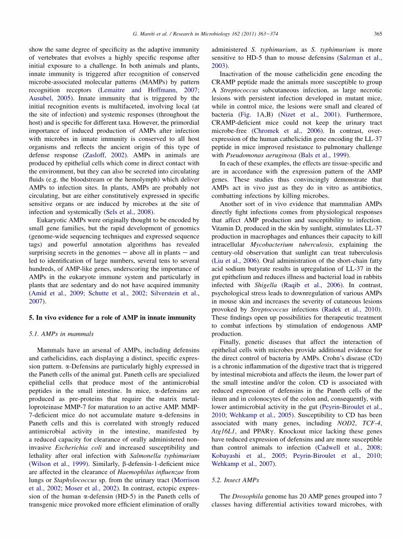
Inactivation of the mouse cathelicidin gene encoding theCRAMP peptide made the animals more susceptible to groupA Streptococcus subcutaneous infection, as large necroticlesions with persistent infection developed in mutant mice,while in control mice, the lesions were small and cleared ofbacteria (Fig. 1A,B) (Nizet et al., 2001). Furthermore,CRAMP-deficient mice could not keep the urinary tractmicrobe-free (Chromek et al., 2006). In contrast, over-expression of the human cathelicidin gene encoding the LL-37peptide in mice improved resistance to pulmonary challengewith Pseudomonas aeruginosa (Bals et al., 1999).
In each of these examples, the effects are tissue-specific andare in accordance with the expression pattern of the AMPgenes. These studies thus convincingly demonstrate thatAMPs act in vivo just as they do in vitro as antibiotics,combatting infections by killing microbes.
Another sort of in vivo evidence that mammalian AMPsdirectly fight infections comes from physiological responsesthat affect AMP production and susceptibility to infection.Vitamin D, produced in the skin by sunlight, stimulates LL-37production in macrophages and enhances their capacity to killintracellular Mycobacterium tuberculosis, explaining thecentury-old observation that sunlight can treat tuberculosis(Liu et al., 2006). Oral administration of the short-chain fattyacid sodium butyrate results in upregulation of LL-37 in thegut epithelium and reduces illness and bacterial load in rabbitsinfected with Shigella (Raqib et al., 2006). In contrast,psychological stress leads to downregulation of various AMPsin mouse skin and increases the severity of cutaneous lesionsprovoked by Streptococcus infections (Radek et al., 2010).These findings open up possibilities for therapeutic treatmentto combat infections by stimulation of endogenous AMPproduction.
Finally, genetic diseases that affect the interaction ofepithelial cells with microbes provide additional evidence forthe direct control of bacteria by AMPs. Crohn’s disease (CD)is a chronic inflammation of the digestive tract that is triggeredby intestinal microbiota and affects the ileum, the lower part ofthe small intestine and/or the colon. CD is associated withreduced expression of defensins in the Paneth cells of theileum and in colonocytes of the colon and, consequently, withlower antimicrobial activity in the gut (Peyrin-Biroulet et al.,2010; Wehkamp et al., 2005). Susceptibility to CD has beenassociated with many genes, including NOD2, TCF-4,Atg16L1, and PPARg. Knockout mice lacking these geneshave reduced expression of defensins and are more susceptiblethan control animals to infection (Cadwell et al., 2008;Kobayashi et al., 2005; Peyrin-Biroulet et al., 2010;Wehkamp et al., 2007).
5.2. Insect AMPs
The Drosophila genome has 20 AMP genes grouped into 7classes having differential activities toward microbes, with

Fig. 1. Immune effects attributed to AMPs. A,B. Lesions in mouse skin provoked by Streptococcus infection in wild-type (A, arrows) and cathelicidin-deficient (Cnlp-
null) mice (B). Reprinted by permission from Macmillan Publishers Ltd: Nature (Nizet et al., 2001), copyright (2001). C. Expression of defensin-GFP fusion in
transgenic Drosophila flies. The systemic response of the flies to infection with bacteria (þ) can be observed as a strong green fluorescent signal. Reprinted from
Immunity, Tzou et al., 2000, Copyright (2000), with permission from Elsevier. D. Infection of a mutant fly unable to produce antifungal AMPs (deficient for the Toll
receptor; Tl-deficient) with Aspergillus fumigatus. Death is associated with uncontrolled development of fungal hyphae covering the fly’s body (arrow). Reprinted
fromCell, Lemaitre et al., 1996, Copyright (1996), with permission fromElsevier. E. Transgenic potato plants (FR13-15, FR-11, FR13-08) overexpressing aMedicago
sativa antifungal AMP (alfAFP) at increasing levels are protected from infection by the fungal pathogen Verticillium dahliae in a field test, while control plants (RB)
die. Reprinted by permission fromMacmillan Publishers Ltd: Nature Biotechnology (Gao et al., 2000), copyright (2000). F,G. Lesions in the skin of mice produced by
wild-type (F) and an AMP-resistant Streptococcusmutant (crgR-deficient) (G). Reprinted by permission fromMacmillan Publishers Ltd: Nature (Nizet et al., 2001),
Copyright (2001).
366 G. Maroti et al. / Research in Microbiology 162 (2011) 363e374
some being more active toward Gram-negative bacteria,others toward Gram-positive bacteria or fungi. The expressionof these peptides is inducible in the fat body (the functionalequivalent of the liver) which secretes AMPs in the hemo-lymph circulatory system (Fig. 1C) as well as in epithelialcells in a tissue-specific manner (Tzou et al., 2000). Theexpression of all AMP genes is dependent on Toll and/or Imdpathways that link perception of MAMPs by pattern recog-nition receptors to AMP gene transcription (Lemaitre andHoffmann, 2007). Mutants in the Toll or Imd pathway ordouble mutants in both pathways which do not express anyAMPs are highly susceptible to systemic infections (Fig. 1D)(Hedengren et al., 1999; Lemaitre et al., 1996). The ectopicexpression of individual AMP genes rescued the suscepti-bility of the double mutants, and the rescued activity intransgenic flies correlated with the known specificity ofAMPs toward Gram-negative bacteria, Gram-positive bacteriaand fungi (Tzou et al., 2002). This demonstrates theprimordial role of AMPs in fighting systemic infections of thehemolymph.
However, despite the fact that AMP gene expression isinduced in the gut epithelia of wild-type flies upon infection(Tzou et al., 2000), Ryu and colleagues found that they play no
dominant role in gut immunity after infection with yeast-contaminated food (Ryu et al., 2006). Instead, dynamicproduction of reactive oxygen species (ROS) generated bya NADPH oxidase was found to be critical for gut immunity(Ha et al., 2005). Nevertheless, AMPs provide an essentialcomplement to ROS-mediated gut defense in the case ofinfections that escape killing by ROS (Ryu et al., 2006), andAMP production in the gut has a critical role against food-borne infections with Gram-negative bacteria (Liehl et al.,2006). Thus, the relative contribution of AMPs and ROSmight depend on the infecting microorganism.
Careful quantification of clearance of infecting bacteria andinduction of AMP activity in the beetle Tenebrio molitorshowed that the vast majority of invading bacteria are alreadyeliminated when induction of AMP activity starts (Haine et al.,2008). This suggests that the first immediate defense linecomprises phagocytosis of bacteria by hemocytes and theirkilling by ROS. The inducible AMPs are then the second lineof defense that serves to eliminate residual microbes whichescaped the first wave of immune effectors. In spite ofcontinuous AMP production, bacteria remained in the insectseven for several weeks (Haine et al., 2008), suggesting thatAMPs not only kill in acute infections but, during chronic

367G. Maroti et al. / Research in Microbiology 162 (2011) 363e374
infections, might also alter the physiology of the bacteria,reducing their pathogenicity.
5.3. Plant AMPs
Plants built their innate immune system against microbialattack via several lines of protection, including local andsystemic production of secondary metabolites, proteins andROS with antimicrobial activity. Plants inhibit spreading ofpathogens by constructing physical barriers with lignin andpolysaccharides and by a programmed-cell-death-type reactioncalled the hypersensitive response. The plant immune systemalso relies on production of various classes of AMPs includingdefensins, thionins, and lipid transfer proteins (Sels et al.,2008). Several lines of evidence strongly suggest that thesepeptides indeed contribute to plant innate immunity. The plantAMPs have the capability to inhibit in vitro growth of bacteriaand/or fungi. The peptides are secreted and can thus attackpathogens before infecting cells. The genes are often expressedconstitutively, principally in flowers and seeds, the reproductivetissues that are particularly sensitive to infections. AMPexpression can also be induced locally and systemically uponpathogen infection. The few studies that report on knockout orknock-down plants of AMP genes failed to detect any alteredphenotype upon infection (De Coninck et al., 2010; Stotz et al.,2009), but there are many examples of constitutive AMPoverexpression in plants that resulted in significantly improvedresistance (Fig. 1E). The absence of phenotype in singleknockout AMP mutants is not surprising due to the possibleredundancy of effectors, especially in light of the high numberof putative AMP genes in plants. An interesting approach toevaluating the potential contribution of AMPs in plant defensecould be provided by the constitutive expression of AMP genesin an immune-deficient genetic background.
Most plant AMPs are characterized by typical arrange-ments of cysteine residues and belong to an extremely largegroup of small cysteine-rich peptides (CRPs) identified largelyby bioinformatic analyses of genome sequences and expressedsequence tags (Silverstein et al., 2007). For example, Arabi-dopsis thaliana has a surprising prolific 825 CRP genes, whilerice has 598 of them. These genes are subject to divergentselection and the arrangement of the genes on the genome inclusters is compatible with a high capacity for evolution ofthese genes so as to generate sequence diversity, new peptidestructures and fusions with other proteins. Naturally, for thelarge majority of these genes, no functions have been identi-fied. Nevertheless, this abundance of AMP-like genes suggeststhat plants have a formidable repertoire of AMPs to fightpathogens as well as the capacity to evolve new AMPs withnew specificities. Remarkably, plants have also developed newfunctions for CRPs other than immunity. For example, severaldefensin-like genes are involved in complex signalingprocesses between male and female gametophytes (Schopferet al., 1999; Okuda et al., 2009; Amien et al., 2010;Higashiyama, 2010), and other CRPs are involved incontrolling the density of stomata, pores in the leaves for gasexchange (Sugano et al., 2010).
5.4. Bacterial resistance mechanisms
Another set of arguments highlighting the critical functionof AMPs is provided by bacterial genetic experiments showingthat the degree of resistance or sensitivity of a pathogen toAMPs produced by the host strongly correlates with the path-ogenicity of the microbe. Because most AMPs are cationic andinteract initially with negatively charged bacterial membranes,pathogenic bacteria, during infection and in response to thehost environment, can modify their anionic lipids or wallcomponents to reduce the net negative charge of their envelope(Peschel and Sahl, 2006). This results in decreased affinity ofthe AMPs for their target and resistance of the pathogen toAMPs. Pathogenic g-proteobacteria for mammals, insects andplants are equipped with a two-component regulatory system,PhoPQ, that functions as a sensor for the host environment and,upon activation, initiates a transcriptional program leading tomodifications in the lipopolysaccharide (LPS) constituent ofthe bacterial outer membrane and increased AMP resistance. InSalmonella, PhoPQ-mediated LPS modifications are essentialfor survival in macrophages and resistance to AMPs producedby these host cells (Fields et al., 1989; Rosenberger et al.,2004). In the plant pathogen Erwinia chrysanthemi or theinsect pathogen Photorhabdus luminescens, the PhoPQ regu-lator also confers resistance to AMPs and thereby increasessurvival in the host and bacterial virulence (Llama-Palacioset al., 2003; Derzelle et al., 2004).
Gram-positive pathogens such as group A and B Strepto-coccus and Staphylococcus species also sense the host envi-ronment using various two-component regulators to induceAMP resistance by surface modifications. The negative chargeof lipoteichoic acids of the cell wall or phospholipids in themembranes is neutralized through the action of DltABCD orMprF gene products, respectively. DltABCD or MprF mutantsin these bacteria have lost their AMP resistance and haveattenuated virulence (Peschel et al., 2001; Kristian et al.,2003).
Remarkably, AMPs themselves are among the host factorsthat are directly recognized by two-component sensors inmany bacteria. For instance, the Salmonella PhoPQ sensor canrecognize cathelicidin LL-37, thereby stimulating LPS modi-fications (Bader et al., 2005). The Staphylococcus Aps sensorrecognizes cationic AMPs such as human b-defensin andLL-37 upon which the DltABCD andMprF genes are activated(Li et al., 2007). In group A Streptococcus, CsrRS (also knownas CovRS) recognizes AMPs and stimulates virulence factorsand AMP resistance (Froehlich et al., 2009; Gryllos et al.,2008).
Finally, it is worth pointing out that bacteria can useadditional resistance strategies such as expulsion of AMPs bypumps, cleavage by proteases or trapping by extracellularbinding proteins or anionic extracellular polysaccharides(Peschel and Sahl, 2006). Some of these mechanisms havebeen shown to be associated with increased bacterial virulence(Lauth et al., 2009; Cole et al., 2010).
The picture that emerges from these examples is that manypathogens have a certain degree of resistance to AMPs that

368 G. Maroti et al. / Research in Microbiology 162 (2011) 363e374
they encounter inside the host during infection, and that AMPresistance is an integral part of the pathogenicity of thesebacteria. AMPs and AMP resistance have likely co-evolved ina continuous arms race: pathogen AMP resistance may haveshaped the AMP repertoire in hosts and, conversely, AMPs ofinnate immunity may have shaped the repertoire of virulencefactors (Peschel and Sahl, 2006).
One study reported a pathogen mutant in which higherAMP resistance was coupled with greater virulence. AStreptococcus pyogenes mutant was isolated that could growin the presence of the AMP cathelicidin (Nizet et al., 2001). Itis worth mentioning that this mutant had reduced growthcompared to the parent strain, indicating a fitness cost asso-ciated with AMP resistance. The mutant produced largernecrotic lesions on the skin of infected mice than the wild-typestrain, indicating increased virulence (Fig. 1F,G). Thusbacterial resistance to cathelicidin increases disease symptomsin a similar way as does inactivation of the cathelicidin gene inthe host (see above; Fig. 1A,B,F,G).
6. Symbiotic interactions
Not all close encounters of plants or animals with micro-organisms are sources of conflict, requiring rapid eliminationof the microorganism. Most organisms maintain symbioticrelations with beneficial microbes. In such associations, themicroorganisms form chronic infections within the host, andoften the host may even actively participate in the infectionprocess by creating a selective niche for the symbiont. In thehost, symbiotic interactions affect nutrient acquisition, defenseagainst enemies and immunity, development or reproduction.In most cases, the benefit to the microorganism is privilegedacquisition of nutrients and a growth niche. Symbionts canreside extracellularly, in luminal spaces between cells andtissues, or live mostly an intracellular existence (endosymbi-onts), but they are always in close association with the cells ofthe host. While a key role of AMPs in management of path-ogenic relations is intuitively understandable, AMP control ofsymbiotic relations is less so. A number of recent studiesdescribe how AMPs are employed to keep symbiotic bacteriain check.
6.1. AMPs control differentiation of nitrogen-fixingendosymbionts in legume plants
Among the most economically and ecologically importantsymbiotic associations of plants are the interactions oflegumes with nitrogen-fixing bacteria known as rhizobia. Thesymbiosis leads to formation of new organs, the root nodules(Fig. 2A). These organs house millions of endosymbioticrhizobia (Fig. 2B,D). Within symbiotic nodule cells, therhizobia become capable of reducing atmospheric nitrogen toammonium, which is transferred to the plant and used for itsgrowth. These endosymbiotic rhizobia are called bacteroids.They are differentiated bacteria, adapted to symbiotic life andnitrogen fixation with altered physiology and metabolism. Insome legumes, as in the model plant Medicago truncatula,
bacteroids are dramatically different from soil-dwellingRhizobium bacteria, as they are much larger, elongated orbranched cells with highly amplified genome content andincreased membrane permeability (Fig. 3C,D). These bacte-roids are incapable of cell division and reproduction. Thus,they are irreversibly differentiated, non-cultivable livingbacteria (Mergaert et al., 2006). Although this terminaldifferentiation of bacteroids is not observed in all legumes andis therefore not essential per se for symbiotic nitrogen fixation,it improves the symbiotic efficiency of the bacteroids (Oonoand Denison, 2010).
In M. truncatula, symbiotic nodule cells produce nodule-specific AMPs of a particular family called NCR for nodule-specific cysteine-rich peptides (Mergaert et al., 2003; Alunniet al., 2007). The NCR gene family in M. truncatula is quiteremarkable because it consists of several hundred genes whichare all exclusively expressed in nodules, and for the testedcases, expression was restricted to Rhizobium-infectedsymbiotic nodule cells. The NCR peptides were found to beresponsible for the terminal differentiated state of the endo-symbiont Sinorhizobium meliloti in M. truncatula nodules(Van de Velde et al., 2010). NCRs are transported to thebacteroids (Fig. 3EeG) and some of the NCRs are localized inthe cytosol of bacteroids, indicating that they enter thebacterial cytosol and likely have intracellular bacterial targets.Obstruction of NCR transport in an exocytosis mutant ofM. truncatula correlated with the absence of terminal bacterialdifferentiation (Fig. 3HeK). In contrast, ectopic expression ofNCRs in a legume lacking NCRs as well as terminal bacteroiddifferentiation or challenge of cultured rhizobia with purepeptides provoked symptoms of terminal differentiation.
NCRs resemble AMPs described in plants and otherorganisms, and analysis of in vitro NCR activity demonstratedthat some NCR peptides indeed possess genuine antimicrobialproperties and effectively kill both Gram-positive and Gram-negative bacteria at concentrations similar to those describedfor most classical antimicrobial peptides. The in vivo and invitro effects of NCRs on S. meliloti are dramatically different.The peptides quickly kill the rhizobia in vitro in strikingcontrast to the nodule where bacteroids, despite their inhibi-tion for growth, maintain active metabolism for nitrogenfixation. This could be explained by a concerted in vivo actionof several tens or hundreds of peptides, each likely present atvery low concentrations, hardly comparable to the in vitroeffect of an externally applied peptide added at high concen-trations. Moreover, particular conditions prevalent in nodules,such as the low free oxygen concentration, needed for activityof the oxygen-sensitive nitrogenase enzyme, could modulatethe bacterial responses to the NCRs in such a way that thebacteroids remain alive, although with complete loss of theirreproductive capacity. Some NCR peptides inhibit bacterialdivision in vivo and in vitro, leading to cell elongation. SuchNCRs were localized at the division site of S. meliloti cells,suggesting that these peptides may interfere with bacterial celldivision machinery. However, the high sequence variety ofNCRs suggests diversity in their functions, mode of actionsand bacterial targets interfering with different aspects of

Fig. 2. AMPs controlling differentiation of Rhizobium endosymbionts in legume nodules A. Nitrogen-fixing nodules formed on the roots of the legume M.
truncatula by infection with the bacterium S. meliloti. B. Longitudinal nodule section showing only the tip region where the small dividing apical cells form the
nodule meristem and provide constant nodule growth. The underlying cells stopped dividing and grow and differentiate gradually by endoreduplication after
infection with rhizobia. Mature nodule cells (red arrows) are entirely filled with nitrogen-fixing rhizobia. C. Drawing from 1888 by Beyerinck, one of the pioneers
of microbiology, documenting the differentiation steps of rhizobia in vetch nodules; from rod-shaped to highly elongated and branched bacteria, today known to be
provoked by nodule-specific AMPs. D. A close-up of a mature wild-type symbiotic nodule cell, stained with toluidine blue, containing thousands of differentiated
rhizobia elongated up to 10 mm. EeG. Localization of NCR peptides in a wild-type nodule cell by fluorescence microscopy. NCR peptides are visible by red
fluorescence of NCR-mCherry fusion and co-localize with rhizobia stained green with DNA stain SYTO13. H. Close-up of a nodule cell in a M. truncatula mutant
369G. Maroti et al. / Research in Microbiology 162 (2011) 363e374

Fig. 3. a-Defensin production by Paneth cells regulates intestinal microbial ecology. A. The epithelium of the small intestine is covered with villi surrounded by
crypts of Lieberkuhn. The inset shows a crypt with Paneth cells at its base which secrete defensin-rich granules into the lumen of the crypt. Reprinted by permission
from Macmillan Publishers Ltd: Nature Immunology (Ganz, 2000), Copyright (2000). B. Modulating a-defensin secretion by Paneth cells in transgenic mice alters
microbial homeostasis. DEFA5 (human a-defensin 5) homozygous (þ/þ) or hemizygous (þ/�) transgenic mice produce higher-than-normal levels of a-defensin.
Mmp-7 mutants (�/�) or heterozygous mice (þ/�) secrete no or less than normal defensins. The total bacterial numbers in the intestine are not affected in the five
models. A group of bacteria known as the Firmicutes becomes significantly underrepresented as defensin levels increase, while bacteria of the Bacteroides group
followed the opposite trend (Salzman et al., 2010). C. Bacterial population in the small intestine of wild-type, DEFA5 hemizygous (þ/�) and homozygous (þ/þ)
mice shows that the population shifts gradually from small bacilli and cocci towards elongated bacteria. Reproduced fromWehkamp et al., 2005, Copyright (2005),
The National Academy of Sciences.
370 G. Maroti et al. / Research in Microbiology 162 (2011) 363e374
bacteroid metabolism and perhaps even optimizing the effi-ciency of nitrogen fixation. Another raison d’etre for the highdiversity of NCR peptides could be adaptation to the highdiversity of rhizobia in soils. Analysis of the sequence of NCRpeptides showed that they are subject to diversifying evolutionwhich is compatible with such a hypothesis (Alunni et al.,2007). Moreover, it is remarkable that plants generallycontain hundreds of AMP-like CRP genes, independently oftheir capacity to form symbiosis (Silverstein et al., 2007). Thishigh diversity also remains hitherto unexplained.
As the NCRs are expressed only in nodules and are notinduced during treatment ofM. truncatula with pathogens, it isunlikely that they play a general role in plant defense. On theother hand, they are similar to AMP genes of plant innateimmunity. Thus, despite the fact that the interaction withrhizobia is symbiotic, legumes like M. truncatula adopteffectors of the innate immune system to dominate theirendosymbionts in order to maximize their own profits.
6.2. Control of the gut microbiota by AMPs
Another well-studied and highly important example ofsymbiosis is the interaction of the gut with the microbiota,
(dnf1) where rhizobia infect nodule cells but do not differentiate and are visible as
system that is required for transport to the intracellular bacteroids. IeK. NCR lo
intracellular rhizobia (green), but remain in the endoplasmic reticulum because dnf1
DeK are reproduced from Van de Velde et al., 2010. Reprinted with permission f
a community of microorganisms present in healthy individualsand essential for physiology and health. Notably, the intestinalbacteria enhance digestive efficiency by producing largequantities of hydrolytic enzymes and thereby increase energyharvest from food. But the microbiota also promotes celldifferentiation in the intestine and protection against patho-gens. It is a challenge for the gut to maintain stability of themicrobiota without inducing inflammation, while maintainingthe ability to clear intestinal infections. This is called intestinalhomeostasis. Perturbations in homeostasis are the basis ofvarious diseases such as obesity, diabetes and inflammatorybowel disease such as CD. While homeostasis is complex,relying on several immunological mechanisms (Hooper andMacpherson, 2010), some studies also revealed how AMPsproduced by the epithelia contribute to maintaining the pop-ulation structure of the microbiota.
Unlike the complex human gut, the Drosophila gut isdominated by only five species. These commensals chroni-cally activate the Imd pathway but without activating AMPexpression (Ryu et al., 2008). The AMPs are activated by thepathway only upon pathogen infections. Repression of AMPgenes in the presence of commensal bacteria in the gut ismediated by the homeobox transcription factor called caudal.
small dots in the plant cell. The dnf1 mutant lacks a nodule-specific exocytosis
calization in the dnf1 nodule cells. NCRs (red) do not co-localize with the
mutation blocks NCR transport. Scale bars in D, E, H and I are 10 mm. Images
rom AAAS.

371G. Maroti et al. / Research in Microbiology 162 (2011) 363e374
Inactivation of caudal provoked abnormally high production ofAMPs, with a dramatic shift in the microbiota community asa consequence: a minor species in the wild-type gut emergedas a dominant one in mutant flies. Ectopic AMP expression intransgenic flies had the same result. The dominance of thissingle gut microbe eventually led to gut cell apoptosis andmortality. Thus, a controlled and balanced level of AMPexpression under healthy as well as pathogenic conditions isessential for maintaining normal flora and homeostasis in thefly gut (Ryu et al., 2008).
Each human being harbors in the gut lumen an estimated100 trillion intestinal bacteria belonging to more than 100different species (Qin et al., 2010). The intestinal epithelia mustprevent these microbiota from penetrating the underlyingtissues. The epithelia are covered with a thin mucus layer thatis kept nearly bacteria-free, while bacteria are abundant in theabove-lying lumen (Hooper and Macpherson, 2010). In thesmall intestine, this is accomplished by the Paneth cells whichsecrete into the gut AMPs and other antimicrobial proteins(Kobayashi et al., 2005; Vaishnava et al., 2008) (Fig. 3A).When Paneth cells are specifically ablated in transgenic miceexpressing a toxin under the control of a Paneth cell-specificpromoter, both the commensals and pathogens penetrate themucosal barrier and epithelial tissues (Vaishnava et al., 2008).Thus, the AMPs produced by Paneth cells contribute tohomeostasis by limiting contact between the microbiota andepithelial tissues. But, in addition, these AMPs also regulate thecomposition of the microbiota in the lumen. A series of fivemouse models were created in which the Paneth cells producedeither higher- or lower-than-normal levels of a-defensins(Salzman et al., 2010) (Fig. 3B). These models were wild-typemice, together with transgenic mice, homozygous or hemi-zygous for transgene HD-5 expressing human a-defensin(DEFA5 mice) (Salzman et al., 2003) and mice, heterozygousor homozygous for a mutated Mmp-7 allele, required for pro-cessing of mouse a-defensin (Wilson et al., 1999). Analysis ofthe composition of the microbiota in these complementarymodels with metagenomic methods revealed significantchanges in the microbiota composition of the small intestine,but not in total bacterial numbers (Fig. 3B) (Salzman et al.,2010). In particular, bacteria known as Firmicutes decreasedwith higher defensin production by Paneth cells, while Bac-teroidetes followed an opposite trend (Fig. 3B). This is inagreement with an earlier study in DEFA5 mice that founda shift in the bacterial population in the gut via microscopicobservation (Fig. 3C) (Wehkamp et al., 2005). Thus Panethcells and a-defensins exert not only protection from pathogens,but also control of homeostasis of the intestinal microbiota, ina similar way as what was described in flies.
7. Concluding remarks
Nature seems to be an unlimited source of peptides withantimicrobial activity, and AMPs are undoubtedly attractivesources for the development of new antibiotics. These peptidesare diverse in sequence and in structure. At present, severalhundred AMPs have been described in the literature and
databases, but the advent of high-throughput genomesequencing and the concomitant rapid development ofbioinformatic tools made rapid filtering and proper annotationof sequenced genomes possible for short genes, potentiallyencoding AMPs. Large numbers of such AMP candidates havebeen discovered in genomes of both prokaryotes andeukaryotes, as illustrated by the spectacular example of CRPsin plant genomes (Silverstein et al., 2007). Of course, effortson experimental studies are not able to keep pace with insilico analyses; nevertheless, our knowledge of possible anti-microbial activities and in vivo roles of these peptides iscontinually expanding.
Another interesting feature of AMPs is their mode of action.Traditional antibiotics are generally designed to target a specificessential microbial molecule with the highest possible affinity.This has the drawback of permitting bacteria to rather easilydevelop resistance. Nature has apparently followed a differentstrategy in the evolution of AMPs. Although the exact mode ofaction ofmost AMPs remains to be defined, the view is emergingthat AMPs have multiple low-affinity interactions, both outsideand inside the bacterial cell, and including non-protein targets.The existence of multiple low-affinity targets likely thwarts thedevelopment of bacterial resistance.
Nevertheless, partial resistance to AMPs exists in present-day pathogens. However, these mechanisms have evolved overa long evolutionary period. It is unlikely that in vivo adapta-tion to natural AMPs can appear quickly, since microbes areexposed to a wide range of AMPs that, moreover, may varybetween species, between individuals within a single speciesand between tissues within a single host. However, it ispossible to obtain resistance to AMPs after in vitro exposure toAMPs in much shorter time under laboratory conditions (Nizetet al., 2001; Perron et al., 2006) and this can lead to increasedpathogenicity. Thus, therapeutic application of high concen-trations of a single AMP would almost certainly cause specificadaptations and the appearance of resistance mechanisms. Thedevelopment of therapeutic applications of AMPs needs to beaccompanied by careful evaluation of the consequences ofresistance to AMPs. Will resistance to therapeutic peptidesalso confer resistance to natural AMPs? And will resistance beaccompanied by increased virulence? What are the fitnesscosts, if any, associated with AMP resistance? The numerousexamples of genetic inactivation of AMP activity in animals,leading to dramatic increases in sensitivity to pathogens,constitute a warning that these organisms need their fullarsenal of AMPs to cope with invading microorganisms. Fullunderstanding of how symbionts such as gut microbiota(Salzman et al., 2010), nitrogen-fixing rhizobia in legumenodules (Van de Velde et al., 2010) or pathogens that persist inthe insect hemolymph (Haine et al., 2008) survive continuousexposure to host AMPs should provide further clues forenabling intelligent use of AMP therapeutics.
Acknowledgments
Work in our laboratories is supported by the French AgenceNationale de la Recherche, grant ANR-09-BLAN-0396-01 and

372 G. Maroti et al. / Research in Microbiology 162 (2011) 363e374
the Hungarian National Office for Research and Technology,grants OMFB-00441/2007 and OMFB-00128/2010.
References
Alunni, B., Kevei, Z., Redondo-Nieto, M., Kondorosi, A., Mergaert, P.,
Kondorosi, E., 2007. Genomic organization and evolutionary insights on
GRP and NCR genes, two large nodule-specific gene families in Medicago
truncatula. Mol. Plant Microbe Interact. 20, 1138e1148.
Amid, C., Rehaume, L.M., Brown, K.L., Gilbert, J.G., Dougan, G.,
Hancock, R.E., Harrow, J.L., 2009. Manual annotation and analysis of the
defensin gene cluster in the C57BL/6J mouse reference genome. BMC
Genomics 10, 606.
Amien, S., Kliwer, I., Marton, M.L., Debener, T., Geiger, D., Becker, D.,
Dresselhaus, T., 2010. Defensin-like ZmES4 mediates pollen tube burst in
maize via opening of the potassium channel KZM1. PLoS Biol. 8,
e1000388.
Ausubel, F.M., 2005. Are innate immune signaling pathways in plants and
animals conserved? Nat. Immunol. 6, 973e979.Bader, M.W., Sanowar, S., Daley, M.E., Schneider, A.R., Cho, U., Xu, W.,
Klevit, R.E., Le Moual, H., Miller, S.I., 2005. Recognition of antimicrobial
peptides by a bacterial sensor kinase. Cell 122, 461e472.
Bals, R., Weiner, D.J., Moscioni, A.D., Meegalla, R.L., Wilson, J.M., 1999.
Augmentation of innate host defense by expression of a cathelicidin
antimicrobial peptide. Infect. Immun. 67, 6084e6089.
Bell, G., Gouyon, P.H., 2003. Arming the enemy: the evolution of resistance to
self-proteins. Microbiology 149, 1367e1375.Brogden, K.A., 2005. Antimicrobial peptides: pore formers or metabolic
inhibitors in bacteria? Nat. Rev. Microbiol. 3, 238e250.
Buckling, A., Brockhurst, M., 2005. RAMP resistance. Nature 438, 170e171.Cadwell, K., Liu, J.Y., Brown, S.L., Miyoshi, H., Loh, J., Lennerz, J.K.,
Kishi, C., Kc, W., Carrero, J.A., Hunt, S., Stone, C.D., Brunt, E.M.,
Xavier, R.J., Sleckman, B.P., Li, E., Mizushima, N., Stappenbeck, T.S.,
Virgin 4th, H.W., 2008. A key role for autophagy and the autophagy gene
Atg16l1 in mouse and human intestinal Paneth cells. Nature 456, 259e263.
Chauhan, A., Madiraju, M.V., Fol, M., Lofton, H., Maloney, E., Reynolds, R.,
Rajagopalan, M., 2006. Mycobacterium tuberculosis cells growing in
macrophages are filamentous and deficient in FtsZ rings. J. Bacteriol. 188,
1856e1865.
Chromek, M., Slamova, Z., Bergman, P., Kovacs, L., Podracka, L., Ehren, I.,
Hokfelt, T., Gudmundsson, G.H., Gallo, R.L., Agerberth, B., Brauner, A.,
2006. The antimicrobial peptide cathelicidin protects the urinary tract
against invasive bacterial infection. Nat. Med. 12, 636e641.
Cole, J.N., Pence, M.A., von Kockritz-Blickwede, M., Hollands, A., Gallo, R.
L., Walker, M.J., Nizet, V., 2010. M protein and hyaluronic acid capsule
are essential for in vivo selection of covRS mutations characteristic of
invasive serotype M1T1 group A Streptococcus. mBio 1, e00191-10.
Czaran, T.L., Hoekstra, R.F., Pagie, L., 2002. Chemical warfare between
microbes promotes biodiversity. Proc. Natl. Acad. Sci. U S A 99,
786e790.
De Coninck, B.M., Sels, J., Venmans, E., Thys, W., Goderis, I.J., Carron, D.,
Delaure, S.L., Cammue, B.P., De Bolle, M.F., Mathys, J., 2010. Arabi-
dopsis thaliana plant defensin AtPDF1.1 is involved in the plant response
to biotic stress. New Phytol. 187, 1075e1088.
Derzelle, S., Turlin, E., Duchaud, E., Pages, S., Kunst, F., Givaudan, A.,
Danchin, A., 2004. The PhoPePhoQ two-component regulatory system of
Photorhabdus luminescens is essential for virulence in insects. J. Bacteriol.
186, 1270e1279.
Fields, P.I., Groisman, E.A., Heffron, F., 1989. A Salmonella locus that
controls resistance to microbicidal proteins from phagocytic cells. Science
243, 1059e1062.
Fierer, N., Breitbart, M., Nulton, J., Salamon, P., Lozupone, C., Jones, R.,
Robeson, M., Edwards, R.A., Felts, B., Rayhawk, S., Knight, R.,
Rohwer, F., Jackson, R.B., 2007. Metagenomic and small-subunit rRNA
analyses reveal the genetic diversity of bacteria, archaea, fungi, and viruses
in soil. Appl. Environ. Microbiol. 73, 7059e7066.
Froehlich, B.J., Bates, C., Scott, J.R., 2009. Streptococcus pyogenes CovRS
mediates growth in iron starvation and in the presence of the human
cationic antimicrobial peptide LL-37. J. Bacteriol. 191, 673e677.
Ganz, T., 2000. Paneth cells e guardians of the gut cell hatchery. Nat.
Immunol. 1, 99e100.Ganz, T., 2003. Defensins: antimicrobial peptides of innate immunity. Nat.
Rev. Immunol. 3, 710e720.
Gao, A.G., Hakimi, S.M., Mittanck, C.A., Wu, Y., Woerner, B.M., Stark, D.M.,
Shah, D.M., Liang, J., Rommens, C.M., 2000. Fungal pathogen protection
in potato by expression of a plant defensin peptide. Nat. Biotechnol. 18,
1307e1310.
Gordon, D.M., Riley, M.A., Pinou, T., 1998. Temporal changes in the
frequency of colicinogeny in Escherichia coli from house mice. Micro-
biology 144, 2233e2240.
Gryllos, I., Tran-Winkler, H.J., Cheng, M.F., Chung, H., Bolcome 3rd, R.,
Lu, W., Lehrer, R.I., Wessels, M.R., 2008. Induction of group A Strepto-
coccus virulence by a human antimicrobial peptide. Proc. Natl. Acad. Sci.
U S A 105, 16755e16760.
Ha, E.M., Oh, C.T., Bae, Y.S., Lee, W.J., 2005. A direct role for dual oxidase
in drosophila gut immunity. Science 310, 847e850.
Haine, E.R., Moret, Y., Siva-Jothy, M.T., Rolff, J., 2008. Antimicrobial defense
and persistent infection in insects. Science 322, 1257e1259.
Hale, J.D., Hancock, R.E., 2007. Alternative mechanisms of action of cationic
antimicrobial peptides on bacteria. Expert Rev. Anti. Infect. Ther. 5,
951e959.
Hancock, R.E., Sahl, H.G., 2006. Antimicrobial and host-defense peptides
as new anti-infective therapeutic strategies. Nat. Biotechnol. 24,
1551e1557.
Handler, A.A., Lim, J.E., Losick, R., 2008. Peptide inhibitor of cytokinesis
during sporulation in Bacillus subtilis. Mol. Microbiol. 68, 588e599.
Hasper, H.E., Kramer, N.E., Smith, J.L., Hillman, J.D., Zachariah, C.,
Kuipers, O.P., de Kruijff, B., Breukink, E., 2006. An alternative bacteri-
cidal mechanism of action for lantibiotic peptides that target lipid II.
Science 313, 1636e1637.Hedengren, M., Asling, B., Dushay, M.S., Ando, I., Ekengren, S.,
Wihlborg, M., Hultmark, D., 1999. Relish, a central factor in the control
of humoral but not cellular immunity in Drosophila. Mol. Cell. 4,
827e837.Higashiyama, T., 2010. Peptide signaling in pollen-pistil interactions. Plant
Cell Physiol. 51, 177e189.
Hilpert, K., McLeod, B., Yu, J., Elliott, M.R., Rautenbach, M., Ruden, S.,
Burck, J., Muhle-Gol, C., Ulrich, A.S., Keller, S., Hancock, R.E., 2010.
Short cationic antimicrobial peptides interact with ATP. Antimicrob.
Agents Chemother. 54, 4480e4483.
Hooper, L.V., Macpherson, A.J., 2010. Immune adaptations that maintain
homeostasis with the intestinal microbiota. Nat. Rev. Immunol. 10,
159e169.
Huber, J.A., Mark Welch, D.B., Morrison, H.G., Huse, S.M., Neal, P.R.,
Butterfield, D.A., Sogin, M.L., 2007. Microbial population structures in the
deep marine biosphere. Science 318, 97e100.
Kerr, B., Riley, M.A., Feldman, M.W., Bohannan, B.J., 2002. Local dispersal
promotes biodiversity in a real-life game of rockepaperescissors. Nature
418, 171e174.Kirkup, B.C., Riley, M.A., 2004. Antibiotic-mediated antagonism leads to
a bacterial game of rockepaperescissors in vivo. Nature 428, 412e414.
Kobayashi, K.S., Chamaillard, M., Ogura, Y., Henegariu, O., Inohara, N.,
Nunez, G., Flavell, R.A., 2005. Nod2-dependent regulation of innate and
adaptive immunity in the intestinal tract. Science 307, 731e734.
Kristian, S.A., Durr, M., Van Strijp, J.A., Neumeister, B., Peschel, A., 2003.
MprF-mediated lysinylation of phospholipids in Staphylococcus aureus
leads to protection against oxygen-independent neutrophil killing. Infect.
Immun. 71, 546e549.
Lauth, X., von Kockritz-Blickwede, M., McNamara, C.W., Myskowski, S.,
Zinkernagel, A.S., Beall, B., Ghosh, P., Gallo, R.L., Nizet, V., 2009. M1
protein allows Group A streptococcal survival in phagocyte extracellular
traps through cathelicidin inhibition. J. Innate Immun. 1, 202e214.
Lemaitre, B., Hoffmann, J., 2007. The host defense of Drosophila mela-
nogaster. Annu. Rev. Immunol. 25, 697e743.

373G. Maroti et al. / Research in Microbiology 162 (2011) 363e374
Lemaitre, B., Nicolas, E., Michaut, L., Reichhart, J.M., Hoffmann, J.A., 1996.
The dorsoventral regulatory gene cassette spatzle/Toll/cactus controls the
potent antifungal response in drosophila adults. Cell 86, 973e983.
Li, M., Lai, Y., Villaruz, A.E., Cha, D.J., Sturdevant, D.E., Otto, M., 2007.
Gram-positive three-component antimicrobial peptide-sensing system.
Proc. Natl. Acad. Sci. U S A 104, 9469e9474.
Liehl, P., Blight, M., Vodovar, N., Boccard, F., Lemaitre, B., 2006. Prevalence
of local immune response against oral infection in a Drosophila/Pseudo-
monas infection model. PLoS Pathog. 2, e56.
Liu, P.T., Stenger, S., Li, H., Wenzel, L., Tan, B.H., Krutzik, S.R., Ochoa, M.T.,
Schauber, J., Wu, K., Meinken, C., Kamen, D.L., Wagner, M., Bals, R.,
Steinmeyer, A., Zugel, U., Gallo, R.L., Eisenberg, D., Hewison, M.,
Hollis, B.W., Adams, J.S., Bloom, B.R., Modlin, R.L., 2006. Toll-like
receptor triggering of a vitamin D-mediated human antimicrobial response.
Science 311, 1770e1773.
Llama-Palacios, A., Lopez-Solanilla, E., Poza-Carrion, C., Garcıa-Olmedo, F.,
Rodrıguez-Palenzuela, P., 2003. The Erwinia chrysanthemi phoPephoQ
operon plays an important role in growth at low pH, virulence and bacterial
survival in plant tissue. Mol. Microbiol. 49, 347e357.Lobo, D.S., Pereira, I.B., Fragel-Madeira, L., Medeiros, L.N., Cabral, L.M.,
Faria, J., Bellio, M., Campos, R.C., Linden, R., Kurtenbach, E., 2007.
Antifungal Pisum sativum defensin 1 interacts with Neurospora crassa
cyclin F related to the cell cycle. Biochemistry 46, 987e996.Marlow, V.L., Haag, A.F., Kobayashi, H., Fletcher, V., Scocchi, M., Walker, G.
C., Ferguson, G.P., 2009. Essential role for the BacA protein in the uptake
of a truncated eukaryotic peptide in Sinorhizobium meliloti. J. Bacteriol.
191, 1519e1527.
Mattiuzzo, M., Bandiera, A., Gennaro, R., Benincasa, M., Pacor, S.,
Antcheva, N., Scocchi, M., 2007. Role of the Escherichia coli SbmA in the
antimicrobial activity of proline-rich peptides. Mol. Microbiol. 66,
151e163.
Mergaert, P., Nikovics, K., Kelemen, Z., Maunoury, N., Vaubert, D.,
Kondorosi, A., Kondorosi, E., 2003. A novel family in Medicago trun-
catula consisting of more than 300 nodule-specific genes coding for small,
secreted polypeptides with conserved cysteine motifs. Plant Physiol. 132,
161e173.
Mergaert, P., Uchiumi, T., Alunni, B., Evanno, G., Cheron, A., Catrice, O.,
Mausset, A.E., Barloy-Hubler, F., Galibert, F., Kondorosi, A.,
Kondorosi, E., 2006. Eukaryotic control on bacterial cell cycle and
differentiation in the Rhizobium-legume symbiosis. Proc. Natl. Acad. Sci.
U S A 103, 5230e5235.
Morrison, G., Kilanowski, F., Davidson, D., Dorin, J., 2002. Characterization
of the mouse beta defensin 1, Defb1, mutant mouse model. Infect. Immun.
70, 3053e3060.
Moser, C., Weiner, D.J., Lysenko, E., Bals, R., Weiser, J.N., Wilson, J.M.,
2002. b-Defensin 1 contributes to pulmonary innate immunity in mice.
Infect. Immun. 70, 3068e3072.
Nizet, V., Ohtake, T., Lauth, X., Trowbridge, J., Rudisill, J., Dorschner, R.A.,
Pestonjamasp, V., Piraino, J., Huttner, K., Gallo, R.L., 2001. Innate anti-
microbial peptide protects the skin from invasive bacterial infection.
Nature 414, 454e457.
Okuda, S., Tsutsui, H., Shiina, K., Sprunck, S., Takeuchi, H., Yui, R.,
Kasahara, R.D., Hamamura, Y., Mizukami, A., Susaki, D., Kawano, N.,
Sakakibara, T., Namiki, S., Itoh, K., Otsuka, K., Matsuzaki, M., Nozaki, H.,
Kuroiwa, T., Nakano, A., Kanaoka, M.M., Dresselhaus, T., Sasaki, N.,
Higashiyama, T., 2009. Defensin-like polypeptide LUREs are pollen tube
attractants secreted from synergid cells. Nature 458, 357e361.
Oono, R., Denison, R.F., 2010. Comparing symbiotic efficiency between
swollen versus nonswollen rhizobial bacteroids. Plant Physiol. 154,
1541e1548.Perron, G.G., Zasloff, M., Bell, G., 2006. Experimental evolution of resistance
to an antimicrobial peptide. Proc. Biol. Sci. 273, 251e256.
Peschel, A., Jack, R.W., Otto, M., Collins, L.V., Staubitz, P., Nicholson, G.,
Kalbacher, H., Nieuwenhuizen, W.F., Jung, G., Tarkowski, A., van
Kessel, K.P., van Strijp, J.A., 2001. Staphylococcus aureus resistance to
human defensins and evasion of neutrophil killing via the novel virulence
factor MprF is based on modification of membrane lipids with L-lysine. J.
Exp. Med. 193, 1067e1076.
Peschel, A., Sahl, H.G., 2006. The co-evolution of host cationic antimicrobial
peptides and microbial resistance. Nat. Rev. Microbiol. 4, 529e536.
Peyrin-Biroulet, L., Beisner, J., Wang, G., Nuding, S., Oommen, S.T.,
Kelly, D., Parmentier-Decrucq, E., Dessein, R., Merour, E., Chavatte, P.,
Grandjean, T., Bressenot, A., Desreumaux, P., Colombel, J.F.,
Desvergne, B., Stange, E.F., Wehkamp, J., Chamaillard, M., 2010.
Peroxisome proliferator-activated receptor gamma activation is required
for maintenance of innate antimicrobial immunity in the colon. Proc. Natl.
Acad. Sci. U S A 107, 8772e8777.
Qin, J., Li, R., Raes, J., Arumugam, M., Burgdorf, K.S., Manichanh, C.,
Nielsen, T., Pons, N., Levenez, F., Yamada, T., Mende, D.R., Li, J., Xu, J.,
Li, S., Li, D., Cao, J., Wang, B., Liang, H., Zheng, H., Xie, Y., Tap, J.,
Lepage, P., Bertalan, M., Batto, J.M., Hansen, T., Le Paslier, D.,
Linneberg, A., Nielsen, H.B., Pelletier, E., Renault, P., Sicheritz-Ponten, T.
, Turner, K., Zhu, H., Yu, C., Li, S., Jian, M., Zhou, Y., Li, Y., Zhang, X.,
Li, S., Qin, N., Yang, H., Wang, J., Brunak, S., Dore, J., Guarner, F.,
Kristiansen, K., Pedersen, O., Parkhill, J., Weissenbach, J., MetaHIT
Consortium, Bork, P., Ehrlich, S.D., Wang, J., 2010. A human gut
microbial gene catalogue established by metagenomic sequencing. Nature
464, 59e65.
Radek, K.A., Elias, P.M., Taupenot, L., Mahata, S.K., O’Connor, D.T.,
Gallo, R.L., 2010. Neuroendocrine nicotinic receptor activation increases
susceptibility to bacterial infections by suppressing antimicrobial peptide
production. Cell Host Microbe 7, 277e289.
Raqib, R., Sarker, P., Bergman, P., Ara, G., Lindh, M., Sack, D.A., Nasirul
Islam, K.M., Gudmundsson, G.H., Andersson, J., Agerberth, B., 2006.
Improved outcome in shigellosis associated with butyrate induction of an
endogenous peptide antibiotic. Proc. Natl. Acad. Sci. U S A 103,
9178e9183.
Reichenbach, T., Mobilia, M., Frey, E., 2007. Mobility promotes and jeopar-
dizes biodiversity in rockepaperescissors games. Nature 448,
1046e1049.
Riley, M.A., Gordon, D.M., 1999. The ecological role of bacteriocins in
bacterial competition. Trends Microbiol. 7, 129e133.
Rosenberger, C.M., Gallo, R.L., Finlay, B.B., 2004. Interplay between
antibacterial effectors: a macrophage antimicrobial peptide impairs intra-
cellular Salmonella replication. Proc. Natl. Acad. Sci. U S A 101,
2422e2427.Ryu, J.H., Ha, E.M., Oh, C.T., Seol, J.H., Brey, P.T., Jin, I., Lee, D.G., Kim, J.,
Lee, D., Lee, W.J., 2006. An essential complementary role of NF-kappaB
pathway to microbicidal oxidants in Drosophila gut immunity. EMBO J.
25, 3693e3701.Ryu, J.H., Kim, S.H., Lee, H.Y., Bai, J.Y., Nam, Y.D., Bae, J.W., Lee, D.G.,
Shin, S.C., Ha, E.M., Lee, W.J., 2008. Innate immune homeostasis by the
homeobox gene caudal and commensal-gut mutualism in Drosophila.
Science 319, 777e782.
Salzman, N.H., Ghosh, D., Huttner, K.M., Paterson, Y., Bevins, C.L., 2003.
Protection against enteric salmonellosis in transgenic mice expressing
a human intestinal defensin. Nature 422, 522e526.Salzman, N.H., Hung, K., Haribhai, D., Chu, H., Karlsson-Sjoberg, J.,
Amir, E., Teggatz, P., Barman, M., Hayward, M., Eastwood, D., Stoel, M.,
Zhou, Y., Sodergren, E., Weinstock, G.M., Bevins, C.L., Williams, C.B.,
Bos, N.A., 2010. Enteric defensins are essential regulators of intestinal
microbial ecology. Nat. Immunol. 11, 76e83.
Sass, V., Schneider, T., Wilmes, M., Korner, C., Tossi, A., Novikova, N.,
Shamova, O., Sahl, H.G., 2010. Human b-defensin 3 inhibits cell wall
biosynthesis in Staphylococci. Infect. Immun. 78, 2793e2800.
Schneider, T., Kruse, T., Wimmer, R., Wiedemann, I., Sass, V., Pag, U.,
Jansen, A., Nielsen, A.K., Mygind, P.H., Raventos, D.S., Neve, S.,
Ravn, B., Bonvin, A.M., De Maria, L., Andersen, A.S., Gammelgaard, L.
K., Sahl, H.G., Kristensen, H.H., 2010. Plectasin, a fungal defensin, targets
the bacterial cell wall precursor lipid II. Science 328, 1168e1172.
Schopfer, C.R., Nasrallah, M.E., Nasrallah, J.B., 1999. The male determinant
of self-incompatibility in Brassica. Science 286, 1697e1700.Schutte, B.C., Mitros, J.P., Bartlett, J.A., Walters, J.D., Jia, H.P., Welsh, M.J.,
Casavant, T.L., McCray Jr., P.B., 2002. Discovery of five conserved b-
defensin gene clusters using a computational search strategy. Proc. Natl.
Acad. Sci. U S A 99, 2129e2133.

374 G. Maroti et al. / Research in Microbiology 162 (2011) 363e374
Sels, J., Mathys, J., De Coninck, B.M., Cammue, B.P., De Bolle, M.F., 2008.
Plant pathogenesis-related (PR) proteins: a focus on PR peptides. Plant
Physiol. Biochem. 46, 941e950.
Silverstein, K.A., Moskal Jr., W.A., Wu, H.C., Underwood, B.A., Graham, M.
A., Town, C.D., VandenBosch, K.A., 2007. Small cysteine-rich peptides
resembling antimicrobial peptides have been under-predicted in plants.
Plant J. 51, 262e280.
Stotz, H.U., Spence, B., Wang, Y., 2009. A defensin from tomato with dual
function in defense and development. Plant Mol. Biol. 71, 131e143.
Sugano, S.S., Shimada, T., Imai, Y., Okawa, K., Tamai, A., Mori, M., Hara-
Nishimura, I., 2010. Stomagen positively regulates stomatal density in
Arabidopsis. Nature 463, 241e244.Tait, K., Sutherland, I.W., 2002. Antagonistic interactions amongst bacte-
riocin-producing enteric bacteria in dual species biofilms. J. Appl.
Microbiol. 93, 345e352.
Torsvik, V., Øvreas, L., Thingstad, T.F., 2002. Prokaryotic diversity-magni-
tude, dynamics, and controlling factors. Science 296, 1064e1066.
Tringe, S.G., von Mering, C., Kobayashi, A., Salamov, A.A., Chen, K.,
Chang, H.W., Podar, M., Short, J.M., Mathur, E.J., Detter, J.C., Bork, P.,
Hugenholtz, P., Rubin, E.M., 2005. Comparative metagenomics of
microbial communities. Science 308, 554e557.
Tzou, P., Ohresser, S., Ferrandon, D., Capovilla, M., Reichhart, J.M.,
Lemaitre, B., Jules, A., Hoffmann, J.A., Imler, J.-L., 2000. Tissue-specific
inducible expression of antimicrobial peptide genes in Drosophila surface
epithelia. Immunity 13, 737e748.
Tzou, P., Reichhart, J.M., Lemaitre, B., 2002. Constitutive expression of
a single antimicrobial peptide can restore wild-type resistance to infection
in immunodeficient Drosophila mutants. Proc. Natl. Acad. Sci. U S A 99,
2152e2157.
Vaishnava, S., Behrendt, C.L., Ismail, A.S., Eckmann, L., Hooper, L.V., 2008.
Paneth cells directly sense gut commensals and maintain homeostasis at
the intestinal hostemicrobial interface. Proc. Natl. Acad. Sci. U S A 105,
20858e20863.
Van de Velde, W., Zehirov, G., Szatmari, A., Debreczeny, M., Ishihara, H.,
Kevei, Z., Farkas, A., Mikulass, K., Nagy, A., Tiricz, H., Satiat-
Jeunemaıtre, B., Alunni, B., Bourge, M., Kucho, K., Abe, M., Kereszt, A.,
Maroti, G., Uchiumi, T., Kondorosi, E., Mergaert, P., 2010. Plant peptides
govern terminal differentiation of bacteria in symbiosis. Science 327,
1122e1126.Wehkamp, J., Salzman, N.H., Porter, E., Nuding, S., Weichenthal, M.,
Petras, R.E., Shen, B., Schaeffeler, E., Schwab, M., Linzmeier, R.,
Feathers, R.W., Chu, H., Lima Jr., H., Fellermann, K., Ganz, T., Stange, E.
F., Bevins, C.L., 2005. Reduced Paneth cell a-defensins in ileal Crohn’s
disease. Proc. Natl. Acad. Sci. U S A 102, 18129e18134.
Wehkamp, J., Wang, G., Kubler, I., Nuding, S., Gregorieff, A., Schnabel, A.,
Kays, R.J., Fellermann, K., Burk, O., Schwab, M., Clevers, H., Bevins, C.
L., Stange, E.F., 2007. The Paneth cell a-defensin deficiency of ileal
Crohn’s disease is linked to Wnt/Tcf-4. J. Immunol. 179, 3109e3118.
Wilson, C.L., Ouellette, A.J., Satchell, D.P., Ayabe, T., Lopez-Boado, Y.S.,
Stratman, J.L., Hultgren, S.J., Matrisian, L.M., Parks, W.C., 1999. Regu-
lation of intestinal a-defensin activation by the metalloproteinase matri-
lysin in innate host defense. Science 286, 113e117.
Zasloff, M., 2002. Antimicrobial peptides of multicellular organisms. Nature
415, 389e395.
Related Documents


