Pharmacognosy Reviews | July-December 2013 | Vol 7 | Issue 14 107 Natural proteins: Sources, isolation, characterization and applications Jitendra Y. Nehete, Rajendra S. Bhambar, Minal R. Narkhede 1 , Sonali R. Gawali Departments of Pharmacognosy and 1 Pharmaceutics, MGV’s Pharmacy College, Panchavati, Nashik, India Submitted: 22-03-2013 Revised: 05-04-2013 Published: **-**-**** Address for correspondence: Dr. Jitendra Y. Nehete, Department of Pharmacognosy, MGV’s Pharmacy College, Panchavati, Nashik - 422 003, Maharashtra, India. E-mail: [email protected] Worldwide, plant protein contributes substantially as a food resource because it contains essential amino acids for meeting human physiological requirements. However, many versatile plant proteins are used as medicinal agents as they are produced by using molecular tools of biotechnology. Proteins can be obtained from plants, animals and microorganism cells. The abundant economical proteins can be obtained from plant seeds. These natural proteins are obtained by isolation procedures depending on the physicochemical properties of proteins. Isolation and purification of single protein from cells containing mixtures of unrelated proteins is achievable due to the physical and chemical attributes of proteins. The following characteristics are unique to each protein: Amino acid composition, sequence, subunit structures, size, shape, net charge, isoelectric point, solubility, heat stability and hydrophobicity. Based on these properties, various methods of isolation exist, like salting out and isoionic precipitation. Purification of proteins is quiet challenging and, therefore, several approaches like sodium dodecyl sulfate gel electrophoresis and chromatography are available. Characterization of proteins can be performed by mass spectrometry/liquid chromatography‑mass spectrometry (LC‑MS). The amino acid sequence of a protein can be detected by using tandem mass spectrometry. In this article, a review has been made on the sources, isolation, purification and characterization of natural proteins. Key words: Amino acid, natural proteins, SDS gel electrophoresis, salting out INTRODUCTION For carrying out different body functions, the body regularly needs nutrients like vitamins, minerals, proteins, fiber and carbohydrates, which are obtained from plant or animal sources or both. Among nutrients, the human body requires proteins as the most important compounds because they aid in building cells and tissues and help in repairing the tissues in the body. A high protein diet is recommended for those thinking of building body or muscles. If the body lacks in carbohydrates and fats, the body makes use of proteins for energy production as they are essential for building muscle mass. [1] The term “complete protein” refers to foods that contain all nine essential amino acids in the correct proportion to build protein in the body. In contrast, “incomplete protein” refers to foods that have all essential amino acids but not in the correct proportion, and are termed as “limiting amino acid.” [2] Thus, proteins are not only important in the human body but are also widely used in the industry. Hence, an attempt is made to review naturally obtained proteins and its application in pharmaceutical industries. Building blocks of proteins (Amino acids) Naturally occurring organic compounds containing amino and carboxyl groups, which are the chief constituents of protein and are necessary for human and animal growth and nutrition, are termed as essential amino acids. Hence, essential amino acid-rich food consumption is an option to source these as they are not produced by the human body. [3] Proteins (or polypeptides) are amino acids joined together by peptide bonds. [4] The roles of the various amino acids are highlighted in Table 1. [5] Properties of proteins Solubility Protein solubility properties are summarized as: 1. Forms colloidal solutions in water (due to huge size) PHCOG REV. REVIEW ARTICLE ABSTRACT Access this article online Quick Response Code: Website: www.phcogrev.com DOI: 10.4103/0973-7847.120508

Welcome message from author
This document is posted to help you gain knowledge. Please leave a comment to let me know what you think about it! Share it to your friends and learn new things together.
Transcript
Pharmacognosy Reviews | July-December 2013 | Vol 7 | Issue 14 107
Natural proteins: Sources, isolation, characterization and applicationsJitendra Y. Nehete, Rajendra S. Bhambar, Minal R. Narkhede1, Sonali R. GawaliDepartments of Pharmacognosy and 1Pharmaceutics, MGV’s Pharmacy College, Panchavati, Nashik, India
Submitted: 22-03-2013 Revised: 05-04-2013 Published: **-**-****
Address for correspondence: Dr. Jitendra Y. Nehete, Department of Pharmacognosy, MGV’s Pharmacy College, Panchavati, Nashik - 422 003, Maharashtra, India. E-mail: [email protected]
Worldwide, plant protein contributes substantially as a food resource because it contains essential amino acids for meeting human physiological requirements. However, many versatile plant proteins are used as medicinal agents as they are produced by using molecular tools of biotechnology. Proteins can be obtained from plants, animals and microorganism cells. The abundant economical proteins can be obtained from plant seeds. These natural proteins are obtained by isolation procedures depending on the physicochemical properties of proteins. Isolation and purification of single protein from cells containing mixtures of unrelated proteins is achievable due to the physical and chemical attributes of proteins. The following characteristics are unique to each protein: Amino acid composition, sequence, subunit structures, size, shape, net charge, isoelectric point, solubility, heat stability and hydrophobicity. Based on these properties, various methods of isolation exist, like salting out and isoionic precipitation. Purification of proteins is quiet challenging and, therefore, several approaches like sodium dodecyl sulfate gel electrophoresis and chromatography are available. Characterization of proteins can be performed by mass spectrometry/liquid chromatography‑mass spectrometry (LC‑MS). The amino acid sequence of a protein can be detected by using tandem mass spectrometry. In this article, a review has been made on the sources, isolation, purification and characterization of natural proteins.
Key words: Amino acid, natural proteins, SDS gel electrophoresis, salting out
INTRODUCTION
For carrying out different body functions, the body regularly needsnutrients likevitamins,minerals,proteins,fiber andcarbohydrates, which are obtained from plant or animal sources or both. Among nutrients, the human body requires proteins as the most important compounds because they aid in building cells and tissues and help in repairing the tissues in the body. A high protein diet is recommended for those thinking of building body or muscles. If the body lacks in carbohydrates and fats, the body makes use of proteins for energy production as they are essential for building muscle mass.[1]
The term “complete protein” refers to foods that contain
all nine essential amino acids in the correct proportion to build protein in the body. In contrast, “incomplete protein” refers to foods that have all essential amino acids but not in the correct proportion, and are termed as “limiting amino acid.”[2]
Thus, proteins are not only important in the human body but are also widely used in the industry. Hence, an attempt is made to review naturally obtained proteins and its application in pharmaceutical industries.
Building blocks of proteins (Amino acids)Naturally occurring organic compounds containing amino and carboxyl groups, which are the chief constituents of protein and are necessary for human and animal growth and nutrition, are termed as essential amino acids. Hence, essential amino acid-rich food consumption is an option to source these as they are not produced by the human body.[3] Proteins (or polypeptides) are amino acids joined together by peptide bonds.[4] The roles of the various amino acids are highlighted in Table 1.[5]
Properties of proteinsSolubilityProtein solubility properties are summarized as:1. Forms colloidal solutions in water (due to huge
size)
P H C O G R E V . R E V I E W A R T I C L E
A B S T R A C T
Access this article onlineQuick Response Code: Website:
www.phcogrev.com
DOI:10.4103/0973-7847.120508

Nehete, et al.: Natural proteins
108 Pharmacognosy Reviews | July-December 2013 | Vol 7 | Issue 14
2. Solubility depends on electrostatic charges; net charge depends on number, identity, location of amino acids and pH of solvent
3. It depends upon isoelectric point (range 5-8.5): Isoelectric point depends on seven-charge amino acids, viz. glutamate (δ-carboxyl group), aspartate (ß-carboxyl group), cysteine (thiol group), tyrosine (phenol group), histidine (imidazole side chains), lysine (ε-ammonium group) and arginine (guanidinium group).[5]
Molecular weightProteins’ molecular weight variation depends on the number of amino acid residues. Each amino acid contributes 110 value increases in a proteins’ molecular weight, e.g., Insulin - 5700; Myoglobin - 1700; Hemoglobin - 64,450.
ShapeProtein shape varies as globular (insulin), oval (albumin), fibrousorelongated(fibrinogen).
Acidic and basic proteinsProteins in which the ratio of ( is >1, then it is a basic protein and if the value is <1, then it is an acidic protein.
Color reactions of proteinsUseful to identify the nature of the amino acids present in proteins as given in Table 2.[6]
In vivo half‑lifeThis indicates the means time taken by proteins to disappear after its synthesis in cell to its initial half amount, and is predicted using three model organisms (human, yeast and
E. coli).[7] “N-end rule,” which relates to the half-life of a protein, is used to identity its N-terminal residue.
Extinction coefficientIt indicates at the amount of light absorbed by a protein at a certainwavelength.Thiscoefficientvaluehelpsinestimatingandidentifying a protein when exposed to a spectrophotometer.[7] Themolarextinctioncoefficientofaproteincanbeestimatedby knowing its amino acid composition. By using the following equation, thenativeprotein extinctioncoefficient inwatercanbecomputedusingthemolarextinctioncoefficientvaluesat a given wavelength of tyrosine, tryptophan and cystine (at 280 nm, the extinction value of Tyr is 1490, of Trp is 5500 and of Cys is 125 in water).[8]
E1 = no. of (Tyr) * Ext (Tyr) + no. of (Trp) * Ext (Trp) no. of (Cystine) * Ext (Cystine)
E2 = no. of (Tyr) * Ext (Tyr) + no. of (Trp) * Ext (Trp)
Two values of the proteins produced in water at 280 nm using theaboveequationindicatethatthefirstvalue(E1)isduetocysteine residues appearing as half cystines and that the second value (E2) is due to no cysteine appearing as half cystine.
Aliphatic indexThe aliphatic index of a protein indicates a relative volume occupied by the aliphatic side chains (alanine, valine, isoleucine and leucine), which also increase the thermostability of the globular proteins with increasing value. This is calculated as below.
Aliphatic index = X (Ala) + a * X (Val) + b * {X (Ile) + X (Leu)}
*aandbcoefficientsaretherelativevolumesofthevalineside chain (a = 2.9) and of the Leu/Ile side chains (b = 3.9) to the side chain of alanine.[9]
GRAVY (Grand average of hydropathicity)This is calculated using the Kyte-Doolittle scale[10] as follows:
Table 1: Essential amino acids and their rolesAmino acid RoleIsoleucine Formation of hemoglobin; prevents muscle
wasting in debilitated individualsLeucine Promotes healing of skin and broken bones;
reduces muscle protein breakdownValine Influences brain uptake of other
neurotransmitter precursors (tryptophan, phenylalanine and tyrosine)
Histidine Production of red and white blood cells; treatment of anemia
Lysine Inhibits viruses; treatment of herpes simplex, Lysine and Vitamin C together form L-carnitine, a biochemical that enables muscle tissue to use oxygen more efficiently, delaying fatigue
Methionine Increases the antioxidant levels (glutathione); reduces blood cholesterol levels
Phenylalanine Production of collagen, precursor of tyrosine; enhances learning, memory, mood and alertness
Threonine Prevents fatty build up in the liver; amino detoxifers
Tryptophan Prevents fatty buildup in the liver; precursor of key neurotransmitter serotonin, which exerts a calming effect
Table 2: Color reactions of proteins/amino acidsReaction Observations Specific group or
amino acidBiuret reaction Violet or
purple colorTwo peptide linkages
Ninhydrin reaction Blue color α-amino acidsXanthoproteic reaction Orange color Aromatic amino
acids (phe, Tyr, Trp)Millions reaction Red color Phenolic group (Tyr)Hopkins-Cole reaction Violet ring Indole ring (Trp)Sakaguchi reaction Intense red
colorGuanidio group (Arg)
Nitroprusside reaction - Sulfhydryl groups (Cys)Sulfur test Black ppt Sulfhydryl groups (Cys)Pauly’s test - Imidazole ring (His)Folin-Coicalteau’s test - Phenolic groups

Nehete, et al.: Natural proteins
Pharmacognosy Reviews | July-December 2013 | Vol 7 | Issue 14 109
GRAVY = Sum of AA hydropathy values/residue numbers in sequence
An increasing positive score indicates greater hydrophobicity.
Protein sequencingThe amino acid sequence determination, protein conformation and extent of complexation with any non-peptide molecules in a protein is called as protein sequencing.[11]
Methods of protein sequencing are as follows:1. Mass spectrometry2. Edman degradation reaction.
Edman degradation reactionOrdered amino acid composition of a protein can be determined by using this reaction. Peptide sequences up to 50 amino acids long can be sequenced by the Automated Edman sequencer. The reaction scheme with sequencing steps for protein is as follows:1. Break any disulfide bridges in proteinwith an
oxidizing agent like performic acid or reducing agent like 2-mercaptoethanol. Re-forming of disulfidebridgesispreventedbyusingaprotectinggroup such as iodoacetic acid. Proteins containing morethanonechainareseparatedandpurified
2. Determine the amino acid composition of each chain
3. Determine the terminal amino acid of each chain4. Break each chain into fragments under 50 amino
acids long5. Separate and purify fragments6. Determine the sequence of each fragment7. Repeat with a different pattern of cleavage8. Construct a sequence of the overall protein.
Proteins containing 50-70 amino acids cannot be sequenced reliably by the Edman degradation because long protein chains need to be broken up into small fragments that are sequenced individually. Digestion is performed either by endopeptidases such as trypsin or pepsin or by chemical reagents such as cyanogen bromide.
Mass spectrometryThe protein sequence can be directly determined by this technique using electro-spray ionization. A protein of any size canbe sequencedby thismethod, but difficultyarises as the protein size increases. Liquid samples for mass spectrometry can be easily prepared due to greater solubility of peptides as compared with whole proteins. In solution, the proteins are digested by an endoprotease and passed through a high-pressure liquid chromatography column. At the end of this column, the solution is sprayed out of a narrow nozzle charged to a high positive potential into a mass spectrometer. The charge on the droplets causes them to fragment until
only single ions remain. Peptides are then fragmented and the mass-to-charge ratio of this is measured. This process is repeated with a different digestion enzyme, and overlaps in sequences are used to construct a sequence for protein.[12]
Determining the amino acid sequencesAmino acid sequence can be determined by hydrolysis, separation, quantitation[13]
HydrolysisThe peptide chain is hydrolyzed into its amino acid using 6M hydrochloric acid at 110o C for 24 h.
SeparationSeparation of amino acids from peptides is performed by eluting the mixture of peptide and buffers (with increasing pH) using a sulfonated polystyrene ion-exchange chromatography column.
QuantitationReactions with ninhydrin quantify the amino acids from a peptide in micrograms. This reaction results in an intense blue color for most of the amino acids except proline. Proline gives a yellow color due to the secondary amino group in its structure. The nanogram of an amino acid, determinedbyfluorescamine,isthecomponentthatreactswith the alfa-amino group.
Protein sources: Plant and animalAnimal products, meat, milk, milk products, egg, poultryandfisharerichsourcesofproteincontainingabalanced level of amino acids. Plant food items, legumes and nuts are also a source of the same. Animal protein and plant (vegetable) protein are differentiated as:1. Animal protein is generally associated with high
fat content and, because of this, when consumed in large amounts, it leads to high risks of diseases, including high blood pressure and heart diseases
Table 3: Parts of plants as source of proteins with examplesProtein source as part of plant
Examples
Legume Garbanzo beans, kidney beans, lentils, lima beans, navy beans, soybeans, split peas
Grain Barley, brown rice, buckwheat, millet, oatmeal, Quinoa, rye, wheat germ, wheat, wild rice
Vegetable Barley, brown rice, buckwheat, millet, oatmeal, Quinoa, rye, wheat germ, wheat, wild rice
Fruit Apple, banana, cantaloupe, grape, grapefruit, honeydew melon, orange, papaya, peach, pear, pineapple, strawberry, yangerine, watermelon
Nuts and seeds Almonds, cashews, filberts, hemp seeds, peanuts, pumpkin seeds, sesame seeds, sunflower seeds, walnuts (black)
Nehete, et al.: Natural proteins
110 Pharmacognosy Reviews | July-December 2013 | Vol 7 | Issue 14
2. Animal protein has a balanced combination of all amino acids; hence, it is called complete protein. In contrast, plant (vegetable) protein is incomplete protein; soybean protein is an exception to this.
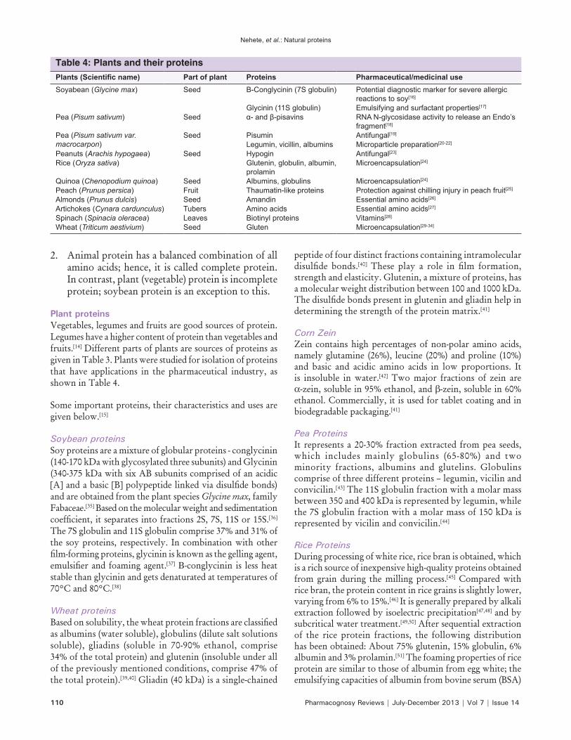
Plant proteinsVegetables, legumes and fruits are good sources of protein. Legumes have a higher content of protein than vegetables and fruits.[14] Different parts of plants are sources of proteins as given in Table 3. Plants were studied for isolation of proteins that have applications in the pharmaceutical industry, as shown in Table 4.
Some important proteins, their characteristics and uses are given below.[15]
Soybean proteinsSoy proteins are a mixture of globular proteins - conglycinin (140-170 kDa with glycosylated three subunits) and Glycinin (340-375 kDa with six AB subunits comprised of an acidic [A]andabasic[B]polypeptidelinkedviadisulfidebonds)and are obtained from the plant species Glycine max, family Fabaceae.[35] Based on the molecular weight and sedimentation coefficient,itseparatesintofractions2S,7S,11Sor15S.[36] The 7S globulin and 11S globulin comprise 37% and 31% of the soy proteins, respectively. In combination with other film‑formingproteins,glycininisknownasthegellingagent,emulsifierandfoamingagent.[37] Β-conglycinin is less heat stable than glycinin and gets denaturated at temperatures of 70°C and 80°C.[38]
Wheat proteinsBased on solubility,thewheatproteinfractionsareclassifiedas albumins (water soluble), globulins (dilute salt solutions soluble), gliadins (soluble in 70-90% ethanol, comprise 34% of the total protein) and glutenin (insoluble under all of the previously mentioned conditions, comprise 47% of the total protein).[39,40] Gliadin (40 kDa) is a single-chained
peptide of four distinct fractions containing intramolecular disulfide bonds.[40] These play a role in film formation,strength and elasticity. Glutenin, a mixture of proteins, has a molecular weight distribution between 100 and 1000 kDa. Thedisulfidebondspresentingluteninandgliadinhelpindetermining the strength of the protein matrix.[41]
Corn ZeinZein contains high percentages of non-polar amino acids, namely glutamine (26%), leucine (20%) and proline (10%) and basic and acidic amino acids in low proportions. It is insoluble in water.[42] Two major fractions of zein are α-zein, soluble in 95% ethanol, and β-zein, soluble in 60% ethanol. Commercially, it is used for tablet coating and in biodegradable packaging.[41]
Pea ProteinsIt represents a 20-30% fraction extracted from pea seeds, which includes mainly globulins (65-80%) and two minority fractions, albumins and glutelins. Globulins comprise of three different proteins – legumin, vicilin and convicilin.[43] The 11S globulin fraction with a molar mass between 350 and 400 kDa is represented by legumin, while the 7S globulin fraction with a molar mass of 150 kDa is represented by vicilin and convicilin.[44]
Rice ProteinsDuring processing of white rice, rice bran is obtained, which is a rich source of inexpensive high-quality proteins obtained from grain during the milling process.[45] Compared with rice bran, the protein content in rice grains is slightly lower, varying from 6% to 15%.[46] It is generally prepared by alkali extraction followed by isoelectric precipitation[47,48] and by subcritical water treatment.[49,50] After sequential extraction of the rice protein fractions, the following distribution has been obtained: About 75% glutenin, 15% globulin, 6% albumin and 3% prolamin.[51] The foaming properties of rice protein are similar to those of albumin from egg white; the emulsifying capacities of albumin from bovine serum (BSA)
Table 4: Plants and their proteinsPlants (Scientific name) Part of plant Proteins Pharmaceutical/medicinal useSoyabean (Glycine max) Seed Β‑Conglycinin (7S globulin) Potential diagnostic marker for severe allergic
reactions to soy[16]
Glycinin (11S globulin) Emulsifying and surfactant properties[17]
Pea (Pisum sativum) Seed α‑ and β‑pisavins RNA N-glycosidase activity to release an Endo’s fragment[18]
Pea (Pisum sativum var. macrocarpon)
Seed Pisumin Antifungal[19]
Legumin, vicillin, albumins Microparticle preparation[20-22]
Peanuts (Arachis hypogaea) Seed Hypogin Antifungal[23]
Rice (Oryza sativa) Glutenin, globulin, albumin, prolamin
Microencapsulation[24]
Quinoa (Chenopodium quinoa) Seed Albumins, globulins Microencapsulation[24]
Peach (Prunus persica) Fruit Thaumatin-like proteins Protection against chilling injury in peach fruit[25]
Almonds (Prunus dulcis) Seed Amandin Essential amino acids[26]
Artichokes (Cynara cardunculus) Tubers Amino acids Essential amino acids[27]
Spinach (Spinacia oleracea) Leaves Biotinyl proteins Vitamins[28]
Wheat (Triticum aestivium) Seed Gluten Microencapsulation[29-34]
Nehete, et al.: Natural proteins
Pharmacognosy Reviews | July-December 2013 | Vol 7 | Issue 14 111
aresignificantlyhigherthanthoseofriceproteins;minimumprotein solubility is close to isoelectric point at pH 4 and maximum at pH 10; main amino acid content of rice proteins is similar to that of casein and soy proteins; and denaturation temperature of the rice protein isolate is about 83.4°C.[44]
Sunflower ProteinsProteins aremajorityconstituents in sunfloweroil cakes.Defattedsunflowerflourcontainsahighquantityofproteins,around 27% in dry weight.[52] The dehulled seed consists of about 20-40% crude protein. Four fractions of protein arepresentinthesunflowerprotein:[53] Globulins, 55-60%; albumins, 17-23% of total proteins; and two minor fractions, glutelins and prolamins, comprising 11-17% and 1-4% of the total protein fractions, respectively. It shows two major fractions: 11S globulins (also named helianthinin) and 2S albumins. Helianthinin has been reported to be present as a globular oligomeric protein with a molecular weight of 300-350 kDa,[54] and this protein mainly exists in the 11S hexametric form.
Animal proteinsProtein from animal sources contains essential amino acids needed for an adult’s diet. The important examples are as given below.
CaseinThere are four main subunits: s1 α casein (23.6 kDa, 4.94 a pI, net charge - 21.9 at a pH of 6.6), s2 αcasein(netcharge−13.8,5.37 pI, hydrophilic due to high-charge density), β-casein (polar N-terminal amphipathic protein with large hydrophobic domain, Ca 2 + sensitive, at 4°C solubility increases) and κ-casein (not Ca 2 + sensitive), which make up 38%, 10%, 36% and 13% of the casein composition, respectively, and has a unique property to form films.[55-57] s1 α casein is amphipathic due to the charge between the hydrophobic N- and C-terminals. It has 8 phosphorylated serine clustered with glutamine residues possessing calcium-binding sites and hence is Ca 2 + sensitive, 17 proline, 25 glutamine residues and no cysteine residues. Here, the Ca 2 + sensitivity means aggregation and precipitation in low ion concentrations. It does notparticipateindisulfidebondformationandcross‑linkingdue to the absence of free cysteine. Caseins are heat stable because they are proline-rich, which interrupt alfa-helix and betastrands,resultingintheabsenceofdisulfidebridgesinthestructure. It has relatively little secondary or tertiary structure. These undergo proteolytic cleavage due their open structure imparted due by the high proline content. This characteristic, along with acid-soluble calcium–phosphate bridging, makes an excellent target-activated release mechanism for unloading drug in the stomach.[58,59]
Whey proteinTechnically, whey proteins are those that remain in milk serum after coagulating caseins at 4.6 pH and 20°C.[60] It contain β-lactoglobulin (18.3 kDa, containing 160 amino
acids),[61] α-lactalbumin (14.2 kDa), BSA (66 kDa, longest single-chain protein) and immunoglobins (thermolabile mixture of proteins).[62,63]
Meat proteinsSarcoplasmic, stromal andmyofibrillar are types ofmeatprotein. Sarcoplasmic proteins contain enzymes myoglobulin and cytoplasmic. Collagen and elastin are the content of stromal proteins while myosin, actin, tropomysin and troponinsarethecontentofmyofibrillarproteins.Stromalandmyofibrillarproteins,solubleinsaltsolutions,areusedformaking ediblefilms and coatings.Collagen, afibrousstromal protein extracted from connective tissue, tendons, skin, bones and the vascular system, and is a waste products of meat processing. Collagen is a superhelical structure formed by a combination of three parallel alfa-chains, and forms gelatine.[64] Collagen exposed to mild heat treatment under acidic or alkaline conditions forms gelatin.[65]
Egg albuminProtein fractions: Ovalbumin (44.5 kDa, contains free sulfhydryl groups for cross-linking); ovotransferrin (77.7 kDa, iron-binding protein); ovomucoid, ovomucin and lysozyme, a gram negative antimicrobial present in albumin.[66] Ovalbumin, ovotransferrin and lysozyme protein on denaturation form strong intermolecular beta-sheet structures as heat-set gels due to their thermolabile nature.
Silk proteinsThe silkworm Bombyx mori produces silk to weave its cocoon. Biocompatibility, slow bio-degradability, self-assembly, excellent mechanical properties, controllable structure and morphology make it promising materials for drug delivery and tissue engineering.[67]Fibrousproteinfibroin,acoreofsilkandaglue‑likeproteinsericinthatenvelopfibroinincocoon formation, are major components of silk.[68]
Isolation of proteins[69]
Selective precipitation of proteins can be used as:1. Bulk method to recover majority of the proteins
from a crude lysate2. Selective method to fractionate a subset of proteins
from a protein solution3. Specificmethod to recover a single protein ofinterestfromapurificationstep.
Selective precipitation methods1. Salting out2. Isoionic precipitation3. Organic co-solvent precipitation4. Two carbon (C2) organic co-solvent precipitation
of proteins5. C4 and C5 organic co-solvent precipitation, phase
partitioning and extraction of proteins6. Protein exclusion and crowding agents (neutral

Nehete, et al.: Natural proteins
112 Pharmacognosy Reviews | July-December 2013 | Vol 7 | Issue 14
polymers) and osmolytes7. Synthetic and semisynthetic polyelectrolyte
precipitation8. Metallic and polyphenolic heteropolyanion
precipitation9. Hydrophobic ion pairing (HIP) entanglement
ligands10. Matrix-stacking ligand co-precipitation11. Di- and trivalent metal cation precipitation.
Salting outProteins are salted out as co-precipitate by ammonium sulfate because the saturation concentration provides high molarity that causes precipitation of most proteins. It does not have a large heat of solution and hence the generated heat get easily dissipated; a saturated solution (4.04 M at 20°C) of proteins has a density of 1.235 g/cm3, which does not interfere with the precipitated protein sedimentation by centrifugation. Its concentrated solutions are generally bacteriostatic and protect most proteins from denaturation in solution state.
Figure illustrates the procedures for a pilot experiment.1. Dialyze protein samples against a pH buffer or a
pH buffer/ammonium sulfate mixture having a sulfate concentration below that needed to start precipitation
2. Set up a pilot experiment to determine the optimal protein concentration, pH, salt concentration, temperature and incubation time
3. Add ammonium sulfate to optimal concentration. Incubate (for an optimal period of time) until a precipitate forms
4. Collect the precipitate by simple decantation5. Re-dissolve the precipitate in a buffer suitable for
the next step6. Perform an assay for the protein of interest.
Isoionic precipitationColumn methodProteins are frequently least soluble and most precipitable when they are isoionic. In an isoionic, salt-free state, the
protein molecules are in their most compact, least-hydrated conformation – a phenomenon that is closely related to the condition of proteins at their isoelectric point. The distinction between isoionic and isoelectric properties is determined by the procedure described by Tanford.[70] Deionization using a column or dialysis aims at rendering proteins isoionic to precipitate them. Two important parameters determine the solubility of many proteins: Solution pH with respect to each proteins’ isoionic point (pI) and low salt concentration (0 to 0.1 to 0.2 M salt). The column method used is appropriate only for proteins that remain soluble at their isoionic point. In addition to adjusting the proteins to their isoionic pH, the general method is using mixed-bed resin deionization to strip away all salts from proteins. Salts, even in small concentration, often have large effects on protein solubility and, therefore, on perceptibility. Inorganic salts tend to salt in many proteins thus enhancing their solubility.
Dialysis methodOne of the older methods of rendering proteins salt free or nearly salt free (i.e., isoionic) is dialysis. However, two problems frequently arise with conventional dialysis: (1) When appreciable amounts of protein are present, osmotic effects result in swelling of the dialysis bags as the salt diffuses outward and (2) often, it is quite uncertain where the isoionic point is even if it is feasible to deionize by dialysis against a buffer. The resin deionization method, sometimes called the Dintzis method,[71] automatically adjusts a protein precisely to its isoionic pH without prior knowledge of the same. Salt ions and protein counterions exchange through the membrane and are trapped outside in the exchanger resins. After exchange is complected, the precipitated protein in the dialysis tubing is recovered by centrifugation of the bag’s contents. This general technique requires several hours. Salting out and diffusion become slow as the protein concentraction inside the dialysis bag decreases; hence, this method is slower than the flow‑throughcolumnmethod.
When proteins are insoluble and precipitate at their isoionic point, deionization is accomplished by placing the dialysis tubing containing the protein sample in a slurry of mixed-bed resin exchangers and incubating with rocking. Proteins insoluble at their isoionic point precipitate inside the bag. The mixed-bed exchange resins remove free salts, forcing the protein to its isoionic pH.
Purification and separationSodium dodecyl sulfate (SDS) gel electrophoresis.[72]
This is a technique in which charged molecules such as protein or DNA are separated according to the physical properties as they are forced through a gel by an electrical current.
Figure 1: Salting out‑small scale basis
Nehete, et al.: Natural proteins
Pharmacognosy Reviews | July-December 2013 | Vol 7 | Issue 14 113
The principal sample applied to the gel has been treated with detergent SDS and ß-mecaptoethanol. This will denature the proteins and the SDS binding tightly to the uncoiled molecule makes it negatively charged. SDS-polyacrylamide gel electrophoresis (PAGE) gel separates proteins primarily according to size because SDS-coated proteins have a uniform charge: Mass ratio.
One‑dimensional gel electrophoresisThis type of electrophoresis can provide information about the molecular size and purity of the proteins as well as the number and molecular size of its subunits. In PAGE, proteinsmigrateinresponsetoanelectricalfieldthroughpores in the gel matrix; the pore size decreases with higher acrylamide concentrations. The combination of gel pore size and protein charge, size and shape determines the migration rate of the protein.
Two‑dimensional gel electrophoresisThis type of electrophoresis separates proteins in the firstdimensionbyisoelectricfocusingandintheseconddimension by electrophoresis in the presence of SDS. By separating proteins in this manner, information is obtained not only about the size of a protein, as in one-dimensional gels, but also about the charge of a protein. Two-dimensional gels are superior for resolving complex mixtures and for assessing protein purity.
Application of 2D‑PAGEApplications of 2D-PAGE are summarized as below.1. Monitor protein accumulation during development2. Comparisons between differentiated organs and
tissues3. Comparisons of genetic variability within and
between species4. Detection of protein synthesis effected by
various environmental stimuli, such as growth substances (ABA, GA), abiotic stresses (high temperature, low temperature, drought, anaerobiosis and salt) and pathogenic attack.
Separating gel/resolving gel and stacking gel are used in electrophoresis. The composition of the same are: Water, acryl amide mix, tris-HCl buffer (pH 6.8), SDS, ammonium per sulfate and N, N, N\N’-tetramethylethylenediamine.
Protein stainingCoomassie blue stainingThis staining requires an acidic medium for generation of an electrostatic attraction between the dye molecules and the amino groups of proteins. Ionic attraction, together with van der Waals forces, binds the dye–protein complex together. Coomassie Blue stains exhibit three-times staining intensity of Fast Green and six-times intensity of Amido Black. Usually, 0.2-0.5 μg of any protein in a sharp band
can be detected using Coomassie Blue staining. Because Coomassie Blue is predominantly non-polar, it is usually used in methanolic solution and excess dye is removed from the gel later by distaining. Stacking gels are usually discarded before staining the resolving gels. Gel slabs are placed in staining solution and are stained fully within 4-6 h at room temperature if only 1.5-mm thick.
Silver stainingSilver staining is 50-100-times more sensitive than Coomassie Blue staining. The principal reactive groups are free amines and sulfur groups (basic and sulfur-containing amino acids) contained on proteins. Most proteins stain with monochromatic brown or black colors. Lipoproteins tend to stain blue while some glycoproteins appear yellow, brown or red. Artefact bands with molecular weights ranging from 50 kDa to 68 kDa have been commonly observed in silver-stained gels. Evidence has been presented indicating that these contaminating bands are due to keratin skin proteins.
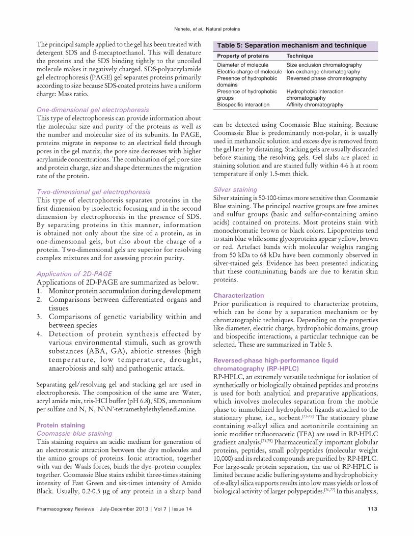
CharacterizationPrior purification is required to characterize proteins, which can be done by a separation mechanism or by chromatographic techniques. Depending on the properties like diameter, electric charge, hydrophobic domains, group andbiospecific interactions,aparticular techniquecanbeselected. These are summarized in Table 5.
Reversed‑phase high‑performance liquid chromatography (RP‑HPLC)RP-HPLC, an extremely versatile technique for isolation of synthetically or biologically obtained peptides and proteins is used for both analytical and preparative applications, which involves molecules separation from the mobile phase to immobilized hydrophobic ligands attached to the stationary phase, i.e., sorbent.[73-75] The stationary phase containing n-alkyl silica and acetonitrile containing an ionicmodifiertrifluoroacetic(TFA)areusedinRP‑HPLCgradient analysis.[74,75] Pharmaceutically important globular proteins, peptides, small polypeptides (molecular weight 10,000)anditsrelatedcompoundsarepurifiedbyRP‑HPLC.For large-scale protein separation, the use of RP-HPLC is limited because acidic buffering systems and hydrophobicity of n-alkyl silica supports results into low mass yields or loss of biological activity of larger polypeptides.[76,77] In this analysis,
Table 5: Separation mechanism and techniqueProperty of proteins TechniqueDiameter of molecule Size exclusion chromatographyElectric charge of molecule Ion-exchange chromatographyPresence of hydrophobic domains
Reversed phase chromatography
Presence of hydrophobic groups
Hydrophobic interaction chromatography
Biospecific interaction Affinity chromatography

Nehete, et al.: Natural proteins
114 Pharmacognosy Reviews | July-December 2013 | Vol 7 | Issue 14
1 μg/μl of the stock sample was prepared in 0.1% (v/v) TFA inwater (bufferA) andfiltered through a 0.22μm filter if anyundissolvedmaterial remained.Baselinewasobtainedusing100%bufferswithaflowrateof1mL/minat 215 nm. Ten microliters of the sample was injected using a linear gradient from 0% to 100% buffer B: 100% CH3CN containing 0.1% (v/v) TFA over 30 min to elute the sample. Ten microliters of Milli-Q water was used as the blank. In the literature, the HPLC system with a reversed-phase octadecylsilica (C-18) column (4.6 mm id [internal diameter] ×250 mm length, 5 μm particle size, 300 Å pore size, C18 guard column) had been used.[74]
Mass spectrometry of proteinsLarge and polar biomolecules are not easily ionized and transferred into the gas phase; hence, electrospray (ES) and matrix-assisted laser desorption ionization (MALDI) techniques are used. Mass spectrometry in proteomics is used in three major areas.[78]
1. Recombinant prote ins , macromolecu le characterizationandqualitycontrolinthefieldofbiotechnology
2. Proteinidentification,eitherinclassicalbiochemicalprojects or in large-scale proteomic ones
3. Detection and characterization of post-translational modifications or anymethod like any covalentmodification that altersmass of a protein usingversatility of it to detect the molecular weight of the protein.[79]
Liquid chromatography–mass spectrometry and tandem mass spectrometry of peptides and proteinsProtein and peptide tandem mass spectrometry involves application of two separate stages: LC–MS and LC–MS/MS, to solveproblems that require protein identificationand software tools to identify proteins by matching mass spectrometric data to protein sequence databases.[80,81] Additionally, LC–MS provides high-quality data for peptide mass as compared with MALDI mass spectrometry in fingerprintingsearches. Tessier et al. reported the prediction of plant protein hydrolysate containing small peptide amino acids’ composition performed by LC-MS and capillary electrophoresis-mass spectrometry.[82]
Applications of proteinsProteins are used in drug and gene delivery systems as protein-based nanocarriers. Extended applications include use in controlled delivery, as a film coater,as hydrogels, as composites, as albumin-based nanoparticles, as microparticles and as beads. Some examples are:1. Whey proteins used as hydrogels, nanoparticle
systems for encapsulation and controlled delivery of bioactive compounds[83]
2. Asanti‑hypertensiveuse,likegeneticallymodified
soybean seeds accumulating novokinin[84]
3. As solubility enhancer of curcumin in the food indus t ry due to pro te in–mice l l e structure (beta-casein), acting as a nano vehicle[85]
4. As vehicles for bioactives, like milk proteins5. As a source of bioactive peptides, e.g., casein-derived
four main bioactive peptides act on the cardiovascular system, nervous system, immune system and nutrition system[85]
6. As a novel antifungal, e.g., Pisumin proteins obtained from Pisum sativum. Sugar snap pea legumes.[86]
7. In microencapsulation, e.g., vegetable proteins – soy proteins, pea proteins, wheat proteins, rice proteins, oatproteinsandsunflowerproteins[83]
8. As pest control: Proteinaceous cysteine proteinase inhibitor, an insecticidal protein found in pulses used to control the proteolytic activity of endogenous digestive cystein proteinase in the mid-gut of some insects.
REFERENCES
1. Hermann JR. Protein and the Body. Oklahoma Cooperative Extension Service, Division of Agricultural Sciences and Natural Resources. Oklahoma State University: T–3163–1 – T–3163–4.
2. McArdle WD, Katch FI, Katch VL. Vitamins, minerals, and water, In: Exercise physiology: Energy, Nutrition, and Human Performance, 5th ed. Philadelphia, PA: Lippincott Williams and Wilkins; 2001. p. 47-81.
3. Millward DJ. The nutritional value of plant based diets in relation to human amino acid and protein requirements. Proc Nutr Soc 1999a; 58:249-60.
4. Streitwieser A, Jr. Heathcock CH. “Introduction to organic chemistry” 3rd ed., 1985.
5. Furst P, Stehle P. "What are the essential elements needed for the determination of amino acid requirements in humans?” J Nutr 2004;134 (6 Suppl):1558-65S.
6. Satyanarayana U, Chakrapani U. Books of Biochemistry. 3- ed., 2006.
7. Bachmair A, Finley D, Varshavsky A. In vivo half-life of a protein is a function of its amino-terminal residue. Science 1986;234:179-86.
8. Pace CN, Vajdos F, Fee L, Grimsley G, Gray T. How to measure and predict the molar absorption coefficient of a protein. Protein Sci 1995;11:2411-23.
9. Edelhoch H. Spectroscopic determination of tryptophan and tyrosine in proteins. Biochemistry 1967;6:1948-54.
10. Gill SC, von Hippel PH. Calculation of protein extinction coefficients from amino acid sequence data. Anal Biochem 1989;182:319-26.
11. Ikai A. Thermostability and aliphatic index of globular proteins. J Biochem 1980;88:1895-8.
12. Kyte J. Doolittle RF. A simple method for displaying the hydropathic character of a protein. J Mol Biol 1982;157:105-32.
13. Alterman MA, Hunziker P. Methods in Molecular Biology, Amino Acid Analysis, Methods and Protocols, 2012.
14. Creighton TE. Proteins: Structures and molecular
Nehete, et al.: Natural proteins
Pharmacognosy Reviews | July-December 2013 | Vol 7 | Issue 14 115
properties. New York: Freeman WH; 1993.15. Coon JJ. Collisions or Electrons? Protein Sequence Analysis in
the 21st Century. Anal Chem 1980;81:3208-15.16. Bender W, Smith M. Population, Food and Nutrition. Popul Bull
1997;51:5-25.17. Fink HH, Mikesky AE, Burgoon LA. Practical Applications in
Sports Nutrition. 3rd ed. 2012. p. 1-569.18. Yue H, Defa L, Xianglan P, Xiangshu P. Forsythia suspensa extract
alleviates hypersensitivity induced by soybean β-conglycinin in weaned piglets. J Ethnopharmacol 2010;128:412-8.
19. Gu X, Campbell LJ, Euston SR. Effects of different oils on the properties of soy protein isolate emulsions and gels. Food Res Int 2009;42:925-32.
20. Shirley SL, Wang LH, Ng TB. Purification and characterization of novel ribosome, inactivating proteins, alpha- and beta-pisavins from seeds of the garden pea Pisum Sativum. Biochem Biophys Res Commun 1998;253:135-42.
21. Ye XY, Ng TB. Isolation of pisumin, a novel antifungal protein from legumes of the sugar snap pea Pisum sativum var. macrocarpon. Comp Biochem Physiol C Toxicol Pharmacol 2003;134:235-40.
22. Irache JM, Bergougnoux L, Ezpeleta I, Gueguen J, Orecchioni AM. Optimization and in vitro stability of legumin nanoparticles obtained by a coacervation method. Int J Pharm 1995;126:103-9.
23. Ezpeleta I, Irache JM, Stainmesse S, Chabenat C, Gueguen J, Orecchioni AM. Preparation of lectin–vicilin nanoparticle conjugates using the carbodiimide coupling technique. Int J Pharm 1996a; 142:227-33.
24. Ezpeleta I, Irache JM, Gueguen J, Orecchioni AM. Properties of glutaraldehyde Crosslink’s vicilin nano and microparticles. J Microencapsul 1997;14:557-65.
25. Ng TB. Antifungal proteins and peptides of leguminous and non-leguminous origins, Department of Biochemistry, Faculty of Medicine, The Chinese University of Hong Kong, Shatin, New Territories, Hong Kong, China. Peptides 2004;25:1215-22.
26. Nesterenko A, Alric I, Silvestre F, Durrieu V. Influence of soy protein’s structural modifications on their microencapsulation properties: Atocopherol microparticles preparation. Food Res Int 2012;48:387-96.
27. Dagar A, Friedman H, Lurie S. Thaumatin like proteins and their possible role in protection against chilling injury in peach fruit. Postharvest Biol Technol 2010;57:77-85.
28. Ducel V, Richard J, Saulnier P, Popineau Y, Boury F. Evidence and characterization of complex coacervates containing plant proteins: Application to the microencapsulation of oil droplets. Colloid Surf 2004b; 232:239-47.
29. Ducel V, Richard J, Popineau Y, Boury F. Rheological interfacial properties of plant protein Arabic gum coacervates at the oil–water interface. Biomacromolecules 2005;6:790-6.
30. Ezpeleta I, Irache JM, Stainmesse S, Chabenat C, Gueguen J, Popineau Y, et al. Gliadin nanoparticles for the controlled release of alltransretinoic acid. Int J Pharm 1996b; 131:191-200.
31. Iwami K, Hattori M, Nakatani S, Ibuki F. Spray dried gliadin powders inclusive of linoleic acid (microcapsules): Their preservability, digestibility and application to bread making. Agric Biol Chem 1987;51:3301-7.
32. Mauguet MC, Legrand J, Brujes L, Carnelle G, Larre C, Popineau Y. Gliadin Matrices for microencapsulation processes by simple coacervation method. J Microencapsul 2002;19:377-84.
33. Yu JY, Lee WC. Microencapsulation of pyrrolnitrin from Pseudomonas cepacia using gluten and casein. J Ferment Bioeng 1997;84:444-8.
34. The Wealth of India Raw Material Series, National institute of Science Communication and Information Resources, CSIR,
New Delhi, 2010.35. Cho SY, Rhee C, Wiss L. Mechanical properties and water vapor
permeability of edible films made from fractionated soy proteins with ultrafiltration. U‑Technology 2004;37:833‑9.
36. Subirade M, Kelly I, Gueguen J, Pezolet M. Molecular basis of film formation from a soybean protein: Comparison between the conformation of glycinin in aqueous solution and in Films. Int J Biol Macromol 1998;23:241-9.
37. Renkema JM, Vliet TV. Heat-induced gel formation of soy proteins at neutral pH. J Agric Food Chem 2002;50:1569-73.
38. Haard NF, Chism GW. Characteristics of edible plant tissue. In: Fennema O, editor. Food Chemistry. New York, NY: Marcel Dekker; 1996. p. 1067.
39. Kinsella JE. Relationships between structure and functional properties of food proteins. In: Fon and Codon, editor. Food Proteins. New York: Springer Publishing; 1982. p. 51-104.
40. Kirsten D, Peggy MT, Phoebe Q. Structure and function of Protein Based edible films and coatings. Edible Films and Coatings for Food Applications. 2009. p. 25-56.
41. Shukla R, Cheryan MZ. The industrial protein from corn. Ind Crops Prod 2001;13:171-92.
42. Koyoro H, Powers JR. Functional properties of pea globulin fractions. Cereal Chem 1987;64:97-101.
43. Nesterenko A, Alri I, Silvestre FO, Durrieu V. Vegetable proteins in microencapsulation: A review of recent interventions and their effectiveness. Ind Crops Prod 2013;42:469-79.
44. Hamada JS. Characterization and functional properties of rice bran proteins modified by commercial exoproteases and endoproteases. J Food Sci 2000;65:305-10.
45. Bienvenido OJ. Rice in human nutrition organization united nations ETL, Agriculture power, Rome 1994.
46. Kaewka K, Therakulkait C, Cadwallader KR. Effect of preparation conditions on composition and sensory aroma characteristics of acid hydrolyzed rice bran protein concentrate. J Cereal Sci 2009;50:56-60.
47. Pinciroli M, Vidal AA, Anon MC, Martínez EN. Comparison between protein functional properties of two rice cultivars. Food Sci Technol 2009;42:1605-10.
48. Hata S, Wiboonsirikul J, Maeda A, Kimura Y, Adachi S. Extraction of defatted rice bran by subcritical water treatment. Biochem Eng J 2008;40:44-53.
49. Sereewatthanawut I, Prapintip S. Watchiraruji K, Goto M, Sasaki M, Shotipruk A. Extraction of protein and amino acids from deoiled rice bran by sub-critical water hydrolysis. Bioresour Technol 2008;99:555-61.
50. Chandi GK, Sogi DS. Functional properties of rice bran protein concentrate. J Food Eng 2007;79:592-7.
51. Agboola S, Ng D, Mills D. Characterisation and functional properties of Australian rice protein isolates. J Cereal Sci 2005;41:283-90.
52. Ordonez C, Asenjo MG, Benitez JL, Gonzalez JL. Obtaining a protein con‑centrate from integral deffated sunflower flour. Bioresour Technol 2001;78:187-90.
53. Gonzalez‑Perez S, Vereijken JM. Sunflower proteins: Overview of their physicochemical, structural and functional properties. J Sci Food Agric 2007;87:2173-91.
54. Linden GL. Biochimie agro-industrielle: Valorisation alimentaire de la pro-duction agricole. Masson, Paris. 1994.
55. Audic JL, Chaufer B, Daufin G. Non‑food applications of milk components and dairy co-products: A review. Lair 2003;83:417-38.
56. Swaisgood HE. Review and update of casein chemistry. J Dairy Sci 1993;76:3054-61.

Nehete, et al.: Natural proteins
116 Pharmacognosy Reviews | July-December 2013 | Vol 7 | Issue 14
57. Swaisgood HE. Characteristics of milk. In: Fennema O, editor. Food chemistry. New York: Marcel Dekker; 1996. p. 1067.
58. Rosemberg M, Young SL. Whey proteins as microencapsulating agents. Microencapsulation of anhydrous milk fat structure evaluation. Food Struct 1993;12:31-41.
59. Livney YD. Milk proteins as vehicles for bioactives. Curr Opin Colloid Interface Sci 2010;15:73-83.
60. Morr CV, Ha EY. Whey protein concentrates and isolates: Processing and functional properties. Crit Rev Food Sci Nutr 1993;33:431-76.
61. DeWit JN, Klarenbeek G. Effects of various heat treatments on structure and solubility of whey proteins. J Dairy Sci 1983;67:2701-10.
62. Kinsella JE. Milk proteins: Physicochemical and functional properties. Crit Rev Food Sci Nutr 1984;21:197-262.
63. Sawyer L, Kontopidis G, Wu SY. Beta-lactoglobulin: A three-dimensional perspective. Int J Food Sci Tech 1999;34:409-18.
64. Haug IJ, Draget KI, Smidsrod O. Physical and rheological properties of fish gelatin compared to mammalian gelatin. Food Hydrocoll 2004;18:203-13.
65. Badii F, Howell NK. Fish gelatine: Structure, gelling properties and interaction with egg albumen proteins. Food Hydrocoll 2006;20:630-40.
66. Mine Y. Recent advances in the understanding of egg white protein functionality. Trends Food Sci Technol 1995;6:225-32.
67. Numata K, Kaplan DL. Silk-based delivery systems of bioactive molecules, Adv Drug Deliv Rev 2010;62:1497-508.
68. Altman GH, Diaz F, Jakuba C, Calabro T, Horan RL, Chen J, et al. Silk-based biomaterials. Biomaterials 2003;24:401-16.
69. Rothstein F. Differential precipitation of proteins. In protein purification process Engineering, Harrison RG, editor. New York: Marcel Dekker; 1994. p. 115-208.
70. Tanford C. Multiple equilibria. In Physical Chemistry of Macromolecules. New York: John Wiley and Sons; 1996. p. 561-4.
71. Edsall JT, Wyman I. Biophysical Chemistry. New York: Academic Press; 1958. p. 591-662.
72. Hames BD, Rickwood D. Gel electrophoresis of proteins: A practical approach. 2nd ed. New York: Oxford University Press; 1990.
73. Aguilar MI. Methods in Molecular Biology. Vol. 251, HPLC of Peptides and Proteins: Methods and Protocols. Totowa, NJ: Humana Press Inc.; 1996.
74. Aguilar MI, Hearn MT. High resolution reversed phase high performance liquid chromatography of peptides and proteins. Methods Enzymol 1996;270:3-26.
75. Mant CT, Hodges RS. Analysis of peptides by high performance liquid chromatography. Methods Enzymol 1996;271:3-50.
76. Purcell AW, Aguilar MI, Hearn MT. Conformational effects in the RP-HPLC of polypeptides. II: The role of insulin A and B chains in the chromatographic behavior of insulin. J Chromatogr 1995;711:71-9.
77. Lin S, Karger BL. Reversed phase chromatographic behavior of proteins in different unfolded states. J Chromatogr 1990;499:89-102.
78. Mann M, Hendrickson RC, Pandey A. Analysis of proteins and proteomes by mass spectrometry. Annu Rev Biochem 2001;70:437-73.
79. Yates JR. Database searching using mass spectrometry data. Electrophoresis 1998;19:893-900.
80. Tessier B, Schweizer M, Fournier F, Framboisier I, Chevalot R, Vanderesse C. Harscoat, I. Marc, Prediction of the amino acid composition of small peptides contained in a plant protein hydrolysate by LC–MS and CE–MS. Food Res Int 2005;38:577-84.
81. Elzoghby AO, Abo El-Fotoh WS, Elgindy NA. Casein-based formulations as promising controlled release drug delivery systems. J Control Release 2011;153:206-16.
82. Elzoghby AO, Samy WM, Elgindy NA. Albumin-based nanoparticles as potential controlled release drug delivery systems. J Control Release 2012;157:168-82.
83. Yuko Y, Keito N, Megumi Y, Hui Z, Kunihiko O, Masayoshi T, et al. Anti‑hypertensive activity of genetically modified soybean seeds accumulating novokinin. Peptides 2008;29:331-7.
84. Esmaili M, Ghaffari MS, Moosavi-Movahedi Z, Atri MS, Sharizadeh A, Farhadi M, et al. Beta casein-micelle as a nano vehicle for solubility enhancement of curcumin: Food industry application in LWT. Food Sci Technol 2011;44:2166-72.
85. Silva SV, Malcata FX. Caseins as source of bioactive peptides. Int Dairy J 2005;15:1-15.
86. Ye XY, Ng TB. Isolation of pisumin, a novel antifungal protein from legumes of the sugar snap pea Pisum sativum. Comp Biochem Physiol Part C Toxicol Pharmacol 2003;134:235-40.
How to cite this Article: Nehete JY, Bhambar RS, Narkhede MR, Gawali SR. Natural proteins: Sources, isolation, characterization and applications. Phcog Rev 2013;7:107-16.
Source of Support: Nil, Conflict of Interest: None declared
Related Documents