Please cite this article in press as: Soares, J.F., et al., Natural infection of the wild canid, Cerdocyon thous, with the piroplasmid Rangelia vitalii in Brazil. Vet. Parasitol. (2014), http://dx.doi.org/10.1016/j.vetpar.2014.02.058 ARTICLE IN PRESS G Model VETPAR-7181; No. of Pages 8 Veterinary Parasitology xxx (2014) xxx–xxx Contents lists available at ScienceDirect Veterinary Parasitology jo u r nal homep age: www.elsevier.com/locate/vetpar Natural infection of the wild canid, Cerdocyon thous, with the piroplasmid Rangelia vitalii in Brazil João F. Soares a,1 , Bruno Dall’Agnol b,1 , Francisco B. Costa a , Felipe S. Krawczak a , Alexandra T. Comerlato c , Bruna C.D. Rossato d , Camila M. Linck d , Eduardo K.O. Sigahi e , Rodrigo H.F. Teixeira c , Luciana Sonne f , Mitika K. Hagiwara a , Fabio Gregori a , Maria Isabel B. Vieira d , João R. Martins b , José Reck b , Marcelo B. Labruna a,∗ a Faculdade de Medicina Veterinária e Zootecnia, Universidade de São Paulo, Av. Prof. Orlando Marques de Paiva 87, São Paulo, SP, Brazil b Instituto de Pesquisas Veterinárias Desidério Finamor, Fundac ¸ ão Estadual de Pesquisa Agropecuária, Eldorado do Sul, RS, Brazil c Parque Zoológico Municipal “Quinzinho de Barros”, Soarocaba, SP, Brazil d Faculdade de Agronomia e Medicina Veterinária, Universidade de Passo Fundo, Passo Fundo, RS, Brazil e Departamento de Vigilância em Saúde, Secretaria de Saúde, Prefeitura Municipal de Mogi das Cruzes, SP, Brazil f Universidade Federal do Rio Grande do Sul, Porto Alegre, RS, Brazil a r t i c l e i n f o Article history: Received 20 January 2014 Received in revised form 18 February 2014 Accepted 26 February 2014 Keywords: Rangelia vitalii Piroplasm Cerdocyon thous Domestic dog Babesia Brazil a b s t r a c t Canine rangeliosis, caused by the piroplasmid protozoon Rangelia vitalii, is currently rec- ognized as a reemerging disease that affects domestic dogs in Brazil. In the present study, piroplasmid infection was searched in wild canids (20 Cerdocyon thous and 4 Lycalopex gymnocercus) in Brazil. Molecular analysis, based on PCR and DNA sequencing of a portion of the 18S rRNA gene, revealed that 30% (6/20) C. thous were infected by R. vitalii. Blood and bone marrow samples from one of the R. vitalii-infected C. thous were inoculated into a domestic dog, which developed clinical rangeliosis that was confirmed by molecular tests. However, the C. thous donor showed no clinical, hematological or biochemical alterations, even though its R. vitalii infection status was confirmed for at least 80 days. These obser- vations suggest that R. vitalii is not as highly pathogenic for C. thous as it is for domestic dogs. Phylogenetic analysis inferred by the 18S rRNA gene placed R. vitalii embedded in the clade ‘Babesia sensu stricto’, consisting of a number of species that represent truly the genus Babesia. It is proposed that the species R. vitalii should be transferred to the genus Babesia. The present study expands our knowledge on the natural history of R. vitalii, sug- gesting that it might have a natural cycle involving the wild canid C. thous. Further studies are needed to confirm that C. thous is a natural reservoir of R. vitalii in Brazil. © 2014 Elsevier B.V. All rights reserved. 1. Introduction Piroplasmids are tick-borne protozoan parasites that infect blood cells of numerous wild and domestic ∗ Corresponding author. Tel.: +55 11 3091 1394; fax: +55 11 3091 7928. E-mail address: [email protected] (M.B. Labruna). 1 These authors contributed equally to this work. vertebrates worldwide. The piroplasmid Rangelia vitalii is the etiologic agent of rangeliosis, a canine disease that was described in the beginning of the previous century in the state of São Paulo, southeastern Brazil (Pestana, 1910; Carini and Maciel, 1914). Because a number of sub- sequent authors (Wenyon, 1926; Doflein and Reichenow, 1929; Moreira, 1938, 1939; Levine, 1973; Peirce, 2000) con- sidered R. vitalii a synonym of Babesia vogeli (reported as Babesia canis), rangeliosis was widely neglected during the http://dx.doi.org/10.1016/j.vetpar.2014.02.058 0304-4017/© 2014 Elsevier B.V. All rights reserved.

Welcome message from author
This document is posted to help you gain knowledge. Please leave a comment to let me know what you think about it! Share it to your friends and learn new things together.
Transcript
V
Np
JAEMJa
b
c
d
e
f
ARRA
KRPCDBB
1
i
0
ARTICLE IN PRESSG ModelETPAR-7181; No. of Pages 8
Veterinary Parasitology xxx (2014) xxx–xxx
Contents lists available at ScienceDirect
Veterinary Parasitology
jo u r nal homep age: www.elsev ier .com/ locate /vetpar
atural infection of the wild canid, Cerdocyon thous, with theiroplasmid Rangelia vitalii in Brazil
oão F. Soaresa,1, Bruno Dall’Agnolb,1, Francisco B. Costaa, Felipe S. Krawczaka,lexandra T. Comerlatoc, Bruna C.D. Rossatod, Camila M. Linckd,duardo K.O. Sigahie, Rodrigo H.F. Teixeirac, Luciana Sonnef,itika K. Hagiwaraa, Fabio Gregoria, Maria Isabel B. Vieirad,
oão R. Martinsb, José Reckb, Marcelo B. Labrunaa,∗
Faculdade de Medicina Veterinária e Zootecnia, Universidade de São Paulo, Av. Prof. Orlando Marques de Paiva 87, São Paulo, SP, BrazilInstituto de Pesquisas Veterinárias Desidério Finamor, Fundac ão Estadual de Pesquisa Agropecuária, Eldorado do Sul, RS, BrazilParque Zoológico Municipal “Quinzinho de Barros”, Soarocaba, SP, BrazilFaculdade de Agronomia e Medicina Veterinária, Universidade de Passo Fundo, Passo Fundo, RS, BrazilDepartamento de Vigilância em Saúde, Secretaria de Saúde, Prefeitura Municipal de Mogi das Cruzes, SP, BrazilUniversidade Federal do Rio Grande do Sul, Porto Alegre, RS, Brazil
a r t i c l e i n f o
rticle history:eceived 20 January 2014eceived in revised form 18 February 2014ccepted 26 February 2014
eywords:angelia vitaliiiroplasmerdocyon thousomestic dogabesiarazil
a b s t r a c t
Canine rangeliosis, caused by the piroplasmid protozoon Rangelia vitalii, is currently rec-ognized as a reemerging disease that affects domestic dogs in Brazil. In the present study,piroplasmid infection was searched in wild canids (20 Cerdocyon thous and 4 Lycalopexgymnocercus) in Brazil. Molecular analysis, based on PCR and DNA sequencing of a portionof the 18S rRNA gene, revealed that 30% (6/20) C. thous were infected by R. vitalii. Bloodand bone marrow samples from one of the R. vitalii-infected C. thous were inoculated into adomestic dog, which developed clinical rangeliosis that was confirmed by molecular tests.However, the C. thous donor showed no clinical, hematological or biochemical alterations,even though its R. vitalii infection status was confirmed for at least 80 days. These obser-vations suggest that R. vitalii is not as highly pathogenic for C. thous as it is for domesticdogs. Phylogenetic analysis inferred by the 18S rRNA gene placed R. vitalii embedded inthe clade ‘Babesia sensu stricto’, consisting of a number of species that represent truly the
genus Babesia. It is proposed that the species R. vitalii should be transferred to the genusBabesia. The present study expands our knowledge on the natural history of R. vitalii, sug-gesting that it might have a natural cycle involving the wild canid C. thous. Further studiesare needed to confirm that C. thous is a natural reservoir of R. vitalii in Brazil.. Introduction
Please cite this article in press as: Soares, J.F., et al., Naturalpiroplasmid Rangelia vitalii in Brazil. Vet. Parasitol. (2014), http
Piroplasmids are tick-borne protozoan parasites thatnfect blood cells of numerous wild and domestic
∗ Corresponding author. Tel.: +55 11 3091 1394; fax: +55 11 3091 7928.E-mail address: [email protected] (M.B. Labruna).
1 These authors contributed equally to this work.
http://dx.doi.org/10.1016/j.vetpar.2014.02.058304-4017/© 2014 Elsevier B.V. All rights reserved.
© 2014 Elsevier B.V. All rights reserved.
vertebrates worldwide. The piroplasmid Rangelia vitalii isthe etiologic agent of rangeliosis, a canine disease thatwas described in the beginning of the previous centuryin the state of São Paulo, southeastern Brazil (Pestana,1910; Carini and Maciel, 1914). Because a number of sub-
infection of the wild canid, Cerdocyon thous, with the://dx.doi.org/10.1016/j.vetpar.2014.02.058
sequent authors (Wenyon, 1926; Doflein and Reichenow,1929; Moreira, 1938, 1939; Levine, 1973; Peirce, 2000) con-sidered R. vitalii a synonym of Babesia vogeli (reported asBabesia canis), rangeliosis was widely neglected during the
ING Model
y Parasi
ARTICLEVETPAR-7181; No. of Pages 8
2 J.F. Soares et al. / Veterinar
second half of the 20th century. During the last decade,a number publications from southern Brazil highlighted R.vitalii as a reemerging agent of a severe canine piroplasmo-sis, especially among rural dogs (Krauspenhar et al., 2003;Loretti and Barros, 2005; Fighera, 2007; Fighera et al., 2008,2010; Franc a et al., 2010). In 2011, the validity of the taxonR. vitalii was proposed by molecular methods based on phy-logenetic analyses inferred by partial sequences of the 18SrRNA and hsp70 genes (Soares et al., 2011). The infectionby R. vitalii has been confirmed solely in domestic dogsfrom southern and southeastern Brazil (Soares et al., 2011,2013a,b; Lemos et al., 2012).
The Brazilian fauna of wild canids is composed by sixnative species (Cheida et al., 2011). Literature records onpiroplasmids infecting these native canids are very scarce,and have been based solely on morphological identifi-cation of intraerythrocytic piroplasmid forms in bloodsmears from Chrysocyon brachyurus (Serra-Freire et al.,1995; Cansi et al., 2012), Lycalopex vetulus (Martins et al.,2006), Lycalopex gymnocercus (Ruas et al., 2003), and Cer-docyon thous (Paraense and Vianna, 1948; Massard et al.,1981). The crab-eating fox, C. thous, is widely distributedin South America, from Uruguay and northern Argentinato the lowlands of Bolivia and Venezuela, also occurring inColombia, Guyana and Suriname. In Brazil, it is found in allmajor biomes except for the Amazon (Cheida et al., 2011).The Pampas fox, L. gymnocercus, has a more restricted dis-tribution, occurring in the southern cone of South America,except for Chile (Cheida et al., 2011). In the present study,piroplasmid infection was searched for in C. thous and L.gymnocercus from Brazil. We report the first moleculardetection of a piroplasmid agent in South American free-ranging wild canids, as well as the effects of the inoculationof this agent in a domestic dog.
2. Materials and Methods
2.1. Wild canids
In May 2012, a free-ranging, young, adult female(approximately 5 kg) of C. thous (animal #1) was rescued inCarazinho Municipality, state of Rio Grande do Sul, south-ern Brazil, and taken to the nearby Veterinary TeachingHospital of the University of Passo Fundo due to a traumaticamputating injury in the hind limb. Clinical evaluationwas performed, and blood samples collected 0, 69, and 80days after admission were used for molecular detectionfor piroplasmids (described below), whereas blood sam-ples collected in both dry and EDTA tubes at the 21st daywere used for biochemical and hematological evaluations,respectively. At the 80th day, the animal died of unknownetiology; bone marrow aspirates and spleen samples werecollected.
From July 2012 to August 2013, tissue samples (blood orinternal organs) were collected from 23 wild canids fromdifferent areas of the states of Rio Grande do Sul and SãoPaulo (southeastern Brazil) (Table 1). Animal #2 was res-
Please cite this article in press as: Soares, J.F., et al., Naturalpiroplasmid Rangelia vitalii in Brazil. Vet. Parasitol. (2014), http
cued due to trauma caused by fighting with domestic dogs;animals #3–13 and 21–23 were road-killed, whereas ani-mals #14–20, and 24, were sampled in captivity at zoos.From road-killed animals, the collected material varied
PRESStology xxx (2014) xxx–xxx
accordingly to the availability of tissues found in the car-casses. From living animals sampled at the zoos, only bloodin EDTA was collected.
2.2. Inoculation of domestic dog
Three ml of bone marrow aspiration and 10 ml of bloodwas collected from C. thous #1 on the 80th day after itsadmission to the hospital, and sent to the University of SãoPaulo under refrigeration. Twenty hours later, the sampleswere intravenously inoculated into an 8-month domesticdog, derived from our Beagle experimental kennel, wheredogs have never experienced tick infestations, are regularlyvaccinated and dewormed, and are regularly tested to cer-tify that they are free of tick-borne diseases. The domesticdog was clinically evaluated and had its rectal tempera-ture measured daily, as well as blood samples taken inEDTA-tubes 3 times per week for blood cell and plateletcounts, and PCR targeting piroplasmids during a period of32 days after inoculation. An additional blood collection at69 days post-inoculation (dpi) was used for PCR analysis.During this period, the dog was kept in a room with noenvironmental contamination with ticks.
2.3. Molecular and phylogenetic analyses
Animal blood or tissue samples were subjected to DNAextraction using the DNeasy Blood & Tissue Kit (Qiagen®,Hilden, Germany), according to the manufacturer’s instruc-tions. All DNA samples from wild canids were initiallytested by one of the following two PCR assays, one targetinga ≈700-bp fragment of the Carnivore mitochondrial DNA(mtDNA) control region containing the first hypervariablesegment (HVS-I), using primers MTLPRO2 and CCR-DR1, aspreviously described (Tchaicka et al., 2007); or a PCR assaytargeting a 359-bp fragment of the vertebrate mitochon-drion cytochrome b gene (cyt b), as previously described(Steuber et al., 2005). For detection of piroplasmids, allsamples were tested by a PCR protocol using primersBAB143-167 and BAB694-667, targeting a ≈500-bp frag-ment of the piroplasmid 18S rRNA gene, as previouslydescribed (Soares et al., 2011). PCR products were elec-trophoresed through a 1.5% agarose gel, stained withethidium bromide, and examined by UV transillumina-tion. Amplicons of the expected size were purified withExoSap (USB, Cleveland, OH) and sequenced in an auto-matic sequencer (Applied Biosystems/PerkinElmer, modelABI Prism 310 Genetic, Foster City, CA) according to themanufacturer’s protocol. Generated sequences were sub-mitted to BLAST analysis (Altschul et al., 1990) to determinethe closest similarities in GenBank.
Partial sequences (549-nt) of the 18S rRNA gene of piro-plasmids derived from the wild canids were aligned withcorresponding 18S rRNA sequences from 53 genotypesof the genera Babesia, Theileria, Cytauxzoon and Hepa-tozoon retrieved from Genbank, using Clustal/W v.1.8.1(Thompson et al., 1994). A maximum likelihood phyloge-
infection of the wild canid, Cerdocyon thous, with the://dx.doi.org/10.1016/j.vetpar.2014.02.058
netic tree using GTR+G+I substitution model was generatedusing Mega 5.2.2 software (Tamura et al., 2011) with 100bootstrap replicates. The substitution model was selectusing Mega 5.2.2 software (Tamura et al., 2011) according
Please cite
this
article in
press
as: Soares,
J.F., et
al., N
atural
infection
of th
e w
ild can
id,
Cerdocyon thous,
with
the
pirop
lasmid
Rangelia
vitalii in
Brazil.
Vet.
Parasitol. (2014),
http
://dx.d
oi.org/10.1016/j.vetpar.2014.02.058
AR
TIC
LE
IN P
RE
SS
G M
odelV
ETPAR
-7181;
No.
of Pages
8
J.F. Soares
et al.
/ V
eterinary Parasitology
xxx (2014)
xxx–xxx
3
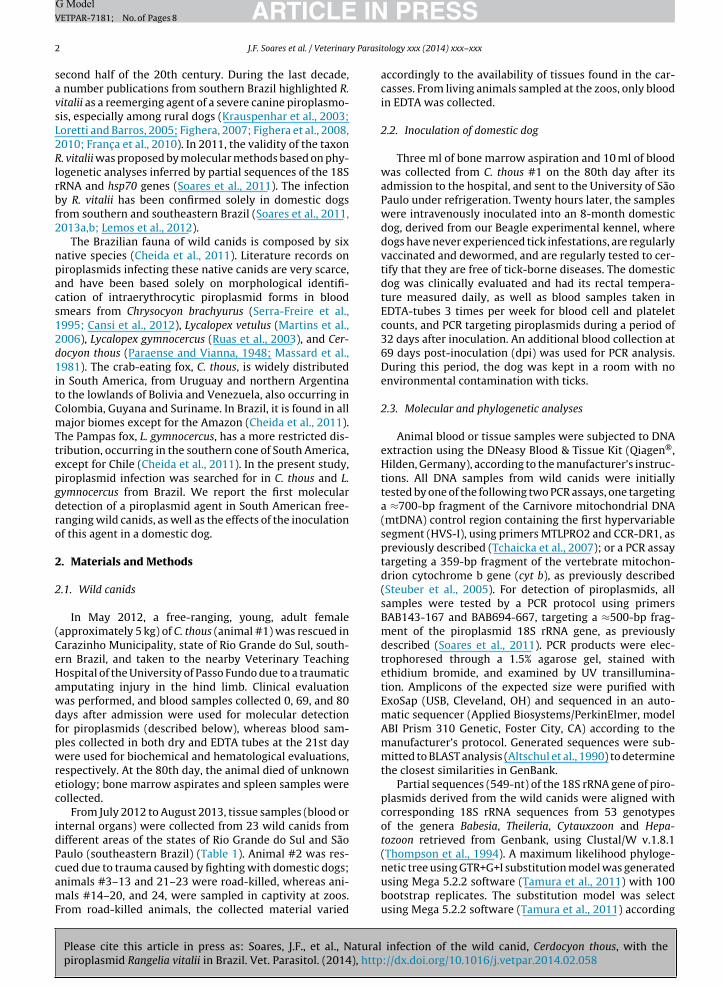
Table 1Wild canids (Cerdocyon thous and Lycalopex gymnocercus) sampled in the states of Rio Grande do Sul (RS) and São Paulo (SP), and tested by PCR for the presence of Rangelia vitalii DNA.
Animal number Species Origin Animalcondition
Collected samplestested by PCR
PCR result
Municipality Coordinates Elevation (m)
South West
1 C. thous Carazinho, RS 28◦17′ 52◦47′ 603 Rescued due totrauma
Blood +Bone marrow +Spleen +
2 C. thous Viamão, RS 30◦03′ 51◦00′ 60 Rescueddue totrauma
Liver +
3 C. thous Cachoeira doSul, RS
30◦02′ 52◦53′ 26 Road-killed Carotid +Muscle −
4 C. thous Cachoeira doSul, RS
30◦02′ 52◦53′ 26 Road-killed Lung +Carotid +
5 C. thous Cachoeira doSul, RS
30◦02′ 52◦53′38′′ 26 Road-killed Lung +Liver −
6 C. thous Itaqui, RS 29◦06′46′′ 56◦17′30′′ 66 Road-killed Muscle −7 C. thous Restinga Seca, RS 29◦48′ 53◦22′ 49 Road-killed Lung −8 C. thous São Borja, RS 28◦38′23′′ 55◦50′00′′ 69 Road-killed Muscle −9 C. thous São Luiz Gonzaga, RS 28◦24′ 54◦57′ 251 Road-killed Muscle −10 C. thous Mogi das
Cruzes, SP23◦31′ 46◦11′ 780 Road-killed Lymph node +
Carotid +Spinal marrow −
11 C. thous Botucatu, SP 22◦53′ 48◦26′ 804 Road-killed Spleen −12 C. thous Sorocaba, SP 23◦30′ 47◦27′ 600 Road-killed Spleen −13 C. thous Sorocaba, SP 23◦30′ 47◦27′ 600 Road-killed Spleen −14 C. thous Sorocaba, SP 23◦30′19′′ 47◦26′15′′ 580 Zoo Blood −15 C. thous Sorocaba, SP 23◦30′19′′ 47◦26′15′′ 580 Zoo Blood −16 C. thous Sorocaba, SP 23◦30′19′′ 47◦26′15′′ 580 Zoo Blood −17 C. thous Sorocaba, SP 23◦30′19′′ 47◦26′15′′ 580 Zoo Blood −18 C. thous Sorocaba, SP 23◦30′19′′ 47◦26′15′′ 580 Zoo Blood −19 C. thous Sorocaba, SP 23◦30′19′′ 47◦26′15′′ 580 Zoo Blood −20 C. thous Sorocaba, SP 23◦30′19′′ 47◦26′15′′ 580 Zoo Blood −21 L. gymnocercus Cachoeira do Sul, RS 30◦02′ 52◦53′ 26 Road-killed Liver −22 L. gymnocercus Cachoeira do
Sul, RS30◦02′ 52◦53′ 26 Road-killed Heart −
Liver −23 L. gymnocercus Uruguaiana, RS 29◦35′13′′ 56◦51′36′′ 65 Road-killed Blood −
Liver −Heart −
24 L. gymnocercus Cachoeira do Sul, RS 30◦02′ 52◦53′ 26 Zoo Blood −+: positive; −: negative; RS: Rio Grande do Sul; SP: São Paulo.
IN PRESSG Model
y Parasitology xxx (2014) xxx–xxx
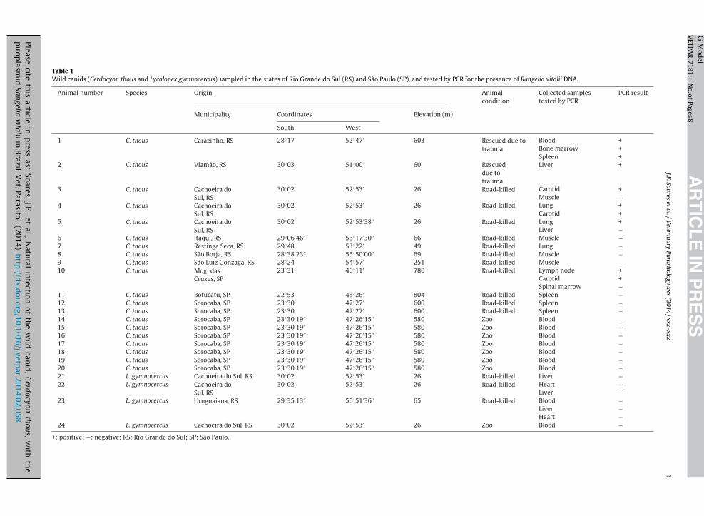
Table 2Results of the hematological and biochemical analyses performed onblood samples of Cerdocyon thous #1 at the 21st day after admission tothe hospital.
Parameters C. thous #1 Reference values(Gomes, 2006)
Erytrocytes (×106/�L) 4.8 4.31–6.77Hemoglobin (g/dL) 14.6 12.96–16.88Packed cell volume % 44 38–49VCM (fL) 91.7 68–95CHCM (%) 33.2 31–38Total Leukocytes (×103/�L) 10 8.1–13.9Segmented (/�L) 6700 5758–10,387Eosinophils(/�L) 1300 189–1336Lymphocytes(/�L) 1900 1062–2357Monocytes (/�L) 100 0–354Platelets (×103/�L) 165 n.a.Total Protein (g/dL) 7.6 5.47–7.09Albumine 34.1 n.a.ALT 17 n.a.Creatinine 0.8 0.37–1.11FA 51 n.a.
ARTICLEVETPAR-7181; No. of Pages 8
4 J.F. Soares et al. / Veterinar
to the lowest Bayesian Information Criterion (BIC) score.The sequence of Hepatozoon canis was used as outgroup.
2.4. Hematological and biochemical evaluation
EDTA-blood samples were processed within 24 h aftercollection. Blood serum samples were separated by cen-trifugation and kept at −20 ◦C until use. Packed cell volume,hemoglobin, erythrocytes, leukocytes and platelets weredetermined in an automatic hematology analyzer (bc2800– Mindray, France). Differential leukocyte count was per-formed on blood smears stained with Rosenfeld staining(Rosenfeld, 1947). Blood smears were prepared within10 min after blood collection. Biochemical analyses weredetermined in an automatic analyzer (Labmax240, Japan)using the commercial kits Randox® (serum urea, totalprotein, and albumin); Labtest® (serum creatinine) andByosistems® (ALT, FA, and bilirubin). The parametersreported by Gomes (2006) and Feldman et al. (2000a,b)were taken as references values for C. thous and domesticdogs, respectively.
2.5. Ethics statements
This work was authorized by the ICMbio Cetas (state ofRio Grande do Sul), and was approved by the Ethical Com-mittee of Animal Use of the Faculty of Veterinary Medicineof the University of São Paulo (protocol 2248/2011).
3. Results
3.1. Wild canids
The C. thous #1 (Table 1) was found infested by Ambly-omma aureolatum adult ticks, but had no clinical signsof tick-borne diseases; fever, anemia, lymphadenopathy,skin hemorrhagic lesions, or jaundice were not observed.In addition, hematological and biochemical analyses per-formed on day 21 showed no abnormalities, apart from aslight increase in total protein (Table 2). Blood samples col-lected on 0 and 69 days after admission, and bone marrowand spleen samples collected on day 80 were PCR-positivefor piroplasmids.
Regarding the remaining wild canids (animals #2 to#24), 5 out of 19 C. thous were PCR-positive for piroplas-mids, whereas the 4 L. gymnocercus were negative. Overall,30% (6/20) C. thous were PCR-positive. Considering only thefree-ranging C. thous, 46.2% (6/13) were PCR-positive, sinceall captive animals were PCR-negative. Amplicons gener-ated in all piroplasma-PCR assays were DNA sequenced,and showed to be 99–100% identical to corresponding18S rRNA sequences of R. vitalii available in GenBank(HQ150006, JN880430, JN880431, JN880432, JN880433,KF218605, KF218606). DNA samples from all 24 wild canidsyielded amplicons of the expected size by the mtDNA con-trol region or the cyt b PCR assay. Taxonomic identification
Please cite this article in press as: Soares, J.F., et al., Naturalpiroplasmid Rangelia vitalii in Brazil. Vet. Parasitol. (2014), http
of 13 free-ranging canids (including the five ones that wereR. vitalii-PCR positive) were confirmed by sequencing theirmtDNA control region or cyt b partial sequence, which were98–100% identical to corresponding sequences of C. thous
Urea 41 22.46–71.84
n.a.: not available.
(DQ309764, EF107009, EF107010, EF107022, EF107030) orL. gymnocercus (EF107034, EF107037) in GenBank.
DNA sequences generated in the present study havebeen deposited in GenBank under the accession num-bers KF964146–KF964151 for 18S rRNA partial sequencesof R. vitalii, KF964152–KF964154 for mtDNA cyt b, andKF964155–KF964164 for mtDNA control region partialsequences of wild canids.
3.2. Domestic dog
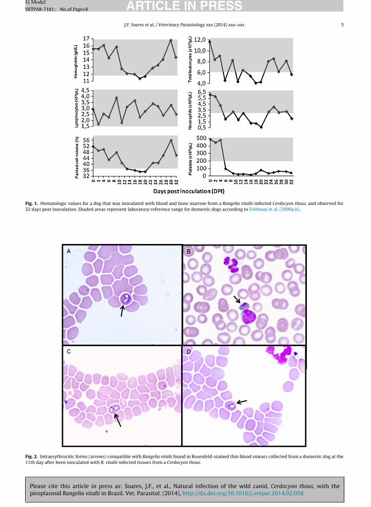
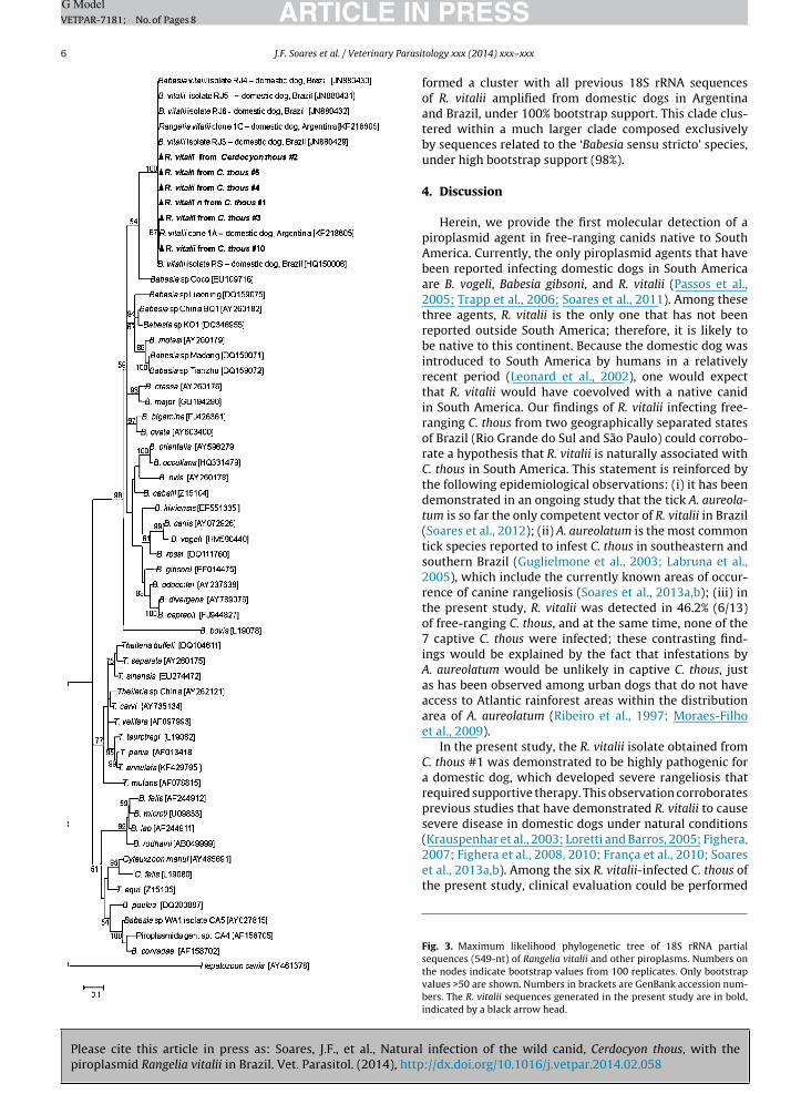
The domestic dog that was inoculated with C. thous#1-blood and bone marrow showed a number of clinicalabnormalities, i.e., anorexia, apathy, decreased body con-dition, vomiting, mild dehydration, mildly pale mucousmembranes, mild fever, and bloody diarrhea, which startedon the 11th dpi and lasted until 17–20 dpi. The mostmarked hematological alteration was thrombocytopenia,which started on the 6th dpi and lasted the whole obser-vation period (Fig. 1). Other hematological alterationsobserved during this period were decreased values forpacked cell volume, hemoglobin, neutrophils, and total leu-cocytes, as shown in Fig. 1. Blood samples were tested bythe Piroplasma-PCR on 0, 2, 4, 32, and 69 dpi; except fordays 0 and 2, all samples were PCR-positive. PCR prod-ucts were DNA sequenced and showed to be 100% identicalto R. vitalii (HQ150006, JN880433). Blood smears revealedintraerythrocitic forms compatible with R. vitalii from 6to 20 dpi (Fig. 2). Within this period, blood smears alsorevealed anisocytosis, polychromasia, erythroblasts, largerplatelets, and reactive lymphocytes. Supportive therapy,initiated at 12 dpi, was continued until 20 dpi, when theanimal improved its clinical condition.
infection of the wild canid, Cerdocyon thous, with the://dx.doi.org/10.1016/j.vetpar.2014.02.058
3.3. Phylogenetic analysis
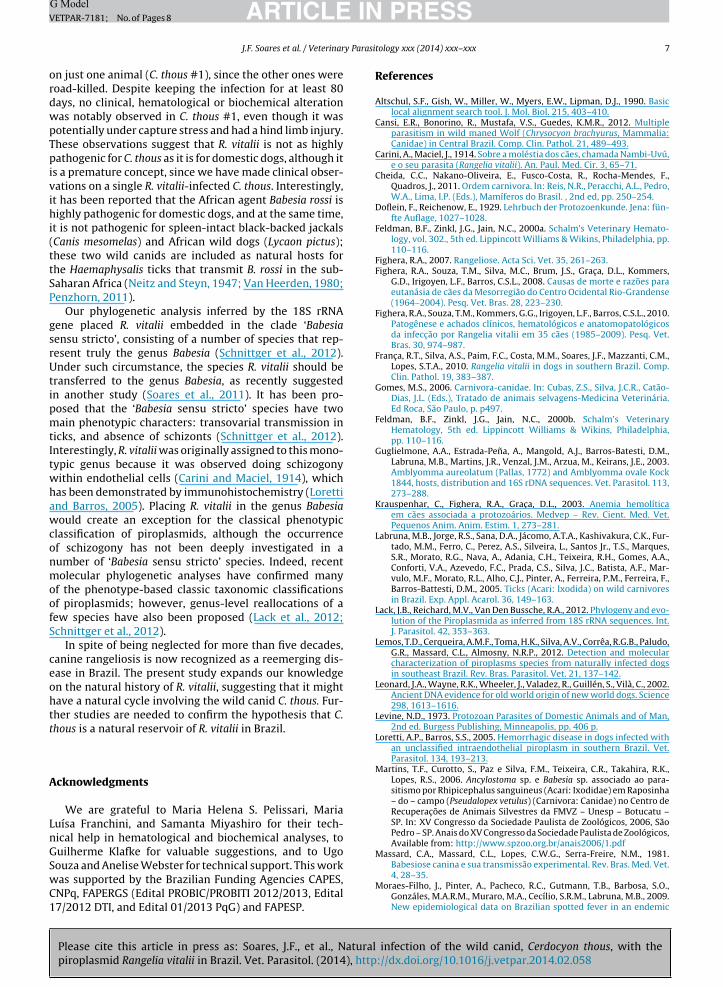
The phylogenetic tree (Fig. 3) depicts that all sixsequences of R. vitalii generated in the present study
Please cite this article in press as: Soares, J.F., et al., Natural infection of the wild canid, Cerdocyon thous, with thepiroplasmid Rangelia vitalii in Brazil. Vet. Parasitol. (2014), http://dx.doi.org/10.1016/j.vetpar.2014.02.058
ARTICLE IN PRESSG ModelVETPAR-7181; No. of Pages 8
J.F. Soares et al. / Veterinary Parasitology xxx (2014) xxx–xxx 5
Fig. 1. Hematologic values for a dog that was inoculated with blood and bone marrow from a Rangelia vitalii-infected Cerdocyon thous, and observed for32 days post inoculation. Shaded areas represent laboratory reference range for domestic dogs according to Feldman et al. (2000a,b).
Fig. 2. Intraerythrocitic forms (arrows) compatible with Rangelia vitalii found in Rosenfeld-stained thin blood smears collected from a domestic dog at the11th day after been inoculated with R. vitalii-infected tissues from a Cerdocyon thous.
Please cite this article in press as: Soares, J.F., et al., Naturalpiroplasmid Rangelia vitalii in Brazil. Vet. Parasitol. (2014), http
ARTICLE ING ModelVETPAR-7181; No. of Pages 8
6 J.F. Soares et al. / Veterinary Parasi
PRESStology xxx (2014) xxx–xxx
formed a cluster with all previous 18S rRNA sequencesof R. vitalii amplified from domestic dogs in Argentinaand Brazil, under 100% bootstrap support. This clade clus-tered within a much larger clade composed exclusivelyby sequences related to the ‘Babesia sensu stricto’ species,under high bootstrap support (98%).
4. Discussion
Herein, we provide the first molecular detection of apiroplasmid agent in free-ranging canids native to SouthAmerica. Currently, the only piroplasmid agents that havebeen reported infecting domestic dogs in South Americaare B. vogeli, Babesia gibsoni, and R. vitalii (Passos et al.,2005; Trapp et al., 2006; Soares et al., 2011). Among thesethree agents, R. vitalii is the only one that has not beenreported outside South America; therefore, it is likely tobe native to this continent. Because the domestic dog wasintroduced to South America by humans in a relativelyrecent period (Leonard et al., 2002), one would expectthat R. vitalii would have coevolved with a native canidin South America. Our findings of R. vitalii infecting free-ranging C. thous from two geographically separated statesof Brazil (Rio Grande do Sul and São Paulo) could corrobo-rate a hypothesis that R. vitalii is naturally associated withC. thous in South America. This statement is reinforced bythe following epidemiological observations: (i) it has beendemonstrated in an ongoing study that the tick A. aureola-tum is so far the only competent vector of R. vitalii in Brazil(Soares et al., 2012); (ii) A. aureolatum is the most commontick species reported to infest C. thous in southeastern andsouthern Brazil (Guglielmone et al., 2003; Labruna et al.,2005), which include the currently known areas of occur-rence of canine rangeliosis (Soares et al., 2013a,b); (iii) inthe present study, R. vitalii was detected in 46.2% (6/13)of free-ranging C. thous, and at the same time, none of the7 captive C. thous were infected; these contrasting find-ings would be explained by the fact that infestations byA. aureolatum would be unlikely in captive C. thous, justas has been observed among urban dogs that do not haveaccess to Atlantic rainforest areas within the distributionarea of A. aureolatum (Ribeiro et al., 1997; Moraes-Filhoet al., 2009).
In the present study, the R. vitalii isolate obtained fromC. thous #1 was demonstrated to be highly pathogenic fora domestic dog, which developed severe rangeliosis thatrequired supportive therapy. This observation corroboratesprevious studies that have demonstrated R. vitalii to causesevere disease in domestic dogs under natural conditions
infection of the wild canid, Cerdocyon thous, with the://dx.doi.org/10.1016/j.vetpar.2014.02.058
(Krauspenhar et al., 2003; Loretti and Barros, 2005; Fighera,2007; Fighera et al., 2008, 2010; Franc a et al., 2010; Soareset al., 2013a,b). Among the six R. vitalii-infected C. thous ofthe present study, clinical evaluation could be performed
Fig. 3. Maximum likelihood phylogenetic tree of 18S rRNA partialsequences (549-nt) of Rangelia vitalii and other piroplasms. Numbers onthe nodes indicate bootstrap values from 100 replicates. Only bootstrapvalues >50 are shown. Numbers in brackets are GenBank accession num-bers. The R. vitalii sequences generated in the present study are in bold,indicated by a black arrow head.
ING ModelV
y Parasi
ordwpTpivihi(ttSP
gsrUtipmtItwhawconmoofS
ceohtt
A
LnGSwC1
ARTICLEETPAR-7181; No. of Pages 8
J.F. Soares et al. / Veterinar
n just one animal (C. thous #1), since the other ones wereoad-killed. Despite keeping the infection for at least 80ays, no clinical, hematological or biochemical alterationas notably observed in C. thous #1, even though it wasotentially under capture stress and had a hind limb injury.hese observations suggest that R. vitalii is not as highlyathogenic for C. thous as it is for domestic dogs, although it
s a premature concept, since we have made clinical obser-ations on a single R. vitalii-infected C. thous. Interestingly,t has been reported that the African agent Babesia rossi isighly pathogenic for domestic dogs, and at the same time,
t is not pathogenic for spleen-intact black-backed jackalsCanis mesomelas) and African wild dogs (Lycaon pictus);hese two wild canids are included as natural hosts forhe Haemaphysalis ticks that transmit B. rossi in the sub-aharan Africa (Neitz and Steyn, 1947; Van Heerden, 1980;enzhorn, 2011).
Our phylogenetic analysis inferred by the 18S rRNAene placed R. vitalii embedded in the clade ‘Babesiaensu stricto’, consisting of a number of species that rep-esent truly the genus Babesia (Schnittger et al., 2012).nder such circumstance, the species R. vitalii should be
ransferred to the genus Babesia, as recently suggestedn another study (Soares et al., 2011). It has been pro-osed that the ‘Babesia sensu stricto’ species have twoain phenotypic characters: transovarial transmission in
icks, and absence of schizonts (Schnittger et al., 2012).nterestingly, R. vitalii was originally assigned to this mono-ypic genus because it was observed doing schizogonyithin endothelial cells (Carini and Maciel, 1914), whichas been demonstrated by immunohistochemistry (Lorettind Barros, 2005). Placing R. vitalii in the genus Babesiaould create an exception for the classical phenotypic
lassification of piroplasmids, although the occurrencef schizogony has not been deeply investigated in aumber of ‘Babesia sensu stricto’ species. Indeed, recentolecular phylogenetic analyses have confirmed many
f the phenotype-based classic taxonomic classificationsf piroplasmids; however, genus-level reallocations of aew species have also been proposed (Lack et al., 2012;chnittger et al., 2012).
In spite of being neglected for more than five decades,anine rangeliosis is now recognized as a reemerging dis-ase in Brazil. The present study expands our knowledgen the natural history of R. vitalii, suggesting that it mightave a natural cycle involving the wild canid C. thous. Fur-her studies are needed to confirm the hypothesis that C.hous is a natural reservoir of R. vitalii in Brazil.
cknowledgments
We are grateful to Maria Helena S. Pelissari, Mariauísa Franchini, and Samanta Miyashiro for their tech-ical help in hematological and biochemical analyses, touilherme Klafke for valuable suggestions, and to Ugo
Please cite this article in press as: Soares, J.F., et al., Naturalpiroplasmid Rangelia vitalii in Brazil. Vet. Parasitol. (2014), http
ouza and Anelise Webster for technical support. This workas supported by the Brazilian Funding Agencies CAPES,NPq, FAPERGS (Edital PROBIC/PROBITI 2012/2013, Edital7/2012 DTI, and Edital 01/2013 PqG) and FAPESP.
PRESStology xxx (2014) xxx–xxx 7
References
Altschul, S.F., Gish, W., Miller, W., Myers, E.W., Lipman, D.J., 1990. Basiclocal alignment search tool. J. Mol. Biol. 215, 403–410.
Cansi, E.R., Bonorino, R., Mustafa, V.S., Guedes, K.M.R., 2012. Multipleparasitism in wild maned Wolf (Chrysocyon brachyurus, Mammalia:Canidae) in Central Brazil. Comp. Clin. Pathol. 21, 489–493.
Carini, A., Maciel, J., 1914. Sobre a moléstia dos cães, chamada Nambi-Uvú,e o seu parasita (Rangelia vitalii). An. Paul. Med. Cir. 3, 65–71.
Cheida, C.C., Nakano-Oliveira, E., Fusco-Costa, R., Rocha-Mendes, F.,Quadros, J., 2011. Ordem carnivora. In: Reis, N.R., Peracchi, A.L., Pedro,W.A., Lima, I.P. (Eds.), Mamíferos do Brasil. , 2nd ed, pp. 250–254.
Doflein, F., Reichenow, E., 1929. Lehrbuch der Protozoenkunde. Jena: fün-fte Auflage, 1027–1028.
Feldman, B.F., Zinkl, J.G., Jain, N.C., 2000a. Schalm’s Veterinary Hemato-logy, vol. 302., 5th ed. Lippincott Williams & Wikins, Philadelphia, pp.110–116.
Fighera, R.A., 2007. Rangeliose. Acta Sci. Vet. 35, 261–263.Fighera, R.A., Souza, T.M., Silva, M.C., Brum, J.S., Grac a, D.L., Kommers,
G.D., Irigoyen, L.F., Barros, C.S.L., 2008. Causas de morte e razões paraeutanásia de cães da Mesorregião do Centro Ocidental Rio-Grandense(1964–2004). Pesq. Vet. Bras. 28, 223–230.
Fighera, R.A., Souza, T.M., Kommers, G.G., Irigoyen, L.F., Barros, C.S.L., 2010.Patogênese e achados clínicos, hematológicos e anatomopatológicosda infecc ão por Rangelia vitalii em 35 cães (1985–2009). Pesq. Vet.Bras. 30, 974–987.
Franc a, R.T., Silva, A.S., Paim, F.C., Costa, M.M., Soares, J.F., Mazzanti, C.M.,Lopes, S.T.A., 2010. Rangelia vitalii in dogs in southern Brazil. Comp.Clin. Pathol. 19, 383–387.
Gomes, M.S., 2006. Carnivora-canidae. In: Cubas, Z.S., Silva, J.C.R., Catão-Dias, J.L. (Eds.), Tratado de animais selvagens-Medicina Veterinária.Ed Roca, São Paulo, p. p497.
Feldman, B.F., Zinkl, J.G., Jain, N.C., 2000b. Schalm’s VeterinaryHematology, 5th ed. Lippincott Williams & Wikins, Philadelphia,pp. 110–116.
Guglielmone, A.A., Estrada-Pena, A., Mangold, A.J., Barros-Batesti, D.M.,Labruna, M.B., Martins, J.R., Venzal, J.M., Arzua, M., Keirans, J.E., 2003.Amblyomma aureolatum (Pallas, 1772) and Amblyomma ovale Kock1844, hosts, distribution and 16S rDNA sequences. Vet. Parasitol. 113,273–288.
Krauspenhar, C., Fighera, R.A., Grac a, D.L., 2003. Anemia hemolíticaem cães associada a protozoários. Medvep – Rev. Cient. Med. Vet.Pequenos Anim. Anim. Estim. 1, 273–281.
Labruna, M.B., Jorge, R.S., Sana, D.A., Jácomo, A.T.A., Kashivakura, C.K., Fur-tado, M.M., Ferro, C., Perez, A.S., Silveira, L., Santos Jr., T.S., Marques,S.R., Morato, R.G., Nava, A., Adania, C.H., Teixeira, R.H., Gomes, A.A.,Conforti, V.A., Azevedo, F.C., Prada, C.S., Silva, J.C., Batista, A.F., Mar-vulo, M.F., Morato, R.L., Alho, C.J., Pinter, A., Ferreira, P.M., Ferreira, F.,Barros-Battesti, D.M., 2005. Ticks (Acari: Ixodida) on wild carnivoresin Brazil. Exp. Appl. Acarol. 36, 149–163.
Lack, J.B., Reichard, M.V., Van Den Bussche, R.A., 2012. Phylogeny and evo-lution of the Piroplasmida as inferred from 18S rRNA sequences. Int.J. Parasitol. 42, 353–363.
Lemos, T.D., Cerqueira, A.M.F., Toma, H.K., Silva, A.V., Corrêa, R.G.B., Paludo,G.R., Massard, C.L., Almosny, N.R.P., 2012. Detection and molecularcharacterization of piroplasms species from naturally infected dogsin southeast Brazil. Rev. Bras. Parasitol. Vet. 21, 137–142.
Leonard, J.A., Wayne, R.K., Wheeler, J., Valadez, R., Guillén, S., Vilà, C., 2002.Ancient DNA evidence for old world origin of new world dogs. Science298, 1613–1616.
Levine, N.D., 1973. Protozoan Parasites of Domestic Animals and of Man,2nd ed. Burgess Publishing, Minneapolis, pp. 406 p.
Loretti, A.P., Barros, S.S., 2005. Hemorrhagic disease in dogs infected withan unclassified intraendothelial piroplasm in southern Brazil. Vet.Parasitol. 134, 193–213.
Martins, T.F., Curotto, S., Paz e Silva, F.M., Teixeira, C.R., Takahira, R.K.,Lopes, R.S., 2006. Ancylostoma sp. e Babesia sp. associado ao para-sitismo por Rhipicephalus sanguineus (Acari: Ixodidae) em Raposinha– do – campo (Pseudalopex vetulus) (Carnivora: Canidae) no Centro deRecuperac ões de Animais Silvestres da FMVZ – Unesp – Botucatu –SP. In: XV Congresso da Sociedade Paulista de Zoológicos, 2006, SãoPedro – SP. Anais do XV Congresso da Sociedade Paulista de Zoológicos,Available from: http://www.spzoo.org.br/anais2006/1.pdf
Massard, C.A., Massard, C.L., Lopes, C.W.G., Serra-Freire, N.M., 1981.Babesiose canina e sua transmissão experimental. Rev. Bras. Med. Vet.
infection of the wild canid, Cerdocyon thous, with the://dx.doi.org/10.1016/j.vetpar.2014.02.058
4, 28–35.Moraes-Filho, J., Pinter, A., Pacheco, R.C., Gutmann, T.B., Barbosa, S.O.,
Gonzáles, M.A.R.M., Muraro, M.A., Cecílio, S.R.M., Labruna, M.B., 2009.New epidemiological data on Brazilian spotted fever in an endemic
ING Model
y Parasi
(Lycaon pictus) (Temminck) and black-backed jackal Canis mesomelas
ARTICLEVETPAR-7181; No. of Pages 8
8 J.F. Soares et al. / Veterinar
area of the state of São Paulo, Brazil. Vector-Borne Zoonotic Dis. 9,73–78.
Moreira, J., 1938. Sobre a natureza do nambiuvú dos cães. Arq. Inst. Biol.de S. Paulo 9, 315–319.
Moreira, J., 1939. O Nambiuvú. O Biológico 6, 113–116.Neitz, W.O., Steyn, H.P., 1947. The transmission of Babesia canis (Piana
and Galli-Valerio, 1895) to the black-backed jackal [Thos mesomelasmesomelas (Schreber)] with a discussion of the classification of thepiroplasms of the Canidae. J. S. Afr. Vet. Med. Assoc. 18, 1–12.
Paraense, W.L., Vianna, Y.L., 1948. Algumas observac ões sobre a babesiosedos cães no Rio de Janeiro. Mem. Inst. Oswaldo Cruz 46, 595–603.
Passos, L.M., Geiger, S.M., Ribeiro, M.F.B., Pfister, K., Zahlerrinder, M., 2005.First molecular detection of Babesia vogeli in dogs from Brazil. Vet.Parasitol. 127, 81–85.
Trapp, S.M., Messick, J.B., Vidotto, O., Jojima, F.S., Morais, H.S.A., 2006.Babesia gibsoni genotype Asia in dogs from Brazil. Vet. Parasitol. 141,177–180.
Peirce, M.A., 2000. Order piroplasmorida. In: Lee, J.J., Leedale, G.F., Brad-bury, P. (Eds.), The Illustrated Guide to the Protozoa, vol. I, 2nd ed.Society of Protozoologists, Lawrence, pp. 347–353.
Penzhorn, B.L., 2011. Why is Southern African canine babesiosis so viru-lent? An evolutionary perspective. Parasit. Vect. 51, 1–6.
Pestana, B.R., 1910. O nambiuvú. Rev. Méd. S. Paulo 22, 423–426.Ribeiro, V.L.S., Weber, M.A., Fetzer, L.O., Vargas, C.R.B., 1997. Espécies e
prevalência das infestac ões por carrapatos em cães de rua da cidadede Porto Alegre, RS, Brasil. Ciênc. Rural. 27, 285–289.
Rosenfeld, G., 1947. Corante pancrômico para hematologia e citologiaclínica, Nova combinac ão dos componentes do May-Grunwald e doGiemsa num só corante de emprego rápido. Mem. Inst. Butantan 20,329–335.
Ruas, J.L., Farias, N.A.R., Soares, M.P., Brum, J.G.W., 2003. Babesia sp. emgraxaim do campo (Lycalopex gymnocercus) no Sul do Brasil. Arq. Inst.Biol. 70, 113–114.
Schnittger, L., Rodriguez, A.E., Florin-Christensen, M., Morrison, D.A., 2012.Babesia: a world emerging. Infect. Genet. Evol. 12, 1788–1809.
Serra-Freire, N.M., Teixeira, R.H.F., Amorim, M., Gazeta, G.S., Nunes, A.L.V.,Yada, H.S., Teixeira, C., 1995. Babesiose associada ao parasitismo porcarrapatos em lobo guará. In: XIX Congresso da Sociedade de Zoológi-
Please cite this article in press as: Soares, J.F., et al., Naturalpiroplasmid Rangelia vitalii in Brazil. Vet. Parasitol. (2014), http
cos do Brasil: Foz do Iguac ú, Arquivos da Sociedade de Zoológicos doBrasil, 9 p.
Soares, J.F., Girotto, A., Brandão, P.E., Da Silva, A.S., Franc a, R.T., Lopes, S.T.A.,Labruna, M.B., 2011. Detection and molecular characterization of acanine piroplasm from Brazil. Vet. Parasitol. 180, 203–208.
PRESStology xxx (2014) xxx–xxx
Soares, J.F., Costa, F.B., Soares, H.S., Da Silva, A.S., Franc a, R.T., Miyashiro,S., Lopes, S.T.A., Monteiro, S.G., Hagiwara, M.K., Labruna, M.B.,2012. Caracterizac ão morfológica, molecular e estudos dos ixodídeosvetores de Rangelia vitalii. In: XVII Congresso Brasileiro de Parasitolo-gia Veterinária Set/2012 São Luiz do Maranhão, Brasil, Anais XVII Con-gresso Brasileiro de Parasitologia Veterinária, p. 221, Available from:http://www.cbpv.com.br/congressos/Anais XVII CBPV FINAL.pdf
Soares, J.F., Girotto, A., Dalmolin, M.L., Franc a, R.T., Hlavac, N.R.C., Moroz,L.R., Alves, C.B.R., Salomão, E.L., Pelissari, M.H.S., Franchini, M.L.,Miyashiro, S., Lopes, S.T.A., Lacerda, L.A., Hagiwara, M.K., Labruna,M.B., 2013a. Deteccão molecular de Rangelia vitalii nos Estados de SãoPaulo, Santa Catarina e Rio Grande do Sul. In: IV Simpósio Brasileirode Acarologia: Maio/2013 Bento Gonc alves-RS, Brasil 2013, Anais doIV Simpósio Brasileiro de Acarologia, CD-ROM.
Soares, J.F., Corrêa, S., Pelissari, M.H.S., Franchini, M.L., Miyashiro, S.,Hagiwara, M.K., Labruna, M.B.,2013b. Detecc ão molecular de Rangeliavitalii no Estado de Minas Gerais. In: III Simpósio Estadual de Doenc asTransmitidas por Carrapatos: Outubro/2013 Campinas-RS, Brasil2013. Boletim Epidemiológico Paulista, vol. 10, Anais, p. 25, Availableat: http://www.saude.sp.gov.br/resources/ccd/homepage/bepa/edicoes-2013/edicao 118 - outubro especial carrapatos parte 2 2.pdf
Steuber, S., Abdel-Rady, A., Clausen, P., 2005. PCR-RFLP analysis: a promis-ing technique for host species identification of blood meals from tsetseflies (Diptera: Glossinidae). Parasitol. Res. 97, 247–254.
Tamura, K., Peterson, D., Peterson, N., Stecher, G., Nei, M., Kumar, S., 2011.MEGA5: molecular evolutionary genetics analysis using maximumlikelihood, evolutionary distance, and maximum parsimony method.Mol. Biol. Evol. 28, 2731–2739.
Tchaicka, L., Eizirik, E., Oliveira, T.G., Candido Jr., J.F., Freitas, T.R.O., 2007.Phylogeography and population history of the crab-eating fox (Cerdo-cyon thous). Mol. Ecol. 16, 819–838.
Thompson, J.D., Higgins, D.G., Gibson, T.J., 1994. CLUSTAL W: improvingthe sensitivity of progressive multiple sequence alignment throughsequence weighting, position specific gap penalties and weight matrixchoice. Nucleic Acids Res. 22, 4673–4680.
Van Heerden, J., 1980. The transmission of Babesia canis to the wild dog
infection of the wild canid, Cerdocyon thous, with the://dx.doi.org/10.1016/j.vetpar.2014.02.058
(Schreber). J. S. Afr. Vet. Assoc. 18, 119–120.Wenyon, C.M., 1926. Protozoology: A Manual for Medical Men, Veterinar-
ians and Zoologists, vol. II., 1st ed. Baillière Tindall and Cox, London,pp. 991–1022.
Related Documents


