Natural Antisense Transcript for Hyaluronan Synthase 2 (HAS2-AS1) Induces Transcription of HAS2 via Protein O-GlcNAcylation * Received for publication, August 12, 2014 Published, JBC Papers in Press, September 2, 2014, DOI 10.1074/jbc.M114.597401 Davide Vigetti ‡1 , Sara Deleonibus ‡1,2 , Paola Moretto ‡ , Timothy Bowen § , Jens W. Fischer ¶ , Maria Grandoch ¶ , Alexander Oberhuber , Dona C. Love**, John A. Hanover**, Raffaella Cinquetti ‡‡ , Eugenia Karousou ‡ , Manuela Viola ‡ , Maria Luisa D’Angelo ‡2 , Vincent C. Hascall §§ , Giancarlo De Luca ‡ , and Alberto Passi ‡3 From the ‡ Dipartimento di Scienze Chirurgiche e Morfologiche, Università degli Studi dell’Insubria, Varese, Italy, § Department of Nephrology, Cardiff University School of Medicine, Heath Park, Cardiff CF14 4XN, United Kingdom, ¶ Institute of Pharmacology and Clinical Pharmacology, University Hospital Düsseldorf, Heinrich-Heine-University Düsseldorf, 40225 Düsseldorf, Germany, Department of Vascular and Endovascular Surgery, University Hospital Düsseldorf, Heinrich-Heine-University Düsseldorf, Düsseldorf, Germany, **Laboratory of Cell Biochemistry and Biology, NIDDK, National Institutes of Health, Bethesda, Maryland 20892, ‡‡ Dipertimento di Biotecnologia e Scienze della Vita, Università degli Studi dell’Insubria, 21100 Varese, Italy, and §§ Biomedical Engineering ND20, The Cleveland Clinic, Cleveland, Ohio 44195 Background: Intracellular proteins glycosylation with O-GlcNAc is able to influence cell microenvironment. Results: O-GlcNAcylation increases hyaluronan synthase 2 (HAS2) transcription via its natural antisense transcript HAS2-AS1. Conclusion: A novel mechanism to regulate hyaluronan synthesis via long non-coding RNA is described. Significance: This finding highlights a new target to regulate HA synthesis, critical in many pathophysiological processes. Changes in the microenvironment organization within vascu- lar walls are critical events in the pathogenesis of vascular pathologies, including atherosclerosis and restenosis. Hyaluro- nan (HA) accumulation into artery walls supports vessel thick- ening and is involved in many cardiocirculatory diseases. Exces- sive cytosolic glucose can enter the hexosamine biosynthetic pathway, increase UDP-N-acetylglucosamine (UDP-GlcNAc) availability, and lead to modification of cytosolic proteins via O-linked attachment of the monosaccharide -N-GlcNAc (O-GlcNAcylation) from UDP-GlcNAc by the enzyme O-GlcNAc transferase. As many cytoplasmic and nuclear proteins can be glycosylated by O-GlcNAc, we studied whether the expression of the HA synthases that synthesize HA could be controlled by O-GlcNAcylation in human aortic smooth muscle cells. Among the three HAS isoenzymes, only HAS2 mRNA increased after O-GlcNAcylation induced by glucosamine treatments or by inhibiting O-GlcNAc transferase with PUGNAC (O-(2-acet- amido-2-deoxy-D-glucopyranosylidene)amino- N-phenylcarbamate). We found that the natural antisense transcript of HAS2 (HAS2- AS1) was absolutely necessary to induce the transcription of the HAS2 gene. Moreover, we found that O-GlcNAcylation modu- lated HAS2-AS1 promoter activation by recruiting the NF-B subunit p65, but not the HAS2 promoter, whereas HAS2-AS1 natural antisense transcript, working in cis, regulated HAS2 transcription by altering the chromatin structure around the HAS2 proximal promoter via O-GlcNAcylation and acetylation. These results indicate that HAS2 transcription can be finely reg- ulated not only by recruiting transcription factors to the pro- moter as previously described but also by modulating chromatin accessibility by epigenetic modifications. HA 4 is an ubiquitous linear macromolecule composed of glu- curonic acid and N-acetylglucosamine (GlcNAc) without any additional chemical modifications typical of the other glycos- aminoglycans. HA can modulate a plethora of cellular functions in both physiological as well as pathological conditions (1), interacting with many receptors and proteins (2). HA is synthe- sized on the plasma membrane by three isoenzymes, HA syn- thase 1, 2, and 3 (HAS1, -2, and -3), that have several transmem- brane domains, use cytosolic UDP-glucuronic acid, and UDP-GlcNAc as precursors and extrude the elongating chain through the plasma membrane into the extracellular matrix. HA synthesis is known to be finely regulated by several factors, including growth factors and proinflammatory cytokines, at both transcriptional and post-translational levels (3, 4). Cardiovascular diseases are the leading causes of death in western countries and are characterized by a strong extracellu- lar matrix remodeling with high deposition of HA in the neointima (5–7). HA has been described to have a critical role favoring vessel wall thickening and neointima formation (8 –10), and inhibition of HA synthesis can be considered a new strategy to face vascular pathologies (11, 12). * This work was supported by FAR, PRIN, Fondazione Comunitaria del Vare- sotto-ONLUS, and European Union Grant IRSES INFLAMA (to A. P.). 1 Both authors contributed equally to this work. 2 Recipient of a fellowship from the Ph.D. School in Biological and Medical Sciences. 3 To whom correspondence should be addressed: Dipartimento di Scienze Chirurgiche e Morfologiche, Università degli Studi dell’Insubria, via J. H. Dunant 5, 21100 Varese, Italy. Tel.: 39-0332-397142; Fax: 39-0332-397119; E-mail: [email protected]. 4 The abbreviations used are: HA, hyaluronan; O-GlcNAcylation, O-linked N-acetylglucosamine; OGA, O-GlcNAcase; HAS, hyaluronan synthase; AoSMCs, human aortic smooth muscle cells; NAT, natural antisense tran- script; PUGNAC, O-(2-acetamido-2-deoxy-d-glucopyranosylidene)amino- N-phenylcarbamate; PDTC, pyrrolidine dithiocarbamate; ActD, actinomy- cin D; qPCR, quantitative PCR; Ct, threshold cycle; IP, immunoprecipitation; L, long; S, short. THE JOURNAL OF BIOLOGICAL CHEMISTRY VOL. 289, NO. 42, pp. 28816 –28826, October 17, 2014 Published in the U.S.A. 28816 JOURNAL OF BIOLOGICAL CHEMISTRY VOLUME 289 • NUMBER 42 • OCTOBER 17, 2014 by guest on March 27, 2016 http://www.jbc.org/ Downloaded from

Welcome message from author
This document is posted to help you gain knowledge. Please leave a comment to let me know what you think about it! Share it to your friends and learn new things together.
Transcript

Natural Antisense Transcript for Hyaluronan Synthase 2(HAS2-AS1) Induces Transcription of HAS2 via ProteinO-GlcNAcylation*
Received for publication, August 12, 2014 Published, JBC Papers in Press, September 2, 2014, DOI 10.1074/jbc.M114.597401
Davide Vigetti‡1, Sara Deleonibus‡1,2, Paola Moretto‡, Timothy Bowen§, Jens W. Fischer¶, Maria Grandoch¶,Alexander Oberhuber�, Dona C. Love**, John A. Hanover**, Raffaella Cinquetti‡‡, Eugenia Karousou‡,Manuela Viola‡, Maria Luisa D’Angelo‡2, Vincent C. Hascall§§, Giancarlo De Luca‡, and Alberto Passi‡3
From the ‡Dipartimento di Scienze Chirurgiche e Morfologiche, Università degli Studi dell’Insubria, Varese, Italy, §Department ofNephrology, Cardiff University School of Medicine, Heath Park, Cardiff CF14 4XN, United Kingdom, ¶Institute of Pharmacology andClinical Pharmacology, University Hospital Düsseldorf, Heinrich-Heine-University Düsseldorf, 40225 Düsseldorf, Germany,�Department of Vascular and Endovascular Surgery, University Hospital Düsseldorf, Heinrich-Heine-University Düsseldorf,Düsseldorf, Germany, **Laboratory of Cell Biochemistry and Biology, NIDDK, National Institutes of Health, Bethesda, Maryland20892, ‡‡Dipertimento di Biotecnologia e Scienze della Vita, Università degli Studi dell’Insubria, 21100 Varese, Italy, and§§Biomedical Engineering ND20, The Cleveland Clinic, Cleveland, Ohio 44195
Background: Intracellular proteins glycosylation with O-GlcNAc is able to influence cell microenvironment.Results: O-GlcNAcylation increases hyaluronan synthase 2 (HAS2) transcription via its natural antisense transcript HAS2-AS1.Conclusion: A novel mechanism to regulate hyaluronan synthesis via long non-coding RNA is described.Significance: This finding highlights a new target to regulate HA synthesis, critical in many pathophysiological processes.
Changes in the microenvironment organization within vascu-lar walls are critical events in the pathogenesis of vascularpathologies, including atherosclerosis and restenosis. Hyaluro-nan (HA) accumulation into artery walls supports vessel thick-ening and is involved in many cardiocirculatory diseases. Exces-sive cytosolic glucose can enter the hexosamine biosyntheticpathway, increase UDP-N-acetylglucosamine (UDP-GlcNAc)availability, and lead to modification of cytosolic proteins viaO-linked attachment of the monosaccharide �-N-GlcNAc(O-GlcNAcylation) from UDP-GlcNAc by the enzyme O-GlcNActransferase. As many cytoplasmic and nuclear proteins can beglycosylated by O-GlcNAc, we studied whether the expressionof the HA synthases that synthesize HA could be controlled byO-GlcNAcylation in human aortic smooth muscle cells. Amongthe three HAS isoenzymes, only HAS2 mRNA increased afterO-GlcNAcylation induced by glucosamine treatments or byinhibiting O-GlcNAc transferase with PUGNAC (O-(2-acet-amido-2-deoxy-D-glucopyranosylidene)amino-N-phenylcarbamate).We found that the natural antisense transcript of HAS2 (HAS2-AS1) was absolutely necessary to induce the transcription of theHAS2 gene. Moreover, we found that O-GlcNAcylation modu-lated HAS2-AS1 promoter activation by recruiting the NF-�Bsubunit p65, but not the HAS2 promoter, whereas HAS2-AS1natural antisense transcript, working in cis, regulated HAS2transcription by altering the chromatin structure around theHAS2 proximal promoter via O-GlcNAcylation and acetylation.
These results indicate that HAS2 transcription can be finely reg-ulated not only by recruiting transcription factors to the pro-moter as previously described but also by modulating chromatinaccessibility by epigenetic modifications.
HA4 is an ubiquitous linear macromolecule composed of glu-curonic acid and N-acetylglucosamine (GlcNAc) without anyadditional chemical modifications typical of the other glycos-aminoglycans. HA can modulate a plethora of cellular functionsin both physiological as well as pathological conditions (1),interacting with many receptors and proteins (2). HA is synthe-sized on the plasma membrane by three isoenzymes, HA syn-thase 1, 2, and 3 (HAS1, -2, and -3), that have several transmem-brane domains, use cytosolic UDP-glucuronic acid, andUDP-GlcNAc as precursors and extrude the elongating chainthrough the plasma membrane into the extracellular matrix.HA synthesis is known to be finely regulated by several factors,including growth factors and proinflammatory cytokines, atboth transcriptional and post-translational levels (3, 4).
Cardiovascular diseases are the leading causes of death inwestern countries and are characterized by a strong extracellu-lar matrix remodeling with high deposition of HA in theneointima (5–7). HA has been described to have a critical rolefavoring vessel wall thickening and neointima formation(8 –10), and inhibition of HA synthesis can be considered a newstrategy to face vascular pathologies (11, 12).
* This work was supported by FAR, PRIN, Fondazione Comunitaria del Vare-sotto-ONLUS, and European Union Grant IRSES INFLAMA (to A. P.).
1 Both authors contributed equally to this work.2 Recipient of a fellowship from the Ph.D. School in Biological and Medical
Sciences.3 To whom correspondence should be addressed: Dipartimento di Scienze
Chirurgiche e Morfologiche, Università degli Studi dell’Insubria, via J. H.Dunant 5, 21100 Varese, Italy. Tel.: 39-0332-397142; Fax: 39-0332-397119;E-mail: [email protected].
4 The abbreviations used are: HA, hyaluronan; O-GlcNAcylation, O-linkedN-acetylglucosamine; OGA, O-GlcNAcase; HAS, hyaluronan synthase;AoSMCs, human aortic smooth muscle cells; NAT, natural antisense tran-script; PUGNAC, O-(2-acetamido-2-deoxy-d-glucopyranosylidene)amino-N-phenylcarbamate; PDTC, pyrrolidine dithiocarbamate; ActD, actinomy-cin D; qPCR, quantitative PCR; Ct, threshold cycle; IP, immunoprecipitation;L, long; S, short.
THE JOURNAL OF BIOLOGICAL CHEMISTRY VOL. 289, NO. 42, pp. 28816 –28826, October 17, 2014Published in the U.S.A.
28816 JOURNAL OF BIOLOGICAL CHEMISTRY VOLUME 289 • NUMBER 42 • OCTOBER 17, 2014
by guest on March 27, 2016
http://ww
w.jbc.org/
Dow
nloaded from

Angiopathies are one of the main complications of diabetes(13), and although the molecular mechanisms involved in suchdiseases are poorly understood, the hexosamine biosyntheticpathway appears to have a crucial role (14). In fact, the elevatedflux of nutrients that enters in the hexosamine biosyntheticpathway in hyperglycemic conditions increases the formationof UDP-GlcNAc (15). High availability of UDP-sugars inducesthe synthesis of HA (16 –18).
Elevated levels of UDP-GlcNAc induces the activity ofO-GlcNAc transferase, which catalyzes the transfer of GlcNAcfrom UDP-GlcNAc to serine and threonine residues (O-GlcNA-cylation) (19). This intracellular glycosylation controls manycellular processes and is also involved in diabetes, cardiovascu-lar diseases, and tumors (20, 21). O-GlcNAcylation can be easilyhydrolyzed from proteins by O-GlcNAcase (OGA) (19). Severalnucleo-cytoplasmic proteins are known to be regulated by
O-GlcNAcylation including RNA polymerase 2 (22) and HAS2(16).
HAS2 is mainly responsible for HA synthesis in adult mam-malian tissues, and at the genetic level, HAS2 possesses a natu-ral antisense transcript (NAT) named HAS2-AS1 (23). NAT ispart of long non-coding RNA (24, 25), which has a crucial rolein the modulation of gene transcription (24, 26 –30).
HAS2-AS1 NAT is transcribed in the opposite strand of theHAS2 gene localized on chromosome 8 (Fig. 1). HAS2 exon 1and HAS2-AS1 exon 2 are partially complementary. Further-more, an alternative splicing can generate two HAS2-AS1 NATisoforms, named long (L) and short (S), that have 257 or 174nucleotides of perfect complementary sequence to the regionstarting �70 bp from the presumed transcription start site ofhuman HAS2, respectively (23). This complementarity permitsHAS2 mRNA�HAS2-AS1 NAT duplex formation that stabilizes
FIGURE 1. Genomic organization of HAS2 and HAS2 genes at locus 8q24.13, localization of primers, luciferase constructs, and NF-�B sites. A, schematicrepresentation of HAS2 (RefSeq gene NM_005328.2 on the minus DNA strand) and HAS2-AS1 (RefSeq gene NR_002835 on the plus DNA strand) genes. Thescheme reports the starting and ending positions of the genes as indicated by UCSC site (Genome Bioinformatics). Light blue arrows indicate the transcriptiondirection, and boxes are exons. The scheme is in scale. B, representation of the first exon of HAS2 gene and the exon number 1 and 2 of HAS2-AS1 gene. Thescheme is not in scale, and black arrows indicate the position of the primers used in this work. The three reporter constructs used in this work containing theluciferase (Luc.) under the control of HAS2 promoter (�2118 � 43) or HAS2 promoter with the HAS2�HAS2-AS1 complementarity region (�2118 � 475; HAS2exon 1) or HAS2-AS1 promoter (�700) are indicated. C, genomic sequence around HAS2 transcription start site that is indicated as the �1 position. Lowercaseletters indicate the first 370 nucleotides of the proximal promoter region, whereas uppercase letters indicate part of HAS2 exon 1. The primer position is alsoindicated by arrows. D, the genomic sequence around HAS2-AS1 transcription start site is indicated as the �1 position. Lowercase letters indicate the first 444nucleotides of the proximal promoter region, whereas uppercase letters indicate part of HAS2-AS1 exon 1. The position of primers is also indicated by arrows.The NF-�B subunit p65 binding sites are highlighted in green.
HAS2-AS1 Induces HAS2 Expression
OCTOBER 17, 2014 • VOLUME 289 • NUMBER 42 JOURNAL OF BIOLOGICAL CHEMISTRY 28817
by guest on March 27, 2016
http://ww
w.jbc.org/
Dow
nloaded from

the HAS2 transcript (31) as already described for other mRNA(32).
The aim of this study was to investigate whether O-GlcNA-cylation could modulate HAS2 gene transcription. We foundthat O-GlcNAcylation induces HAS2 mRNA accumulation,and this event is strictly dependent to HAS2-AS1, which canmodulate the chromatin aperture around HAS2 promoterfavoring transcription.
EXPERIMENTAL PROCEDURES
Cell Cultures, Treatments, and Mammalian Samples—Pri-mary human aortic smooth muscle cells (AoSMCs) were pur-chased from Lonza and grown for 5– 8 passages in completeSmGm2 culture medium (Lonza) supplemented with 5% FBS aspreviously described (16, 33). Briefly, 3 � 105 AoSMCs wereincubated with DMEM (Euroclone) supplemented with 0.2%FBS to induce quiescence. After 48 h the medium was changedto normal glucose (5 mM) DMEM-F-12 with 10% FBS, and thecells were left untreated or treated with 2 mM GlcN or 5 mM
alloxan or 100 �M O-(2-acetamido-2-deoxy-D-glucopyrano-sylidene) amino-N-phenylcarbamate (PUGNAC) for 24 h (allfrom Sigma) to modulate protein O-GlcNAcylation (16). Insome experiments 5 �g/ml actinomycin D (ActD) was used toinhibit RNA synthesis as previously described (34), and pyrrol-idine dithiocarbamate (PDTC) was used to block NF-�B activ-ity (35, 36).
Eight-week-old male apolipoprotein E knock-out (apoEknock-out (KO); The Jackson Laboratory, Bar Harbor, ME)mice were fed a Western type diet containing 21% saturated fatand 0.15% cholesterol or normal chow for 4 weeks. At the end ofthe treatment mice were euthanized by asphyxiation, and theaortas were dissected.
cDNA from atherectomy specimens were derived frompatients of both genders undergoing carotid endarterectomywith a disease severity ranging from elective, asymptomatic(stage I), and patients with transitory ischemic attack (stage II)to stroke patients (stage 4). Tissue collection and analysis wereapproved by the Ethics Committee of the Heinrich-Heine-Uni-versity (ethics approvals number 3944) based on the patients’consent. Atherectomy specimens were directly snap-frozen inliquid nitrogen, and total RNA was extracted using peqGOLDTriFast (PEQLAB Biotechnologie GmbH, Erlangen, Germany)according to the manufacturer’s instructions. 1 �g of total RNAwas transcribed into cDNA using the QuantiTect ReverseTranscription kit (Qiagen, Hilden, Germany). Hearts fromwild-type and OGA knock-out newborn mice (37) wereobtained from newborns animals and stored at �80 °C inRNAlater.
Quantitative RT-PCR and Silencing—Total RNAs wereextracted from AoSMCs or from mouse hearts with TRI re-agent (Invitrogen), retro-transcribed using the High CapacitycDNA synthesis kit (Invitrogen), and amplified on an Abi Prism7000 instrument (Applied Biosystems). Pre-developed Taqmangene expression assays (Invitrogen) were used to quantify tran-scripts coding for human HAS2, HAS2-AS1, HAS3, and mouseHAS2 and HAS2-AS1. �-Actin was used as a reference. Therelative gene expression was determined by comparing �Ct(38). Short interfering RNA (siRNA) from Ambion (assay ID
n265529 or a scramble control) were used to abrogate theexpression of HAS2-AS1 by using a nucleoporator (Nucleofec-tor, AMAXA) (36). After 24 h of incubation, AoSMCs weretreated with GlcN as described above to induce O-GlcNA-cylation, and the next day, RNA was extracted to measure geneexpression.
Luciferase Gene Reporter Assay—The reporter vector (basedon pGL3, Promega) expressing luciferase cloned downstreamfrom the HAS2 promoter (�2118/�43 bp) was a gift of KatriMakkonen (39), whereas the luciferase under the control of theHAS2-AS1 promoter (�700 bp) was previously described (31).The promoter of HAS2 and part of HAS2 exon 1 (�2118/�475)were synthesized by Biomatik and cloned in the pGL3 vector(Promega) by using the KpnI and SmaI restriction sites. Thisconstruct contains all the region of HAS2 exon 1 complemen-tary to HAS2-AS1.
One million AoSMCs were nucleofected with the two HAS2promoter-reported plasmids, split 1:2, and seeded in 35-mmdishes wells. After 24 h of incubation, AoSMCs were harvested,and 3 � 105 cells were divided in six-well plates and treated withGlcN or alloxan to modulate protein O-GlcNAcylation. CMVpromoter-luciferase reporter and a pRL-TK vector (Renillaluciferase under the control of thymidine kinase promoter; Pro-mega) were used as a positive control and nucleofection effi-ciency normalization, respectively. The pcDNA3-SP1 express-ing vector (a gift of Professor Stephen Safe) was nucleofectedin AoSMCs with the �2118 � 43 HAS2 promoter-luciferasereporter construct as an additional control. In other experi-ments the HAS2 gene promoter-luciferase plasmids werenucleofected with pcDNA3 vectors expressing HAS2-AS1 (seebelow). After 24 h of incubation, cell extracts were assayed forluciferase activity using Dual-GLO luciferase assay kit (Pro-mega). Luciferase activities were normalized to both the pro-tein content and the nucleofection efficiency calculated byRenilla luciferase activity as previously described (31).
Chromatin Immunoprecipitation (ChIP)—The Pierce aga-rose ChIP kit was used to immunoprecipitate chromatin fol-lowing the manufacturer’s instructions. Briefly, formaldehydewas added to 2 � 106 cells/ChIP reaction to cross-link DNA andproteins. After an incubation of 10 min at room temperature,glycine was added to quench the excess of formaldehyde. Cellswere scraped into cold PBS, pelleted, and resuspended in LysisBuffer 1 containing a 1� Halt mixture. Nuclei were isolated andresuspended in MNase Digestion Buffer. Cross-linked DNAwas digested with 0.25 �l of micrococcal Nuclease (10 units/�l)at 37 °C for 15 min and centrifuged to recover the nuclei.Digested chromatin was resuspended in Lysis Buffer 2. For eachIP, diluted chromatin was incubated overnight with a specificmonoclonal antibody against O-GlcNAcylated proteins (HGAC85;Pierce) (40) or with anti-p65 monoclonal antibody (D14E12,Cell Signal) or with anti RNA polymerase 2 monoclonal anti-body (Pierce; used as positive control) or with normal rabbitIgG (used as negative control) at 4 °C. Each immunoprecipi-tated complex was column-purified using the agarose resin andeluted in IP Elution Buffer. Reverse cross-linked DNA was puri-fied by using a DNA clean-up column according to the manufac-turer’s instructions. 5 �l of each of the purified DNAs was usedas a template for 48 cycles of quantitative PCR (qPCR) amplifi-
HAS2-AS1 Induces HAS2 Expression
28818 JOURNAL OF BIOLOGICAL CHEMISTRY VOLUME 289 • NUMBER 42 • OCTOBER 17, 2014
by guest on March 27, 2016
http://ww
w.jbc.org/
Dow
nloaded from

cation using primers designed on the proximal promoter(��300 bp from the transcription start site) of HAS2 (5�-CTCA-GGGTTCCCCAGTCCACACCTC-3�, 5�-TCTCTGGTTCA-ATGGGCTGCTCGAA-3�), HAS2-AS1 (5�-AGCGGCCTCA-CTCCTTCAGCAAAG-3�, 5�-GACCGTTGCTGCCTGTTG-GGTCTC-3�), HAS2 exon 1 (5�-TTTTAAAGTGGGGAAGA-ATCAAACA-3�, 5�- GGCAGTTTCCAAAATTGAGGTA-ATA-3�) (see Fig. 1 for primer positions), and glyceraldehyde-3-phosphate dehydrogenase (GAPDH, primers provided withthe ChIP kit). HAS2, HAS2-AS1, and HAS2 exon 1 primerswere designed using Primer3 internet site setting parameters tohave an annealing temperature of 60 °C and an amplicon size of100 bp. qPCRs were carried out in two steps: denaturation at95 °C for 30 s and annealing/elongation at 60 °C for 1 min usingthe Abi Prism 7000 instrument (Applied Biosystems) and theSYBR Green qPCR Mastermix (Invitrogen). All primers weretested on genomic DNA to avoid primer dimers and to checkpolymerase efficiency. Normalization was done using the ��Ctmethod. Briefly, IP samples and total input threshold cycles(Ct) for each treatment were subtracted from the Ct of thecorresponding control IP (rabbit IgG). The resulting correctedvalue for the total input was then subtracted from the correctedexperimental IP value (��Ct), and these values were raised tothe power of 2 (2��Ct). These values were then expressed as arelative promoter binding �S.D. as previously described (41).
Nuclease Accessibility Assay—EpiQ chromatin analysis kit(Bio-Rad) was used to perform nuclease accessibility assays fol-lowing the manufacturer’s protocol to measure open and closedchromatin (42). Briefly, AoSMCs were treated with reagents tomodulate O-GlcNAcylation and processed in two treatmentgroups: undigested and digested (each in triplicate). The undi-gested group was not digested with the EpiQ nuclease, and thedigested group was treated with limited EpiQ nuclease diges-tion (1 h at 37 °C) according to the kit instruction. The genomicDNA samples for both groups were isolated and subjected toqPCR to amplify the proximal promoter of HAS2, HAS2-AS1(see sequences above), and HAS3 (5�-GCCCCACTGCGGAATT-CAAAGCTAA-3�, 5�-CCCGCAAAACCTACTCAC-3�) genesby using the conditions described in ChIP section. qPCR results forall samples were normalized to an internal control, a fragment ofan unexpressed rhodopsin gene (RHO) according to the kit in-structions. Nuclease accessibility index was calculated as the ratiobetween the undigested sample and the digested sample at thesame time point. The equation used was: accessibility index (testregion undigested/RHO region undigested):(test region digested/RHO region digested) as previously reported (43).
HAS2-AS1 Overexpression—The two variants of HAS2-AS1(long (L) and short (S)) were generated by an alternative splic-ing within the exon 1 of HAS2-AS1. L-HAS2-AS1 and S-HAS2-AS1 have a complementary region of 257 and 174 bp with exon2 of HAS2, respectively. L-HAS2-AS1 was synthesized byEpoch Life Science Inc. and subcloned in pcDNA3 expressingvector, whereas S-HAS2-AS1 in pcDNA3 was already availablein the Bowen laboratory. Three micrograms of expressing con-struct were nucleofected in AoSMCs. After 24 h of incubation,the gene expression and the nuclease accessibility assays wereperformed as described above.
Statistical Analyses—Statistical analysis of the data was doneusing analysis of variance (analysis of variance) followed by posthoc tests (Bonferroni) using Origin 7.5 software (OriginLab).Probability values of p 0.01 or 0.05 were considered statisti-cally significant. Experiments were repeated three times, eachtime in duplicate, and data are expressed as the means � S.E.
RESULTS
O-GlcNAcylation Induces HAS2 mRNA and HAS2-AS1Transcript Accumulation—As we previously reported (16),treatments with 2 mM GlcN or 100 �M PUGNAC led to anaccumulation of O-GlcNAcylated proteins in AoSMCs after24 h of incubation. The O-GlcNAcylation of HAS2 enzyme inthe plasma membrane together with an increase of UDP-GlcNAc availability induced HA accumulation �3-fold overthe controls (16). As O-GlcNAcylation is also present in manytranscription factors that regulate HA metabolism (i.e. NF-�B,SP1, and YY1) (3, 44), we investigated whether expression of theHA synthases could be controlled by O-GlcNAcylation. Toincrease protein O-GlcNAcylation, quiescent (i.e. serum-starved) AoSMCs were incubated with DMEM-F-12 with 10%FBS, and the cells were left untreated or treated with 2 mM GlcNor 100 �M PUGNAC (an OGA inhibitor). On the other hand, toreduce protein O-GlcNAcylation, quiescent AoSMCs wereincubated with DMEM-F-12 with 10% FBS, and the cells wereleft untreated or treated with 5 mM alloxan (an O-GlcNActransferase inhibitor). As a further control, we treated AoSMCswith alloxan � GlcN to consider the effect augmentingUDP-GlcNAc without O-GlcNAcylation (16). After 24 h ofincubation the expressions of HAS2 and HAS3 (the main HASisoenzymes present in AoSMCs) were assayed by quantitativeRT-PCR (Fig. 2A). The expression of HAS3 was not signifi-cantly modified by O-GlcNAcylation and was �8-fold lowerthan HAS2 in the control. HAS2 mRNA significantly accumu-lated after treatments with both GlcN and PUGNAC thatincrease O-GlcNAcylation. Interestingly, increasing UDP-GlcNAc without O-GlcNAcylation (alloxan � GlcN treatment)did not induce HAS2 mRNA over the control level.
Recently a positive effect of HAS2-AS1 NAT on HAS2mRNA stability was described (31). Therefore, we quantifiedthis transcript and surprisingly found that HAS2-AS1 NATexpression was also up-regulated by O-GlcNAcylation similarto HAS2 mRNA (Fig. 2B).
HAS2-AS1 NAT Is Expressed in Healthy and PathologicalMammalian Tissues—Although HAS2-AS1 NAT was reportedin several tumor cell lines, we investigated whether HAS2-AS1expression could also be detected in normal mammalian tissues(23, 31). Because of the importance of HA in the onset andprogression of atherosclerosis, we analyzed the HAS2 andHAS2-AS1 expression by qRT-PCR in atherectomy samples ofcarotid arteries at different stages of the disease. We found thatboth transcripts were significantly increased in atherectomyspecimens derived from more severe lesions (Fig. 2C).
To study the expression of HAS2-AS1 in an animal model ofatherosclerosis, we fed apoE KO mice with normal chow orwestern diet (high cholesterol) for 4 weeks as this treatment isknown to strongly induce plaque formation. Interestingly, wefound significant increases of both HAS2 and HAS2-AS1 tran-
HAS2-AS1 Induces HAS2 Expression
OCTOBER 17, 2014 • VOLUME 289 • NUMBER 42 JOURNAL OF BIOLOGICAL CHEMISTRY 28819
by guest on March 27, 2016
http://ww
w.jbc.org/
Dow
nloaded from

scripts in aortas from atherosclerotic mice (Fig. 2D), highlight-ing a putative role of such a NAT in atherosclerosis.
To study the relationship of O-GlcNAcylation with theexpressions of HAS2 and HAS2-AS1, we quantified their tran-scripts in hearts of OGA KO and wild-type newborn mice. Fig.2E shows that HAS2-AS1 expression was significantly higher inhearts from mice with elevated levels of O-GlcNAcylation com-pared with hearts from wild-type or heterozygote mice. Sur-
prisingly, in hearts from OGA KO mice, HAS2 expression wasnot increased relative to hearts from control animals, highlight-ing the specific effect of O-GlcNAcylation on HAS2-AS1expression.
HAS2-AS1 NAT Is Required for HAS2 mRNA Accumulationafter GlcN Treatment—A previous study revealed that HAS2-AS1 NAT is necessary for HAS2 mRNA accumulation in renalproximal tubular epithelial cells after cytokine treatments (31).To assess whether HAS2-AS1 NAT was involved in HAS2mRNA accumulation after O-GlcNAcylation induction, weefficiently abrogated HAS2-AS1 NAT in AoSMCs by siRNArelative to the control and the siSCR treatment (Fig. 3A). Theabrogation of HAS2-AS1 also eliminated the NAT augmentafter the treatment with GlcN, in contrast with the siSCR plusGlcN treatment. As shown in Fig. 3B, the HAS2-AS1 NATsilencing prevented the HAS2 mRNA increase due to GlcNtreatment. The abrogation of HAS2-AS1 NAT reduced theHAS2 mRNA expression by �50%, indicating that it could alsobe involved in HAS2 basal transcription. The effects of HAS2-AS1 NAT knockdown were specific, as the treatment with ascrambled siRNA did not alter the expected results and because
FIGURE 2. HAS2, HAS3, and HAS2-AS1 expression after modulatingO-GlcNAcylation in AoSMCs and mammalian tissues. A, relative quantifi-cation of transcripts coding for HAS2 (red bars) or HAS3 (gray bars) inuntreated (Control) and 48 h after treating AoSMCs with 2 mM GlcN, 100 �M
PUGNAC, 5 mM alloxan, or combinations of alloxan and GlcN. The results ofthe relative quantification were calculated by setting the control HAS3expression as 1, and the S.E. is shown on each bar. *, p 0.01 control versustreated samples. B, relative quantification of HAS2 mRNA (red bars) and theHAS2-AS1 NAT (blue bars) in AoSMCs treated as described above. The resultswere calculated by setting the control HAS2 and HAS2-AS1 expressions as 1,and the S.E. is shown on each bar. *, p 0.01 control versus treated samples. C,relative quantification of HAS2 mRNA and the HAS2-AS1 NAT in humanatherectomy specimen at different stages of severity (Stage I, n 22; Stage II,n 9; Stage IV, n 9). The results were calculated setting stage I HAS2 andHAS2-AS1 expressions as 1, and the S.E. is shown on each bar. *, p 0.01 stageI versus the other stages. D, relative quantification of HAS2 mRNA and theHAS2-AS1 NAT in aortas dissected from ApoE KO mice fed for 4 weeks withnormal chow (n 4) or western diet (WD) (n 7). The results were calculatedby setting normal chow HAS2 and HAS2-AS1 expressions as 1, and the S.E. isshown on each bar. *, p 0.01 chow versus western diet. E, relative quantifi-cation of HAS2 mRNA and the HAS2-AS1 NAT in hearts dissected from new-born OGA KO (n 2), heterozygotes (n 6), and wild-type (n 5) mice. Theresults were calculated by setting normal chow HAS2 and HAS2-AS1 expres-sions as 1, and the S.E. is shown on each bar. *, p 0.01 wild-type versus OGAKO.
FIGURE 3. HAS2-AS1 NAT is necessary to increase HAS2 mRNA transcrip-tion after O-GlcNAcylation. A, relative expression of HAS2-AS1 after HAS2-AS1 abrogation by siRNA (siHAS2-AS1). AoSMCs were nucleofected withsiHAS2-AS1 or a scrambled control (siSCR) and after 24 h treated with or with-out 2 mM GlcN. After 24 h, total RNA was extracted, retrotranscribed, andsubjected to qRT-PCR. The results of the relative quantification are calculatedby setting the HAS2-AS1 expression in control samples as 1, and the S.E. isshown on each bar. *, p 0.01 siSCR�GlcN- versus siHAS2-AS1-treated sam-ples. B, relative expression of HAS2 (red bars) and HAS3 (gray bars) mRNAsafter HAS2-AS1 abrogation by siRNA. AoSMCs were treated as describedabove, and the results were calculated setting HAS2 and HAS3 expression incontrol samples as 1. The S.E. is shown on each bar. *, p 0.01 siSCR�GlcN-versus siHAS2-AS1-treated samples. C, HAS2-AS1 overexpression increasesexpression of HASs mRNA. AoSMCs were nucleofected with no DNA (control)or with 3 �g of pcDNA3 vector or with 3 �g of pcDNA3 that expresses thesplice variant of HAS2-AS1 short (S) the splice variant long (L). After 48 h therelative expressions of HAS2 and HAS3 were assayed by qRT-PCR. The resultsare calculated setting HAS2 and HAS3 expression in control samples as 1, andthe S.E. is shown on each bar. *, p 0.01 control versus treated samples.
HAS2-AS1 Induces HAS2 Expression
28820 JOURNAL OF BIOLOGICAL CHEMISTRY VOLUME 289 • NUMBER 42 • OCTOBER 17, 2014
by guest on March 27, 2016
http://ww
w.jbc.org/
Dow
nloaded from
the HAS3 expression was not influenced by HAS2-AS1 NATsilencing.
To confirm the positive role of HAS2-AS1 NAT, we nucleo-fected pcDNA3 based vectors that drive the expression of thetwo splice variants HAS2-AS1 short (S) and long (L), whichhave a complementarity region of 174 and 257 bp, respectively,with the HAS2 exon 1. Overexpression of both HAS2-AS1 Sand L NATs induced a significant increase of HAS2 mRNA (Fig.3C) confirming the positive effects on HAS2 mRNA favoring itsaccumulation probably through stabilization of HAS2 messen-ger or facilitating its transcription.
mRNA Stabilization Is Not Involved in HAS2 MessengerAccumulation—NATs can form duplexes with their cognatemRNAs, and it has been demonstrated that such complexes areprotected from ribonuclease degradation (31, 32). To investi-gate this possibility, we treated AoSMCs with GlcN and withActD, a common inhibitor of transcription that has alreadybeen used in these cells (34). AoSMCs were treated withoutGlcN or with GlcN for 24 h (Fig. 4A, first and second bars,respectively). ActD was then added or not to the cultures, andthe expression of HAS2 mRNA was assayed after 60 and 120min (Fig. 4A, red bars). Interestingly, the inhibition of tran-scription by ActD prevented the HAS2 messenger increase at120 min, suggesting that RNA stabilization was not involved inHAS2 mRNA accumulation after GlcN treatment. Similarly,the increase of HAS2 mRNA due to HAS2-AS1 L overexpres-sion was strictly dependent on transcription as shown by theability of ActD to block the increase of HAS2 transcription after24 h of GlcN treatment of AoSMCs nucleofected with theHAS2-AS1 L coding vector (Fig. 4B).
HAS2 and HAS2-AS1 Promoters Are Activated by O-GlcNA-cylation—As mRNA stability was not involved in HAS2 mes-senger accumulation after O-GlcNAcylation, we investigatedwhether the GlcN treatments could activate HAS2 and HAS2-AS1 promoters. Luciferase reporter assays were performed by
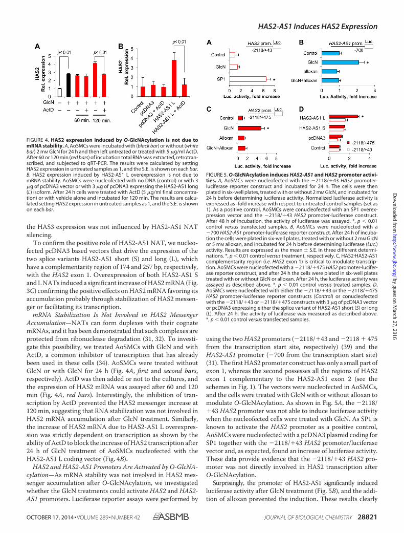
using the two HAS2 promoters (�2118/�43 and �2118 � 475from the transcription start site, respectively) (39) and theHAS2-AS1 promoter (�700 from the transcription start site)(31). The first HAS2 promoter construct has only a small part ofexon 1, whereas the second possesses all the regions of HAS2exon 1 complementary to the HAS2-AS1 exon 2 (see theschemes in Fig. 1). The vectors were nucleofected in AoSMCs,and the cells were treated with GlcN with or without alloxan tomodulate O-GlcNAcylation. As shown in Fig. 5A, the �2118/�43 HAS2 promoter was not able to induce luciferase activitywhen the nucleofected cells were treated with GlcN. As SP1 isknown to activate the HAS2 promoter as a positive control,AoSMCs were nucleofected with a pcDNA3 plasmid coding forSP1 together with the �2118/�43 HAS2 promoter/luciferasevector and, as expected, found an increase of luciferase activity.These data provide evidence that the �2118/�43 HAS2 pro-moter was not directly involved in HAS2 transcription afterO-GlcNAcylation.
Surprisingly, the promoter of HAS2-AS1 significantly inducedluciferase activity after GlcN treatment (Fig. 5B), and the addi-tion of alloxan prevented the induction. These results clearly
FIGURE 4. HAS2 expression induced by O-GlcNAcylation is not due tomRNA stability. A, AoSMCs were incubated with (black bar) or without (whitebar) 2 mM GlcN for 24 h and then left untreated or treated with 5 �g/ml ActD.After 60 or 120 min (red bars) of incubation total RNA was extracted, retrotran-scribed, and subjected to qRT-PCR. The results were calculated by settingHAS2 expression in untreated samples as 1, and the S.E. is shown on each bar.B, HAS2 expression induced by HAS2-AS1 L overexpression is not due tomRNA stability. AoSMCs were nucleofected with no DNA (control) or with 3�g of pcDNA3 vector or with 3 �g of pcDNA3 expressing the HAS2-AS1 long(L) isoform. After 24 h cells were treated with ActD (5 �g/ml final concentra-tion) or with vehicle alone and incubated for 120 min. The results are calcu-lated setting HAS2 expression in untreated samples as 1, and the S.E. is shownon each bar.
FIGURE 5. O-GlcNAcylation induces HAS2-AS1 and HAS2 promoter activi-ties. A, AoSMCs were nucleofected with the �2118/�43 HAS2 promoter-luciferase reporter construct and incubated for 24 h. The cells were thenplated in six-well plates, treated with or without 2 mM GlcN, and incubated for24 h before determining luciferase activity. Normalized luciferase activity isexpressed as -fold increase with respect to untreated control samples (set as1). As a positive control, AoSMCs were conucleofected with an SP1 overex-pression vector and the �2118/�43 HAS2 promoter-luciferase construct.After 48 h of incubation, the activity of luciferase was assayed. *, p 0.01control versus transfected samples. B, AoSMCs were nucleofected with a�700 HAS2-AS1 promoter-luciferase reporter construct. After 24 h of incuba-tion the cells were plated in six-well plates, treated with or without 2 mM GlcNor 5 mM alloxan, and incubated for 24 h before determining luciferase (Luc.)activity. Results are expressed as the mean � S.E. in three different determi-nations. *, p 0.01 control versus treatment, respectively. C, HAS2�HAS2-AS1complementarity region (i.e. HAS2 exon 1) is critical to modulate transcrip-tion. AoSMCs were nucleofected with a �2118/�475 HAS2 promoter-lucifer-ase reporter construct, and after 24 h the cells were plated in six-well platestreated with or without GlcN or alloxan. After 24 h, the luciferase activity wasassayed as described above. *, p 0.01 control versus treated samples. D,AoSMCs were nucleofected with either the �2118/�43 or the �2118/�475HAS2 promoter-luciferase reporter constructs (Control) or conucleofectedwith the �2118/�43 or �2118/�475 constructs with 3 �g of pcDNA3 vectoror pcDNA3 expressing either the splice variant of HAS2-AS1 short (S) or long(L). After 24 h, the activity of luciferase was measured as described above.*, p 0.01 control versus transfected samples.
HAS2-AS1 Induces HAS2 Expression
OCTOBER 17, 2014 • VOLUME 289 • NUMBER 42 JOURNAL OF BIOLOGICAL CHEMISTRY 28821
by guest on March 27, 2016
http://ww
w.jbc.org/
Dow
nloaded from

indicate that O-GlcNAcylation can directly regulate the tran-scription of HAS2-AS1 by modulating its promoter activity.
We then determined whether the HAS2 exon 1 could have arole in the activation of transcription after inducing O-GlcNA-cylation as it is partially complementary to the HAS2-AS1 exon2 (see Fig. 1). AoSMCs nucleofected with the �2118 � 475HAS2 promoter-luciferase construct increased luciferase activ-ity significantly after inducing O-GlcNAcylation with GlcNtreatment (Fig. 5C). To further demonstrate the critical role ofexon 1 in the regulation of HAS2 gene transcription, weconucleofected the �2118 � 475 or the �2118/�43 HAS2 pro-moter-luciferase plasmids with the pcDNA3 vectors expressingHAS2-AS1 S or L and found that only the HAS2 promoterconstruct containing the exon 1 was able to drive luciferaseactivity when nucleofected with the HAS2-AS1 L expressingvector (Fig. 5D).
p65 and O-GlcNAcylated Factor(s) Binds to HAS2-AS1 andHAS2 Promoters—As HAS2-AS1 promoter was activated byGlcN, whereas the HAS2 (�2118/�43) promoter was not ableto drive luciferase activity after GlcN treatment, we hypothe-sized that a transcription factor(s) could interact with HAS2-AS1 but not with �2118/�43 HAS2 promoters after O-GlcNA-cylation. To test this we performed ChIP analyses using or theHGAC85 monoclonal antibody that is specific for O-GlcNAcmodifications on proteins or anti-p65 (also known as RelA, oneof the major components of the NF-�B complex) or anti RNApolymerase 2 antibodies. The cross-linked and digested chro-matin was immunoprecipitated with the antibodies or rabbitIgG (control), and the amount of promoter DNA associatedwith the IP chromatin was quantified by quantitative PCR withprimers specific to the human HAS2-AS1 or HAS2 proximalpromoter regions. After GlcN treatments, we found significantincreases of O-GlcNAcylated proteins associated to the HAS2-AS1 proximal promoter, whereas no significant changes wereobservable in O-GlcNAcylated proteins bound to the HAS2proximal promoter (Fig. 6A). Interestingly, we found that a sig-nificant increase of O-GlcNAcylated proteins associated withthe overlapping region between HAS2 exon 1 and HAS2-AS1exon 2 after GlcN treatment (Fig. 6A), highlighting the crucialrole of this genomic region in the regulation of HAS2transcription.
As previous analyses revealed the presence of NF-�B bindingsites on the HAS2-AS1 promoter (31) (see Fig. 1), we investi-gated whether p65 was present on the HAS2-AS1 promoterafter GlcN treatments. As shown in Fig. 6B, ChIP analysesrevealed that GlcN treatment induced a strong and specificrecruitment of p65 on HAS2-AS1 proximal promoter, whereasp65 was not detected on the HAS2 proximal promoter or in theoverlapping region. As the positive control experiment, we per-formed ChIP with anti-RNA polymerase 2 antibody and foundan increased recruitment of this enzyme on HAS2-AS1 andHAS2 promoters as well as on the overlapping region asexpected.
NF-�B Is Required after GlcN Treatment to Induce HAS2-AS1 NAT Accumulation—To demonstrate the role of NF-�Bsubunit p65 in the accumulation of HAS2-AS1 NAT afterinduction of O-GlcNAcylation with GlcN treatments, quies-cent AoSMCs were untreated or treated with GlcN or treated
with GlcN and PDTC, an inhibitor of NF-�B, at 0.5 and 2 �M asfinal concentrations. PDTC specifically prevented the GlcN-induced HAS2-AS1 NAT accumulation in a dose-responsemanner without altering HAS3 expression (Fig. 7) and, there-fore, highlighted the involvement of NF-�B in the regulation ofHAS2-AS1 NAT.
HAS-AS1 Induces Chromatin Remodeling in the HAS2 Prox-imal Promoter—NATs can regulate their cognate transcript inseveral ways (26). We focused on the possibility that HAS2-AS1NAT could interact in cis with the HAS2 gene and modulatechromatin structure. To investigate this point, we performednuclease accessibility assays on AoSMCs treated with reagentsthat modulate O-GlcNAcylation. The nuclease accessibilityassay can be used to determine the sensitivity of DNA regions tonucleases revealing the location of open or closed chromatin.The method is based on the more rapid nuclease-catalyzedhydrolysis observed with open chromatin structures than withclosed chromatin structures. As shown in Fig. 8A, the inductionof O-GlcNAcylation by GlcN or PUGNAC treatmentsincreased the nuclease accessibility index of the proximal pro-moters of HAS2 and HAS2-AS1, suggesting an open chromatinstructure. Conversely, the chromatin structure around the
FIGURE 6. O-GlcNAcylated factor(s) and p65 are associated with HAS2-AS1 proximal promoter. AoSMCs were left untreated (Control) or treatedwith 2 mM GlcN. After 48 h, chromatin was purified, and ChIP experimentswere performed by using specific monoclonal antibodies against O-GlcNAc(A), p65 (B), RNA polymerase 2 (RNA pol 2) (C) or rabbit IgG. PCR conducted onDNA derived from the input chromatin template served as a positive control,and that of the IgG-precipitated template served as a specificity control. Theamount of proximal promoter DNAs (�300 bp with respect to the transcrip-tion start site) associated with the IP chromatin was quantified by qPCR withspecific primers for HAS2-AS1 proximal promoter or HAS2�HAS2-AS1 comple-mentarity region or HAS2 proximal promoter. ChIP promoter binding in con-trol AoSMCs was set at 1 � S.D. Results are expressed as the mean � S.E. inthree different determinations. *, p 0.01 untreated versus treatment,respectively. The IgG control ChIP assays produced only 0.05� of the anti-body binding levels observed with untreated as well as treated AoSMCs.
HAS2-AS1 Induces HAS2 Expression
28822 JOURNAL OF BIOLOGICAL CHEMISTRY VOLUME 289 • NUMBER 42 • OCTOBER 17, 2014
by guest on March 27, 2016
http://ww
w.jbc.org/
Dow
nloaded from

HAS3 promoter, which shares no complementarity with HAS2-AS1 NAT, was not influenced by O-GlcNAcylation.
To determine whether the compaction status of chromatinaround the HAS2 proximal promoter could depend on HAS2-AS1 NAT, we silenced HAS2-AS1 by siRNA in AoSMCs andtreated the cells with GlcN and alloxan to modulate proteinO-GlcNAcylation. Abrogation of HAS2-AS1 did not permit theopening of chromatin around the HAS2 proximal promoterafter inducing O-GlcNAcylation (Fig. 8B). To confirm the roleof HAS2-AS1 in modulating the chromatin structure, wenucleofected pcDNA3 vectors expressing HAS2-AS1 S or Lvariants and found that overexpression of these NATs was suf-ficient to increase the aperture of the region around the HAS2proximal promoter (Fig. 8C). This change in chromatin struc-ture was specific for the HAS2 gene as the region around theHAS2-AS1 proximal promoter was not altered by HAS2-AS1.
DISCUSSION
O-GlcNAcylation is an intracellular glycosylation involved inmany pathologies as diabetic vascular diseases (21). At thenuclear level, O-GlcNAcylation has been already described tomodify histones as well as several transcription factors andRNA polymerase 2 (46). Therefore, the alteration in chromatinstructure could be due to histone glycosylation and, in thisstudy we found that O-GlcNAcylation increased the accessibil-ity index of the proximal promoters of both HAS2 and HAS2-AS1. Furthermore, the chromatin opening of the HAS2 pro-moter required the HAS2-AS1 NAT, and the overexpression ofsuch a NAT was able to induce similar chromatin remodelingspecifically at HAS2 promoter.
Although all histones can by modified by O-GlcNAc (47),GlcN treatment is known to induce histone H3 O-GlcNA-cylation with a reduction of H3 phosphorylation. As O-GlcNA-cylated H3 was found in association with both H3K4me3 orH3K9me3, which are active or inactive histone marks, respec-tively, the role of histone glycosylation in chromatin regulationis still under investigation (48).
To alter chromatin structure many factors can be involved,but epigenetic modifications represent the most commonmechanism that allows the aperture of nucleosomes, permit-ting a more efficient interaction between DNA and the tran-
scription machinery (49, 50). Recently, O-GlcNAc transferasehas been found associated with promoters where it regulatesten-eleven translocation (TET) family enzymes that have a rolein DNA demethylation process (22, 51–53).
Based on the observation that many histone-modifyingenzymes lack specific DNA binding domain (54) and that longnon-coding RNAs and NATs might interact with ubiquitouslyexpressed histone modifying enzymes providing the requiredlevel of binding specificity (24), we speculated that HAS2-AS1could bind an uncharacterized factor(s) able to modify histonesand mediate chromatin structure change (Fig. 9). Several epi-genetic modification proteins have been described to interactwith long non-coding RNA as the histone methyltransferasemixed-lineage leukemia that is able to activate transcription atHOXA locus (55).
The specific activity of HAS2-AS1 NAT to induce HAS2expression could be due to the complementarity region that isclose to HAS2 promoter. HAS2-AS1 could interact in cis withHAS2 exon 1 and permit the modification of chromatin specif-ically on HAS2 promoter as outlined in the scheme of Fig. 9.
FIGURE 8. O-GlcNAcylation and HAS2-AS1 induce chromatin remodeling.A, AoSMCs were left untreated or treated with 2 mM GlcN, 100 �M PUGNAC, 40�M DON, 5 mM alloxan, or combinations of alloxan and GlcN. After 48 h, thenuclease accessibility index was calculated for the proximal promoter of HAS2(red bars), HAS2-AS1 (blue bars), and HAS3 (gray bars). The nuclease accessibil-ity index was set at 1 in control samples. Results are expressed as the mean �S.E. in three different determinations. *, p 0.01 untreated versus treatment,respectively. B, HAS2-AS1 abrogation prevented the aperture of chromatin inproximal promoter (Prom.) of HAS2. AoSMCs were nucleofected with siHAS2-AS1 (white bars) or a scrambled control (red bars). After 24 h of incubation, thecells were left untreated (Control) or treated with 2 mM GlcN or 5 mM alloxan ora combination of alloxan and GlcN. After 24 h nuclease accessibility assayswere performed as described above. Results are expressed as the mean � S.E.in three different determinations. *, p 0.01 untreated versus treatment,respectively. C, overexpression of HAS2-AS1 S and L induced aperture of chro-matin around HAS2 proximal promoter. AoSMCs were nucleofected with noDNA (Control) or with 3 �g of pcDNA3 vector or with 3 �g of pcDNA3 express-ing either splice variant of HAS2-AS1 short (S) or long (L). After 48 h the acces-sibility of nuclease to HAS2 (red bars) or HAS2-AS1 (blue bars) proximal promot-ers was determined. Results are expressed as the mean � S.E. in threedifferent determinations. *, p 0.01 control versus treatment, respectively.
FIGURE 7. HAS2-AS1 expression is mediated by NF-�B. Relative quantifica-tion of HAS2-AS1 (blue bars) and HAS3 (gray bars) in untreated (Control) andafter 48 h of treatment of AoSMCs with 2 mM GlcN or with 2 mM GlcN plus 0.5or 2 �M PDTC (an NF-�B inhibitor). The results of the relative quantificationwere calculated by setting control HAS2-AS1 and HAS3 expression as 1, andthe S.E. is shown on each bar. *, p 0.01 GlcN- versus PDTC-treated samples.
HAS2-AS1 Induces HAS2 Expression
OCTOBER 17, 2014 • VOLUME 289 • NUMBER 42 JOURNAL OF BIOLOGICAL CHEMISTRY 28823
by guest on March 27, 2016
http://ww
w.jbc.org/
Dow
nloaded from

How such a complementary region between HAS2 and HAS2-AS1 controls transcription is still under investigation, but as thesimple overexpression of HAS-AS1 (in particular the long iso-form that has an extended complementarity region respect tothe short isoform) without any treatment is able to induce tran-scriptional activity, we hypothesize a RNA-DNA interactionbetween HAS2-AS1 NAT and HAS2 gene at exon 1 level. Anintriguing point to investigate would be whether HAS2 couldcontrol HAS2-AS1 expression in an opposite regulation.
The importance of HAS-AS1 is supported by the fact thatHAS2-AS1 promoter is finely tuned by SP1 And SMAD2/3 (31).Our results show that this promoter is activated by O-GlcNA-cylation and that NF-�B subunit p65 is associated with theHAS2-AS1 promoter after GlcN treatment. Interestingly, p65 isknown to increase its transcriptional activity after O-GlcNA-cylation (45, 56). On the other hand, although HAS2 promoteris known to be finely regulated by several transcription factors(3), we found that the activity of HAS2 promoter was not alteredby O-GlcNAcylation, and no O-GlcNAcylated factor(s) wasdetectable bound with it. Interestingly, the addition of the com-plementarity region between HAS2 and HAS2-AS1 to theHAS2 promoter (construct �2218/�475) led to an incrementof luciferase activity after induction of O-GlcNAcylation, high-lighting a crucial role of this region (i.e. HAS2 exon 1) in tran-scription regulation. This issue is also supported by the fact thatthis region was able to interact with the O-GlcNAcylatedfactor(s).
As the overexpression of HAS2 and HA accumulation can bedetrimental in vivo in the case of malignancies and vascularpathologies (10), we found that the expression of HAS2-AS1NAT is augmented in human atheromatous plaque with anincreased grade of severity known as the proatheroscleroticeffect of HA. Furthermore, in a mouse model of atherosclerosisinduced by high cholesterol, the accumulation of both HAS2and HAS2-AS1 is evident highlighting the potential role in thepathology of such NAT. Although our in vivo data cannotexplore the molecular mechanism of HAS2-AS1 action, theexpression of HAS2-AS1 in mice with a high level ofO-GlcNAcylation was double with respect to wild-type orheterozygous animals, confirming the role such glycosylationto control the NAT expression.
In conclusion, our data identify HAS2-AS1 as new importantplayer in the regulation of HAS2 gene transcription and high-light the coordinated expression of such NAT and its sensecounterpart in healthy and atherosclerotic human carotids aswell as in murine model of atherosclerosis.
Acknowledgments—We thank Stephen Safe (Texas A&M University,for pcDNA3-SP1 vector), Katri Makkonen (University of Eastern Fin-land, for HAS2 promoter luciferase vectors), Stuart Schreiber (How-ard Hughes Medical Institute, for pcDNA3-HADC1 vector), HendrikStunnenberg (Radboud University, The Netherlands, for pcDNA3-p300 vector). We acknowledge the “Centro Grandi Attrezzature per laRicerca Biomedica,” Università degli Studi dell’Insubria, for instru-ment availability.
REFERENCES1. Jiang, D., Liang, J., and Noble, P. W. (2011) Hyaluronan as an immune
regulator in human diseases. Physiol. Rev. 91, 221–2642. Vigetti, D., Karousou, E., Viola, M., Deleonibus, S., De Luca, G., and Passi,
A. (2014) Hyaluronan: biosynthesis and signaling. Biochim. Biophys. Acta1840, 2452–2459
3. Tammi, R. H., Passi, A. G., Rilla, K., Karousou, E., Vigetti, D., Makkonen,K., and Tammi, M. I. (2011) Transcriptional and post-translational regu-lation of hyaluronan synthesis. FEBS J. 278, 1419 –1428
4. Vigetti, D., Viola, M., Karousou, E., De Luca, G., and Passi, A. (2014)Metabolic control of hyaluronan synthases. Matrix Biol. 35, 8 –13
5. Ross, R. (1999) Atherosclerosis: an inflammatory disease. N. Engl. J. Med.340, 115–126
6. Vigetti, D., Moretto, P., Viola, M., Genasetti, A., Rizzi, M., Karousou, E.,Pallotti, F., De Luca, G., and Passi, A. (2006) Matrix metalloproteinase 2and tissue inhibitors of metalloproteinases regulate human aortic smoothmuscle cell migration during in vitro aging. FASEB J. 20, 1118 –1130
7. Riessen, R., Wight, T. N., Pastore, C., Henley, C., and Isner, J. M. (1996)Distribution of hyaluronan during extracellular matrix remodeling in hu-man restenotic arteries and balloon-injured rat carotid arteries. Circula-tion 93, 1141–1147
8. Chai, S., Chai, Q., Danielsen, C. C., Hjorth, P., Nyengaard, J. R., Ledet, T.,Yamaguchi, Y., Rasmussen, L. M., and Wogensen, L. (2005) Overexpres-sion of hyaluronan in the tunica media promotes the development ofatherosclerosis. Circ. Res. 96, 583–591
9. Cuff, C. A., Kothapalli, D., Azonobi, I., Chun, S., Zhang, Y., Belkin, R., Yeh,C., Secreto, A., Assoian, R. K., Rader, D. J., and Puré, E. (2001) The adhe-sion receptor CD44 promotes atherosclerosis by mediating inflammatorycell recruitment and vascular cell activation. J. Clin. Invest. 108,1031–1040
10. Vigetti, D., Viola, M., Karousou, E., Genasetti, A., Rizzi, M., Clerici, M.,Bartolini, B., Moretto, P., De Luca, G., and Passi, A. (2008) Vascular pa-thology and the role of hyaluronan. ScientificWorldJournal 8, 1116 –1118
11. Vigetti, D., Rizzi, M., Moretto, P., Deleonibus, S., Dreyfuss, J. M., Karou-sou, E., Viola, M., Clerici, M., Hascall, V. C., Ramoni, M. F., De Luca, G.,and Passi, A. (2011) Glycosaminoglycans and glucose prevent apoptosis in4-methylumbelliferone-treated human aortic smooth muscle cells. J. Biol.Chem. 286, 34497–34503
12. Vigetti, D., Rizzi, M., Viola, M., Karousou, E., Genasetti, A., Clerici, M.,Bartolini, B., Hascall, V. C., De Luca, G., and Passi, A. (2009) The effects of4-methylumbelliferone on hyaluronan synthesis, MMP2 activity, prolifer-ation, and motility of human aortic smooth muscle cells. Glycobiology 19,537–546
13. Nigro, J., Osman, N., Dart, A. M., and Little, P. J. (2006) Insulin resistanceand atherosclerosis. Endocr. Rev. 27, 242–259
14. Brownlee, M. (2001) Biochemistry and molecular cell biology of diabeticcomplications. Nature 414, 813– 820
15. Copeland, R. J., Bullen, J. W., and Hart, G. W. (2008) Cross-talk betweenGlcNAcylation and phosphorylation: roles in insulin resistance and glu-cose toxicity. Am. J. Physiol. Endocrinol. Metab. 295, E17–E28
FIGURE 9. Working model of HAS2-AS1 mechanism of action. In basal con-ditions the accessibility of both HAS2 and HAS2-AS1 promoters are low allow-ing a limited production of transcripts. After induction of O-GlcNAcylation orin hyperglycemic conditions in vivo, O-GlcNAcylated p65 binds to the HAS2-AS1 promoter, increasing HAS2-AS1 NAT accumulation. HAS2-AS1, pairingwith the complementarity region on HAS2 exon 1, could bring the enzyme(s)(circle with question mark) involved in epigenetic modification of histones(indicated by the triangle flags on cylinders), permitting chromatin apertureon HAS2 promoter and allowing a more efficient transcription.
HAS2-AS1 Induces HAS2 Expression
28824 JOURNAL OF BIOLOGICAL CHEMISTRY VOLUME 289 • NUMBER 42 • OCTOBER 17, 2014
by guest on March 27, 2016
http://ww
w.jbc.org/
Dow
nloaded from

16. Vigetti, D., Deleonibus, S., Moretto, P., Karousou, E., Viola, M., Bartolini,B., Hascall, V. C., Tammi, M., De Luca, G., and Passi, A. (2012) Role ofUDP-N-acetylglucosamine (GlcNAc) and O-GlcNAcylation of hyaluro-nan synthase 2 in the control of chondroitin sulfate and hyaluronan syn-thesis. J. Biol. Chem. 287, 35544 –35555
17. Vigetti, D., Ori, M., Viola, M., Genasetti, A., Karousou, E., Rizzi, M., Pal-lotti, F., Nardi, I., Hascall, V. C., De Luca, G., and Passi, A. (2006) Molec-ular cloning and characterization of UDP-glucose dehydrogenase fromthe amphibian Xenopus laevis and its involvement in hyaluronan synthe-sis. J. Biol. Chem. 281, 8254 – 8263
18. Jokela, T. A., Makkonen, K. M., Oikari, S., Kärnä, R., Koli, E., Hart, G. W.,Tammi, R. H., Carlberg, C., and Tammi, M. I. (2011) Cellular content ofUDP-N-acetylhexosamines controls hyaluronan synthase 2 expressionand correlates with O-GlcNAc modification of transcription factors YY1and SP1. J. Biol. Chem. 286, 33632–33640
19. Hart, G. W., Housley, M. P., and Slawson, C. (2007) Cycling of O-linked�-N-acetylglucosamine on nucleocytoplasmic proteins. Nature 446,1017–1022
20. Buse, M. G. (2006) Hexosamines, insulin resistance, and the complicationsof diabetes: current status. Am. J. Physiol. Endocrinol. Metab. 290, E1–E8
21. Slawson, C., Copeland, R. J., and Hart, G. W. (2010) O-GlcNAc signaling:a metabolic link between diabetes and cancer? Trends Biochem. Sci. 35,547–555
22. Hanover, J. A., Krause, M. W., and Love, D. C. (2012) Bittersweet memo-ries: linking metabolism to epigenetics through O-GlcNAcylation. Nat.Rev. Mol. Cell Biol. 13, 312–321
23. Chao, H., and Spicer, A. P. (2005) Natural antisense mRNAs to hyaluronansynthase 2 inhibit hyaluronan biosynthesis and cell proliferation. J. Biol.Chem. 280, 27513–27522
24. Magistri, M., Faghihi, M. A., St Laurent, G., 3rd, and Wahlestedt, C. (2012)Regulation of chromatin structure by long noncoding RNAs: focus onnatural antisense transcripts. Trends Genet. 28, 389 –396
25. Rinn, J. L., and Chang, H. Y. (2012) Genome regulation by long noncodingRNAs. Annu. Rev. Biochem. 81, 145–166
26. Wang, K. C., and Chang, H. Y. (2011) Molecular mechanisms of longnoncoding RNAs. Mol. Cell 43, 904 –914
27. Yoon, J. H., Abdelmohsen, K., and Gorospe, M. (2013) Posttranscriptionalgene regulation by long noncoding RNA. J. Mol. Biol. 425, 3723–3730
28. Mercer, T. R., and Mattick, J. S. (2013) Structure and function of longnoncoding RNAs in epigenetic regulation. Nat. Struct. Mol. Biol. 20,300 –307
29. Lee, J. T. (2012) Epigenetic regulation by long noncoding RNAs. Science338, 1435–1439
30. Faghihi, M. A., and Wahlestedt, C. (2009) Regulatory roles of natural an-tisense transcripts. Nat. Rev. Mol. Cell Biol. 10, 637– 643
31. Michael, D. R., Phillips, A. O., Krupa, A., Martin, J., Redman, J. E., Altaher,A., Neville, R. D., Webber, J., Kim, M. Y., and Bowen, T. (2011) The humanhyaluronan synthase 2 (HAS2) gene and its natural antisense RNA exhibitcoordinated expression in the renal proximal tubular epithelial cell. J. Biol.Chem. 286, 19523–19532
32. Faghihi, M. A., Modarresi, F., Khalil, A. M., Wood, D. E., Sahagan, B. G.,Morgan, T. E., Finch, C. E., St Laurent, G., 3rd, Kenny, P. J., and Wahlest-edt, C. (2008) Expression of a noncoding RNA is elevated in Alzheimer’sdisease and drives rapid feed-forward regulation of �-secretase. Nat. Med.14, 723–730
33. Vigetti, D., Clerici, M., Deleonibus, S., Karousou, E., Viola, M., Moretto, P.,Heldin, P., Hascall, V. C., De Luca, G., and Passi, A. (2011) Hyaluronansynthesis is inhibited by adenosine monophosphate-activated protein ki-nase through the regulation of HAS2 activity in human aortic smoothmuscle cells. J. Biol. Chem. 286, 7917–7924
34. Raman, P., Krukovets, I., Marinic, T. E., Bornstein, P., and Stenina, O. I.(2007) Glycosylation mediates up-regulation of a potent antiangiogenicand proatherogenic protein, thrombospondin-1, by glucose in vascularsmooth muscle cells. J. Biol. Chem. 282, 5704 –5714
35. Schreck, R., Meier, B., Männel, D. N., Dröge, W., and Baeuerle, P. A. (1992)Dithiocarbamates as potent inhibitors of nuclear factor �B activation inintact cells. J. Exp. Med. 175, 1181–1194
36. Vigetti, D., Genasetti, A., Karousou, E., Viola, M., Moretto, P., Clerici,
M., Deleonibus, S., De Luca, G., Hascall, V. C., and Passi, A. (2010)Proinflammatory cytokines induce hyaluronan synthesis and mono-cyte adhesion in human endothelial cells through hyaluronan synthase2 (HAS2) and the nuclear factor-�B (NF-�B) pathway. J. Biol. Chem.285, 24639 –24645
37. Yang, Y. R., Song, M., Lee, H., Jeon, Y., Choi, E. J., Jang, H. J., Moon, H. Y.,Byun, H. Y., Kim, E. K., Kim, D. H., Lee, M. N., Koh, A., Ghim, J., Choi, J. H.,Lee-Kwon, W., Kim, K. T., Ryu, S. H., and Suh, P. G. (2012) O-GlcNAcaseis essential for embryonic development and maintenance of genomic sta-bility. Aging Cell 11, 439 – 448
38. Livak, K. J., and Schmittgen, T. D. (2001) Analysis of relative gene expres-sion data using real-time quantitative PCR and the 2(���C(T)) method.Methods 25, 402– 408
39. Makkonen, K. M., Pasonen-Seppänen, S., Törrönen, K., Tammi, M. I., andCarlberg, C. (2009) Regulation of the hyaluronan synthase 2 gene by con-vergence in cyclic AMP response element-binding protein and retinoidacid receptor signaling. J. Biol. Chem. 284, 18270 –18281
40. Love, D. C., Ghosh, S., Mondoux, M. A., Fukushige, T., Wang, P., Wilson,M. A., Iser, W. B., Wolkow, C. A., Krause, M. W., and Hanover, J. A. (2010)Dynamic O-GlcNAc cycling at promoters of Caenorhabditis elegans genesregulating longevity, stress, and immunity. Proc. Natl. Acad. Sci. U.S.A.107, 7413–7418
41. Wang, I. C., Chen, Y. J., Hughes, D., Petrovic, V., Major, M. L., Park, H. J.,Tan, Y., Ackerson, T., and Costa, R. H. (2005) Forkhead box M1 regulatesthe transcriptional network of genes essential for mitotic progression andgenes encoding the SCF (Skp2-Cks1) ubiquitin ligase. Mol. Cell. Biol. 25,10875–10894
42. Rao, S., Procko, E., and Shannon, M. F. (2001) Chromatin remodeling,measured by a novel real-time polymerase chain reaction assay, acrossthe proximal promoter region of the IL-2 gene. J. Immunol. 167,4494 – 4503
43. Yuan, W., Wu, T., Fu, H., Dai, C., Wu, H., Liu, N., Li, X., Xu, M., Zhang, Z.,Niu, T., Han, Z., Chai, J., Zhou, X. J., Gao, S., and Zhu, B. (2012) Densechromatin activates Polycomb repressive complex 2 to regulate H3 lysine27 methylation. Science 337, 971–975
44. Ozcan, S., Andrali, S. S., and Cantrell, J. E. (2010) Modulation of transcrip-tion factor function by O-GlcNAc modification. Biochim. Biophys. Acta1799, 353–364
45. Allison, D. F., Wamsley, J. J., Kumar, M., Li, D., Gray, L. G., Hart, G. W.,Jones, D. R., and Mayo, M. W. (2012) Modification of RelA by O-linkedN-acetylglucosamine links glucose metabolism to NF-�B acetylation andtranscription. Proc. Natl. Acad. Sci. U.S.A. 109, 16888 –16893
46. Slawson, C., and Hart, G. W. (2011) O-GlcNAc signalling: implications forcancer cell biology. Nat. Rev. Cancer 11, 678 – 684
47. Sakabe, K., Wang, Z., and Hart, G. W. (2010) �-N-Acetylglucosamine(O-GlcNAc) is part of the histone code. Proc. Natl. Acad. Sci. U.S.A. 107,19915–19920
48. Zhang, S., Roche, K., Nasheuer, H. P., and Lowndes, N. F. (2011) Modifi-cation of histones by sugar �-N-acetylglucosamine (GlcNAc) occurs onmultiple residues, including histone H3 serine 10, and is cell cycle-regu-lated. J. Biol. Chem. 286, 37483–37495
49. Chi, P., Allis, C. D., and Wang, G. G. (2010) Covalent histone modifica-tions: miswritten, misinterpreted and mis-erased in human cancers. Nat.Rev. Cancer 10, 457– 469
50. Bannister, A. J., and Kouzarides, T. (2011) Regulation of chromatin byhistone modifications. Cell Res. 21, 381–395
51. Chen, Q., Chen, Y., Bian, C., Fujiki, R., and Yu, X. (2013) TET2 promoteshistone O-GlcNAcylation during gene transcription. Nature 493,561–564
52. Deplus, R., Delatte, B., Schwinn, M. K., Defrance, M., Méndez, J., Mur-phy, N., Dawson, M. A., Volkmar, M., Putmans, P., Calonne, E., Shih,A. H., Levine, R. L., Bernard, O., Mercher, T., Solary, E., Urh, M.,Daniels, D. L., and Fuks, F. (2013) TET2 and TET3 regulateGlcNAcylation and H3K4 methylation through OGT and SET1/COM-PASS. EMBO J. 32, 645– 655
53. Tahiliani, M., Koh, K. P., Shen, Y., Pastor, W. A., Bandukwala, H., Brudno,Y., Agarwal, S., Iyer, L. M., Liu, D. R., Aravind, L., and Rao, A. (2009)
HAS2-AS1 Induces HAS2 Expression
OCTOBER 17, 2014 • VOLUME 289 • NUMBER 42 JOURNAL OF BIOLOGICAL CHEMISTRY 28825
by guest on March 27, 2016
http://ww
w.jbc.org/
Dow
nloaded from

Conversion of 5-methylcytosine to 5-hydroxymethylcytosine in mamma-lian DNA by MLL partner TET1. Science 324, 930 –935
54. Bernstein, E., and Allis, C. D. (2005) RNA meets chromatin. Genes Dev. 19,1635–1655
55. Du, Z., Fei, T., Verhaak, R. G., Su, Z., Zhang, Y., Brown, M., Chen, Y., andLiu, X. S. (2013) Integrative genomic analyses reveal clinically relevant
long noncoding RNAs in human cancer. Nat. Struct. Mol. Biol. 20,908 –913
56. Yang, W. H., Park, S. Y., Nam, H. W., Kim do, H., Kang, J. G., Kang, E. S.,Kim, Y. S., Lee, H. C., Kim, K. S., and Cho, J. W. (2008) NF�B activation isassociated with its O-GlcNAcylation state under hyperglycemic condi-tions. Proc. Natl. Acad. Sci. U.S.A. 105, 17345–17350
HAS2-AS1 Induces HAS2 Expression
28826 JOURNAL OF BIOLOGICAL CHEMISTRY VOLUME 289 • NUMBER 42 • OCTOBER 17, 2014
by guest on March 27, 2016
http://ww
w.jbc.org/
Dow
nloaded from

Hascall, Giancarlo De Luca and Alberto PassiCinquetti, Eugenia Karousou, Manuela Viola, Maria Luisa D'Angelo, Vincent C.
Maria Grandoch, Alexander Oberhuber, Dona C. Love, John A. Hanover, Raffaella Davide Vigetti, Sara Deleonibus, Paola Moretto, Timothy Bowen, Jens W. Fischer,
-GlcNAcylationOTranscription of HAS2 via Protein Natural Antisense Transcript for Hyaluronan Synthase 2 (HAS2-AS1) Induces
doi: 10.1074/jbc.M114.597401 originally published online September 2, 20142014, 289:28816-28826.J. Biol. Chem.
10.1074/jbc.M114.597401Access the most updated version of this article at doi:
Alerts:
When a correction for this article is posted•
When this article is cited•
to choose from all of JBC's e-mail alertsClick here
http://www.jbc.org/content/289/42/28816.full.html#ref-list-1
This article cites 56 references, 30 of which can be accessed free at
by guest on March 27, 2016
http://ww
w.jbc.org/
Dow
nloaded from
Related Documents