1 National University of Medical Science, Spain (NUMSS) Title: The effect of acetic acid (2%) iontophoresis on the calcification deposit of the shoulder BY Anoushiravan Mohammadi, Student NO, S 1502122 For Doctor of Physical Therapy Degree (DPT) March,2016 Supervisors: Dr. Mehran Razavipour,MD, orthopedist, Hand Surgery Fellowship, faculty members of Mazandaran University of Medical Sciences Dr. Ismail Shafiee, MD, orthopedist, Knee Surgery Fellowship, faculty members of Mazandaran University of Medical Sciences

Welcome message from author
This document is posted to help you gain knowledge. Please leave a comment to let me know what you think about it! Share it to your friends and learn new things together.
Transcript
1
National University of Medical Science, Spain
(NUMSS)
Title: The effect of acetic acid (2%) iontophoresis on the
calcification deposit of the shoulder
BY
Anoushiravan Mohammadi,
Student NO, S 1502122
For
Doctor of Physical Therapy Degree (DPT)
March,2016
Supervisors:
Dr. Mehran Razavipour,MD, orthopedist, Hand Surgery Fellowship,
faculty members of Mazandaran University of Medical Sciences
Dr. Ismail Shafiee, MD, orthopedist, Knee Surgery Fellowship,
faculty members of Mazandaran University of Medical Sciences

2
Case-research article
Title: The effect of acetic acid (2%) iontophoresis on the calcification
deposit of the shoulder
Anoushiravan Mohammadi, Doctor of physical therapy student at the national university of
medical sciences in Spain (NUMSS)
Supervisors:
Dr. Sayyed Mehran Razavipour, orthopedic specialist and Hand Surgery Fellowship, faculty
members of Mazandaran University of Medical Sciences
Dr. Sayyed Ismail Shafiee, orthopedic specialist and Knee Surgery Fellowship, faculty
members of Mazandaran University of Medical Sciences
Introduction:
One of the worst shoulder pain that causes dysfunction of mechanical type around the
shoulder joint is the deposition of hydroxyapatite crystals in the rotator tendons of the
shoulder (Rotator cuff) that is known calcified deposit or shoulder calcific or calcification
tendinitis (1,3, 9).
Calcium deposits around the shoulder most commonly occur in the burse (outside of rotator
cuff) and into supraspinatus tendon at the people over 30 to 60 years (most people over 40
years) (1,2,3,4,5,6,9,13,18 24).
So far, the real cause of this condition is unknown ( 2,3,4 ) , but several mechanisms have
been proposed for its incidence .
Codman and his colleagues, pointed that the cause of degeneration of the rotator cuff tendon
are wear and tear process in that cell necrosis and tissue hypoxia create the endochondral
ossification process and it transforms tenosit cells into chondrocyte cells and it causes the
formation of calcification. ( 6 , 7 )
Uhthoff and sarkar expressed other theories except degeneration process ; they proposed
reactive and dynamic process by triggering a cell-mediated in the area, followed by
phagocytose of multi-nuclear cells leads to the formation of osteoblastic cells ( 3,6 ).
The third theory is the extra bone formation by mesenchymal stem cell metaplasia, so the
normal cells exist in tendon tissue, can change into bone form ( 7,19 ).
Calcium deposits despite of normal metabolism of calcium and the normal amount in the
blood, happens.
3
In any case, the delay in the process of repair and recovery is one of the major causes calcium
deposit in addition to the aforementioned causes (delay healing) (4).
This problem is mainly with sudden and surprising pain and is not related by position or
activity and sometimes can be severe like tooth pain. Pain is extreme and intensified within
24 to 48 hours so that the patient holds his arm with the other hand for alleviate and no desire
for doing any active or passive movement.
The pain worsens by the elevation of arm and sleeping on involving shoulder. Even in such
condition, the patient may induce the narcotic drug for pain relief ( 2 ) . There are other
complaints with this disorder such as muscle weakness in shoulder elevation, catching and
snapping.
Despite of this complication, some patients may be asymptomatic on radiographic image.
The incidence of this complication has been reported 3-20% in which 25-30% of cases are
bilateral (2, 13 ) .
Despite of many similarities, the differences of these disorders with other common disorders
in the shoulder such as impingement syndrome or shoulder capsulitis, is that in recent cases,
the pain usually begins gradually and are directly related with activity and status of arm, it
means that there is a painful arc of movement. However, the deposition of calcium in the
shoulder, the pain can be severe without motion in the shoulder, unlike capsulitis that
beginning of the gradual and takes months, the pattern of capsular restriction is not present in
calcium deposits, however, long-term sustainable disorders in the shoulder could eventually
lead to capsulitis in the joint of shoulder.
The symptoms of acute phase of the calcium deposit maybe subside spontaneously (3, 4) but
the symptoms of the condition may be chronic and the existence of calcium deposits takes in
the months and even years (5 to 15 years) (9, 8).
Commonly the detection of calcification takes place by using a plain radiograph A/P and
Lateral view (outlet view) and also internal and external rotation of the shoulder (6, 13).
Calcification on X-ray images has been divided into three types. 1- (Type I) deposition of
high density (High density) 2- (Type II) deposits with distinct borders (well circumscribed
borders) 3- (Type III) cloudy sediment (cloudy) (13).
Plain X-ray Imaging is one of the strongest diagnostic methods approved by calcium deposit
in the shoulder. MRI is not necessary to detect calcium deposit but more than 95% accuracy
in finding calcifications in the pathology associated with complications such as rupture of the
tendon, and for this reason it can be valuable (6).
It should be remembered that the diagnosis of this complication is more clinical until
radiological (2), Although this idea, makes it difficult to judge about prevalence of this
disease, but in fact we judged the patients based on x-ray radiographs and thus we could not
have been controlled study in incidence of the disease among people, and therefore providing

4
the estimation. This study is an attempt to establish the effect of acetic acid iontophoresis
treatment and access to research study to document it.
Ionto means ion, phoresis means the transmission or transfer, a physical process in which
ions (typically "just (µmol / cm2 /h), flow by an electric field, by dispersing in a medium.
The main factor of this approach is that the Polarity of chemical substance or drugs for
transferring should be clear. In this study the acetic acid is a material with a negative polarity
so we should transfer it through exposure to the negative pole of the direct current (DC).
Iontophoresis is a physical therapy modality that although development has long been
suggested but for some reason has failed, one of the main reasons are the issue of chemicals
and chemical reactions due to unfamiliarity and the other reason is that the direct current use
very little in physiotherapy.
The lack of effect of conventional physiotherapy modalities in the treatment of calcium
deposit of the shoulder, we were decided to re-flow ionthophoresis approach as an effective
modality in the physiotherapy of this condition.
With examples and practical actions in this area found that acetic acid iontophoresis for the
treatment of calcium deposits can be considered as a strategy of treatment.
The Patients of this study: In this study 10 patients were included with a mean age of 53/7
year and 7 months of the length of complications by following protocol.
Materials and Methods:
Acetic acid solution: Acetic acid solution (manufactured by Merck Germany with the purity
of 98%), 2% solution of acetic acid were taken to a volume of 500 cc. The solution were kept
in the bottle sealed and covered to prevent light.
For Applying an electrical direct current (DC), we used Power Stim machine for transmission
and transfusion solution of acetic acid ion .The machine was manufactured by Engineering of
modern medicine (Iran,Isfahan co) . The maximum intensity at this process was 7 mA current
output, which was begun 5 mA and the flow was controlled by display. For doing this way,
at first we examined the patient and we recorded the range of motion and the limitation of
movement in flexion and abduction , as well as pain based on the degree of pain visual analog
scale (VAS) and finally the radiographic consideration and size of calcium deposits was
recorded . The patient slept on non- infected shoulder (side lying) and infected shoulder is
above and was placed for 15 minutes under infra-red radiation on the scapula and trapezius
muscles of the shoulder with posterior radiation.
How placement of the electrodes: The patient was in a sitting position and the place of
electrodes were cleaned with alcohol. The negative or cathode electrode (active electrode) in
the procedure for implantation in the best place on greater tuberosity between the humerus
and the acromion process, were put in the gap of glenohumeral joint, or in the place of
calcification. Positive electrode or anode at a distance of about 20 to 25 cm from the negative
electrode in the supra spinatus muscle was set . Before fixing electrodes on the skin, soaking

5
a pad floating in acetic acid solution (8 layers of conventional cotton gas) below the cathode
electrode placed on the skin in the shoulder. The electrode pad sponge soaked in tap water
were put under the positive pole on supraspinatus muscle. The size of both electrodes ( 4 * 6
cm ) and a rubber electrodes were selected . Each electrode were separately fixed and tighten
with wide elastic bands on their place
figure1: Power Stim machine, Rubber electrodes, cotton gas, syringe
figure 2: The first patient Figure
After the patient's readiness to start with power and flow to the patient's threshold (5 mA)
was increased gradually over 25 to 35 minutes then flow to a maximum of 7 mA scored. At
the end of the course and turn off the device and open the electrodes, it was used the current
pulse mode ultrasound 1 MHz and intensity of 1.5 watts per square centimeter for 4 minutes
on the negative electrode area. In conclusion, patients with pendulum codman,s exercise , left
the treatment site. The number of treatment sessions: in the first two weeks we had 5 sessions
in each week and in the next weeks we had three sessions. The maximum number of sessions
was 30 sessions.
The first case ( fig. 3 , 4 and 5 ) : The patient , 51 -year-old man with right shoulder pain and
movement disorders with symptoms of abnormal function of the shoulder about 9 months ago
. e He had intermittent pain and was uncomfortable sleeping on the right shoulder . Shoulder
range of motion didn’t have significant limitation unless the feeling of entrapment click on
the angle ( 80 -100 degrees of Abduction) and 110 degrees of flexion . Isometric muscle tests
of the shoulder in abduction and rotation, especially external rotation, was a little painful. But
the test of passive internal rotation of shoulder was producing more painful. The degree of
pain based on pain scores was 7 of 10. The following radiograph showed a large piece
measuring 10 * 28 mm calcium deposit. During each 10 sessions, the size of deposition
decreased and pain intensity was reduced to one.

6
Fig3:The beginning of treatment fig 4: After 10 sessions fig 5: After 30 sessions
The outcome of treatment: Pain score after 30 sessions were reported 1 to 10 . Shoulder range
of motion was normal and there was no sense clicking on the shoulder. Abduction isometric
testing and passive internal rotation showed a small amount of pain. According to the above
figure disappearance of calcium deposits is shown during the treatment of process.
The second case (Figure 6 and 7): 58-year-old male patient with left shoulder pain, from a
year ago due to a fall on the hand no matter skeletal damage. Pain has been intensified from a
month ago after agricultural activities. Pain in forearm and wrist even has been referred.
Shoulder flexion in the range of 170 degrees and abduction to 120 degrees has been
associated with pain and catching. Pain intensity was measured on a pain score 6 of 10.
Isometric muscle tests around the shoulder will not cause major pain. The measurement of
calcification was 9 * 5 mm. After 10 sessions of pain score decreased from 10 to 3. The
following figure shows a decrease in calcification point density.
fig 6: The beginning of treatment fig7: After 10 sessions
The third case (Figure 8 and 9): 51-year-old female patient with right shoulder pain and
movement limitation around six months ago. The patient also has a history of diabetes. Click
and felt pain in the shoulder flexion range of 90-100 degrees and 70 degrees in abduction.
Pre-treatment pain score determine 8 of 10 and in end of session pain decreases to1 of 10.
The size of calcification was 11 * 9 mm. The number of treatment sessions was 20 sessions
and 10 sessions of that first period, 5 times a week and then continued 3 times a week.
The results of treatment based on radiographic findings are shown as follows.

7
Fig 8: the beginning of treatment fig 9: After 20 sessions of treatment
The fourth case (figure. 10 and 11): 60-year-old female patient with left shoulder pain and
loss of function from 5 months ago. Range of motion in flexion after 90 degrees and
abduction after 60 degree was with a sense of clicking and pain. Pain score at baseline was 7
of 10.The size of calcification was 6 * 4 mm. The number of treatment sessions has continued
10 sessions. The result of treatment, the range of motion without pain and pain scores were
determined 1 to 10.
Fig 10: The beginning of treatment Fig 11: after treatment
The fifth case (Figure 12 and 13): The patient, 68-year-old with a history of nearly a year of
weakness and dysfunction on shoulder about 4 months was intensifying. Shoulder abduction
to 65 ° and flexion in the higher range of 80 ° to create intense pain and the sense of snapping
was associated. Pain score according to the VAS 7 of 10 and patients was upset to sleep on
any shoulder .The size of calcification was 22 * 6 mm. After 20 sessions 5 times on week,
pain decreased to 2 of 10 and radiograph´s symptoms disappear.
8
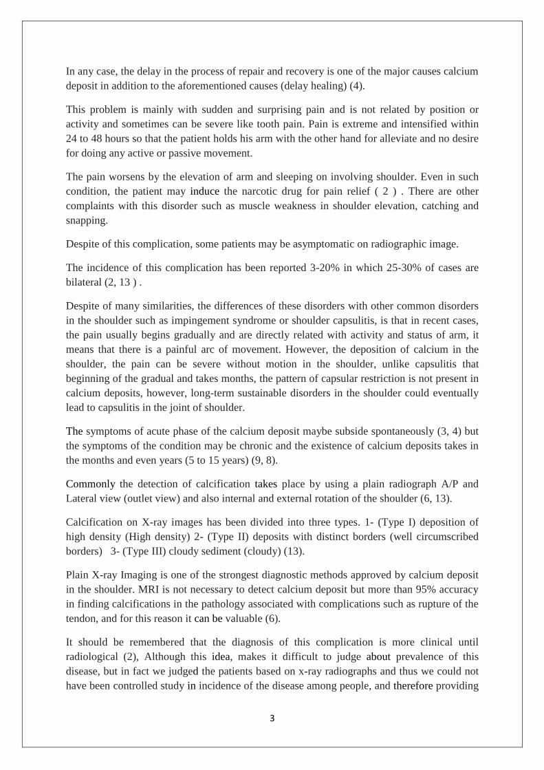
Fig 12:The begining of treatment Fig 13: after 20 sessions
The sixth case (figure. 14 and 15): A female 45-year-old with a history of shoulder pain than
five months ago due to the intensification of pain from 10 days ago for 10 sessions of two
weeks was treated for. pain score at the beginning of treatment was 8 of 10 and the abduction
of active shoulder 40 degrees and shoulder flexion above 55 degrees was associated with
severe pain and also internal rotation as active and passive associated with severe pain. The
size of calcification was 20 * 5mm. After 10 sessions, the size of calcification decreased and
the intensity of pain decreased to 2 of 10.
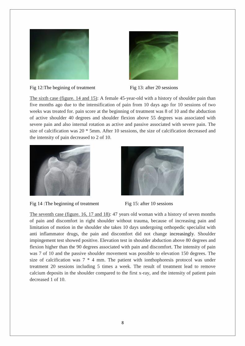
Fig 14 :The beginning of treatment Fig 15: after 10 sessions
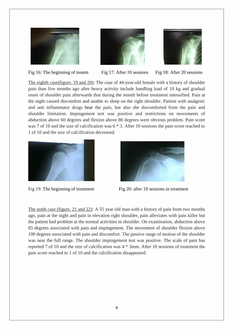
The seventh case (figure. 16, 17 and 18): 47 years old woman with a history of seven months
of pain and discomfort in right shoulder without trauma, because of increasing pain and
limitation of motion in the shoulder she takes 10 days undergoing orthopedic specialist with
anti inflammator drugs, the pain and discomfort did not change increasingly. Shoulder
impingement test showed positive. Elevation test in shoulder abduction above 80 degrees and
flexion higher than the 90 degrees associated with pain and discomfort. The intensity of pain
was 7 of 10 and the passive shoulder movement was possible to elevation 150 degrees. The
size of calcification was 7 * 4 mm. The patient with ionthophoresis protocol was under
treatment 20 sessions including 5 times a week. The result of treatment lead to remove
calcium deposits in the shoulder compared to the first x-ray, and the intensity of patient pain
decreased 1 of 10.
9
Fig 16: The beginning of treatm Fig 17: After 10 sessions Fig 18: After 20 sessions
The eighth case(figure. 19 and 20): The case of 44-year-old female with a history of shoulder
pain than five months ago after heavy activity include handling load of 10 kg and gradual
onset of shoulder pain afterwards that during the month before treatment intensified. Pain at
the night caused discomfort and unable to sleep on the right shoulder. Patient with analgesic
and anti inflammator drugs bear the pain, but also she discomforted from the pain and
shoulder limitation. Impingement test was positive and restrictions on movements of
abduction above 60 degrees and flexion above 80 degrees were obvious problem. Pain score
was 7 of 10 and the size of calcification was 6 * 3. After 10 sessions the pain score reached to
1 of 10 and the size of calcification decreased.
Fig 19: The beginning of treatment Fig 20: after 10 sessions in treatment
The ninth case (figure. 21 and 22): A 55 year old man with a history of pain from two months
ago, pain at the night and pain in elevation right shoulder, pain alleviates with pain killer but
the patient had problem at the normal activities in shoulder. On examination, abduction above
85 degrees associated with pain and impingement. The movement of shoulder flexion above
100 degrees associated with pain and discomfort. The passive range of motion of the shoulder
was near the full range. The shoulder impingement test was positive. The scale of pain has
reported 7 of 10 and the size of calcification was 4 * 3mm. After 10 sessions of treatment the
pain score reached to 1 of 10 and the calcification disappeared.

10
Fig 21: The beginning of treatment Fig 22: After 10 sessions in treatment
The tenth case (figure. 23 and 24): A female 58-year-old with severe pain and limited
movement of the right shoulder, the pain gradually begins seven months ago after sports
activities bodybuilders in that her pain intensified from two months ago. In the examination,
there is tenderness in palpation of the humeral greater tuberosity. The tests of impingement
and active and passive in the shoulder is very painful. The range of active shoulder abduction
was 25 ° and flexion was 40 ° .The size of calcium deposits was 12 * 6 mm. The scale of
pain reported 9 of 10. After 20 sessions, attracting deposits, and pain intensity was reduced
from 10 to 1.
Fig 23: The beginning of treatment Fig 24: After 10 sessions in treatment
The results: Based on the clinical and radiographic signs, the results of treatment in 10 cases
after 10 sessions, were positive and clearly showed the graphs of controlling the absorption of
calcification as compared with the first graph of treatment. After the phase of treatment, the
patients satisfied of pain decrease and facilitated the return of motion and muscle strength.

11
[DataSet1] D:\sherkat\anoushiravan mohammvadi\data.
SEX
Frequency Percent Valid Percent Cumulative Percent
Valid
male 4 40.0 40.0 40.0
female 6 60.0 60.0 100.0
Total 10 100.0 100.0
Frequencies
[DataSet1] D:t\anoushiravan mohammadi\data.

12
Statistic
SEX Duration of Pain ROM limitation,
Abduction
ROM limitation,
Flexion
Intencity of pain
(After treatment)
N
Valid 10 10 10 10 10
Missing 0 0 0 0 0
Mean 53.7000 7.0000 68.5000 90.5000 1.4000
Median 53.0000 6.5000 67.5000 90.0000 1.0000
Mode 51.00a 5.00 60.00a 90.00 1.00
Std. Deviation 7.54321 3.19722 25.93260 34.67708 .69921
Minimum 44.00 2.00 25.00 40.00 1.00
Maximum 68.00 12.00 120.00 170.00 3.00
Statistics
Intencity of pain(Before treatment) The number of treatment sessions
N
Valid 10 10
Missing 0 0
Mean 7.3000 16.0000
Median 7.0000 15.0000
Mode 7.00a 10.00
Std. Deviation .82327 6.99206
Minimum 6.00 10.00
Maximum 9.00 30.00
13
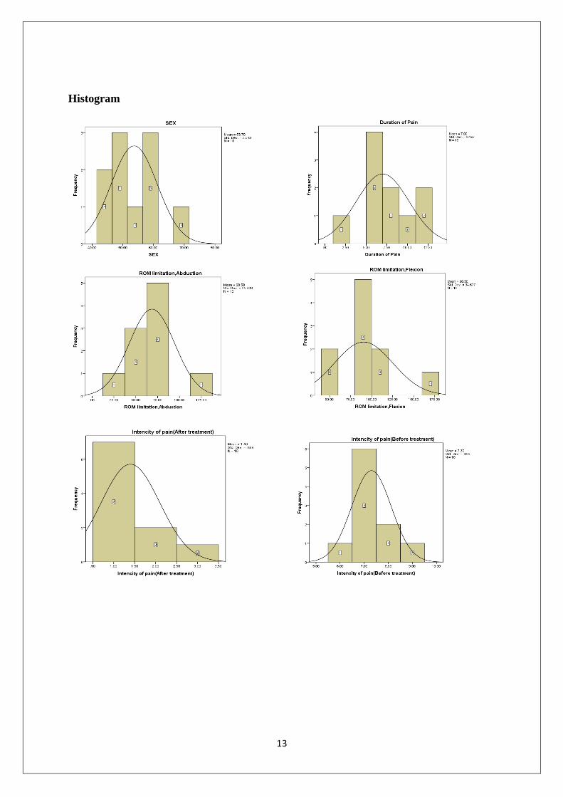
Histogram

14
Discussion:
Codman and other researcher have been reported common disorders in the shoulder higher
than 10 % from 1930(23). Mechanical causes of pain around the shoulder joint and tendon
involvement, constitute the majority of cases. Due to anatomical characteristics and high
range of motion in different directions and relative instability of the shoulder buildings, the
main act sets on tendons and muscles, and therefore, the injuries of repetitive motion are
occurring the most common. Although the cause of calcium deposit is unclear in the shoulder
(2,3), but the changes tendon wear, necrosis and metaplasia and after the event fibrocartilage
in tendon, the tissue hypoxia, are such theories pointed for the foreground crystal material
hydroxyapatite (1.4.3). Deposition creates a high pressure sealed container like a cyst on one
hand and on the other hand acts as a foreign body.
Although this complication is characterized improvable and they believed that the sediment
itself will be absorbed over time, however, as shown in this study were patients treated in
chronic phase and changes in the density of calcium deposits with the beginning of treatment
quickly occurred.
There are several treatments for calcium deposit on the shoulder, Including: 1- medication
drugs (NSAID) 2-physicaltherapy 3-local steroid injection 4- injected by a needle for
breakdown and lavage of the deposit (with 60-74 % success rate) (2,3,7,8) 5- shock wave
therapy ECSW (30 to 70 % relief of pain and 20 to 77 % absorption of calcification in studies
in Germany) ( 7,8,16 ) 6-deprival of calcium in food , such as dairy and nuts. It seam,
calcium deficiency of the body is the factor that cause to absorb of the pathological calcium
deposits (7, 10), but this method should not be used as a stand-alone therapy so that the
balanced nutrition with more than 1,000 mg of calcium supplementation in healthy people is
necessary especially in women during natural menopause (18) 7-The use of supplements of
magnesium ( magnesium deficiency increases the phosphor and calcium) (10) 8-in a study
by yokoyama in 2003, in 16 patients, cimetedine used in calcium deposit for the treatment of
chronic shoulder, pain relieved in 10 patients and deposits disappeared in 9 patients( 7 ) 9-
More recently, the use of EDTA include intravenous administration of amino acid in which a
substance that is used in the treatment of lead poisoning and heavy metals , have been
15
proposed ( 14 ) 10-ionthophoresis 11-Surgery (with a success rate of about 90 %) and about
10 % likelihood of further surgery ( 7 ). In Wolk and wittenberg reported that conservative
treatment was 70% successful (8).
Successful treatment of needling for this problem is mentioned in the medical literature,
however hazards such as probability of further damage to the tendon or infection with a
needle manipulation compared to treatment of this study makes it risky. New study using
shock wave also has taken far the results and it did not result in detailed and clear to solve
this problem.
Iontophoresis is not a new technique (11), iontophoresis has begun since 1700 but most of its
researchers recognize it since 1900, after working Leduc (19,20,25).Using iontophoresis
acetic acid in the treatment of patients with calcium deposits, described first in 1955 by psaki
and carroll and then in 1977 by Kabn (19).
Iontophoresis is a technique in which an electric current to drive (deliver) a chemical or drug
is given through the skin (25, 26). In fact, it is a needle -free injection and it is used as a non-
invasive, painless, low- cost. Iontophoresis is used in laboratory research and particularly
useful in Neuropharmacology (12). It is also common treatments for hyperhydrosis.
Iontophoresis is an alternative method in the treatment of calcium deposit that it is used this
method instead of acetic acid injection. It has no side effects of the needle injection so that
entering the needle causes the puncture, bleeding and it could be a field (precursor)
calcification forming. This method prohibits in patients with wounds or skin lesions in the
treated area, electric current or the chemical hyper sensivity, patients with Pace maker and in
patients with brain disorders, but it can be used in diabetic patients with caution (11). The
most problem in the iontophoresis approach is chemical burns at electrodes of treatment site.
Considering into the necessary precautions we observed small surface vesicles burned in
some patients. In iontophoresis, acetic acid ionized negative charge of Acetate, it penetrates
through the skin and can be combined with calcium ions in calcium deposit (calcium
carbonate) and creates soluble Calcium Acetate that solved through local circulation ( 4 ).
CaCo3+2H(C2H3O2)= Ca(C2H3O2)2 + H2O + CO2
The presence of calcification in the shoulder can press the rotator cuff tendon and cause
permanent damage in the parts of tendon (18).
In the present study, although other interventions of physical therapy modalities commonly
used for treatment, but so far the lack of acetic acid iontophoresis has poor outcomes and
with the addition of this modality the result has surprisingly been different.there are
inconsistency reports about iontophoresis ( 15,16,21,22,26 )
In this study it is used the flow rate to 7 mA and time more to 35 minutes. The study has been
done in the past, they used time and intensity lower than this present study. Moreover the
type of acid produced and the amount dipped in the acid acetic solution (as in treatment time,
drying pad was again wet by syringe including acetic acid) and 5 sessions per week of
treatment were different in this research into available articles. In this study Because of pain

16
intensity and lack of precision in Active shoulder rotation, it was used flexion and abduction
just in the shoulder.
One of the problems of the present study was the lack of number of patients with clearly
calcium deposits in radiographs. As The number of aforementioned samples provide with the
assistance of the professional orthopedists in during 1 year. Therefore in this study we
couldn´t use control group for comparison because there weren´t enough patients. In this
research the difference has shown in radiographic and clinical improvement as well as
compared to the beginning of treatment. In doing study the researcher faced with a
calcification of the hand area that was used this approach and the result of treatment for both
therapists and patients is amazing.
33 -year-old male patient with a history of 10 months of trauma to the fourth metacarpo
phalangeal joint during volleyball playing with collateral ligament sprain of forth finger in
that after the following radiography to eliminate calcification was treated for 10 sessions. x-
ray results after 10 sessions of treatment for the patient and therapist, was stunning.
The beginning of treatment After 10 sessions
Conclusion: In the present study with a limited number it was seen a supremacy between
women with the mean age lower as compared to males (statistical analysis and histogram).
The average age of about 53 years of involvement in both sexes, which is consistent with
research done in this field.
The mean duration of the onset of pain was about 5-7 months. Pain intensity based on the
scale (VAS) at the beginning of treatment, about 7 to 10 at the end of treatment to around
1.05 from 10 declined significantly.
The average of joint limitation of motion in the shoulder Abduction about 75 degrees and the
Flexion about100-80 degree has obtained, and it is indicated that the priority of the limitation
of shoulder Abduction relative to Flexion.
17
In this study the average of number of treatment sessions, it has been obtained 10 sessions.
The measurement of calcium deposits in the shoulder has strictly declined after treatment.
How to change the size and extent of calcium deposits showed that a decrease in the density
and calcium reabsorption increased by the iontophoresis acetic acid during the course of
treatment, and with the increasing the number of sessions. With the final treatment, the signs
of dense calcification were not observed. On the other hand it can be stated that this is age-
related complication occurs mainly in middle-aged or older and on the other hand it is
considered as a chronic condition. Also click and clinical particularly felt trapped shoulder
rotator cuff tendon depended to two factors, one as much as the deposits, other is to the
amount of space available of acromio humeral joint. It is considered that the patient has
enough space in this area despite a larger deposit, the impingement symptoms were mild
In the present study with a limited number it was seen a supremacy between women with the
mean age lower as compared to males (statistical analysis and histogram).
The average age of about 53 years of involvement in both sexes, which is consistent with
research done in this field.
The mean duration of the onset of pain was about 5-7 months. Pain intensity based on the
scale (VAS) at the beginning of treatment, about 7 to 10 at the end of treatment to around
1.05 from 10 declined significantly.
The average of joint limitation of motion in the shoulder Abduction about 75 degrees and the
Flexion about100-80 degree has obtained, and it is indicated that the priority of the limitation
of shoulder Abduction relative to Flexion.
In this study the average of number of treatment sessions, it has been obtained 10 sessions.
The measurement of calcium deposits in the shoulder has strictly declined after treatment.
.
Recommendation: more systematic research with a larger sample and different methods
without interfering with other physical modalities for evaluating the effect of this treatment is
recommended.
This method can be used in similar diseases such as myositis ossificant, calcium deposits in
other pheriferal joint , frozen shoulder , heel spur.
Keywords: shoulder,calcific,calcium deposit, calcification , iontophoresis, acetic acid
References:
1- Bernard E. Leduc,MD,FRCPC, Jocelyne Caya,PT, Sylvie Tremblay, PT, Nathalie J,
Bureau, MD,FRCPC, Marc Dumont, MSc. Treatment of Calcific Tendinitis of the Shoulder

18
by Acetic Acid Ionthophoresis: A Double-Blind Randomized Controlled Trial. Arch Phys
Med Rehabil 2003, 84:1523-7
the physician and sport medicin Vol 27 No 9 september 1999ellman,MD
2- William B. Wolf III,MD. Calcific Tendinitis of the Shoulder,Diagnosis and
Simple,Effective Treatment. The Physician And SportsMedicine- VOL 27- NO. 9 –
September 1999
3- Rima Aina,MD,Etienne Cardinal,MD,Nathalie J.Bureau,MD. Calcific Shoulder
tendinitis:treatment with modified US-guided Fine-Needle Technique. Department of
radiology,CHUM-Hospital Saint-Luc,1058 Saint-Denis,Montreal,Quebec,Canada H2X
3j4(R.A.,E.C.,N.J.B.,B.A.). March 21,2001
4- John Miller. What is Rotator Cuff Calcific Tendinitis. Physio Works-Physiotherapy
Brisbane. Sep 7 ,2015
5- e orthopod MEDIA.Humpal Physical Therapy&Sport Medicine Centers. Calcific
Tendonitis of the Shoulder. MMG 2001
6- John S. Rogerson,MD,SC. Calcific Tendinosis Of The shoulder. Excellence in
Orthopaedics,Uncompromised Care
7- Peter Symonds. Calcific Tendinitis,From Wikipedia,the free encyclopedia, May 2008
8- Anthony H Woodward,MD:chief editor,Harris Gellman,MD.Calcifying Tendonitis
Treatment &Management, Nov 21, 2015
9- Shoulderdoc.CO.uk, Calcific tendonitis, feb 2014
10- Peter Symonds. Calcific Tendinitis,From Wikipedia,the free encyclopedia, May 2008
11- Stephanie L. Stradley: ATC and Thomas W.Kaminski,PHD,ATC/L. New Uses for
Iontophoresis. Advance Health Care network for Physical Therapy&Rehab Medicine, June 1,
1999
12- Ionthophoresis from Wikipedia, the free encyclopedia 25 october 2015
13- Bernard E. Leduc MD treatment of calcifying tendonitis of the shoulder by acetic acid
iontophoresis Arch.phy med rehabil Vol 84 october 2003 pages 1523-1527
14- Angelo Cacchio, Elisabetta De Blasis. Effectiveness of treatment of calcific tendinitis of
the shoulder by disodium EDTA. Artheritis Care&Research vol 61 issue 1 pages 84-91. 15
january 2009
15- Perron&Maluin F. acetic acid iontophoresis(AAI) and ultrasound on calcifying tendinitis
of the shoulder: a randomized control trial. Arch Phys Med Rehabil 1997 78: 379-384
16- John S.Rogerson,orthopaedic surgeon. Calcific tendinitis of the shoulder htm 1/1/2003
19
2- John Rogerson Calcific tendonitis of the shoulder Jan 2003
17- Jonathan Cluett, M.D. Calcific Tendonitis, What is calcific tendonitis? About.Com
Orthopedics August,12,2012
18-Bakersfield . Calcium Deposits in the Shoulder. Southern California Orthopedic Institute
Appointments(661) 328-5565…………..
19- Deborah L Wieder. Treatment of traumatic myositis ossificans with acetic acid
iontophoresis. Physical Therapy,Journal of the American physical therapy
association,1992,72:133-137
20- Kevin Gard,DPT.OCS and David Ebaugh,PHD,PT. The use of acetic acid iontophoresis
in the management of a soft tissue injury. N Am J Sports Phys Ther.2010
December,5(4):220-226
21-Charles D Ciccone,PT,PHD. Dose acetic acid iontophoresis accelerate the resorption of
calcium depositis in calcific tendinitis of the shoulder? Physical therapy,Journal of the
American Physical therapy association. January 2003 vol. 83 no. 1 68-74
22- S.shetty, T.L.Moore, S.Jackson, D.Brettle and A.L.Herrick. A pilot study of acetic acid
ionthophoresis and ultrasound in the treatment of systemic sclerosis related calcinosis.
Rheumatology(2005) 44(4):536-538,January 11
23- Stephen C Weber MD Arthroscopic treatment of calcific tendonitis Feb 2007/02/05
24- Sushil G Kachewar and Devidas S Kulkarni. Calcific Tendinitis of the R-Cuff, A review.
J.Clin Diagn Res 2013 jul 7(7)
25- Tim Waston. Electrotherapy:Evidence Based Practice. 2008(march) Elsevier
26- Eddy Krueger,Jose Luiz Claudino Junior,Eduardo Mendonca Scheeren. Iontophoresis:
Principles and applications. Fisioter Mov.2014 Jul/set;27(3):469-81
Related Documents