National Academy of Clinical Biochemistry Laboratory Medicine Practice Guidelines for Use of Tumor Markers in Testicular, Prostate, Colorectal, Breast, and Ovarian Cancers Catharine M. Sturgeon, 1* Michael J. Duffy, 2 Ulf-Håkan Stenman, 3 Hans Lilja, 4 Nils Bru ¨ nner, 5 Daniel W. Chan, 6 Richard Babaian, 7 Robert C. Bast, Jr., 8 Barry Dowell, 9 Francisco J. Esteva, 10 Caj Haglund, 11 Nadia Harbeck, 12 Daniel F. Hayes, 13 Mads Holten-Andersen, 5 George G. Klee, 14 Rolf Lamerz, 15 Leendert H. Looijenga, 16 Rafael Molina, 17 Hans Jørgen Nielsen, 18 Harry Rittenhouse, 19 Axel Semjonow, 20 Ie-Ming Shih, 6 Paul Sibley, 21 Gyo ¨ rgy So ¨ le ´ tormos, 22 Carsten Stephan, 23 Lori Sokoll, 6 Barry R. Hoffman, 24 and Eleftherios P. Diamandis 24 NACB SUB-COMMITTEE MEMBERS Testicular Cancer: Ulf-Håkan Stenman, Chair; Rolf Lamerz; and Leendert H. Looijenga; Prostate Cancer: Hans Lilja, Chair ; Richard Babaian; Barry Dowell; George G. Klee; Harry Rittenhouse; Axel Semjonow; Paul Sibley; Lori Sokoll; and Carsten Stephan; Colorectal Cancer: Nils Bru ¨ nner, Chair ; Michael J. Duffy; Caj Haglund; Mads Holten-Andersen; and Hans Jørgen Nielsen; Breast Cancer: Michael J Duffy, Chair ; Francisco J. Esteva; Nadia Harbeck; Daniel F. Hayes; and Rafael Molina; Ovarian Cancer: Daniel W. Chan, Chair; Robert C. Bast, Jr.; Ie-Ming Shih; Lori J. Sokoll; and Gyo ¨ rgy So ¨ le ´ tormos BACKGROUND: Updated National Academy of Clinical Biochemistry (NACB) Laboratory Medicine Practice Guidelines for the use of tumor markers in the clinic have been developed. METHODS: Published reports relevant to use of tumor markers for 5 cancer sites—testicular, prostate, colo- rectal, breast, and ovarian—were critically reviewed. RESULTS: For testicular cancer, -fetoprotein, human chorionic gonadotropin, and lactate dehydrogenase are recommended for diagnosis/case finding, staging, prognosis determination, recurrence detection, and ther- apy monitoring. -Fetoprotein is also recommended for differential diagnosis of nonseminomatous and semi- nomatous germ cell tumors. Prostate-specific antigen (PSA) is not recommended for prostate cancer screen- ing, but may be used for detecting disease recurrence and monitoring therapy. Free PSA measurement data are useful for distinguishing malignant from benign prostatic disease when total PSA is 10 g/L. In colo- rectal cancer, carcinoembryonic antigen is recommended (with some caveats) for prognosis determination, postop- erative surveillance, and therapy monitoring in advanced disease. Fecal occult blood testing may be used for 1 Department of Clinical Biochemistry, Royal Infirmary of Edinburgh, Edinburgh, UK; 2 Department of Pathology and Laboratory Medicine, St Vincent’s University Hospital and UCD School of Medicine and Medical Science, Conway Institute of Biomolecular and Biomedical Research, University College Dublin, Dublin, Ire- land; 3 Department of Clinical Chemistry, Helsinki University Central Hospital, Helsinki, Finland; 4 Departments of Clinical Laboratories, Urology, and Medi- cine, Memorial Sloan-Kettering Cancer Center, New York, NY; 5 Section of Biomedicine, Department of Veterinary Pathobiology, Faculty of Life Sciences, University of Copenhagen, Denmark; 6 Departments of Pathology and Oncology, Johns Hopkins Medical Institutions, Baltimore, MD; 7 Department of Urology, The University of Texas Anderson Cancer Center, Houston, TX; 8 Depart- ment of Experimental Therapeutics, University of Texas Anderson Cancer Cen- ter, Houston, Texas, USA.; 9 Abbott Laboratories, Abbott Park, IL; 10 Depart- ments of Breast Medical Oncology, Molecular and Cellular Oncology, University of Texas M.D. Anderson Cancer Center, Houston TX; 11 Department of Surgery, Helsinki University Central Hospital, Helsinki, Finland; 12 Frauenklinik der Tech- nischen Universita ¨t Mu ¨ nchen, Klinikum rechts der Isar, Munich, Germany; 13 Breast Oncology Program, University of Michigan Comprehensive Cancer Center, Ann Arbor, MI; 14 Department of Laboratory Medicine and Pathology, Mayo Clinic College of Medicine, Rochester, MN; 15 Department of Medicine, Klinikum of the University of Munich, Grosshadern, Germany; 16 Laboratory of Experimental Patho-Oncology, Erasmus MC-University Medical Center Rotter- dam, and Daniel den Hoed Cancer Center, Rotterdam, the Netherlands; 17 Lab- oratory of Biochemistry, Hospital Clinico Provincial, Barcelona, Spain; 18 De- partment of Surgical Gastroenterology, Hvidovre Hospital, Copenhagen, Denmark; 19 Gen-Probe, San Diego, CA; 20 Prostate Center, Department of Urology, University Clinic Muenster, Muenster, Germany; 21 Siemens Medical Solutions Diagnostics, Glyn Rhonwy, Llanberis, Gwynedd, UK; 22 Department of Clinical Biochemistry, Hillerød Hospital, Hillerød, Denmark; 23 Department of Urology, Charite ´ Hospital, Universita ¨ tsmedizin Berlin, Berlin, Germany; 24 De- partment of Pathology and Laboratory Medicine, Mount Sinai Hospital, and Department of Laboratory Medicine and Pathobiology, University of Toronto, Ontario, Canada. * Address correspondence to this author at: the Department of Clinical Biochem- istry, Royal Infirmary, Edinburgh EH16 4SA, UK. Fax 44 131 242 6882; e-mail [email protected]. All relationships with industry for the guidelines committee are reported in the online supplement. Received February 19, 2008; accepted August 27, 2008. Previously published online at DOI: 10.1373/clinchem.2008.105601 Clinical Chemistry 54:12 e11–e79 (2008) Special Report e11

Welcome message from author
This document is posted to help you gain knowledge. Please leave a comment to let me know what you think about it! Share it to your friends and learn new things together.
Transcript
National Academy of ClinicalBiochemistry Laboratory Medicine
Practice Guidelines for Use of TumorMarkers in Testicular, Prostate,
Colorectal, Breast, and Ovarian CancersCatharine M. Sturgeon,1* Michael J. Duffy,2 Ulf-Håkan Stenman,3 Hans Lilja,4 Nils Brunner,5 Daniel W. Chan,6
Richard Babaian,7 Robert C. Bast, Jr.,8 Barry Dowell,9 Francisco J. Esteva,10 Caj Haglund,11 Nadia Harbeck,12
Daniel F. Hayes,13 Mads Holten-Andersen,5 George G. Klee,14 Rolf Lamerz,15 Leendert H. Looijenga,16
Rafael Molina,17 Hans Jørgen Nielsen,18 Harry Rittenhouse,19 Axel Semjonow,20 Ie-Ming Shih,6 Paul Sibley,21
Gyorgy Soletormos,22 Carsten Stephan,23 Lori Sokoll,6 Barry R. Hoffman,24 and Eleftherios P. Diamandis24
NACB SUB-COMMITTEE MEMBERSTesticular Cancer: Ulf-Håkan Stenman, Chair; Rolf Lamerz; and Leendert H. Looijenga; Prostate Cancer:Hans Lilja, Chair ; Richard Babaian; Barry Dowell; George G. Klee; Harry Rittenhouse; Axel Semjonow;Paul Sibley; Lori Sokoll; and Carsten Stephan; Colorectal Cancer: Nils Brunner, Chair; Michael J. Duffy;Caj Haglund; Mads Holten-Andersen; and Hans Jørgen Nielsen; Breast Cancer: Michael J Duffy, Chair ;
Francisco J. Esteva; Nadia Harbeck; Daniel F. Hayes; and Rafael Molina; Ovarian Cancer: Daniel W. Chan,Chair; Robert C. Bast, Jr.; Ie-Ming Shih; Lori J. Sokoll; and Gyorgy Soletormos
BACKGROUND: Updated National Academy of ClinicalBiochemistry (NACB) Laboratory Medicine PracticeGuidelines for the use of tumor markers in the clinichave been developed.
METHODS: Published reports relevant to use of tumormarkers for 5 cancer sites—testicular, prostate, colo-rectal, breast, and ovarian—were critically reviewed.
RESULTS: For testicular cancer, �-fetoprotein, humanchorionic gonadotropin, and lactate dehydrogenaseare recommended for diagnosis/case finding, staging,prognosis determination, recurrence detection, and ther-
apy monitoring. �-Fetoprotein is also recommended fordifferential diagnosis of nonseminomatous and semi-nomatous germ cell tumors. Prostate-specific antigen(PSA) is not recommended for prostate cancer screen-ing, but may be used for detecting disease recurrenceand monitoring therapy. Free PSA measurement dataare useful for distinguishing malignant from benignprostatic disease when total PSA is �10 �g/L. In colo-rectal cancer, carcinoembryonic antigen is recommended(with some caveats) for prognosis determination, postop-erative surveillance, and therapy monitoring in advanceddisease. Fecal occult blood testing may be used for
1 Department of Clinical Biochemistry, Royal Infirmary of Edinburgh, Edinburgh,UK; 2 Department of Pathology and Laboratory Medicine, St Vincent’s UniversityHospital and UCD School of Medicine and Medical Science, Conway Institute ofBiomolecular and Biomedical Research, University College Dublin, Dublin, Ire-land; 3 Department of Clinical Chemistry, Helsinki University Central Hospital,Helsinki, Finland; 4 Departments of Clinical Laboratories, Urology, and Medi-cine, Memorial Sloan-Kettering Cancer Center, New York, NY; 5 Section ofBiomedicine, Department of Veterinary Pathobiology, Faculty of Life Sciences,University of Copenhagen, Denmark; 6 Departments of Pathology and Oncology,Johns Hopkins Medical Institutions, Baltimore, MD; 7 Department of Urology,The University of Texas Anderson Cancer Center, Houston, TX; 8 Depart-ment of Experimental Therapeutics, University of Texas Anderson Cancer Cen-ter, Houston, Texas, USA.; 9 Abbott Laboratories, Abbott Park, IL; 10 Depart-ments of Breast Medical Oncology, Molecular and Cellular Oncology, Universityof Texas M.D. Anderson Cancer Center, Houston TX; 11 Department of Surgery,Helsinki University Central Hospital, Helsinki, Finland; 12 Frauenklinik der Tech-nischen Universitat Munchen, Klinikum rechts der Isar, Munich, Germany;13 Breast Oncology Program, University of Michigan Comprehensive CancerCenter, Ann Arbor, MI; 14 Department of Laboratory Medicine and Pathology,Mayo Clinic College of Medicine, Rochester, MN; 15 Department of Medicine,
Klinikum of the University of Munich, Grosshadern, Germany; 16 Laboratory ofExperimental Patho-Oncology, Erasmus MC-University Medical Center Rotter-dam, and Daniel den Hoed Cancer Center, Rotterdam, the Netherlands; 17 Lab-oratory of Biochemistry, Hospital Clinico Provincial, Barcelona, Spain; 18 De-partment of Surgical Gastroenterology, Hvidovre Hospital, Copenhagen,Denmark; 19 Gen-Probe, San Diego, CA; 20 Prostate Center, Department ofUrology, University Clinic Muenster, Muenster, Germany; 21 Siemens MedicalSolutions Diagnostics, Glyn Rhonwy, Llanberis, Gwynedd, UK; 22 Department ofClinical Biochemistry, Hillerød Hospital, Hillerød, Denmark; 23 Department ofUrology, Charite Hospital, Universitatsmedizin Berlin, Berlin, Germany; 24 De-partment of Pathology and Laboratory Medicine, Mount Sinai Hospital, andDepartment of Laboratory Medicine and Pathobiology, University of Toronto,Ontario, Canada.
* Address correspondence to this author at: the Department of Clinical Biochem-istry, Royal Infirmary, Edinburgh EH16 4SA, UK. Fax �44 131 242 6882; [email protected].
All relationships with industry for the guidelines committee are reported in theonline supplement.Received February 19, 2008; accepted August 27, 2008.Previously published online at DOI: 10.1373/clinchem.2008.105601
Clinical Chemistry 54:12e11–e79 (2008)
Special Report
e11
screening asymptomatic adults 50 years or older. Forbreast cancer, estrogen and progesterone receptors aremandatory for predicting response to hormone ther-apy, human epidermal growth factor receptor-2 mea-surement is mandatory for predicting response to tras-tuzumab, and urokinase plasminogen activator/plasminogen activator inhibitor 1 may be used fordetermining prognosis in lymph node–negative pa-tients. CA15-3/BR27–29 or carcinoembryonic antigenmay be used for therapy monitoring in advanced dis-ease. CA125 is recommended (with transvaginal ultra-sound) for early detection of ovarian cancer in womenat high risk for this disease. CA125 is also recom-mended for differential diagnosis of suspicious pelvicmasses in postmenopausal women, as well as for detec-tion of recurrence, monitoring of therapy, and deter-mination of prognosis in women with ovarian cancer.
CONCLUSIONS: Implementation of these recommenda-tions should encourage optimal use of tumor markers.© 2008 American Association for Clinical Chemistry
We present here to clinical chemists, clinicians, andother practitioners of laboratory and clinical medicinethe latest update of the National Academy of ClinicalBiochemistry (NACB)25 Laboratory Medicine PracticeGuidelines for the use of tumor markers in testicular,prostate, colorectal, breast, and ovarian cancers. Theseguidelines are intended to encourage more appropriateuse of tumor marker tests by primary care physicians,hospital physicians and surgeons, specialist oncolo-gists, and other health professionals.
Clinical practice guidelines are systematically de-veloped statements intended to assist practitioners and
patients in making decisions about appropriate health-care for specific clinical circumstances (1 ). An expla-nation of the methodology used when developing theseguidelines is provided in an accompanying preamble(2 ). As might be expected, many of the NACB recom-mendations are similar to those made by other groups,as is made clear from the tabular comparisons pre-sented for each malignancy (2 ). The disciplines of allauthors and statements of conflicts of interest, declaredaccording to NACB requirements, are provided in anonline data supplement (Supplemental Data Disclo-sures Table that accompanies this Special Report athttp://www.clinchem.org/content/vol54/issue12). Thelatter are also listed at the end of this manuscript. Allcomments received about these guidelines are also re-corded in an online data supplement (SupplementalData Comments Received Table), together with re-sponses to these comments.
To prepare these guidelines, the literature relevantto the use of tumor markers was reviewed. Particularattention was given to reviews, including the few rele-vant systematic reviews, and to guidelines issued byexpert panels. If possible, the consensus recommenda-tions of the NACB Panels reported here were based onavailable evidence, i.e., were evidence based. An ac-companying paper presents NACB recommendationsrelating to general quality requirements for tumormeasurements and includes tabulation of importantcauses of false-positive tumor marker results that mustalso be taken into account (e.g., heterophilic antibodyinterference, high-dose “hooking,” etc.) (3 ).
Tumor Markers in Testicular Cancers26,27
BACKGROUND
About 95% of all malignant testicular tumors are ofgerm-cell origin; most of the rest are lymphomas, Leydigor Sertoli cell tumors, and mesotheliomas. Germ celltumors of adolescents and adults are classified into 2main types, seminomas and nonseminomatous germcell cancers of the testis (NSGCT). Testicular cancersrepresent about 1% of all malignancies in males, butthey are the most common tumors in men age 15–35years. Testicular cancers are a significant cause ofdeath in this age group in spite of the fact that presentlymore than 90% of the cases are cured (4 ). Germ celltumors may also originate in extragonadal sites, e.g.,
25 Nonstandard abbreviations: NACB, National Academy of Clinical Biochemistry;NSGCT, nonseminomatous germ cell cancers of the testis; AFP, �-fetoprotein; hCG,human chorionic gonadotropin; LDH, lactate dehydrogenase; LOE, level of evi-dence; ITGCNU, intratubular germ cell neoplasia unclassified; MSI, microsatelliteinstability; PLAP, placental/germ cell alkaline phosphatase; SOR, strength of rec-ommendation; PSA, prostate-specific antigen; NICE, United Kingdom NationalInstitute for Health and Clinical Excellence; DRE, digital rectal examination; fPSA,free PSA; EGTM, European Group on Tumour Markers; cPSA, complexed PSA;ERSPC, European Randomized Screening for Prostate Cancer; SEER, Surveillance,Epidemiology and End Results; CTCs, circulating tumor cells; CRC, colorectalcancer; ASCO, American Society of Clinical Oncology; CEA, carcinoembryonicantigen; TIMP-1, tissue inhibitor of metalloproteinases type 1; uPA; urokinaseplasminogen activator; PAI-1, plasminogen activator inhibitor 1; EGFR, epidermalgrowth-factor receptor; FOBT, fecal occult blood test; FIT, fecal immunochemicaltest; NCCN, National Comprehensive Cancer Network; ER, estrogen receptor; PR,progesterone receptors; IHC, immunohistochemical analysis or immunohistochem-istry; MINDACT, Microarray for Node-Negative Disease Avoids Chemotherapy(trial); RS, recurrence score; TAILORx, Trial Assigning Individualized Options forTreatment; FIGO, International Federation of Gynecology and Obstetrics; GCIG,Gynecologic Cancer Intergroup; TPA, tissue polypeptide antigen; LPA, lysophos-phatidic acid; TATI, tumor-associated trypsin inhibitor; CASA, cancer-associatedserum antigen; hCG�cf, hCG �-core fragment.
26 NACB Testicular Cancer Sub-Committee members: Ulf-Hakan Stenman, Chair;Rolf Lamerz; and Leendert H. Looijenga.
27 All comments received about the NACB Recommendations for TesticularCancer are included in the online Data Supplement. Professor George Bosl,Professor Barry Hancock, Dr. Grahame Howard, and Professor Michael Secklwere invited expert reviewers.
e12 Clinical Chemistry 54:12 (2008)
the sacrococcygeal region, mediastinum, and pinealgland (5 ). Those of the sacrum are predominantlyfound in young males. Based on the histology, age ofthe patient at diagnosis, clinical behavior, and chromo-somal constitution, these tumors can be subdividedinto 3 distinct entities with different clinical and bio-logical characteristics (6 –9 ): (a) teratomas and yolk sactumors of newborns and infants, (b) seminomas andnonseminomas of adolescents and young adults, and(c) spermatocytic seminoma of the elderly. Seminomasand nonseminomas in adolescence and adulthoodwere the focus of attention when developing theserecommendations.
The incidence of testicular cancers varies consid-erably in different countries. In the US about 7200 newcases are diagnosed each year (4 ), and the age-adjustedincidence is 5.2 per 100 000. The incidence is about4-fold higher in white than in black men. In Europe,the age-adjusted incidence is lowest in Lithuania (0.9per 100 000), intermediate in Finland (2.5 per 100 000)and highest in Denmark (9.2 per 100 000) (10 ). Theincidence in various European countries has in-creased by 2%–5% per year. In the US the incidenceincreased by 52% from the mid-1970s to the mid-1990s(11 ). The cause of germ cell tumors is unknown,but familial clustering has been observed, and cryp-torchidism and Klinefelters syndrome are predispos-ing factors (4 ).
At presentation most patients have diffuse testicu-lar swelling, hardness, and pain. At an earlystage a painless testicular mass is a pathognomonicfinding, but testicular masses are most often causedby infectious epididymitis or orchitis. The diagnosiscan usually be confirmed by ultrasonography. If testic-ular cancer is suspected, the serum concentrations of�-fetoprotein (AFP), human chorionic gonadotropin(hCG), and lactate dehydrogenase (LDH) should be de-termined before therapy. As a rule, orchiectomyis performed before any further treatment, but may bedelayed until after chemotherapy in individuals withlife-threatening metastatic disease. After orchiectomy,additional therapy depends on the type and stage of thedisease.
Surveillance is increasingly used for seminoma pa-tients with stage I disease, but radiation to the retro-peritoneal and ipsilateral pelvic lymph nodes, which isstandard treatment for stage IIa and IIb disease, isalso used, as is short (single)-course carboplatin (12 ).About 4%–10% of patients relapse, and more than90% of patients who relapse are cured by chemother-apy. About 15%–20% of stage I seminoma patients un-der surveillance have a relapse and must be treated withchemotherapy. Patients with stage I nonseminomatoustumors are treated by orchiectomy. After this treat-ment, surveillance and nerve-sparing retroperitoneal
lymph-node dissection are accepted treatment op-tions. About 20% of patients under surveillance willhave a relapse and require chemotherapy. Patients withstage II nonseminomatous tumors are treated with ei-ther chemotherapy or retroperitoneal lymph node dis-section. Testicular cancer patients with advanced dis-ease are treated with chemotherapy (4 ).
Serum tumor markers have an important role inthe management of patients with testicular cancer,contributing to diagnosis, staging and risk assessment,evaluation of response to therapy, and early detectionof relapse. Increasing marker concentrations aloneare sufficient findings for treatment initiation. AFP,hCG, and LDH are established serum markers. Inmost cases of NSGCT, serum levels of one or more ofthese markers are increased, and in seminomas LDHand hCG are useful indicators. Other markers havebeen evaluated but provide limited additional clinicalinformation.
To prepare these guidelines, we reviewed the liter-ature relevant to the use of tumor markers for testicularcancer. Particular attention was given to reviews, pro-spective randomized trials that included the use of mark-ers, and guidelines issued by expert panels. Only onerelevant systematic review was identified. When pos-sible, the consensus recommendations of the NACBpanel were based on available evidence, i.e., were evi-dence based.
CURRENTLY AVAILABLE MARKERS FOR TESTICULAR CANCER
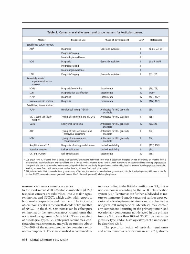
The most widely investigated tissue-based and serum-based tumor markers for testicular cancer are listed inTable 1. Also listed is the phase of development of eachmarker as well as the level of evidence (LOE) for itsclinical use.
TUMOR MARKERS IN TESTICULAR CANCER: NACB
RECOMMENDATIONS
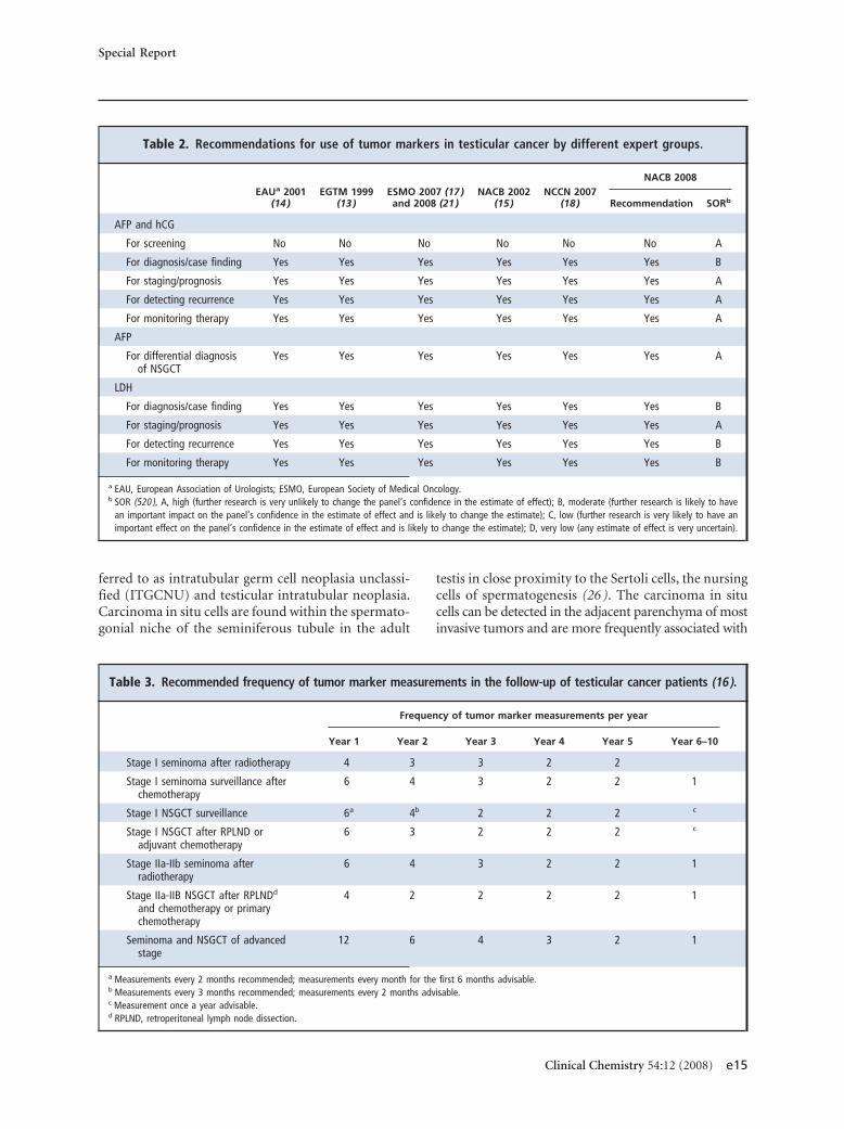
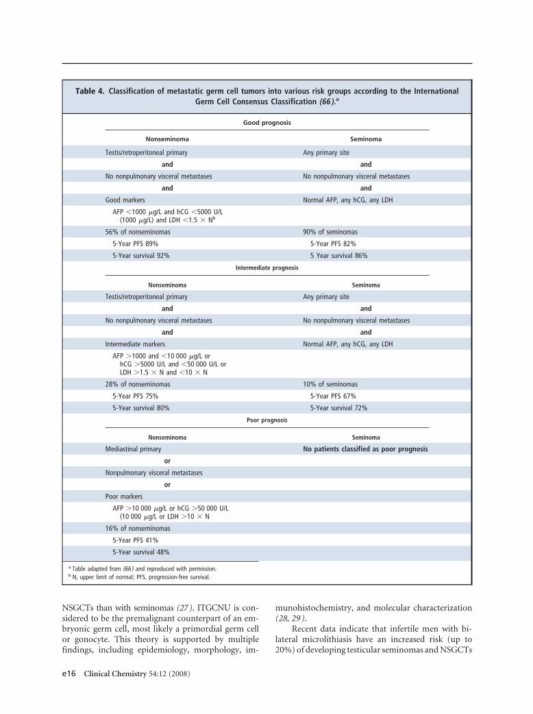
A summary of recommendations from representativeguidelines published on the use of tumor markers intesticular cancer is presented in Table 2. This table alsosummarizes the NACB guidelines for the use of mark-ers in this malignancy. A number of groups have madedetailed recommendations regarding the managementof testicular cancer (13–21 ), with some of those re-lating to tumor marker use summarized in Table 3.Table 4 summarizes the prognostic significance of se-rum tumor markers in metastatic testicular cancer, ac-cording to the consensus statement of the InternationalGerm Cell Consensus Group Classification, which re-mains the cornerstone for diagnosis and treatment oftesticular germ cell tumors. Below, we briefly reviewthe histological types of testicular cancer and present amore detailed discussion on the markers listed in thesetables.
Special Report
Clinical Chemistry 54:12 (2008) e13
HISTOLOGICAL TYPES OF TESTICULAR CANCER
In the most recent WHO-Mostofi classification (8, 22),testicular cancers are subdivided into 2 major types,seminomas and NSGCT, which differ with respect toboth marker expression and treatment. The incidenceof seminoma peaks in the fourth decade of life and thatof NSGCT in the third. Seminomas can be either pureseminomas or the rare spermatocytic seminomas thatoccur in older age groups. Most NSGCTs are a mixtureof histological types, i.e., embryonal carcinomas, cho-riocarcinomas, teratomas, and yolk sac tumors. About10%–20% of the nonseminomas also contain a semi-noma component. These are classified as combined tu-
mors according to the British classification (23 ), but asnonseminomas according to the WHO classificationsystem (22 ). Teratomas are further subdivided as ma-ture or immature. Somatic cancers of various types oc-casionally develop from a teratoma and are classified asnongerm cell malignancies. Metastases may containany component occurring in the primary tumor, andoccasionally components not detected in the primarytumor (22 ). Fewer than 10% of NSGCT contain a sin-gle tissue type, and all histological types of tissue shouldbe described (24 ).
The precursor lesion of testicular seminomasand nonseminomas is carcinoma in situ (25 ), also re-
Table 1. Currently available serum and tissue markers for testicular tumors.
Marker Proposed use Phase of development LOEa References
Established serum markers
AFPb Diagnosis Generally available II (4, 65, 73, 89 )
Prognosis/staging I
Monitoring/surveillance II
hCG Diagnosis Generally available II (4, 89, 103 )
Prognosis/staging I
Monitoring/surveillance II
LDH Prognosis/staging Generally available I (63, 109 )
Potentially usefulexperimental serummarkers
hCG� Diagnosis/monitoring Experimental IV (96, 103 )
LDH-1 Diagnosis/risk stratification Experimental IV (109 )
PLAP Diagnosis Experimental IV (111, 112 )
Neuron-specific enolase Diagnosis Experimental IV (116, 117 )
Established tissue markers
PLAP Histological typing ITGCNU Antibodies for IHC generallyavailable
II (24 )
c-KIT, stem cell factorreceptor
Typing of seminoma and ITGCNU Antibodies for IHC available II (28 )
CD30 Embryonal carcinoma Antibodies for IHC generallyavailable
IV (60, 519 )
AFP Typing of yolk sac tumors andembryonal carcinoma
Antibodies for IHC generallyavailable
II (24 )
hCG Typing of seminoma andchoriocarcinoma
Antibodies for IHC generallyavailable
II (24 )
Amplification of 12p Diagnosis of extragonadal tumors Limited availability II (107, 108 )
Vascular invasion Risk stratification Limited availability II (54 )
OCT3/4, POU5F1 Risk stratification Experimental IV (58 )
a LOE (120), level 1, evidence from a single, high-powered, prospective, controlled study that is specifically designed to test the marker, or evidence from ameta-analysis, pooled analysis or overview of level II or III studies; level II, evidence from a study in which marker data are determined in relationship to prospectivetherapeutic trial that is performed to test therapeutic hypothesis but not specifically designed to test marker utility; level III, evidence from large prospective studies;level IV, evidence from small retrospective studies; level V, evidence from small pilot studies.
b AFP, �-fetoprotein; hCG, human chorionic gonadotropin; hCB�, free �-subunit of human chorionic gonadotropin; LDH, lactic dehydrogenase; NSE, neuron specificenolase NSGCT, nonseminomatous germ cell tumors; PLAP, placental (germ cell) alkaline phosphatase.
e14 Clinical Chemistry 54:12 (2008)
ferred to as intratubular germ cell neoplasia unclassi-fied (ITGCNU) and testicular intratubular neoplasia.Carcinoma in situ cells are found within the spermato-gonial niche of the seminiferous tubule in the adult
testis in close proximity to the Sertoli cells, the nursingcells of spermatogenesis (26 ). The carcinoma in situcells can be detected in the adjacent parenchyma of mostinvasive tumors and are more frequently associated with
Table 2. Recommendations for use of tumor markers in testicular cancer by different expert groups.
EAUa 2001(14)
EGTM 1999(13)
ESMO 2007 (17)and 2008 (21)
NACB 2002(15)
NCCN 2007(18)
NACB 2008
Recommendation SORb
AFP and hCG
For screening No No No No No No A
For diagnosis/case finding Yes Yes Yes Yes Yes Yes B
For staging/prognosis Yes Yes Yes Yes Yes Yes A
For detecting recurrence Yes Yes Yes Yes Yes Yes A
For monitoring therapy Yes Yes Yes Yes Yes Yes A
AFP
For differential diagnosisof NSGCT
Yes Yes Yes Yes Yes Yes A
LDH
For diagnosis/case finding Yes Yes Yes Yes Yes Yes B
For staging/prognosis Yes Yes Yes Yes Yes Yes A
For detecting recurrence Yes Yes Yes Yes Yes Yes B
For monitoring therapy Yes Yes Yes Yes Yes Yes B
a EAU, European Association of Urologists; ESMO, European Society of Medical Oncology.b SOR (520 ), A, high (further research is very unlikely to change the panel’s confidence in the estimate of effect); B, moderate (further research is likely to have
an important impact on the panel’s confidence in the estimate of effect and is likely to change the estimate); C, low (further research is very likely to have animportant effect on the panel’s confidence in the estimate of effect and is likely to change the estimate); D, very low (any estimate of effect is very uncertain).
Table 3. Recommended frequency of tumor marker measurements in the follow-up of testicular cancer patients (16).
Frequency of tumor marker measurements per year
Year 1 Year 2 Year 3 Year 4 Year 5 Year 6–10
Stage I seminoma after radiotherapy 4 3 3 2 2
Stage I seminoma surveillance afterchemotherapy
6 4 3 2 2 1
Stage I NSGCT surveillance 6a 4b 2 2 2 c
Stage I NSGCT after RPLND oradjuvant chemotherapy
6 3 2 2 2 c
Stage IIa-IIb seminoma afterradiotherapy
6 4 3 2 2 1
Stage IIa-IIB NSGCT after RPLNDd
and chemotherapy or primarychemotherapy
4 2 2 2 2 1
Seminoma and NSGCT of advancedstage
12 6 4 3 2 1
a Measurements every 2 months recommended; measurements every month for the first 6 months advisable.b Measurements every 3 months recommended; measurements every 2 months advisable.c Measurement once a year advisable.d RPLND, retroperitoneal lymph node dissection.
Special Report
Clinical Chemistry 54:12 (2008) e15
NSGCTs than with seminomas (27). ITGCNU is con-sidered to be the premalignant counterpart of an em-bryonic germ cell, most likely a primordial germ cellor gonocyte. This theory is supported by multiplefindings, including epidemiology, morphology, im-
munohistochemistry, and molecular characterization(28, 29 ).
Recent data indicate that infertile men with bi-lateral microlithiasis have an increased risk (up to20%) of developing testicular seminomas and NSGCTs
Table 4. Classification of metastatic germ cell tumors into various risk groups according to the InternationalGerm Cell Consensus Classification (66 ).a
Good prognosis
Nonseminoma Seminoma
Testis/retroperitoneal primary Any primary site
and and
No nonpulmonary visceral metastases No nonpulmonary visceral metastases
and and
Good markers Normal AFP, any hCG, any LDH
AFP �1000 �g/L and hCG �5000 U/L(1000 �g/L) and LDH �1.5 � Nb
56% of nonseminomas 90% of seminomas
5-Year PFS 89% 5-Year PFS 82%
5-Year survival 92% 5 Year survival 86%
Intermediate prognosis
Nonseminoma Seminoma
Testis/retroperitoneal primary Any primary site
and and
No nonpulmonary visceral metastases No nonpulmonary visceral metastases
and and
Intermediate markers Normal AFP, any hCG, any LDH
AFP �1000 and �10 000 �g/L orhCG �5000 U/L and �50 000 U/L orLDH �1.5 � N and �10 � N
28% of nonseminomas 10% of seminomas
5-Year PFS 75% 5-Year PFS 67%
5-Year survival 80% 5-Year survival 72%
Poor prognosis
Nonseminoma Seminoma
Mediastinal primary No patients classified as poor prognosis
or
Nonpulmonary visceral metastases
or
Poor markers
AFP �10 000 �g/L or hCG �50 000 U/L(10 000 �g/L or LDH �10 � N
16% of nonseminomas
5-Year PFS 41%
5-Year survival 48%
a Table adapted from (66 ) and reproduced with permission.b N, upper limit of normal; PFS, progression-free survival.
e16 Clinical Chemistry 54:12 (2008)
(30 ). Surgical biopsy to assess the presence of ITGCNU(31 ) is indicated in this condition.
Tissue Markers for Testicular CancerGENETIC ABERRATIONS
A gain of chromosomal 12p sequences is observed ingerm cell tumors both of testicular and extragonadalorigin, a finding that indicates that gain of 12p se-quences may be of crucial importance for the develop-ment of this cancer. Indeed, this finding is used todiagnose germ cell tumors at extragonadal sites (32 ).The expression level of 12p sequences, however, doesnot correlate with stage of the disease or treatmentsensitivity/resistance (33–35 ). The crucial determinantof response to cisplatin-based compounds appears tooccur downstream of DNA binding in the intrinsic orextrinsic pathways of apoptosis or DNA repair (36–38).
Although the majority of germ cell tumors showan intact DNA mismatch-repair pathway, a defect lead-ing to microsatellite instability (MSI) has been ob-served in tumors refractory to cisplatin (39 – 41 ). Otherpotentially relevant findings in the context of treat-ment sensitivity and resistance relate to a possible de-fect in caspase 9 function (42 ). All these factors mightbe important, and it is unlikely that a single factor de-termines treatment sensitivity or resistance. The multi-factorial nature of treatment response is illustrated bythe finding that mature teratomas are resistant to var-ious DNA-damaging treatment protocols (38 ), possi-bly due to epigenetic changes occurring during somaticdifferentiation.
The majority of invasive seminomas and nonsemi-nomas contain additional copies of the X chromosome(43 ). This finding is interesting, because during nor-mal (female) development, X-chromosome inactiva-tion can occur in these tumors, in which X (inactive)-specific transcript (non-protein coding) (XIST)28 is theregulatory gene (6 ). Detection of unmethylated XISTDNA in plasma has been suggested to be useful formolecular diagnosis and the monitoring of testicularGCT-patients (44 ). This observation merits furtherinvestigation.
A number of studies have linked the developmentof germ cell tumors to a deregulated G1/S checkpoint,possibly related to the lack of a functional retinoblas-toma [retinoblastoma 1 (RB1) gene] cell cycle regula-tor (45 ), and consequently no upregulation of p21 afterinduction of DNA damage. Cells without p21 show re-duced cisplatin-induced DNA damage–repair capacityand increased sensitivity to cisplatin (46 ). The treat-ment-resistant mature teratomas show, in contrast toother invasive components, positive staining for mul-tiple proteins potentially related to treatment resis-tance. In addition, they are positive for RB1 and p21,allowing them to go into G1/S cycle arrest (47, 48 ).These characteristics might explain the observationthat residual mature teratoma is found in about30%– 40% of remnants of initial metastases afterchemotherapy.
A predictive model for the histology of a residualretroperitoneal mass, based on primary tumor histol-ogy, prechemotherapy markers, mass size, and size re-duction under chemotherapy, has been developed(49 ). Absence of teratoma elements or viable cancercells in the primary tumor has been identified as themost powerful predictor for benign residual tissue(50 ). Caution is warranted, however, because smallteratoma areas may be missed in the primary tumor,and absence of teratoma elements does not exclude oc-currence of malignant cells in residual masses. Thesefindings may again be related to the origin of thesetumors (51 ), because RB1 expression is not found inhuman fetal gonocytes or ITGCNU (52, 53 ).
VASCULAR INVASION
Particular attention must be paid to the presence orabsence of vascular invasion as a predictor of meta-static spread and occult metastases (54 ). Distinguish-ing venous from lymphatic invasion does not add in-formation as to the risk of occult metastasis. Besidesvascular invasion, high proliferative activity (assessedwith the monoclonal antibody MIB-1), and to a lesserextent the presence of embryonal carcinoma in the pri-mary tumor and a high pathologic stage, have beenreported to be predictors of systemic spread in clinicalstage I NSGCT [for review, see (55 )]. However, thepredictive value of this model is limited, because thegroup defined as high risk in fact has a 50% risk ofoccult metastasis, and the low risk group a 16% risk.
Prospective assessment of risk factors for relapse inclinical stage I NSGCT also showed that vascular inva-sion was the strongest predictive factor (56 ). With theaddition of 2 other risk parameters (MIB-1 score�70% and embryonal carcinoma �50%), the positivepredictive value increased to 63.6%. Thus, even with anoptimal combination of prognostic factors and refer-ence pathology, more than one-third of patients pre-
28 Human genes: XIST, X (inactive)-specific transcript (non-protein coding); RB1,retinoblastoma 1; APC, adenomatous polyposis coli; MLH1, mutL homolog 1, coloncancer, nonpolyposis type 2 (E. coli ); MSH2, mutS homolog 2, colon cancer,nonpolyposis type 1 (E. coli ); MSH6, mutS homolog 6 (E. coli ); PMS2, postmeioticsegregation increased 2 (S. cerevisiae); HER-2 and NEU, aliases for ERBB2 [v-erb-b2erythroblastic leukemia viral oncogene homolog 2, neuro/glioblastoma derivedoncogene homolog (avian)]; BRCA1, breast cancer 1, early onset; BRCA2, breastcancer 2, early onset; BRAF, v-raf murine sarcoma viral oncogene homolog B1;KRAS, v-Ki-ras2 Kirsten rat sarcoma viral oncogene homolog; �-catenin; PTEN,phosphatase and tensin homolog; MUC16, mucin 16, cell surface associated;prostasin [alias for PRSS8 (protease, serine, 8)]; AKT2, v-akt murine thymoma viraloncogene homolog 2; RSF-1, remodeling and spacing factor 1; NAC-1 [alias forNACC1 (nucleus accumbens associated 1, BEN and BTB (POZ) domain containing)];PCA3, prostate cancer antigen 3 (non-protein coding).
Special Report
Clinical Chemistry 54:12 (2008) e17
dicted to have pathologic stage II or a relapse duringfollow-up will not have metastatic disease and will beover-treated with adjuvant therapy. On the other hand,patients at low risk can be predicted with better accu-racy (86.5%), suggesting that surveillance may be anoption for highly compliant patients. Recently, clusteranalysis has been used to identify prognostic subgroupsin patients with embryonal carcinoma (57 ).
Serum Markers for Testicular Cancer
MARKER EXPRESSION AND TUMOR TYPE
Certain markers have been found to be informative forthe classification of seminomas and NSGCT. Placental/germ cell alkaline phosphatase (PLAP) is detected inmost seminomas and embryonal carcinomas and in50% of yolk sac tumors and choriocarcinomas, but onlyrarely in teratomas. HCG is expressed by syncytiotropho-blasts, choriocarcinoma, and approximately 30% of sem-inomas. Of the other tissue markers, the stem cell factorreceptor (c-KIT) has been used mainly to detect ITGCNUand seminoma, CD30 to detect embryonal carcinoma,and AFP to detect yolk sac tumors and a 10%–20% sub-set of embryonal carcinomas and teratomas. Recently,a potentially valuable marker, OCT3/4, also known asPOU5F1, has been identified (58–61).
Although a large number of serum markers havebeen studied, only hCG, AFP, and LDH have thus far
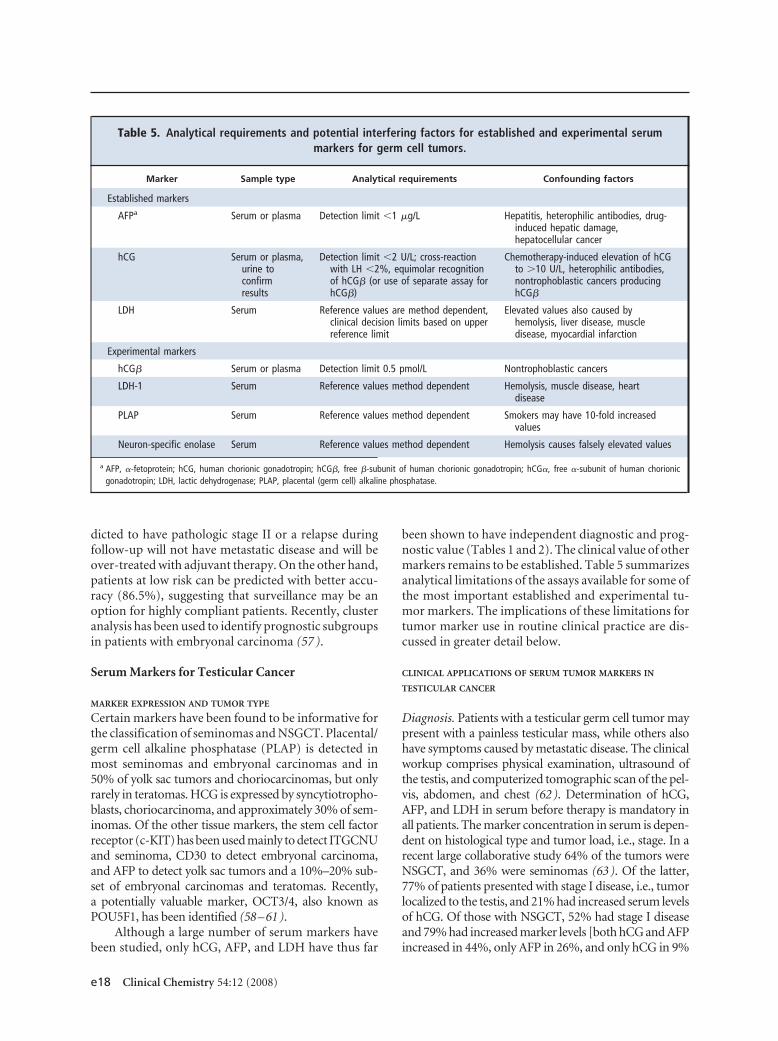
been shown to have independent diagnostic and prog-nostic value (Tables 1 and 2). The clinical value of othermarkers remains to be established. Table 5 summarizesanalytical limitations of the assays available for some ofthe most important established and experimental tu-mor markers. The implications of these limitations fortumor marker use in routine clinical practice are dis-cussed in greater detail below.
CLINICAL APPLICATIONS OF SERUM TUMOR MARKERS IN
TESTICULAR CANCER
Diagnosis. Patients with a testicular germ cell tumor maypresent with a painless testicular mass, while others alsohave symptoms caused by metastatic disease. The clinicalworkup comprises physical examination, ultrasound ofthe testis, and computerized tomographic scan of the pel-vis, abdomen, and chest (62). Determination of hCG,AFP, and LDH in serum before therapy is mandatory inall patients. The marker concentration in serum is depen-dent on histological type and tumor load, i.e., stage. In arecent large collaborative study 64% of the tumors wereNSGCT, and 36% were seminomas (63). Of the latter,77% of patients presented with stage I disease, i.e., tumorlocalized to the testis, and 21% had increased serum levelsof hCG. Of those with NSGCT, 52% had stage I diseaseand 79% had increased marker levels [both hCG and AFPincreased in 44%, only AFP in 26%, and only hCG in 9%
Table 5. Analytical requirements and potential interfering factors for established and experimental serummarkers for germ cell tumors.
Marker Sample type Analytical requirements Confounding factors
Established markers
AFPa Serum or plasma Detection limit �1 �g/L Hepatitis, heterophilic antibodies, drug-induced hepatic damage,hepatocellular cancer
hCG Serum or plasma,urine toconfirmresults
Detection limit �2 U/L; cross-reactionwith LH �2%, equimolar recognitionof hCG� (or use of separate assay forhCG�)
Chemotherapy-induced elevation of hCGto �10 U/L, heterophilic antibodies,nontrophoblastic cancers producinghCG�
LDH Serum Reference values are method dependent,clinical decision limits based on upperreference limit
Elevated values also caused byhemolysis, liver disease, muscledisease, myocardial infarction
Experimental markers
hCG� Serum or plasma Detection limit 0.5 pmol/L Nontrophoblastic cancers
LDH-1 Serum Reference values method dependent Hemolysis, muscle disease, heartdisease
PLAP Serum Reference values method dependent Smokers may have 10-fold increasedvalues
Neuron-specific enolase Serum Reference values method dependent Hemolysis causes falsely elevated values
a AFP, �-fetoprotein; hCG, human chorionic gonadotropin; hCG�, free �-subunit of human chorionic gonadotropin; hCG�, free �-subunit of human chorionicgonadotropin; LDH, lactic dehydrogenase; PLAP, placental (germ cell) alkaline phosphatase.
e18 Clinical Chemistry 54:12 (2008)
(63)]. In seminoma patients, hCG concentrations areusually below 300 U/L. Levels �1000 U/L are mostly as-sociated with NSGCT. Levels �10000 U/L are mainlyseen in patients with pure choriocarcinoma but occasion-ally may occur in seminoma. LDH is increased in 40%–60% of patients with seminoma or NSGCT (64). Theclassification of a tumor is based on histological examina-tion, but if serum AFP is increased, a tumor classified as aseminoma is reclassified as NSGCT and treated accord-ingly (4).
NACB TESTICULAR CANCER PANEL RECOMMENDATION 1:
TUMOR MARKERS IN THE DIAGNOSIS OF
TESTICULAR CANCER
When testicular cancer is suspected, pretreatmentdetermination of hCG, AFP, and LDH is manda-tory (Table 2) [LOE, II; strength of recommenda-tion (SOR), B].
STAGING, RISK STRATIFICATION, AND SELECTION OF THERAPY
Increased serum concentrations of AFP, hCG, andLDH are associated with adverse prognosis (65, 66 ). Ahigh serum hCG concentration is a strong prognosticfactor, and the risk of recurrence increases with in-creasing concentration (67 ). The International GermCell Cancer Collaborative Group has incorporated se-rum concentrations of hCG, AFP, and LDH in a schemefor classification of metastatic germ cell tumors (Table4). Tumors are classified as having good, intermediate,or poor prognosis on the basis of marker levels, pri-mary site of the tumor, and presence or absence of non-pulmonary visceral metastases (66 ).
The selection of treatment is based on tumortype and prognostic group. Stage I seminomas may betreated by orchiectomy alone, which leads to cure in80%– 85% of the cases. Orchiectomy in combinationwith radiotherapy of the abdominal lymph nodesleads to cure in 97%–99% of the cases, and this ap-proach is routinely used in many centers. Without ra-diotherapy 15%–20% of the patients relapse, but mostof these are cured by second line therapy. Thereforesurveillance at increased frequency is an alternative toradiotherapy.
When treated by orchiectomy only, patients withstage I NSGCT have a 30% risk of relapse. The risk ishigher (50%) if perivascular infiltration is presentthan if it is absent (risk 15%–20%). The relapse risk isvery low if retroperitoneal lymph node dissection isperformed in connection with primary therapy. Thisprocedure is associated with morbidity and thereforesurveillance is used as an alternative to retroperitoneallymph node dissection. Chemotherapy is another al-ternative to retroperitoneal lymph node dissection butpatients who undergo chemotherapy often have resid-
ual retroperitoneal tumors consisting of teratomas,which must be treated by surgery. If serum marker lev-els do not normalize or increase after retroperitoneallymph node dissection, positive retroperitoneal lymphnodes or systemic disease requiring chemotherapy aremost likely present (68, 69 ).
FURTHER RISK STRATIFICATION
Embryonal carcinoma is the most common cell type inNSGCT. Embryonal carcinoma is totipotential, and tu-mors with pure embryonal carcinoma are associatedwith early metastatic disease. Therefore, more accurateestimation of prognosis is needed for tumors contain-ing this cell type. Cluster analysis of the serum markersAFP and hCG in combination with the tissue markersp53, Ki67, and apoptosis index suggest that a patternwith high Ki67, low apoptosis, and low p53 is associ-ated with better survival than other patterns. Classifi-cation with this algorithm has been reported to be in-dependent of the International Germ Cell CollaborativeGroup Classification (67 ). Confirmation of these re-sults could provide a tool for more precise tailoring oftherapy.
NACB TESTICULAR CANCER PANEL RECOMMENDATION 2:
TUMOR MARKERS IN THE STAGING OF TESTICULAR CANCER
Measurement of hCG, AFP, and LDH is mandatoryfor staging and risk stratification according to theInternational Germ Cell Consensus Classification(Table 4) [LOE, I; SOR, A].
MONITORING OF RESPONSE TO THERAPY
If AFP or hCG in serum is increased before therapy, therate of marker decline reflects the response to therapy.Persistent marker elevation after chemotherapy indi-cates residual disease and the need for further therapy(70, 71 ). Chemotherapy may induce a transient in-crease or surge in marker concentrations during thefirst week of treatment (72 ).
In the absence of residual disease after orchidec-tomy, the half-life of hCG is approximately 1.5 daysand that of AFP 5 days (73, 74 ). During chemotherapy,half-lives �3.5 days for hCG or �7 days for AFP pre-dict recurrence and adverse prognosis (75 ). Markerhalf-life is calculated from the slope of the logarithm ofthe marker concentration vs time. It is preferable to usemarker concentrations from several time points and tocalculate the half-life from the slope of the regressionline (64 ). The half-life should be determined after theinitial marker surge during 2 cycles of chemotherapybetween days 7 and 56. A slow rate of marker decline isof potential use in poor-risk patients and may imply aneed for more aggressive therapy (75 ).
Special Report
Clinical Chemistry 54:12 (2008) e19
NACB TESTICULAR CANCER PANEL RECOMMENDATION 3:
TUMOR MARKERS IN MONITORING RESPONSE TO
TREATMENT IN PATIENTS WITH TESTICULAR CANCER
If raised before therapy, serum markers (AFP, hCG,and/or LDH) should be monitored weekly untilconcentrations are within the reference interval.Wherever possible, the marker half-life should bedetermined. Marker levels exceeding the upper ref-erence limit after therapy suggest residual disease,which should be confirmed or excluded by othermethods [LOE, II; SOR, A].
SURVEILLANCE
After successful primary therapy, all patients are mon-itored with physical examination, tumor marker deter-minations and computed tomographic scan. Withsuch surveillance, relapse is in most cases detectedbefore clinical symptoms appear. Most relapses occurwithin the first year and relapses after 2 years are rarebut in some cases relapse may occur even after 10 years.The surveillance is tailored to take into account tumortype, stage, treatment, and likelihood of relapse (Table3). Patients with low-risk disease treated with surgeryalone are monitored most frequently, e.g., every 1–2weeks during the first 6 months. Some centers recom-mend weekly monitoring to detect a relapse before thetumor grows to a size associated with adverse progno-sis, as estimated by serum concentrations of AFP �500kU/L and of hCG �1000 U/L (76 ). In all patients,monitoring is continued for 5 years (16 ).
NACB TESTICULAR CANCER PANEL RECOMMENDATION 4:
TUMOR MARKERS IN SURVEILLANCE OF PATIENTS WITH
TESTICULAR CANCER
Serial monitoring with AFP, hCG, and LDH is rec-ommended even when these are not raised beforetherapy, because marker expression can changeduring therapy. Frequency of measurement de-pends on the stage and pathology of disease butshould be determined according to agreed proto-cols (e.g., as in Table 3). Because baseline levels areindividual, increases are more important than ab-solute concentrations. A single increasing valuemust be confirmed with a second sample and thepossibility of transient elevation due to nonspecificinterference (e.g., iatrogenic hypogonadism)should be actively considered [LOE, II; SOR, A].
ANALYTICAL CONSIDERATIONS
Tumor marker measurements are mandatory in themanagement of testicular cancer patients. It is there-fore appropriate to review analytical requirements forthese important tests in more detail.
�-FETOPROTEIN
Biochemistry and biology. AFP is a homolog of albuminand is thought to act as a carrier protein in the fetus.During pregnancy, AFP is initially produced by theyolk sac and later by the fetal liver (77 ). Concentrationsin fetal plasma reach levels of 3 g/L in the week 12–14 ofpregnancy and decrease thereafter to 10 –200 mg/L atterm (78 ). After birth, circulating concentrations de-crease, with a half-life of 5 days, falling to adult levels at8 –10 months of age (79, 80 ). The high values that arenormal in early childhood must be remembered whenusing AFP as a marker for testicular yolk sac tumors,which are the most common testicular neoplasms ininfants (81, 82 ).
Assay methods, standardization, and reference values.AFP is quantified by 2-site immunometric assays em-ploying monoclonal antibodies or combinations ofmonoclonal and polyclonal antibodies. Results aregenerally comparable to those obtained with the com-petitive radioimmunoassay (RIA) format used previ-ously. The WHO standard 72/225, in which 1 Interna-tional Unit (U) of AFP corresponds to 1.21 ng, is usedfor calibration. Laboratories report values in mass units(ng/mL or �g/L) or kU/L. Reference values should beestablished for each assay to reflect differences in assaybias. Most centers quote an upper reference limit forAFP in the range of 10 –15 �g/L. Circulating concen-trations increase slightly with age; in one study the up-per reference limit increased from 9.3 kU/L in patientsyounger than 40 years to 12.6 kU/L in those older than40 years (83 ).
False-positive results. Rising levels of serum AFP indi-cate persistent germ cell tumor, even in the absence ofradiographic evidence of disease, provided other pos-sible causes can be excluded (see below) (4 ). Moder-ately increased AFP levels may persist even after che-motherapy, particularly when persistent disease has alarge cystic component, serving as a reservoir leakingAFP into the circulation (84 ). Increased serum con-centrations of AFP occur in most hepatocellular carci-nomas and 10%–30% of other gastrointestinal cancers,but these diseases are rare in patients with testicularcancer. Increased AFP values may not reflect cancer,and it is therefore important to identify positive resultscaused by other diseases and by nonspecific interfer-ence. Benign liver disease, in particular hepatitis, andliver damage induced by chemotherapy are often asso-ciated with moderately increased serum AFP levels,and may result in misinterpretation especially if levelsare rising (85, 86 ).
The carbohydrate compositions of AFP derivedfrom the liver and the yolk sac are different (87 ). Lectinbinding can differentiate increased levels caused by tes-
e20 Clinical Chemistry 54:12 (2008)
ticular cancer and liver disease (88 ), but such methodsare not routinely used. Patients who initially have in-creased AFP levels may have normal levels during arelapse if therapy has eliminated AFP-producing ele-ments but not all other components (89 ). Moderatelyincreased values that remain stable do not usually indi-cate relapse (86 ).
NACB TESTICULAR CANCER PANEL RECOMMENDATION 5:
ANALYTICAL REQUIREMENTS FOR MEASUREMENT OF AFP
AFP methods should be calibrated against WHOStandard 72/225 and the units in which results arereported (�g/L or kU/L) clearly stated. The detec-tion limit for AFP assays should be �1 �g/L (i.e.,�0.8 kU/L). Reference values should be establishedto reflect method bias. AFP may be raised due tobenign diseases, malignancies other than testicularcancer, or nonspecific interferences, and these pos-sibilities must be considered when interpreting re-sults [LOE, not applicable; SOR, A].
hCG AND hCG�
Biochemistry and biology. hCG is a member of the gly-coprotein hormone family, which includes luteinizinghormone, follicle-stimulating hormone and thyroidstimulating hormone. All 4 contain a common �-sub-unit. The distinct �-subunits confer biological activityand display various degrees of homology, with that be-tween the �-subunits of luteinizing hormone and hCG(hCG�) being about 80%. hCG� contains a 24 –aminoacid C-terminal extension not present in luteinizinghormone �, so antibodies to this part of the moleculeare specific for hCG. Although the subunits lack hCGactivity, hCG� has been shown to enhance the growthof tumor cells in culture by preventing apoptosis (90 ).hCG is expressed at very high concentrations by theplacenta and trophoblastic tumors, including chorio-carcinoma of the testis. hCG is heavily glycosylated,hCG� containing 6 and hCG� 2 carbohydrate chains.The glycosylation of hCG secreted by tumors is oftendifferent from that of hCG found in pregnant women.An antibody, B152, detects only a hyperglycosylatedvariant of hCG. This form predominates in earlypregnancy and is possibly more cancer-specific than“normal” hCG (91 ).
Nomenclature, assay methods, standardization, and ref-erence values. Specific determination of hCG is basedon antibodies reacting with hCG� (92 ). This practicehas caused confusion in the nomenclature of hCG as-says: the expressions “�-hCG” or “hCG-� assay” maydenote assays measuring both hCG and hCG� or onlyhCG�. According to the nomenclature recommendedby the IFCC, hCG denotes the intact �� heterodimer,
hCG� the free �-subunit, and hCG� the free �-subunit(93 ). Assays should be defined according to what theymeasure, i.e., hCG and hCG� separately or hCG andhCG� together (64, 94 ).
Assays for hCG are currently calibrated against theFourth International Standard (IS 75/589), in whichconcentrations are expressed in International Units(IU) based on bioactivity. It is difficult, however, tocompare concentrations of hCG with those of hCG�and hCG�, which are expressed in different arbitraryunits of the relevant International Standards (IS 75/551and IRP 75/569, respectively). Recently establishedWHO Reference Reagents have values assigned in mo-lar concentrations, which should facilitate direct com-parison of hCG and hCG� concentrations in the future(93, 95 ).
Because seminomas may produce solely hCG�and not intact hCG, it is essential that both hCG andhCG� are measured when monitoring testicular cancer(14, 96 ) Recommendations about antibody combina-tions that recognize most important forms of hCG-related isoforms and are appropriate for use in oncol-ogy have been published (94 ). Assays recognizing bothhCG and hCG� often use antibodies to epitopes on theC-terminal peptides of hCG�, but the relatively lowaffinities of these antibodies may limit assay sensitivity(94 ). Theoretically it should be possible to improvedetection of testicular cancer by using separate assaysfor hCG and hCG� (64, 96 ) but this remains to beconfirmed.
hCG is secreted at low levels by the pituitary, pro-ducing plasma levels that are measurable by sensitivemethods. The serum concentrations may increase withpatient age, particularly in women after menopause(97, 98 ). For most assays, the upper reference limit ofhCG is stated to be 5–10 U/L. When determined byultrasensitive methods, the upper limit in postmeno-pausal women is 5 U/L and in menstruating women is3 U/L. The upper reference limit for men younger than50 years is 0.7 U/L and for men older is 2.1 U/L (98 ).Cutoff values lower than the commonly used 5–10 U/Lcan be used to diagnose patients with testicular cancer.However, although most men with testicular cancerare young, their hCG levels may be increased due totesticular malfunction. Therefore diagnosis of activedisease in a patient with a history of a germ cell tumorrequires sequential determinations and rising values.The detection limit of most commercial assays doesnot allow reliable measurement of levels below 5 U/Land the utility of ultrasensitive assays and lower cutoffvalues needs to be determined (64 ). When expressedin molar concentrations, 5 U/L of hCG corresponds to15 pmol/L. The upper reference limit for hCG� is2 pmol/L and is independent of age and sex (98 ).
Special Report
Clinical Chemistry 54:12 (2008) e21
Specificity and confounding factors. It is important tonote that chemotherapy often causes gonadal suppres-sion that increases the hCG levels. Such hypogonadismcan also be spontaneous. This can be confirmed bymeasurement of serum luteinizing hormone and folli-cle-stimulating hormone and, when necessary, sup-pression with testosterone replacement (99 ). There-fore, hCG levels increasing from below 2 up to 5– 8 U/Lduring chemotherapy are often iatrogenic and do notnecessarily indicate relapse. Moderately increased lev-els of hCG may be of pituitary origin, especially if ac-companying serum levels of luteinizing hormone andfollicle-stimulating hormone exceed 30 –50 U/L, andare attributed to interrupted feedback inhibition fromthe gonads. This can be confirmed by short-term tes-tosterone treatment, which suppresses pituitary secre-tion of hCG (100, 101 ).
Nontrophoblastic tumors may in extremely rarecases produce hCG, whereas hCG� is often expressedat moderate levels by a large variety of tumors, includ-ing ovarian, gastrointestinal, bladder, lung, and headand neck cancers (101 ). Some patients with such tu-mors will have increased hCG levels when measure-ment is carried out by an assay recognizing both hCGand hCG�.
Falsely increased results for serum hCG can becaused by heterophilic antibodies. This phenomenonhas been reported only in women (102 ) but there is noreason why it should not also occur in men. False-positive results can be identified by analysis of hCG inurine or by repeating the assay after adding a blockingagent (e.g., nonimmune mouse IgG) to the sample toblock the interference (64, 102 ).
Apparently false-negative results will be obtainedwith assays measuring only hCG if the tumor produceshCG� but not hCG. Although this situation is morecommon in seminoma patients (103 ), it may also oc-cur in NSGCT patients (104 ).
NACB TESTICULAR CANCER PANEL RECOMMENDATION 6:
ANALYTICAL REQUIREMENTS FOR MEASUREMENT OF HCG
It is essential that both hCG and hCG� be measuredwhen using hCG to monitor testicular cancer pa-tients, either using a method recognizing a broadspectrum of hCG-related isoforms or separate spe-cific assays. hCG and hCG� should be recognizedon an equimolar basis with a detection limit of �1U/L. IFCC hCG nomenclature should be used todescribe the method used. The possibility of inter-ferences (e.g., from heterophilic antibodies) andtransient increases (e.g., due to chemotherapy)must be considered when interpreting hCG results[LOE, not applicable; SOR, A].
LACTATE DEHYDROGENASE
Biochemistry and biology. LDH in the circulation existsas a tetramer that may contain various combinations of2 subunits, LDH-A and LDH-B. The various subunitscan combine in 5 isoenzymes, LDH-1 [consisting of 4 Bsubunits (B4)], LDH-2 (B3A1), LDH-3 (B2A2), LDH-4(B1A3), and LDH-5 (A4). The gene encoding LDH-A islocated on chromosome 11, whereas the gene forLDH-B is located on the short arm of chromosome 12(i.e., 12p) (105 ). Interestingly, all invasive seminomasand NSGCTs show additional copies of this chromo-somal arm (106 ), suggesting that it may play a role indisease progression. No gain of 12p is detected inITGCNU (107, 108 ). A correlation between copynumber of 12p, tumor invasiveness, and the serumlevel of LDH-1 has been reported, but thus far the rel-evant 12p-genes have not been identified (109 ). Al-though theoretically interesting, these findings need tobe confirmed.
Specificity and confounding factors. Serum concentra-tions of LDH are measured enzymatically and the val-ues are method-dependent. The degree of elevation istherefore most conveniently expressed relative to theupper reference limit. LDH-1 can be determined byzymography or by immunoprecipitation of the otherisoenzymes and determination of residual catalytic ac-tivity. LDH is expressed in many tissues and increasedlevels may be caused by a wide variety of diseases. De-spite its lack of specificity, LDH is a useful marker, es-pecially for staging of seminoma and NSGCT (108 ).Hemolysis may cause falsely increased values andshould be avoided.
NACB TESTICULAR CANCER PANEL RECOMMENDATION 7:
ANALYTICAL REQUIREMENTS FOR MEASUREMENT OF LDH
Because LDH is measured enzymatically and thevalues are method dependent, the degree of eleva-tion should be expressed relative to the appropriateupper reference limit. Care must be taken to avoidhemolysis, which may cause falsely increased values[LOE, not applicable; SOR, A].
PLACENTAL ALKALINE PHOSPHATASE
Biochemistry and biology. A tumor-associated isoen-zyme of alkaline phosphatase was first described in apatient with lung cancer and later detected in serum ofpatients with other cancers and identified as placentalalkaline phosphatase (PLAP) (110 ). In fact, 2 genes en-code the proteins detected as PLAP activity, i.e., theplacental (PLAP) and germ cell enzymes. Both genesmap to chromosome 2 and the proteins cannot be dis-tinguished from each other using routine enzymatic or
e22 Clinical Chemistry 54:12 (2008)
immunohistochemical methods (111 ). PLAP is in-creased most frequently in patients with seminoma(60%–70%) (112, 113 ) and less frequently in thosewith other germ cell tumors, including ITGCNU (24 ).An enzymatic method can be used to detect ITGCNUcells in frozen tissue sections (114 ).
Assay methods, standardization, and reference values.PLAP has usually been determined by zymography butit can be also be measured by immunoassay or enzy-matically after immunocapture (113 ). The resultshould be compared with locally determined referencevalues. Because of homology with other alkaline phos-phatase isoenzymes, antibody selection is critical.However, the antibodies available so far cannot distin-guish between the PLAP and germ cell alkaline phos-phatase isoenzymes. Therefore, PLAP denotes both ofthese isoenzymes.
Specificity and confounding factors. Serum concentra-tions of PLAP are increased up to 10-fold in smokersand its measurement is therefore of little value in thisgroup (113 ). This fact and the paucity of commercialassays limit its clinical application, and serum assaysfor PLAP are not routinely included in the diagnosticworkup of testicular cancer patients.
OTHER MARKERS
Although pregnancy-specific �-1 glycoprotein andhCG are both expressed in trophoblastic cells, hCG isthe superior marker (115 ). Consequently, pregnancy-specific �-1 glycoprotein is not routinely measured. Neu-ron-specific enolase is increased in about 30%–50% of
patients with seminomas and less often in NSGCT pa-tients (16, 116, 117), but in spite of these promising re-sults the use of neuron-specific enolase is limited.
KEY POINTS: TUMOR MARKERS IN TESTICULAR CANCER
Tumor markers are of central importance in the diagno-sis, staging, risk assessment, and monitoring of patientswith testicular cancer. Several serum markers have beendescribed but only AFP, hCG, and LDH have been thor-oughly validated and shown to have independent prog-nostic value. Several tissue markers may prove to be clin-ically important in the diagnosis and classification oftesticular germ cell tumors. Germ cell tumors also displaytypical chromosomal abnormalities and amplification of12p is sufficiently characteristic to be useful in the clinic toidentify extratesticular germ cell tumors. Developmentsin DNA-based diagnostics have revealed a number ofchanges that may in the future enable more accurate strat-ification of prognosis.
Tumor Markers in Prostate Cancer29,30
BACKGROUND
Prostate cancer is the most common tumor in men inthe US. In 2007, 218 890 new cases and 27 050 deaths
29 NACB Prostate Cancer Sub-Committee members: Hans Lilja, Chair; RichardBabaian; Barry Dowell; George Klee; Harry Rittenhouse; Axel Semjonow; PaulSibley; Lori Sokoll; and Carsten Stephen.
30 All comments received about the NACB Recommendations for Prostate Cancerare included in the online Data Supplement. Prasad Bollina, Professor FritzSchroder, and Professor Hein von Poppel were invited expert reviewers.
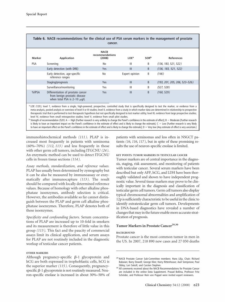
Table 6. NACB recommendations for the clinical use of PSA serum markers in the management of prostatecancer.
Marker Application
NACBrecommendations
(2008) LOEa SORb References
PSA Screening No III B (136, 183, 521, 522 )
Early detection (with DRE) Yes III B (136, 183, 521, 522)
Early detection, age-specificreference ranges
No Expert opinion B (146 )
Staging/prognosis Yes III B (193, 201, 205, 206, 523–526 )
Surveillance/monitoring Yes III B (527, 528 )
%fPSA Differentiation of prostate cancerfrom benign prostatic diseasewhen total PSA is 2–10 �g/L
Yes III B (160, 529 )
a LOE (120 ), level 1, evidence from a single, high-powered, prospective, controlled study that is specifically designed to test the marker, or evidence from ameta-analysis, pooled analysis or overview of level II or III studies; level II, evidence from a study in which marker data are determined in relationship to prospectivetherapeutic trial that is performed to test therapeutic hypothesis but not specifically designed to test marker utility; level III, evidence from large prospective studies;level IV, evidence from small retrospective studies; level V, evidence from small pilot studies.
b Strength of recommendation (520): A � High [Further research is very unlikely to change the Panel’s confidence in the estimate of effect]; B � Moderate [Further researchis likely to have an important impact on the Panel’s confidence in the estimate of effect and is likely to change the estimate]; C � Low [Further research is very likelyto have an important effect on the Panel’s confidence in the estimate of effect and is likely to change the estimate]; D � Very low [Any estimate of effect is very uncertain.]
Special Report
Clinical Chemistry 54:12 (2008) e23
Tabl
e7.
Reco
mm
enda
tion
sby
diff
eren
tex
pert
grou
psfo
rus
eof
PSA
and
%fP
SAas
tum
orm
arke
rsfo
rpr
osta
teca
ncer
.
Mar
ker
Ap
plic
atio
nA
CSa
(138
)A
CP
(530
)A
STR
O(5
27)
AU
A(5
28)
EAU
(531
)EG
TM(1
48)
ESM
O(5
32)
NA
CB
/EG
TM20
02(1
5)
NC
CN
(533
)U
SPST
F(5
34)
NIC
E20
08(1
21,1
39)
NA
CB
2008
b
PSA
Scre
enin
g(w
ithDR
E)Ye
sN
ocN
one pu
blis
hed
Yes
Yes
Noc
Nod
Yes
(NAC
B)Ye
sIn
suffi
cien
tev
iden
ceav
aila
ble
for
men
�75
year
sof
age.
Scre
enin
gfo
rm
en75
year
sor
olde
rno
tre
com
men
ded
(535
)
Insu
ffici
ent
evid
ence
avai
labl
eN
oat pr
esen
t
Early
dete
ctio
n:Ag
e-sp
ecifi
cre
fere
nce
rang
es
Non
e publ
ishe
dN
one pu
blis
hed
Non
e publ
ishe
dN
one pu
blis
hed
Non
e publ
ishe
dN
oN
one pu
blis
hed
Yes
(NAC
B)N
one pu
blis
hed
Non
epu
blis
hed
Non
epu
blis
hed
No
Early
dete
ctio
n:PS
Ave
loci
tyN
one pu
blis
hed
Non
e publ
ishe
dN
one pu
blis
hed
Non
e publ
ishe
dN
one pu
blis
hed
Non
e publ
ishe
dN
one pu
blis
hed
Non
e publ
ishe
dYe
sN
one
publ
ishe
dYe
sYe
s
Stag
ing/
Prog
nosi
sN
one pu
blis
hed
Non
e publ
ishe
dN
one pu
blis
hed
Yes
Yese
Non
e publ
ishe
dYe
sN
one pu
blis
hed
Yese
Non
epu
blis
hed
Yes
Yese
Follo
w-u
pne
gativ
ebi
opsy
(with
DRE)
Non
e publ
ishe
dN
one pu
blis
hed
Non
e publ
ishe
dN
one pu
blis
hed
Non
e publ
ishe
dN
one pu
blis
hed
Non
e publ
ishe
dN
one pu
blis
hed
Yes
Non
epu
blis
hed
Yes
Yes
Surv
eilla
nce/
mon
itorin
gN
one pu
blis
hed
Non
e publ
ishe
dYe
sfYe
sYe
sYe
sYe
sN
one pu
blis
hed
Yes
Non
epu
blis
hed
Yes
Yes
%fP
SAg
Diffe
rent
iatio
nof
PCa
and
beni
gnpr
osta
ticdi
seas
ew
hen
tota
lPSA
isbe
twee
n2–
10�
g/L
Non
e publ
ishe
dN
one pu
blis
hed
Non
e publ
ishe
dN
one pu
blis
hed
Non
e publ
ishe
dYe
sN
one pu
blis
hed
Yes
Yes
Non
epu
blis
hed
Non
epu
blis
hed
Yes
Follo
w-u
pne
gativ
ebi
opsy
(with
DRE)
orpa
tient
sw
ithin
crea
sed
biop
syris
k
Non
e publ
ishe
dN
one pu
blis
hed
Non
e publ
ishe
dN
one pu
blis
hed
Non
e publ
ishe
dN
one pu
blis
hed
Non
e publ
ishe
dN
one pu
blis
hed
Yes
Non
epu
blis
hed
Non
epu
blis
hed
Yes
aAC
S,Am
eric
anCa
ncer
Soci
ety;
ACP,
Amer
ican
Colle
geof
Phys
icia
ns;A
STRO
,Am
eric
anSo
ciet
yfo
rThe
rape
utic
Radi
olog
yan
dO
ncol
ogy;
AUA,
Amer
ican
Uro
logi
calA
ssoc
iatio
n;ES
MO
,Eur
opea
nSo
ciet
yfo
rMed
ical
Onc
olog
y;PC
a,Pr
osta
teca
ncer
;USP
STF,
US
Prev
entiv
eSe
rvic
esTa
skFo
rce.
bFo
rSO
Rsse
eTa
ble
6.c
Not
rout
inel
y,in
divi
dual
deci
sion
.d
Exce
ptin
men
with
urin
ary
sym
ptom
s.e
Aspa
rtof
nom
ogra
ms
with
DRE
and
biop
syG
leas
ongr
ade
(Par
tinta
bles
).fFo
llow
ing
radi
atio
nth
erap
y.g
Inm
enw
itha
tota
lPSA
of4
–10
�g/
Lan
da
nega
tive
DRE.
e24 Clinical Chemistry 54:12 (2008)
were predicted. Although prostate cancer is unequivo-cally lethal in some patients, most men die with ratherthan of their cancer (118 ). Autopsy data suggest that42% of men older than 50 years old have cancerous fociin their prostates but prostate cancer will be diagnosedin only approximately 16% of men during their life-time and only a quarter of these will die from it. Manymore men die with than of prostate cancer (119 ). Cur-rent incidence rates of clinical disease are 15-foldhigher in the US than in Japan despite similar frequen-cies of histological cancer. Hence, the far greater prev-alence of histological than symptomatic cancer hasbeen cited to support a conservative, nonintervention-ist approach to this disease. However, once prostatecancer reaches advanced stages either locally or system-ically with bone metastases, or becomes refractory tohormone therapy, little if any therapeutic means forcure are available.
The optimal management of patients with pros-tate cancer requires the use of the tumor marker pros-tate-specific antigen (PSA) in all instances and diseasestates. The use of PSA-related isoforms is appropriatein certain specific circumstances. Here we present newNational Academy of Clinical Biochemistry guidelineson the use these and other serum-based tumor markersin prostate cancer. A summary of relevant guidelinespublished by other expert panels on this topic is alsoprovided.
To prepare these guidelines, the literature relevantto the use of tumor markers in prostate cancer wasreviewed. Particular attention was given to reviews (in-cluding systematic reviews), prospective randomizedtrials that included the use of markers, and guidelinesissued by expert panels. Where possible, the consensusrecommendations of the NACB Panel were based onavailable evidence, i.e., were evidence based.
CURRENTLY AVAILABLE MARKERS FOR PROSTATE CANCER
Commercially available PSA markers cleared by theFDA for use in the management of patients with pros-tate cancer are listed in Table 6, together with the phaseof development for each marker as well as the LOE fortheir clinical use (120 ).
TUMOR MARKERS IN PROSTATE CANCER:
NACB RECOMMENDATIONS
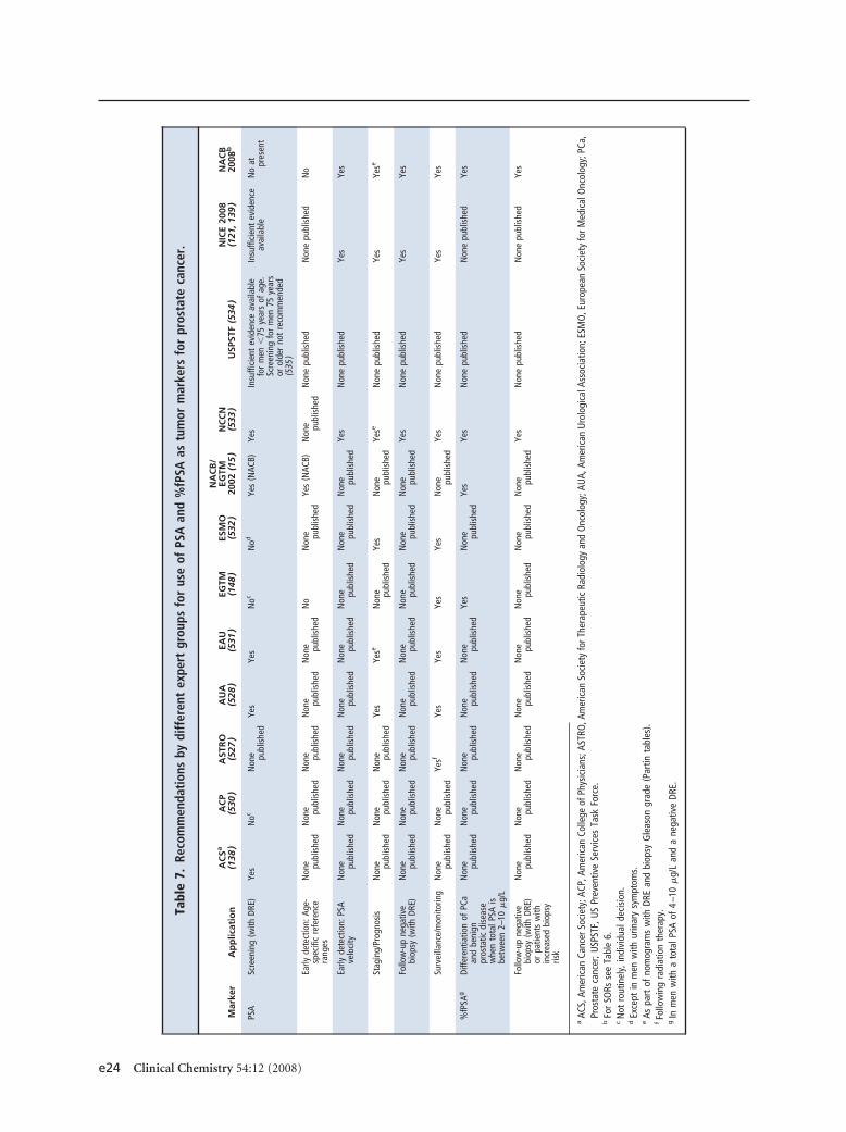
Table 7 summarizes the NACB guidelines for the use ofPSA markers in prostate cancer together with recom-mendations from other representative guidelines pub-lished on the use of tumor markers in prostate cancer,including recently published recommendations issuedby the United Kingdom National Institute for Healthand Clinical Excellence (NICE), which has undertakena systematic review of best available evidence (121 ).Although other markers have been investigated (Table
8), based on currently available evidence only the use ofPSA and its isoforms can be recommended in prostatecancer. Below we present a more detailed discussion ofthe use of these measurements.
PSA Markers in Patient Management
PSA MARKERS IN THE SCREENING AND EARLY DETECTION OF
PROSTATE CANCER
The widespread measurement of serum PSA is largelyresponsible for the increased incidence of prostate can-cer in the US during the past 2 decades. As demon-strated by epidemiological data showing both a markedincrease in the number of men diagnosed with prostatecancer and a profound migration toward earlier stagedisease at the time of diagnosis (122 ), there is strongevidence supporting the growing concern that such“stage migration” causes overdiagnosis and overtreat-ment of men with indolent cancer, a condition thatmay pose little threat to the life or health of the patient(123 ). The usefulness of PSA screening has also beenquestioned owing to poor specificity when serum con-centrations are modestly increased (124 ). Although ex-tensive evidence shows that elevations of PSA in serumare exclusively associated with disease conditions in theprostate, such findings are not cancer specific, occur-ring also in other conditions such as benign prostatichyperplasia and prostatitis. This well-documentedlack of specificity of the conventional PSA test evenprompted researchers to question whether any associ-ation exists between serum PSA levels and prostatecancer (125 ). In contrast, reports from many other in-vestigators have shown that there is very strong evi-dence of a very significant association between serumPSA levels and presence or outcome of prostate cancer(126 –130 ). Also, the lack in specificity of the PSA test isless critical in monitoring patients with a prostate can-cer diagnosis, for whom PSA is the most importantmarker in evaluating response to therapeutic interven-tions and in detecting tumor relapse. Although poten-tially valuable as part of multivariate panels to identifyaggressive cancers and/or cancer recurrence, measure-ment of prostatic acid phosphatase alone does not pro-vide any clinically useful information additional to PSAmeasurement (131, 132 ), and therefore is not recom-mended by the NACB.
NACB PROSTATE CANCER PANEL RECOMMENDATION 1:
CHOICE OF TUMOR MARKER FOR MANAGEMENT OF
PATIENTS WITH PROSTATE CANCER
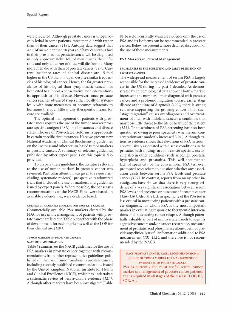
PSA is currently the most useful serum tumormarker in management of prostate cancer patientsand is required in all stages of the disease [LOE, III;SOR, A].
Special Report
Clinical Chemistry 54:12 (2008) e25
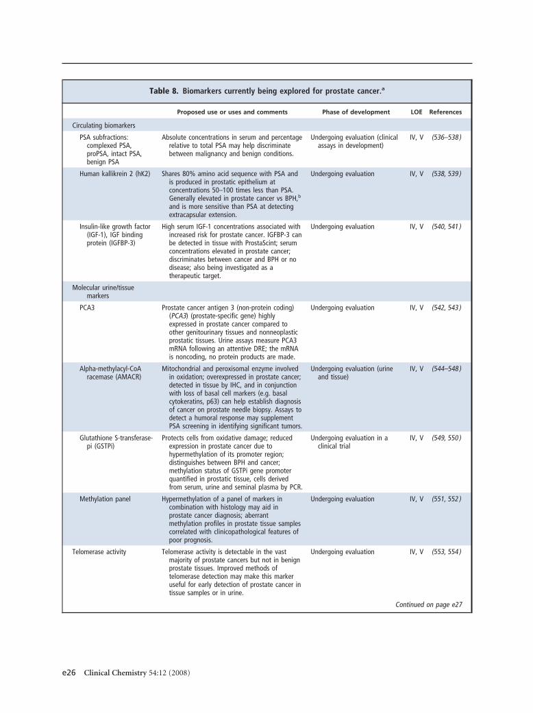
Table 8. Biomarkers currently being explored for prostate cancer.a
Proposed use or uses and comments Phase of development LOE References
Circulating biomarkers
PSA subfractions:complexed PSA,proPSA, intact PSA,benign PSA
Absolute concentrations in serum and percentagerelative to total PSA may help discriminatebetween malignancy and benign conditions.
Undergoing evaluation (clinicalassays in development)
IV, V (536–538 )
Human kallikrein 2 (hK2) Shares 80% amino acid sequence with PSA andis produced in prostatic epithelium atconcentrations 50–100 times less than PSA.Generally elevated in prostate cancer vs BPH,b
and is more sensitive than PSA at detectingextracapsular extension.
Undergoing evaluation IV, V (538, 539 )
Insulin-like growth factor(IGF-1), IGF bindingprotein (IGFBP-3)
High serum IGF-1 concentrations associated withincreased risk for prostate cancer. IGFBP-3 canbe detected in tissue with ProstaScint; serumconcentrations elevated in prostate cancer;discriminates between cancer and BPH or nodisease; also being investigated as atherapeutic target.
Undergoing evaluation IV, V (540, 541 )
Molecular urine/tissuemarkers
PCA3 Prostate cancer antigen 3 (non-protein coding)(PCA3) (prostate-specific gene) highlyexpressed in prostate cancer compared toother genitourinary tissues and nonneoplasticprostatic tissues. Urine assays measure PCA3mRNA following an attentive DRE; the mRNAis noncoding, no protein products are made.
Undergoing evaluation IV, V (542, 543 )
Alpha-methylacyl-CoAracemase (AMACR)
Mitochondrial and peroxisomal enzyme involvedin oxidation; overexpressed in prostate cancer;detected in tissue by IHC, and in conjunctionwith loss of basal cell markers (e.g. basalcytokeratins, p63) can help establish diagnosisof cancer on prostate needle biopsy. Assays todetect a humoral response may supplementPSA screening in identifying significant tumors.
Undergoing evaluation (urineand tissue)
IV, V (544–548 )
Glutathione S-transferase-pi (GSTPi)
Protects cells from oxidative damage; reducedexpression in prostate cancer due tohypermethylation of its promoter region;distinguishes between BPH and cancer;methylation status of GSTPi gene promoterquantified in prostatic tissue, cells derivedfrom serum, urine and seminal plasma by PCR.
Undergoing evaluation in aclinical trial
IV, V (549, 550 )
Methylation panel Hypermethylation of a panel of markers incombination with histology may aid inprostate cancer diagnosis; aberrantmethylation profiles in prostate tissue samplescorrelated with clinicopathological features ofpoor prognosis.
Undergoing evaluation IV, V (551, 552 )
Telomerase activity Telomerase activity is detectable in the vastmajority of prostate cancers but not in benignprostate tissues. Improved methods oftelomerase detection may make this markeruseful for early detection of prostate cancer intissue samples or in urine.
Undergoing evaluation IV, V (553, 554 )
Continued on page e27
e26 Clinical Chemistry 54:12 (2008)
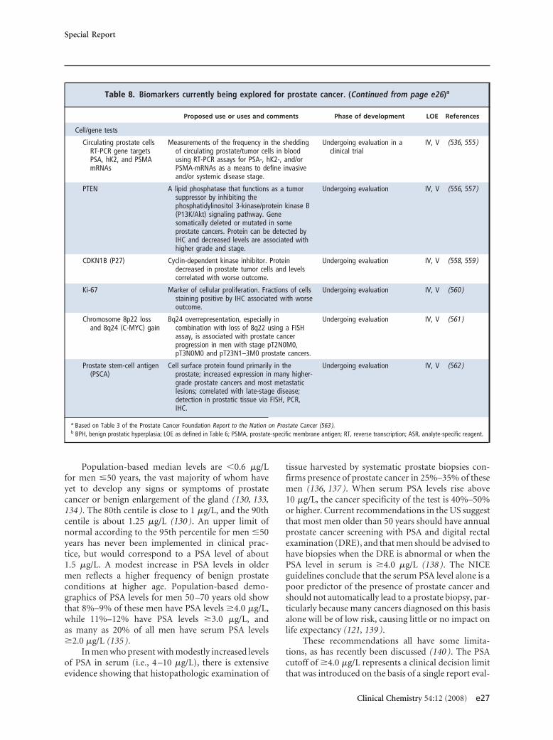
Population-based median levels are �0.6 �g/Lfor men �50 years, the vast majority of whom haveyet to develop any signs or symptoms of prostatecancer or benign enlargement of the gland (130, 133,134 ). The 80th centile is close to 1 �g/L, and the 90thcentile is about 1.25 �g/L (130 ). An upper limit ofnormal according to the 95th percentile for men �50years has never been implemented in clinical prac-tice, but would correspond to a PSA level of about1.5 �g/L. A modest increase in PSA levels in oldermen reflects a higher frequency of benign prostateconditions at higher age. Population-based demo-graphics of PSA levels for men 50 –70 years old showthat 8%–9% of these men have PSA levels �4.0 �g/L,while 11%–12% have PSA levels �3.0 �g/L, andas many as 20% of all men have serum PSA levels�2.0 �g/L (135 ).
In men who present with modestly increased levelsof PSA in serum (i.e., 4 –10 �g/L), there is extensiveevidence showing that histopathologic examination of
tissue harvested by systematic prostate biopsies con-firms presence of prostate cancer in 25%–35% of thesemen (136, 137 ). When serum PSA levels rise above10 �g/L, the cancer specificity of the test is 40%–50%or higher. Current recommendations in the US suggestthat most men older than 50 years should have annualprostate cancer screening with PSA and digital rectalexamination (DRE), and that men should be advised tohave biopsies when the DRE is abnormal or when thePSA level in serum is �4.0 �g/L (138 ). The NICEguidelines conclude that the serum PSA level alone is apoor predictor of the presence of prostate cancer andshould not automatically lead to a prostate biopsy, par-ticularly because many cancers diagnosed on this basisalone will be of low risk, causing little or no impact onlife expectancy (121, 139 ).
These recommendations all have some limita-tions, as has recently been discussed (140 ). The PSAcutoff of �4.0 �g/L represents a clinical decision limitthat was introduced on the basis of a single report eval-
Table 8. Biomarkers currently being explored for prostate cancer. (Continued from page e26)a
Proposed use or uses and comments Phase of development LOE References
Cell/gene tests
Circulating prostate cellsRT-PCR gene targetsPSA, hK2, and PSMAmRNAs
Measurements of the frequency in the sheddingof circulating prostate/tumor cells in bloodusing RT-PCR assays for PSA-, hK2-, and/orPSMA-mRNAs as a means to define invasiveand/or systemic disease stage.
Undergoing evaluation in aclinical trial
IV, V (536, 555 )
PTEN A lipid phosphatase that functions as a tumorsuppressor by inhibiting thephosphatidylinositol 3-kinase/protein kinase B(P13K/Akt) signaling pathway. Genesomatically deleted or mutated in someprostate cancers. Protein can be detected byIHC and decreased levels are associated withhigher grade and stage.
Undergoing evaluation IV, V (556, 557 )
CDKN1B (P27) Cyclin-dependent kinase inhibitor. Proteindecreased in prostate tumor cells and levelscorrelated with worse outcome.
Undergoing evaluation IV, V (558, 559 )
Ki-67 Marker of cellular proliferation. Fractions of cellsstaining positive by IHC associated with worseoutcome.
Undergoing evaluation IV, V (560 )
Chromosome 8p22 lossand 8q24 (C-MYC) gain
Bq24 overrepresentation, especially incombination with loss of 8q22 using a FISHassay, is associated with prostate cancerprogression in men with stage pT2N0M0,pT3N0M0 and pT23N1–3M0 prostate cancers.
Undergoing evaluation IV, V (561 )
Prostate stem-cell antigen(PSCA)
Cell surface protein found primarily in theprostate; increased expression in many higher-grade prostate cancers and most metastaticlesions; correlated with late-stage disease;detection in prostatic tissue via FISH, PCR,IHC.
Undergoing evaluation IV, V (562 )
a Based on Table 3 of the Prostate Cancer Foundation Report to the Nation on Prostate Cancer (563 ).b BPH, benign prostatic hyperplasia; LOE as defined in Table 6; PSMA, prostate-specific membrane antigen; RT, reverse transcription; ASR, analyte-specific reagent.
Special Report
Clinical Chemistry 54:12 (2008) e27
uating the optimal combination of sensitivity andspecificity of the PSA test in a study cohort, and thedistribution of values observed in this original studymay no longer apply (141 ). It is debatable whether aPSA cut-point lower than 4 �g/L should be recom-mended. Also debatable is whether decisions to recom-mend prostate biopsy should be based solely on a singlePSA cut-point value (e.g., �4 �g/L). Lower PSA cutoffsincrease the cancer detection rate at the expense ofincreasing the number of men advised to undergo bi-opsy. It has also been clearly demonstrated, however,that 20% or more of all men who have PSA levels from2.0 (or 3.0) up to 4.0 �g/L are found to have prostatecancer at biopsy (142, 143 ). This finding was con-firmed in a recent study, in which prostate cancer wasdiagnosed by biopsy in as many as 15.2% of all 2950biopsied men with PSA values �4.0 �g/L (128 ). Thisstudy showed that the prevalence of prostate cancer inmen 62–91 years old increased from 6.6% in men withPSA of 0 – 0.5 �g/L, 10% with PSA 0.6 –1.0 �g/L, 17%with PSA 1–2 �g/L, up to 23.9% with PSA 2.1–3.0�g/L, and 26.9% with PSA 3.1– 4.0 �g/L (128 ). Also,the prevalence of high-grade prostate cancer increasedwith increasing PSA values. Hence, the positive predic-tive value of the PSA test in terms of biopsy-proven(histological) prostate cancer is similar for men with aPSA value between 2– 4 �g/L and those with a PSAvalue between 4 –10 �g/L (136, 144 ).
NACB PROSTATE CANCER PANEL RECOMMENDATION 2:
CLINICAL DECISION LIMITS
Given the controversy regarding the use of PSA todetect very small tumors, reported benefits arisingfrom lowering the clinical decision limit for biopsybelow 4 �g/L are too uncertain to mandate any gen-eral recommendation. Cut-points lower than thecommonly used 4 �g/L limit will increase sensitiv-ity with a concomitant decrease in specificity unlessother adjunctive tests or measures are employed toincrease specificity. Conversely, use of clinical deci-sion limits for PSA higher than 4.0 �g/L decreasesthe sensitivity, which results in the missed diag-noses of clinically significant tumors in men whomight potentially benefit from early treatment[LOE, not applicable; SOR, B].
The across-the-board recommendation of annualPSA testing for men older than 50 years (138 ) is overlysimplistic and fails to alter testing frequency based onthe individualized risk imparted by previously deter-mined PSA levels. For example, a 55-year-old man witha baseline PSA of 0.4 �g/L is much less likely to developprostate cancer in the future than a similarly aged man
with a baseline PSA of 3.3 �g/L. Stenman et al. (126 )used frozen serum samples and information from ahealth examination survey in Finland, and Gann et al.(145 ) used information from the Physicians’ HealthStudy to examine the usefulness of PSA for identifyingmen in whom prostate cancer subsequently was or wasnot clinically diagnosed. The data of Gann et al. suggestthat men with PSA levels between 2.0 and 3.0 �g/L have5.5-fold higher relative risk for diagnosis of prostatecancer than men with PSA levels �1.0 �g/L. In theformer group, serum PSA levels reached 2–3 �g/L onaverage more than 5 years before the cancer was de-tected by DRE. Recently, Lilja et al. (130 ) demon-strated a very strong association between PSA levels inblood collected more than 20 years before prostate can-cer diagnosis and the likelihood of that diagnosis in alarge representative population of Swedish men age44 –50 years who had not previously undergone PSAtesting. These data and those reported from others(129 ) suggest that risk stratification during early mid-dle age may be important to consider in refining cur-rent imperfect early cancer detection strategies. Severaladditional issues particularly relevant to screening pro-grams are discussed below.
AGE-SPECIFIC REFERENCE INTERVALS FOR PSA
Because serum PSA levels gradually increase with age inmen older than 40 years, age-specific reference inter-vals have been proposed with the expectation that theirimplementation would increase cancer detection ratesin younger men by lowering the cut-point and wouldincrease specificity in older men by raising the cut-point (146 ). Although there is no consensus, many ex-perts—including a majority of opinion of the NationalComprehensive Cancer Network—favor the use ofclinical decision limits lower than 4.0 �g/L for serumPSA in younger men. The NACB, however, is not yetconvinced of the net benefit of this protocol in the ab-sence of additional test(s) that could significantly in-crease diagnostic specificity (i.e., reduce unnecessarybiopsies). At the same time the NACB advises cautionin increasing the decision limit above 4.0 �g/L, becausethis could result in failure to diagnose clinically signif-icant tumors in men who might potentially benefitfrom early treatment (147 ). Hence, contrary to previ-ously issued recommendations (148 ), the NACB doesnot endorse the use of age-specific reference ranges.
NACB PROSTATE CANCER PANEL RECOMMENDATION 3:
AGE-SPECIFIC REFERENCE INTERVALS FOR PSA
Age-specific reference intervals should not be usedfor PSA [LOE, expert opinion; SOR, B].
e28 Clinical Chemistry 54:12 (2008)
INCREASING PSA SPECIFICITY IN SCREENING FOR PROSTATE
CANCER
The total PSA in circulation roughly corresponds to thesum of circulating free PSA (fPSA) and PSA bound as astable complex to �-1-antichymotrypsin. The free frac-tion constitutes from 5% up to more than 40% of thetotal (149 ). Free and bound forms may be selectivelydetected by commercially available assays without anysignificant interfering cross-reaction (150 ). Severalcomposite measures have been proposed to improvethe specificity of a single serum total PSA concentra-tion for the early detection of prostate cancer. PSA den-sity (151–153 ), PSA velocity (154 ), PSA doubling time(155, 156 ), and percentage of free PSA (%fPSA) (157–161 ) have all been evaluated in this context, but only%fPSA has been widely validated and implemented inclinical practice. Men with benign disease generallypresent with higher %fPSA than men with prostatecancer (and no benign enlargement). Unfortunately,concurrent benign prostatic enlargement and prostatecancer complicates interpretation of %fPSA data (162).Nevertheless, in a systematic review carried out in2005, the use of %fPSA was suggested as a means ofdecreasing the number of unnecessary biopsies, partic-ularly for men with PSA levels of 4 –10 �g/L (163 ). Inaccord with the conclusions of a recent meta-analysis(164 ), the current NACB Panel and the EuropeanGroup on Tumor Markers (EGTM) (148 ) both recom-mend the use of %fPSA as an aid in distinguishingmen with prostate cancer from men with benign dis-ease in selected high-risk groups, e.g., when total PSA is�10 �g/L and DRE is negative. In particular, %fPSAmay be useful in identifying men who have prostatecancer despite initial negative biopsy findings. In menwith low %fPSA suspected to indicate a high risk ofharboring malignant disease, a cancer diagnosis maybecome evident after a repeat biopsy. This recommen-dation is tempered by the need to validate the medicaldecision limit for each fPSA and total PSA commercialassay combination (165 ).
NACB PROSTATE CANCER PANEL RECOMMENDATION 4:
USE OF %fPSA IN DIAGNOSIS
The use of %fPSA is recommended as an aid indistinguishing men with prostate cancer from menwith benign prostatic hypertrophy when the totalPSA level in serum is within the range of 4 –10 �g/Land DRE is negative, most frequently in men un-dergoing repeat biopsy, in selected high-risk groupsand particularly in identifying men who have pros-tate cancer despite initial negative biopsy findings.The clinical decision limit must be properly vali-dated for each combination of free and total PSAassays [LOE, I; SOR, A].
More than 95% of the immunodetectable com-plexed PSA (cPSA) fraction is bound to �-1-antichy-motrypsin with �5% bound to other complex ligands,e.g., �-1-protease inhibitor (157, 166–168). PSA boundto �-2-macroglobulin is not detected by current im-munoassays for PSA. Levels of cPSA in blood can bedetermined either directly using assays for PSA boundas a stable complex to �-1-antichymotrypsin (157,158, 169 ), which first block access to fPSA and thenmeasure levels of cPSA (170 ), or indirectly by subtract-ing fPSA from total PSA levels (171 ) using 2 assaysdesigned to work together and standardized appropri-ately. Measurement of cPSA alone provides compara-ble cancer detection to total PSA but appears to givesomewhat better specificity in a narrow concentrationrange (172). However, cPSA levels alone cannot achievespecificity similar to that of %fPSA (170 ).
GUIDELINES FOR THE EARLY DETECTION OF PROSTATE CANCER
The American Cancer Society has issued guidelinesrelated to the early detection of prostate cancer. Theseguidelines recommend an annual screening with DREand serum PSA measurement beginning at the age of50 in men at average risk with at least 10 years of lifeexpectancy (138 ). Although PSA is considered thebest biochemical test currently available to detectprostate cancer, a DRE should also be included when-ever possible, according to the American Cancer Soci-ety. Screening at earlier age (45 years or even 40 years)is warranted in men at increased risk, including thoseof African-American descent and those with one ormore first-degree relatives with prostate cancer. Bothof these groups often develop prostate cancer severalyears earlier than the general population and also tendto present with a more aggressive type of cancer (173 ).
The recommended follow-up testing of high-riskindividuals initially screened at 40 years of age dependson the PSA result. Those with PSA levels �1 �g/Lwould resume testing at 45 years of age, those with levels�1 but �2.5 �g/L would be tested annually, and thosewith levels �2.5 �g/L would be evaluated further andconsidered for biopsy (138 ).
These guidelines do not endorse a general recom-mendation for mass screening, but support the no-tion that individual men should be informed of thebenefits and limitations of prostate cancer screeningbefore making their decision, as for example is recom-mended in the United Kingdom through the ProstateCancer Risk Management Program (174 ) and by NICE(121, 139, 174 ). Much greater emphasis than previ-ously is being placed on informed decision-making bythe individual. This topic has recently been the subjectof a systematic review in which PSA decision aids andevaluations were identified and appraised (175 ). Theauthors concluded that PSA decision aids improve
Special Report
Clinical Chemistry 54:12 (2008) e29
knowledge about PSA testing, at least in the short term.There are many issues to consider, including the dis-parity between incidence and mortality associated withprostate cancer, because many more men are diag-nosed with prostate cancer than eventually die from it.However, early detection affords the opportunity todetect organ-confined disease when curative treatmentis possible. Metastatic disease now constitutes onlyabout 5% of initial diagnoses in the US, a dramatic fallfrom the 50% incidence rate of the pre-PSA era (122 ).Nevertheless there are still many uncertainties con-cerning treatment of early stage disease, including thepreferred treatment for clinically localized prostatecancer.
MERITS OF EARLY DETECTION OF PROSTATE CANCER
Because of the uncertainties regarding prostate cancertreatment, considerable debate is ongoing regardingthe merits of early detection of prostate cancer, and notall physician organizations advocate routine screening(176 ). Although the American Urological Associationendorses the American Cancer Society policy state-ment on the early detection of prostate cancer, recom-mendations of other organizations differ regarding thebenefit of prostate cancer screening (177, 178 ). Argu-ments against screening are based on the fact that thereis no conclusive evidence from any randomized trialsthat early detection and treatment influence overallmortality, whereas the standard treatments for organ-confined prostate cancer are associated with a signifi-cant frequency of side effects. Currently, the US Pre-ventive Task Force, the American Academy of FamilyPhysicians, the American College of Physicians, theNational Cancer Institute, and the EGTM do not rec-ommend population-based prostate cancer screening(177, 178 ). The overriding concern is that currentscreening modalities result in overdiagnosis and over-treatment of early stage disease that may not be clini-cally significant, as has recently been reviewed (179 ).
The NACB and the EGTM recommend that wide-spread implementation of screening for prostate can-cer in the general population should await the finaloutcome of ongoing prospective randomized studies,in particular the European Randomized Screening forProstate Cancer (ERSPC) trial (180 ), which are suffi-ciently powered to establish whether early detectionand treatment decreases prostate cancer mortality. TheERSPC trial has been underway for 10 years, with re-sults expected in 2010 (181 ). Long-term multicentertrials to determine the impact of prostate cancerscreening on survival are also ongoing in the US underthe aegis of the National Cancer Institute and the USPublic Health Service (182 ).
With no clear-cut evidence as yet that prostatecancer screening is of net benefit, proponents of
screening have pointed to the association of PSA test-ing with earlier cancer stage at detection and reducedmortality arising from prostate cancer. Registry datafrom heavily and sparsely screened male populations inAustria provide a case in point. The expected death ratefrom prostate cancer (183 ) declined much more in theTyrol, a heavily screened section of the country, than inless intensely screened areas (184 ). The decrease in ob-served mortality was associated with a shift toward amore favorable stage at diagnosis, in particular an in-crease in the proportion of organ-confined disease.The inference is that early detection and availabilityof effective treatment resulted in a corresponding im-provement in disease-specific survival. A similar trendhas been observed in data from the National CancerInstitute’s Surveillance, Epidemiology and End Results(SEER) program, from a study conducted in OlmstedCounty, Minnesota (185 ), and from a comparison ofprostate cancer mortality in the US and the UnitedKingdom between 1975 and 2004 (186 ).
Although recent data suggest that the apparentstage shift to early stage disease and subsequent treat-ment of localized prostate cancer detected with PSAhave positively influenced mortality rates, it is still anopen question whether early detection and therapeuticintervention alters the natural history of the disease, asobserved benefits may be the result of selection or lead-time bias(es) (187 ). The stage at diagnosis may be moredependent on the biological behavior of the tumor (ag-gressiveness) than on delay in presentation, and earlydetection may not have a significant impact on mortal-ity. An increase in the proportion of localized prostatecancers that are being treated may account for some ofthe change in the mortality statistics (181 ).
Currently there is insufficient evidence either tosupport or refute the routine use of mass, selective oropportunistic PSA-based screening, and it is equallyunclear whether to advise against the use of PSA-basedscreening, for which success in reducing prostate can-cer mortality has yet to be demonstrated. Currently, norobust evidence from randomized controlled trials isavailable regarding the impact of screening on qualityof life, the disadvantages of screening, or its economicvalue. Results from 2 ongoing large-scale multicenterrandomized controlled trials that will be available in thenext several years are required for evidence-based deci-sion-making regarding prostate cancer screening (188).
NACB PROSTATE CANCER PANEL RECOMMENDATION 5:
PROSTATE CANCER SCREENING
A decision as to whether widespread implementa-tion of PSA screening for prostate cancer in the gen-eral population can be recommended must await-the outcome of ongoing prospective randomized
e30 Clinical Chemistry 54:12 (2008)
screening studies (e.g., the European RandomizedScreening for Prostate Cancer trial in Europe)which are due to be completed by 2010 [LOE, III;SOR, A].
PSA IN PATIENT MANAGEMENT
The optimal treatment of early stage prostate cancerhas yet to be established. Treatment options includeexpectant management (active surveillance or watchfulwaiting), radical prostatectomy, or radiation therapy(external beam radiation or brachytherapy) (139 ). Al-ternative treatment modalities (e.g., cryosurgery orhigh-intensity focused ultrasound) await evaluation oftheir long-term results. Patients with advanced (meta-static) disease are typically offered hormonal therapyto deprive the prostate of androgen stimulation. PSAsynthesis by differentiated prostate cells is greatly im-paired by such treatment and the PSA levels in bloodreflect tumor burden differently from before andro-gen deprivation. When the disease becomes refractoryto either first- or second-line androgen deprivation,patients may be entered into chemotherapy or experi-mental protocols with various agents (e.g., Taxotere).The assessment of PSA levels in the blood plays a car-dinal role in all aspects of the management of prostatecancer from surveillance to selection of optimal treat-ment to estimation of prognosis to posttherapeuticmonitoring. fPSA measurement has not been shown tooffer any advantages over total PSA during the follow-up of prostate cancer (189 ).
The treatment selected after detection of prostatecancer depends critically on whether the disease is con-fined to the prostate. Radical prostatectomy is primar-ily an option for patients with organ-confined disease,although patients with extracapsular disease may alsobenefit from radical surgery (190 ). However, the extentof disease is difficult to predict accurately. PSA alone isnot informative (191 ), but in combination with theclinical stage and Gleason score predicts reasonablywell the pathological stage of localized prostate cancer.Predictive tables that incorporate these parameters havebeen published (192–194 ) and are used by physiciansto estimate the probability of organ-confined diseaseand to determine whether radical prostatectomy is in-dicated. It is recommended by NICE that urologicalmultidisciplinary teams should assign a risk category toall men with newly diagnosed localized prostate cancer,taking these parameters into account (121, 139 ).
Assessment of changes of PSA levels with time(PSA velocity or PSA doubling time) was first intro-duced in 1992 (154 ), with a rapid increase indicating ahigher risk for subsequent development of prostatecancer. Results of several studies further suggested thata more rapid rise in PSA before treatment is correlated
with aggressive disease and early recurrence after treat-ment. In more recent studies reported by D’Amico etal. (195, 196 ) a PSA velocity of more than 2.0 �g/L/yearmeasured during the year before diagnosis was shownto be significantly associated with prostate cancer-specific mortality. Recently, Carter et al. reported evi-dence that total PSA velocity could also be used to pre-dict life-threatening prostate cancer up to 15 yearsbefore diagnosis (197 ). However, to demonstrate thatPSA velocity has important clinical value, it must alsobe unequivocally shown that a multivariable modelthat incorporates both PSA and PSA velocity (e.g., ad-dition of PSA velocity to a model that includes totalPSA, age, and date of diagnosis) is superior to themodel that uses PSA alone. This LOE appears still to belacking, even in the most recently reported studies onthis subject.
Following successful surgery, PSA should decreaseto undetectable levels (198, 199). Persistently increasedPSA provides evidence of residual disease. However,the converse does not always hold, namely that unde-tectable PSA postoperatively indicates a surgical cure.Considerable time may elapse before residual diseasebecomes evident through detectable PSA. Most com-monly, residual disease will become evident within 3years of surgery. Up to 20%–30% of the men who un-dergo radical prostatectomy present with residual dis-ease during the first 10 years after surgery.
A rising PSA level after radical prostatectomy isa biochemical sign of recurrent disease that typicallypredates other signs of progression by many years.However, not all patients with biochemical recurrencewill progress to symptoms of clinical disease and met-astatic spread in their lifetimes and require treatment(200, 201 ). Factors reported to predict the time courseto the development of metastatic disease include timeto biochemical recurrence, tumor grade (Gleasonscore), and PSA doubling time (156, 161 ). These pa-rameters can be used to estimate the likelihood of pa-tients remaining free of overt metastatic disease andallow physicians to stratify patients into low-risk andhigh-risk categories and to make better treatmentdecisions.
Monitoring response after initial treatment andevaluating outcome during subsequent therapy are sig-nificant clinical applications of PSA determinations.Measurement of PSA provides essential informationabout the efficacy of surgery or radiation therapy, helpsestablish the possibility of residual disease (local or dis-tant), signals recurrent metastatic disease before it canbe detected by other conventional diagnostic proce-dures, and provides a useful adjunct in the evaluationof therapeutic response.
Special Report
Clinical Chemistry 54:12 (2008) e31
PSA may provide the earliest measure of treatmentefficacy or disease recurrence, and as such influence thepatient’s perception of well-being. For some patients, itmay be most appropriate to stop measuring PSA, par-ticularly if effective alternative treatments to counteradverse findings are not available (148 ).
PSA MARKERS IN THE POSTTREATMENT MONITORING OF
PROSTATE CANCER
Following treatment, it is the Panel’s view that a singlePSA measurement at or near the lower detection limitof the assay is not sufficient to diagnose recurrence ofprostate cancer. Rising PSA levels demonstrated by re-peat or serial measurements provide much more reli-able evidence (121, 139, 202 ). Following radical pros-tatectomy, circulating PSA declines to undetectablelevels if the prostate cancer was organ-confined and allresidual prostate tissue surgically excised. Sustaineddetection of PSA suggests either incomplete resectionor metastatic deposits. If ultrasensitive PSA assays areused in this setting, the functional detection limit of theassay should be established and should correspond tothe lower reporting limit.
At present, evidence is equivocal regarding theclinical benefit of reporting biochemical recurrence ofprostate cancer at PSA levels below 0.4 �g/L (200 ).Recently, however, salvage radiation therapy followingprostatectomy has been shown to yield best resultswhen PSA levels are still very low (�0.5 �g/L) (203 ).The recurrence limit is less clear following radiationtherapy because of the typically slower decline in circu-lating PSA concentration. The American Society forTherapeutic Radiation and Oncology has defined bio-chemical recurrence as a rise of 2 �g/L or more abovethe nadir PSA, after external beam radiotherapy with orwithout hormonal therapy (204 ).
Monitoring with PSA after treatment for prostatecancer is a mainstay of clinical practice, although theclinical utility of PSA is variable and depends on thedisease stage of the individual patient. As has recentlybeen observed, the lack of high quality information andpaucity of clinical trials hampers development ofguideline recommendations for prostate cancer, butimplementation of available guidelines are likely to im-prove prostate cancer outcomes while reducing unnec-essary, ineffective, and costly care (140 ). PSA has highsensitivity for detecting recurrence after radical prosta-tectomy but is less sensitive in detecting recurrence fol-lowing radiation therapy. For monitoring hormonaltreatment, PSA provides a sensitive tool with which toverify treatment response and detect tumor growth(recurrence). However, in patients with advanced dis-ease who suffer recurrence during androgen depriva-
tion therapy, PSA has only limited usefulness for pre-dicting survival outcome.
NACB PROSTATE CANCER PANEL RECOMMENDATION 6:
USE OF PSA IN THE POSTTREATMENT MONITORING OF
PROSTATE CANCER
PSA is recommended for management of patientswith prostate cancer to monitor disease status fol-lowing treatment [LOE, III; SOR, A].
USE OF NOMOGRAMS INCORPORATING PSA TO MANAGE
PROSTATE CANCER
Nomograms incorporating one or more factors pro-vide the most accurate means of individualizing ther-apy and predicting outcome, and reflect the most re-cent advances in patient management (205 ). Ratherthan relying on physician experience or general riskassessments of patient populations with similar char-acteristics, the nomograms assess treatment optionsor prognosis based on computerized models of Coxproportional hazards regression analysis. Predictiveoutcomes provided by computer models are not per-fect, but nomograms can be extremely useful in assist-ing with treatment decisions. On occasion, it may bedifficult to select the best nomogram when severalcompeting versions apply to the same clinical decision.Kattan and coworkers (205, 206 ) have developed pre-and postoperative nomograms, incorporating PSA to-gether with Gleason score and other variables, to predictdisease recurrence following radical prostatectomy.
PREANALYTICAL, ANALYTICAL, AND POSTANALYTICAL
CONSIDERATIONS
A number of factors in the preanalytical, analytical andpostanalytical stages can affect the clinical interpreta-tion of PSA results and must be carefully considered. Anumber of these factors were the subject of a systematicreview carried out in 2001 (207 ).
PREANALYTICAL SPECIMEN PROCESSING AND STORAGE
It is desirable to collect blood before any manipulationof the prostate by DRE, cystoscopy, or prostate biopsy(166 ). If prior collection is not possible, then it is pru-dent to delay several days after DRE before drawingblood for PSA, although in most men DRE does notcause a clinically relevant change in circulating PSAconcentration (166 ). Following prostate biopsy or sur-gery, the recommended delay is several weeks to permitsufficient time for the PSA bound as a stable complex to�-1-antichymotrypsin to be eliminated from the bloodcirculation, although the kidneys rapidly clear from theblood any fPSA that was liberated from the prostate bythe procedure (208, 209 ).
e32 Clinical Chemistry 54:12 (2008)

NACB PROSTATE CANCER PANEL RECOMMENDATION 7:
PREANALYTICAL REQUIREMENTS FOR PSA—
PROSTATE MANIPULATION
Blood should be drawn before any manipulation ofthe prostate and several weeks after resolution ofprostatitis [LOE, not applicable; SOR, A].
To eliminate in vitro artifacts, blood should becentrifuged within 3 h of collection to isolate the serumor plasma (210 ). Serum and plasma may be kept atrefrigerated temperatures for up to 24 h without loss ofPSA. If analysis is delayed longer, then it is vital to storespecimens frozen, preferably at or below �30 °C toavoid the eutectic point. Long-term storage at temper-atures of at least �70 °C is desirable. Data show thatfPSA is more susceptible to loss of immunoreactivitythan cPSA (166, 211 ), and that for fPSA this is slower inplasma than in serum (210 ).
NACB PROSTATE CANCER PANEL RECOMMENDATION 8:
PREANALYTICAL REQUIREMENTS FOR PSA—
SAMPLE HANDLING
Samples should be centrifuged and refrigeratedwithin 3 h of phlebotomy; this recommendation isparticularly relevant for fPSA, which is more labilethan total PSA. Samples may be stored at refriger-ated temperatures for up to 24 h, but samples thatwill not be analyzed within 24 h of collection shouldbe stored frozen (at least at �20 °C and preferablyat �30 °C or lower). For long-term storage, sam-ples should be frozen at �70 °C or lower [LOE, notapplicable; SOR, B].
PSA ASSAY STANDARDIZATION
Two reference standards currently are commonly usedfor PSA assays: those traceable to the WHO Interna-tional Standards and those traceable to the Hybritech,Inc. standard. Most clinicians assume that all PSA as-says give similar test values and that changes in thesetest values probably are related to pathophysiologicalchanges in prostate glands. It is assumed that PSA mea-surements are consistent between laboratories and be-tween assay manufacturers, but this is not necessarilythe case (212 ). Although practice guidelines and dis-ease management strategies vary in terms of what“number” should be used to follow up specific types ofpatients, these guidelines seldom contain subcategoriesfor various analytical methods.
In practice there are considerable differences be-tween PSA assays. Historically, the Hybritech Tan-dem-R PSA assay (Hybritech) was the first widely usedFDA-cleared commercial assay. This assay was stan-dardized using the absorptivity for PSA of 1.42 mL/
mg/cm reported by Graves et al. in 1990 (213 ). TheHybritech assay was well adopted by the medical com-munity and provided the basis for the traditional4.0 �g/L upper reference limit (141 ). The secondwidely used commercial assay, the Abbott IMx (AbbottLaboratories), was standardized to harmonize with thisinitial Hybritech assay, and other assays also wereclosely aligned with these assays (214 ). However, in1995, Stamey et al. reported that the mean (SD) trueabsorptivity for PSA is 1.84 (0.04) mL/mg/cm, basedon quantitative amino acid analysis (215 ). It was sug-gested that the error in the initial gravimetric analysiswas caused by the presence of bound water, salt, orcarbohydrate in the lyophilized preparations. The netresult of this error is that the initial Hybritech PSA val-ues are about 20% higher than the WHO First Interna-tional Standard for PSA (IRR 96/670) (216 ).
The First International Standards for PSA (IRR 96/670) and Free PSA (IRR 96/688) were established in1999 using the correct absorptivity. The 2 standardscontain PSA derived from seminal plasma. IRR 96/670is a mixture of PSA and ACT in a 90:10 ratio selected tomimic circulating PSA, and IRR 96/688 contains solelyfree (unbound) PSA. An editorial that accompaniedthe standardization report, WHO First InternationalStandards for Prostate-Specific Antigen: The Beginningof the End for Assay Discrepancies, concluded that thisstandard would lead to greater consistency of PSA asmanufacturers began to use this material to calibratePSA assays (217 ). It is now recommended that PSAassays used in the United Kingdom National HealthService must be accurately calibrated against the ap-propriate International Standard and must be equi-molar (218 ), with formal arrangements in place forindependent annual confirmation of satisfactory per-formance. Although several studies suggest that be-tween-method comparability has improved since in-troduction of the International Standards, there arestill differences in PSA assays that may lead to clinicalmisinterpretation if different PSA assays are used whenevaluating a single patient (218 –220 ).
ANALYTICAL AND REPORTING CONCERNS
PSA is most frequently used in conjunction with phys-ical examination to screen for prostate cancer. A singlepositive PSA screen should always be verified by re-peating the PSA measurement in a specimen collectedseparately, before the ordering of confirmatory his-topathological tissue examination, e.g., as obtained bybiopsy. This protocol may substantially reduce thenumber of unnecessary biopsies (221 ). The diagnosisof prostate cancer can be confirmed only by his-topathological tissue examination.
Analytical performance should be monitored withQC material containing PSA at concentrations near
Special Report
Clinical Chemistry 54:12 (2008) e33
clinically relevant decision points. Information on as-say characteristics and utility, including the lowest re-portable concentration of the assay (often defined asthe PSA concentration below which the analytical CVexceeds 20%) and assay CVs at concentrations corre-sponding to relevant clinical decision points should beavailable to clinicians through laboratory test informa-tion sources.
NACB PROSTATE CANCER PANEL RECOMMENDATION 9:
ANALYTICAL REQUIREMENTS FOR PSA—QC
The lowest reportable concentration should be de-termined by the laboratory and reported to physi-cians. QC at these concentrations should be in place[LOE, not applicable; SOR, A].
BIOLOGICAL VARIABILITY
To interpret PSA data from any individual or seriallycollected specimens, PSA variability in the blood shouldalso be taken into account (207, 222 ). The EGTM re-cently reviewed publications concerning the variabilityof PSA and reported that a fair estimate of the biologi-cal variation of PSA is 20% in men older than 50 yearswithin the PSA concentration range of 0.1–20 �g/L(223 ). In healthy men with PSA concentrations�2 �g/L, biological variation was �14%, whereas achange of 30% between successive PSA measurementswas suggested to be clinically significant (224 ). Inmonitoring men with prostate cancer, a critical differ-ence of 50%– 60% has been suggested (225 ). Takinginto account that intraindividual biological variationmay range up to 20% and that analytical variation forPSA assays is 5%, it has been suggested that the baselinePSA level has to change by 50% to be significant at P �0.05 (223 ).
NACB PROSTATE CANCER PANEL RECOMMENDATION 10:
POSTANALYTICAL REQUIREMENTS FOR PSA—
INTRAINDIVIDUAL BIOLOGICAL VARIATION
The contribution of within-individual biologicalvariation must be taken into account when in-terpreting clinical results [LOE, not applicable;SOR, A].
It is prudent to include with the PSA result a re-minder that a single screening blood test result shouldnot be used as the sole evidence of the presence orabsence of malignant disease. The laboratory reportshould include the manufacturer of the PSA assay used,draw attention to any relevant clinical decision limits,and where necessary warn that the results cannot beused interchangeably with those generated by other as-
says unless the interchange of assay values has previ-ously been validated (212, 220 ).
NACB PROSTATE CANCER PANEL RECOMMENDATION 11:
POSTANALYTICAL REQUIREMENTS FOR PSA—INFORMATION
TO BE INCLUDED IN CLINICAL REPORTS
Clinical reports should include the name of the as-say, relevant clinical decision limits, and a reminderthat a single screening blood test result should notbe used as the sole evidence of the presence or ab-sence of malignant disease [LOE, not applicable;SOR, A].
FUTURE DEVELOPMENTS
Future developments in the use of tumor markers forprostate cancer include the use of experimental assaysto measure circulating tumor cells in blood to detectand assess progression of (micro) metastatic stages ofprostate cancer
Assays detecting circulating tumor cells (CTCs) inthe peripheral blood have been developed and clearedfor clinical use by the FDA to provide prognostic infor-mation in women with node-positive breast cancer(226 ). The current ability to detect and profile (micro)metastatic prostate cancer is limited, however. Multi-ple techniques have been developed and tested to iso-late and characterize CTCs. Reverse transcription PCRassays are sensitive and highly specific when the expres-sion of the target gene is limited to the malignant tu-mor cells. Flow cytometry can be used to detect andverify the identity of the cells as CTCs, but does notallow assessments of morphology and does not allowdetection of molecular changes at a subcellular level.Immobilization (e.g., to magnetic beads) of antibodiesto the epithelial cell adhesion molecule allows enrich-ment and inspection by microscopy of circulating epi-thelial derived tumor cells from peripheral blood. Asemiautomated system was recently developed, whichuses epithelial cell adhesion molecule–antibody-basedimmunomagnetic capture and staining methods (227).Factors predictive of detection of CTCs in prostate can-cer have been reported, and for patients with metastaticprostate cancer, the detection of �5 CTCs per 7.5 mLof blood predicts shorter progression-free survivaland shorter overall survival, with CTC counts found tobe more predictive of outcome than standard clinicalparameters (228 ). For prostate cancer, preliminaryanalysis of the correlation of CTC counts with mRNAsfor prostate specific antigen or prostate specific mem-brane antigen and available clinical predictors (229 )are encouraging but are not yet sufficiently evaluatedor validated to warrant recommendations for any usein routine clinical practice.
e34 Clinical Chemistry 54:12 (2008)
NACB PROSTATE CANCER PANEL RECOMMENDATION 12:
MEASUREMENT OF CIRCULATING PROSTATE CANCER CELLS
IN PERIPHERAL BLOOD
Although initial results are encouraging, these tech-niques are not yet sufficiently validated to warrantrecommending their application in routine clinicalpractice [LOE, IV; SOR, C].
KEY POINTS: TUMOR MARKERS IN PROSTATE CANCER
Measurements of serum PSA markers clearly have animportant role in both diagnosis and management ofpatients with prostate cancer. Further improvement inunderstanding of the natural history of the diseaseshould enable better use of these markers in the future.
Tumor Markers in Colorectal Cancer31,32
BACKGROUND
Colorectal cancer (CRC) is the third most commoncancer worldwide, with an estimated one million newcases and half a million deaths each year (230 ). In theUS, CRC is also the third most common malignantdisease, with an estimated 154 000 new cases diagnosedin 2007 (118 ). Most CRC are detected in the rectum(38%), followed by sigmoid (29%), cecum (15%), andtransverse colon and flexures (10%). Only approxi-mately 5% are found in the ascending colon and 3% inthe descending colon (231 ).
Symptoms of colon cancer may include intermit-tent abdominal pain, nausea, vomiting, or bleeding. Apalpable mass may be found in patients with right-sided colon cancer. Rectal and rectosigmoid cancer aremore likely than colonic cancer to be symptomatic be-fore diagnosis becasue these patients frequently haverectal bleeding. It is important to point out that earlycolon cancers are rarely symptomatic and that theabove-mentioned symptoms are nonspecific.
Disease stage at initial diagnosis is the most widelyused prognostic indicator for patients with CRC. Al-though the original Dukes staging system has beenmodified several times, the extent of cancer invasionthrough the bowel wall and extent of regional lymphnode invasion is still the mainstay of staging systems. Inpractice, the most widely used staging system is theTNM system of the International Union against Can-cer (232 ) and the American Joint Committee on Can-cer (233 ) system. In the TNM system, “T” refers to the
local extent of the untreated primary tumor at the timeof initial diagnosis. The designation “N” refers to thestatus of the regional lymph nodes and “M” refers tothe presence of distant metastasis at initial presentation(234 ).
Although surgery is the first-line treatment formost patients with CRC, some patients with rectalcancer may receive radiation and/or chemotherapy be-fore surgery. In 1990, an NIH Consensus Conferencerecommended that stage III colon cancer patientsshould be treated with adjuvant chemotherapy (235 ).A subsequent pooled analysis of patients with stage IIICRC confirmed that adjuvant chemotherapy increasedboth the probability of remaining free of tumor recur-rence after 5 years and the probability of surviving for5 years (236 ).
The value of adjuvant chemotherapy following re-section of stage II (Dukes B) colon cancer is unclear,however. In 2004, an American Society of Clinical On-cology (ASCO) Expert Panel recommended that adju-vant chemotherapy should not, in general, be given topatients with stage II colon cancer (237 ). However, thepanel also stated that “there are populations of patientswith stage II disease that could be considered for adju-vant therapy, including patients with inadequatelysampled nodes, T4 lesions, perforation, or poorly dif-ferentiated histology” (237 ).
The 1990 NIH Consensus Conference recom-mended combined adjuvant chemotherapy and high-dose external-beam radiotherapy for patients withstage II or III rectal cancer (235 ). Although radiationtherapy does not appear to affect overall survival, itdecreases local recurrence, which is a cause of consid-erable morbidity in patients with rectal cancer.
Despite potentially curative surgery, 40%–50% ofpatients with CRC develop recurrent or metastatic dis-ease (238 ). In an attempt to detect these relapses whenthey are resectable, most patients with either stage II orstage III disease currently undergo follow-up or sur-veillance. Surveillance strategies may include one ormore of the following: clinical examination, radiology(e.g., chest x-ray, ultrasound, computed tomography,and magnetic resonance imaging), endoscopy, clinicalchemistry testing, and the use of tumor markers.
CRC was one of the first cancers in which a tumormarker, i.e., carcinoembryonic antigen (CEA), wasused to aid management. We present NACB guidelineson the use of CEA as well as other markers in the de-tection and management of patients with CRC. In do-ing so, we also summarize the guidelines from otherexpert panels on the use of tumor markers in CRC.
To prepare these guidelines, the literature relevantto the use of tumor markers in CRC was reviewed. Par-ticular attention was given to reviews, including sys-tematic reviews, prospective randomized trials that in-
31 NACB Colorectal Cancer Sub-Committee members: Nils Brunner, Chair; Mi-chael J. Duffy; Caj Haglund; Mads Holten-Anderson; and Hans J. Nielsen.
32 All comments received about the NACB Recommendations for ColorectalCancer are included in the online Data Supplement. Professor Robert Bast,Professor Duncan Jodrell, and Professor Callum Fraser were invited expertreviewers.
Special Report
Clinical Chemistry 54:12 (2008) e35
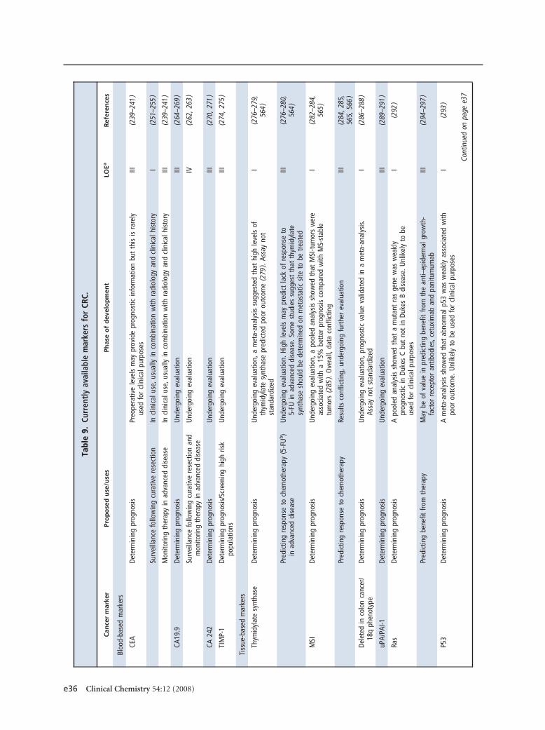
Tabl
e9.
Curr
entl
yav
aila
ble
mar
kers
for
CRC.
Can
cer
mar
ker
Pro
po
sed
use
/use
sPh
ase
of
dev
elo
pm
ent
LOEa
Ref
eren
ces
Bloo
d-ba
sed
mar
kers
CEA
Dete
rmin
ing
prog
nosi
sPr
eope
rativ
ele
vels
may
prov
ide
prog
nost
icin
form
atio
nbu
tth
isis
rare
lyus
edfo
rcl
inic
alpu
rpos
esIII
(239
–241
)
Surv
eilla
nce
follo
win
gcu
rativ
ere
sect
ion
Incl
inic
alus
e,us
ually
inco
mbi
natio
nw
ithra
diol
ogy
and
clin
ical
hist
ory
I(2
51–2
55)
Mon
itorin
gth
erap
yin
adva
nced
dise
ase
Incl
inic
alus
e,us
ually
inco
mbi
natio
nw
ithra
diol
ogy
and
clin
ical
hist
ory
III(2
39–2
41)
CA19
.9De
term
inin
gpr
ogno
sis
Und
ergo
ing
eval
uatio
nIII
(264
–269
)
Surv
eilla
nce
follo
win
gcu
rativ
ere
sect
ion
and
mon
itorin
gth
erap
yin
adva
nced
dise
ase
Und
ergo
ing
eval
uatio
nIV
(262
,263
)
CA24
2De
term
inin
gpr
ogno
sis
Und
ergo
ing
eval
uatio
nIII
(270
,271
)
TIM
P-1
Dete
rmin
ing
prog
nosi
s/Sc
reen
ing
high
risk
popu
latio
nsU
nder
goin
gev
alua
tion
III(2
74,2
75)
Tiss
ue-b
ased
mar
kers
Thym
idyl
ate
synt
hase
Dete
rmin
ing
prog
nosi
sU
nder
goin
gev
alua
tion,
am
eta-
anal
ysis
sugg
este
dth
athi
ghle
vels
ofth
ymid
ylat
esy
ntha
sepr
edic
ted
poor
outc
ome
(279
).As
say
not
stan
dard
ized
I(2
76–2
79,
564
)
Pred
ictin
gre
spon
seto
chem
othe
rapy
(5-F
Ub )
inad
vanc
eddi
seas
eU
nder
goin
gev
alua
tion.
High
leve
lsm
aypr
edic
tla
ckof
resp
onse
to5-
FUin
adva
nced
dise
ase.
Som
est
udie
ssu
gges
tth
atth
ymid
ylat
esy
ntha
sesh
ould
bede
term
ined
onm
etas
tatic
site
tobe
trea
ted
III(2
76–2
80,
564
)
MSI
Dete
rmin
ing
prog
nosi
sU
nder
goin
gev
alua
tion,
apo
oled
anal
ysis
show
edth
atM
SI-t
umor
sw
ere
asso
ciat
edw
itha
15%
bett
erpr
ogno
sis
com
pare
dw
ithM
S-st
able
tum
ors
(285
).O
vera
ll,da
taco
nflic
ting
I(2
82–2
84,
565
)
Pred
ictin
gre
spon
seto
chem
othe
rapy
Resu
ltsco
nflic
ting,
unde
rgoi
ngfu
rthe
rev
alua
tion
III(2
84,2
85,
565,
566
)
Dele
ted
inco
lon
canc
er/
18q
phen
otyp
eDe
term
inin
gpr
ogno
sis
Und
ergo
ing
eval
uatio
n,pr
ogno
stic
valu
eva
lidat
edin
am
eta-
anal
ysis
.As
say
not
stan
dard
ized
I(2
86–2
88)
uPA/
PAI-1
Dete
rmin
ing
prog
nosi
sU
nder
goin
gev
alua
tion
III(2
89–2
91)
Ras
Dete
rmin
ing
prog
nosi
sA
pool
edan
alys
issh
owed
that
am
utan
tra
sge
new
asw
eakl
ypr
ogno
stic
inDu
kes
Cbu
tno
tin
Duke
sB
dise
ase.
Unl
ikel
yto
beus
edfo
rcl
inic
alpu
rpos
es
I(2
92)
Pred
ictin
gbe
nefit
from
ther
apy
May
beof
valu
ein
pred
ictin
gbe
nefit
from
the
anti–
epid
erm
algr
owth
-fa
ctor
rece
ptor
antib
odie
s,ce
tuxi
mab
and
pani
tum
umab
III(2
94–2
97)
P53
Dete
rmin
ing
prog
nosi
sA
met
a-an
alys
issh
owed
that
abno
rmal
p53
was
wea
kly
asso
ciat
edw
ithpo
orou
tcom
e.U
nlik
ely
tobe
used
for
clin
ical
purp
oses
I(2
93)
Cont
inue
don
page
e37
e36 Clinical Chemistry 54:12 (2008)
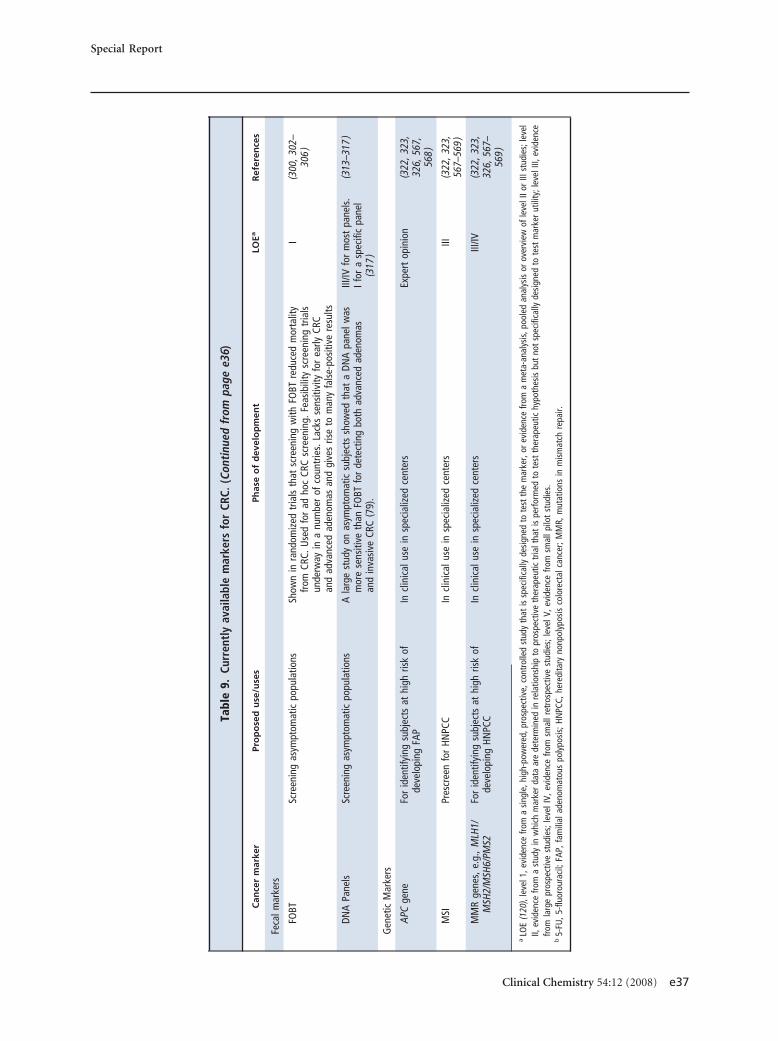
Tabl
e9.
Curr
entl
yav
aila
ble
mar
kers
for
CRC.
(Con
tinu
edfr
ompa
gee3
6)
Can
cer
mar
ker
Pro
po
sed
use
/use
sPh
ase
of
dev
elo
pm
ent
LOEa
Ref
eren
ces
Feca
lmar
kers
FOBT
Scre
enin
gas
ympt
omat
icpo
pula
tions
Show
nin
rand
omiz
edtr
ials
that
scre
enin
gw
ithFO
BTre
duce
dm
orta
lity
from
CRC.
Use
dfo
rad
hoc
CRC
scre
enin
g.Fe
asib
ility
scre
enin
gtr
ials
unde
rway
ina
num
ber
ofco
untr
ies.
Lack
sse
nsiti
vity
for
early
CRC
and
adva
nced
aden
omas
and
give
sris
eto
man
yfa
lse-
posi
tive
resu
lts
I(3
00,3
02–
306
)
DNA
Pane
lsSc
reen
ing
asym
ptom
atic
popu
latio
nsA
larg
est
udy
onas
ympt
omat
icsu
bjec
tssh
owed
that
aDN
Apa
nelw
asm
ore
sens
itive
than
FOBT
for
dete
ctin
gbo
thad
vanc
edad
enom
asan
din
vasi
veCR
C(7
9).
III/IV
for
mos
tpa
nels
.If
ora
spec
ific
pane
l(3
17)
(313
–317
)
Gen
etic
Mar
kers
APC
gene
For
iden
tifyi
ngsu
bjec
tsat
high
risk
ofde
velo
ping
FAP
Incl
inic
alus
ein
spec
ializ
edce
nter
sEx
pert
opin
ion
(322
,323
,32
6,56
7,56
8)
MSI
Pres
cree
nfo
rHN
PCC
Incl
inic
alus
ein
spec
ializ
edce
nter
sIII
(322
,323
,56
7–56
9)
MM
Rge
nes,
e.g.
,MLH
1/M
SH2/
MSH
6/PM
S2Fo
rid
entif
ying
subj
ects
athi
ghris
kof
deve
lopi
ngHN
PCC
Incl
inic
alus
ein
spec
ializ
edce
nter
sIII
/IV(3
22,3
23,
326,
567–
569
)
aLO
E(1
20),
leve
l1,e
vide
nce
from
asi
ngle
,hig
h-po
wer
ed,p
rosp
ectiv
e,co
ntro
lled
stud
yth
atis
spec
ifica
llyde
sign
edto
test
the
mar
ker,
orev
iden
cefro
ma
met
a-an
alys
is,p
oole
dan
alys
isor
over
view
ofle
velI
IorI
IIst
udie
s;le
vel
II,ev
iden
cefro
ma
stud
yin
whi
chm
arke
rda
taar
ede
term
ined
inre
latio
nshi
pto
pros
pect
ive
ther
apeu
tictr
ialt
hat
ispe
rform
edto
test
ther
apeu
tichy
poth
esis
but
not
spec
ifica
llyde
sign
edto
test
mar
ker
utili
ty;l
evel
III,e
vide
nce
from
larg
epr
ospe
ctiv
est
udie
s;le
velI
V,ev
iden
cefro
msm
allr
etro
spec
tive
stud
ies;
leve
lV,e
vide
nce
from
smal
lpilo
tst
udie
s.b
5-FU
,5-fl
uoro
urac
il;FA
P,fa
mili
alad
enom
atou
spo
lypo
sis;
HNPC
C,he
redi
tary
nonp
olyp
osis
colo
rect
alca
ncer
;MM
R,m
utat
ions
inm
ism
atch
repa
ir.
Special Report
Clinical Chemistry 54:12 (2008) e37
cluded the use of markers, and guidelines issued byexpert panels. Where possible, the consensus recom-mendations of the NACB Panel were based on availableevidence, i.e., were evidence based.
CURRENTLY AVAILABLE MARKERS FOR CRC
The most widely investigated tumor markers for CRCand the phase of development of each marker and theLOE for its clinical use are listed in Table 9.
TUMOR MARKERS IN CRC: NACB RECOMMENDATIONS
Table 10 presents a summary of recommendationsfrom representative guidelines published on the use oftumor markers in CRC. This table also summarizes theNACB guidelines for the use of markers in this malig-nancy. Below, we present a more detailed discussion ofthe most widely investigated markers listed in Table 10.
CARCINOEMBRYONIC ANTIGEN
CEA in screening. Lack of sensitivity and specificitywhen combined with the low prevalence of CRC inasymptomatic populations preclude the use of CEA inscreening for CRC (239 –241 ). In agreement withASCO (242–244 ) and EGTM recommendations(245, 246 ), the NACB Panel states that CEA cannot beused in screening healthy individuals for early CRC.
NACB CRC PANEL RECOMMENDATION 1:
SERUM CEA IN SCREENING HEALTHY INDIVIDUALS
CEA cannot be used in screening of healthy subjectsfor early CRC [LOE, IV/V; SOR, A].
CEA in determining prognosis. As mentioned above,disease stage at initial diagnosis is universally used todetermine prognosis in patients with CRC. Severalstudies, however, have demonstrated that preoperativeconcentrations of CEA can also provide prognostic in-formation, which in some situations was found to beindependent of stage (239 –241, 247–249 ). The NACBPanel therefore states that preoperative concentrationsof CEA might be used in combination with other fac-tors in planning surgical treatment. Preoperative CEAconcentrations, however, should not be used at presentto select patients for adjuvant therapy. These guidelinesare broadly in agreement with those previously pub-lished by ASCO and EGTM (242, 244 –246 ).
A College of American Pathologists Expert Panelrecently ranked preoperative serum CEA together withTNM stage, regional lymph node metastasis, blood orlymphatic vessel invasion, and residual tumor follow-ing surgery with curative intent as a category I prognos-tic marker for CRC (250 ). According to the College ofAmerican Pathologists Panel, Category I prognostic
factors are those “definitely proven to be of prognosticimportance based on evidence from multiple statisti-cally robust published trials and generally used in pa-tient management”.
NACB CRC PANEL RECOMMENDATION 2:
SERUM CEA IN PROGNOSIS AND PREDICTION
Preoperative CEA concentrations might be used incombination with other factors in planning surgicaltreatment. Patients with increased concentrationsof CEA (e.g., �5 �g/L) should be evaluated for thepresence of distant metastases [LOE, III; SOR, C].Preoperative CEA concentrations should not beused at present to select patients for adjuvant che-motherapy [LOE, III; SOR, C].
CEA in postoperative surveillance. The main aims ofsurveillance following curative resection of CRC are toprovide reassurance, address possible complicationsdue to therapy, and identify resectable recurrences ormetastases. Six separate metaanalyses have comparedoutcome in patients with intensive follow-up vs thosewith minimal or no follow-up (251–256 ). All con-cluded that the use of an intensive follow-up regimeresulted in a modest but statistically significant im-proved outcome when compared with regimes withminimal follow-up. In one of these metaanalyses, it wasshown that only the studies including CEA demon-strated a significant impact on survival (254 ).
The most recent ASCO guidelines state that CEAshould be measured every 3 months in patients withstage II or III CRC for at least 3 years after diagnosis, ifthe patient is a candidate for surgery or systemic ther-apy of metastatic disease (244, 257 ). The NACB Panelsupports this recommendation.
Although serial measurements of CEA are widelyused in surveillance, no agreement exists as to the mag-nitude of concentration change that constitutes a clin-ically significant increase in CEA during serial moni-toring. According to the EGTM Panel, a significantincrease in CEA occurs if the elevation is at least 30%over that of the previous value. This increase, however,must be confirmed by a second sample taken within 1month. If this latter sample is also increased, the patientshould undergo further investigations (246 ). This 30%increase, however, has not been clinically validated.Furthermore, it should not be regarded as exclusive.For example, small increases in CEA (e.g., 15%–20%,maintained over at least 3 successive assays) may alsoprompt intervention (246 ). It should also be remem-bered that low CEA concentrations do not necessarilyexclude progression, and in patients with clinical
e38 Clinical Chemistry 54:12 (2008)
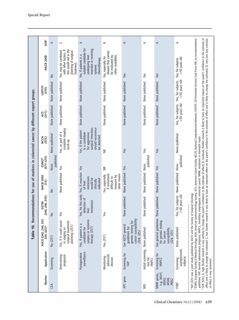
Tabl
e10
.Re
com
men
dati
ons
for
use
ofm
arke
rsin
colo
rect
alca
ncer
bydi
ffer
ent
expe
rtgr
oups
.
Mar
ker
Ap
plic
atio
nA
SCO
(242
,244
,257
,32
4,32
5)a
EGTM
(245
,246
,570
)N
AC
B20
02(1
5)
ESM
Ob
(571
–574
)N
CC
N(5
75)
AC
S(3
11)
USP
STF
(310
)N
AC
B20
08SO
Rc
CEA
Scre
enin
gN
o(2
57)
No
No
Non
e publ
ishe
dN
one
publ
ishe
dN
one
publ
ishe
dN
one
publ
ishe
dN
oA
Dete
rmin
ing
prog
nosi
sYe
s,if
itco
uld
assi
stin
stag
ing
orsu
rgic
altr
eatm
ent
plan
ning
(257
)
Yes
Non
epu
blis
hed
Yes
Yes,
aspa
rtof
aco
mpl
ete
stag
ing
wor
k-up
Non
epu
blis
hed
Non
epu
blis
hed
Yes,
may
beco
mbi
ned
with
othe
rfa
ctor
s,if
this
wou
ldai
din
the
plan
ning
ofsu
rgica
ltre
atm
ent
C
Post
oper
ativ
esu
rvei
llanc
eYe
s,if
patie
ntis
aca
ndid
ate
for
surg
ery
orsy
stem
icth
erap
y(2
57)
Yes,
for
the
early
dete
ctio
nof
liver
met
asta
sis
Yes,
ifre
sect
ion
ofliv
erm
etas
tasi
sw
ould
becl
inic
ally
indi
cate
d
Yes
Yes,
ifth
epa
tient
isa
cand
idat
efo
rag
gres
sive
surg
ical
rese
ctio
n,sh
ould
recu
rren
cebe
dete
cted
Non
epu
blis
hed
Non
epu
blis
hed
Yes,
ifpa
tient
sis
asu
itabl
eca
ndid
ate
for
unde
rgoi
ngliv
erre
sect
ion
orre
ceiv
ing
syst
emic
chem
othe
rapy
A
Mon
itorin
gad
vanc
eddi
seas
e
Yes
(257
)Ye
sYe
s,es
peci
ally
ifm
etas
tasi
sdi
fficu
ltto
mea
sure
byot
her
mea
ns
NR
NR
Non
epu
blis
hed
Non
epu
blis
hed
Yes,
espe
cially
for
dise
ase
that
cann
otbe
eval
uate
dby
othe
rm
odal
ities
B
APC
gene
Scre
enin
gfo
rFA
PSe
eAS
COge
nera
lgu
idel
ines
for
gene
ticte
stin
gfo
rca
ncer
susc
eptib
ility
(324
,325
)
Non
epu
blis
hed
Non
epu
blis
hed
Yes
Yes
Non
epu
blis
hed
Non
epu
blis
hed
Yes
A
MSI
Initi
alsc
reen
ing
test
for
HNPC
C
Non
epu
blis
hed
Non
epu
blis
hed
Non
epu
blis
hed
Non
e publ
ishe
dYe
sN
one
publ
ishe
dN
one
publ
ishe
dYe
sA
MM
Rge
nes,
e.g.
MLH
1,M
SH2,
MSH
6,PM
S2
Scre
enin
gfo
rHN
PCC
See
gene
ralg
uide
lines
for
gene
ticte
stin
gfo
rca
ncer
susc
eptib
ility
(324
,325
)
Non
epu
blis
hed
Non
epu
blis
hed
Yes
Yes
Non
epu
blis
hed
Non
epu
blis
hed
Yes
A
FOBT
Scre
enin
gas
ympt
omat
icsu
bjec
ts
Non
epu
blis
hed
Yes,
for
subj
ects
�50
year
sol
dN
one
publ
ishe
dN
one publ
ishe
dN
one
publ
ishe
dYe
s,fo
rsu
bjec
ts�
50ye
ars
old
Yes,
for
subj
ects
�50
year
sol
dYe
s,fo
rsu
bjec
ts�
50ye
ars
old
A
aRe
f(3
25)
was
ajo
int
stud
ypu
blis
hed
byAS
COan
dth
eSo
ciet
yof
Surg
ical
Onc
olog
yb
ESM
O,E
urop
ean
Soci
ety
ofM
edic
alO
ncol
ogy;
AGA,
Amer
ican
Gas
troe
nter
olog
ySo
ciet
y,AC
S,Am
eric
anCa
ncer
Soci
ety;
NCC
N,N
atio
nalC
ompr
ehen
sive
Net
wor
k;U
SPST
F,U
SPr
even
tive
Serv
ices
Task
Forc
e;N
R,no
reco
mm
enda
tion
publ
ishe
d;FA
P,fa
mili
alad
enom
atou
spo
lypo
sis;
HNPC
C,he
redi
tary
nonp
olyp
osis
colo
rect
alca
ncer
;MM
R,m
utat
ions
inm
ism
atch
repa
ir.c
SOR
(520
),A,
high
(furt
her
rese
arch
isve
ryun
likel
yto
chan
geth
epa
nel’s
conf
iden
cein
the
estim
ate
ofef
fect
);B,
mod
erat
e(fu
rthe
rre
sear
chis
likel
yto
have
anim
port
ant
impa
cton
the
pane
l’sco
nfid
ence
inth
ees
timat
eof
effe
ctan
dis
likel
yto
chan
geth
ees
timat
e);C
,low
(furt
her
rese
arch
isve
rylik
ely
toha
vean
impo
rtan
tef
fect
onth
epa
nel’s
conf
iden
cein
the
estim
ate
ofef
fect
and
islik
ely
toch
ange
the
estim
ate)
;D,v
ery
low
(any
estim
ate
ofef
fect
isve
ryun
cert
ain)
.
Special Report
Clinical Chemistry 54:12 (2008) e39
symptoms of disease recurrence, additional tests suchas computed tomographic scan, x-rays, and colonos-copy are required, irrespective of the CEA concentra-tion (246 ).
NACB CRC PANEL RECOMMENDATION 3:
SERUM CEA IN POSTOPERATIVE SURVEILLANCE
CEA should be measured every 3 months in pa-tients with stage II or III CRC for at least 3 yearsafter diagnosis if the patient is a candidate for sur-gery or systemic therapy of metastatic disease [LOE,I; SOR, A].
CEA in monitoring therapy in advanced disease. Theprognosis for patients with advanced CRC has greatlyimproved in recent years owing to the introduction ofnew cytotoxic agents such as irinotecan and oxaliplatinand monoclonal antibodies such as bevacuzimab(Avastin�), panitumumab (Vectibix�), and cetuximab(Erlotinib�), as has recently been reviewed (258, 259 ).Indeed, the median survival for patients with meta-static CRC has almost doubled in the past 10 years as aresult of these new treatments (258 –260 ). However,because these treatments are potentially toxic as well asexpensive, it is important to establish as quickly as pos-sible that they are effective in halting tumorprogression.
According to the 2006 ASCO guidelines, CEA isthe marker of choice for monitoring metastatic CRCduring systemic therapy (244 ). CEA should be mea-sured at the start of treatment for metastatic diseaseand every 1–3 months during active treatment. Persis-tently increasing concentrations suggest progressivedisease even in the absence of corroborating radio-graphs (242, 243 ). In 2003, the EGTM Panel recom-mended that serial CEA concentrations should be mea-sured every 2–3 months while patients are receivingsystemic therapy (246 ). Both the ASCO and EGTMguidelines stated that caution should be used when in-terpreting increasing CEA concentrations during theearly phase of systemic treatment (16, 18 ). This is be-cause certain treatments (e.g., 5-fluorouracil and le-vamisole; oxaliplatin) can cause transient elevations inCEA levels in the absence of disease progression (246 ).
For monitoring patients with advanced CRC un-dergoing systemic therapy, the NACB Panel recom-mends that regular CEA determinations should be car-ried out. In agreement with the ASCO Panel(242, 243 ), a confirmed CEA increase (e.g., �30%)may be regarded as evidence of progressive disease. Ofcourse, it should be established that the increases arenot false-positive elevations due to either chemothera-py-mediated release of marker or the development of abenign disease that produces CEA.
NACB CRC PANEL RECOMMENDATION 4:
SERUM CEA IN MONITORING PATIENTS WITH
ADVANCED DISEASE
In patients with advanced CRC undergoing sys-temic therapy, regular CEA determinations shouldbe carried out. A confirmed CEA increase (e.g.,�30%) suggests progressive disease provided thepossibility of false-positive elevations can be ex-cluded [LOE, III; SOR, B].
OTHER SERUM MARKERS
CA 19 –9. The CA 19 –9 assay detects a mucin contain-ing the sialated Lewis-a pentasacharide epitope, fuco-pentaose II [for review, see ref (261 )]. CA 19 –9 is a lesssensitive marker than CEA for CRC (262, 263 ). Pre-liminary findings suggest that like CEA, preoperativeconcentrations of CA 19 –9 are also prognostic in pa-tients with CRC (264 –268 ). Based on available data,routine measurement of CA 19 –9 cannot be recom-mended for patients with CRC.
CA 242. The CA 242 assay also detects a mucinlikemolecule. Although less sensitive than CEA for CRC,assay of CA 242 may complement CEA in the surveil-lance of patients with CRC (263, 269 ). Furthermore, anumber of preliminary reports suggest that preopera-tive concentrations of CA 242 are prognostic in CRC(270, 271 ). Routine determinations of CA 242 shouldnot be used at present in patients with CRC.
TISSUE INHIBITOR OF METALLOPROTEINASES TYPE 1
Tissue inhibitor of metalloproteinases type 1 (TIMP-1)is a 25-kDa glycoprotein with multiple activities in-cluding inhibition of matrix metalloproteinases, pro-motion of cell proliferation, and inhibition of apopto-sis. With use of a research ELISA that detects totalTIMP-1 (i.e., the noncomplex form as well as TIMP-1complexed to matrix metalloproteinases), plasma con-centrations of the inhibitor were found to be signifi-cantly higher in patients with CRC than in healthy con-trols or patients with inflammatory bowel diseases,adenomas, or breast cancer (272, 273 ). For patientswith Dukes A and B colon cancers, TIMP-1 appeared tobe more sensitive than CEA for the detection of cancer,i.e., 58% vs 40% at 95% specificity and 56% vs 30% at98% specificity. For patients with early rectal cancer,TIMP-1 and CEA had similar sensitivity (272 ). Otherstudies have shown that preoperative plasma TIMP-1concentration is an independent prognostic factor inpatients with CRC, i.e., independent of Dukes stageand tumor location (274, 275 ). Of particular note wasthe finding that stage II patients with low plasmaTIMP-1 concentrations (dichotomized at the 70% per-
e40 Clinical Chemistry 54:12 (2008)
centile) exhibited a survival pattern similar to an ageand sex-matched background population.
Although these preliminary findings with TIMP-1are promising, the marker cannot be recommended atpresent either for detecting early CRC or for evaluatingprognosis in patients with this malignancy.
NACB CRC PANEL RECOMMENDATION 5:
CA19.9, CA 242 AND TIMP-1 IN CRC
Routine measurement of CA19.9, CA 242, orTIMP-1 is not recommended [LOE, III/IV; SOR,B/C].
TISSUE MARKERS
Several tumor tissue markers have been evaluated forpotential prognostic and predictive value in patientswith CRC. These include thymidylate synthase (276 –280 ), MSI (281–285 ), deleted in colon cancer (286 –288 ), urokinase plasminogen activator (uPA)/plas-minogen activator inhibitor 1 (PAI-1) (289 –291 ),mutant ras (292 ), and mutant/overexpression of p53(293 ). Based on available evidence, none of thesemarkers can at present be recommended for routinelydetermining prognosis or for therapy prediction.Emerging evidence however, suggests that the presenceof wild type k-ras is associated with benefit from theanti– epidermal growth-factor receptor (EGFR) anti-bodies cetuximab and panitumumab (294 –297 ).
NACB CRC PANEL RECOMMENDATION 6:
TISSUE MARKERS IN CRC
The use of thymidylate synthase, MSI, deleted incolon cancer, uPA, PAI-1, or p53 for determiningprognosis or predicting response to therapy is notrecommended [LOE, III; SOR, B]. Determinationof the mutation status of k-ras may in the future beused for predicting benefit from specific anti-EGFRantibodies.
FECAL MARKERS
The most widely used fecal marker involves testing foroccult blood, i.e., the fecal occult blood test (FOBT)Two of the most widely described FOBTs are the guaiactest and the fecal immunochemical test (FIT) (298 –301 ). The guaiac test measures the pseudoperoxidaseactivity of heme in hemoglobin, and the immuno-chemical test detects human globin. Because peroxi-dase activity is also present in certain fruits and vegeta-bles, intake of these foods may give rise to false-positiveresults in the guaiac test. Certain medicines such asnonsteroidal antiinflammatory drugs can also interferewith this test. Despite these limitations, a number of
large randomized trials have shown that screening withthe guaiac test reduced mortality from CRC (302–306 ).
The efficacy of the FIT in reducing either the inci-dence or mortality form CRC has not yet been investi-gated in large population-based studies. However,based on available evidence, it should be at least as ac-curate if not more accurate than guaiac-based tests inscreening for CRC (298, 301, 307 ). The advantages ofthe immunochemical test over the guaiac tests includethe following (for review, see refs (298, 299, 307 ):
• FITs have better sensitivity for human blood.• FITs are not affected by diet or medications.• Some FITs can be automated.• Evidence suggests that the use of FITs increases pa-
tient participation in screening for CRC.• FITs can be quantitated, enabling adjustment of sen-
sitivity, specificity and positivity rates.• Because digested blood from the upper gastrointesti-
nal tract is not usually detected by FITs, the latter arebetter for detecting bleeding from the lower gastro-intestinal tract.
In agreement with other expert panels (308 –310 ),the NACB Panel recommends that all individuals 50years or older should undergo screening for CRC. Mul-tiple screening procedures for CRC exist, however (306–308), and to date no one procedure has been shown tobe significantly superior to the others. The option cho-sen may therefore depend on availability, personalpreference, and risk of developing CRC (311 ).
According to the National Comprehensive CancerNetwork (NCCN), FOBT should be performed on 3successive stool specimens that are obtained while thepatient adheres to a prescribed diet (308 ). This organi-zation specifically recommends the Hemoccult SENSAas the testing method. Both the NCCN and the Amer-ican Cancer Society recommend against use of FOBTof a specimen obtained during a digital rectal examina-tions (308, 311 ).
Although screening has been shown to result inreduced mortality from CRC (302–305, 312), it may beassociated with certain harmful effects. These includethe psychosocial consequence of false-positive results,potential complications of colonoscopy, a false nega-tive result, or the possibility of over-diagnosis (312 ).Overdiagnosis could give rise to unnecessary investiga-tions or treatment.
Because of the lack of sensitivity and specificity ofFOBT for adenomas and early CRC, a considerableamount of research in recent years has focused on otherfecal markers, especially on the genes that undergomutation during CRC carcinogenesis. Among themost widely investigated DNA markers are mutant ras,mutant p53, mutant APC, specific methylated genes,MSI, and long DNA (231, 313–316 ). Almost all of
Special Report
Clinical Chemistry 54:12 (2008) e41
the studies published to date on fecal DNA markerscontained small numbers of patients. Following anoverview of the literature, Allison and Lawson (298 )found that the sensitivities of the different DNA panelsfor invasive CRC varied from 52%–98% (mean, 64%),and the specificity varied from 93%–97% (mean,95%).
Although most of the studies that evaluated DNAmarkers for the detection of CRC included only smallnumbers of patients, a specific panel was recently in-vestigated as a screening test for CRC in a large asymp-tomatic population (317 ). Of the 31 invasive CRCs de-tected, the DNA panel diagnosed 16, whereas FOBTdetected only 4 (51.6% vs 12.9%, P � 0.003). Of the 71invasive cancers and adenomas with high-grade dys-plasia, the DNA panel diagnosed 29, while FOBT de-tected only 10 (P � 0.001). Although the DNA paneldisplayed a higher sensitivity than FOBT, clearly nei-ther test detected the majority of advanced adenomasor carcinomas (317 ). However, because the DNA-based test was superior to FOBT, it might be expectedto be at least as good as the latter in reducing mortalityfrom CRC. However, it should be pointed out thatcompared to FOBTs, measurement of fecal DNAmarkers is more expensive and technically demanding.Furthermore, it is not clear which combination of DNAmarkers provides the optimum balance of sensitivityand specificity (231 ).
One of the main arguments against the use of aDNA panel at present, especially when applied to largepopulations, is the relative cost compared to FOBT(318, 319 ). In 2004, Song et al. (318 ), using a model-ling approach, compared the cost-effectiveness of fecalDNA to that of standard CRC screening methods. Themain conclusions were as follows:
• Compared with no screening, all screening strategiesincreased life expectancy at what was regarded as rea-sonable cost.
• Compared with no screening, the use of fecal DNAtesting gained 4560 life-years per 100 000 persons atan incremental cost of $47 700/life-year gained.
• The use of colonoscopy and FOBT/flexible sigmoid-oscopy were more effective strategies, gaining an in-cremental 6190 and 6270 life-years per 100 000 per-sons compared to no screening, at incremental costsper life-year gained of $17 010 and $17 000.
• All the conventional approaches gained more life-years at lower cost than fecal DNA testing.
Despite their relatively high costs, the technicallydemanding nature of the assays, and the fact that thesetests have not been validated in a prospective random-ized trial, recent joint guidelines from the AmericanCancer Society, the US Multi-Society Task Force, andthe American College of Radiology state that there is
now sufficient data to include fecal DNA “as an accept-able option for CRC screening” (320, 321 ).
NACB CRC PANEL RECOMMENDATION 7:
USE OF FECAL MARKERS IN SCREENING FOR CRC
The NACB recommends that all individuals 50years or older should undergo screening for CRC.Because the most effective screening test is un-known, the method chosen is likely to depend onrisk of CRC, local availability, and personal prefer-ence. Although FOBT is the best-validated stool-based method for screening for CRC [LOE, I; SOR,A], fecal DNA testing may also be an option. Poten-tial harmful consequences of screening includecomplications due to colonoscopy and treatment,the possibility of overdiagnosis leading to unneces-sary investigations, and false-negative and false-positive results.
GENETIC TESTS
For genetic testing for CRC susceptibility, i.e., familialadenomatous polyposis coli and hereditary nonpol-yposis CRC, the NACB Panel supports previously pub-lished guidelines (308, 322–326 ).
NACB CRC PANEL RECOMMENDATION 8:
GENETIC TESTING FOR CRC
Screening for genetic susceptibility to CRC shouldcommence with a detailed family history. Beforeundergoing testing, individuals should receive ge-netic counseling. For persons with suspected famil-ial adenomatous polyposis, genetic testing can beused both to confirm diagnosis in a suspected pro-band and to assess risk in presymptomatic familymembers. Provided the mutation responsible forfamilial adenomatous polyposis within a family isknown, testing for adenomatous polyposis coli(APC) gene mutations can be considered for at-riskfamily members. [LOE, Expert opinion; SOR, A].
MSI testing and/or immunohistochemistry forspecific mismatch repair enzymes can be used as aprescreen for hereditary nonpolyposis CRC. If anindividual is found to possess high MSI, genetictesting for mutations in MLH1, MLH2, MSH6, orPMS2 genes should be carried out [LOE, III/IV;SOR, A].33
33 MLH1, mutL homolog 1, colon cancer, nonpolyposis type 2 (E. coli); MSH2,mutS homolog 2, colon cancer, nonpolyposis type 1 (E. coli); MSH6, mutShomolog 6 (E. coli); PMS2, postmeiotic segregation increased 2 (S. cerevisiae).
e42 Clinical Chemistry 54:12 (2008)
KEY POINTS: TUMOR MARKERS IN CRC
Although many different markers have been evaluatedfor CRC, only a small number can be recommended forclinical use. These include CEA in the postoperativesurveillance of patients who may be suitable candidatesfor either surgical resection or systemic chemotherapy,FOBT in screening for early CRC in persons 50 years orolder, MSI as a surrogate marker for identifying per-sons who should undergo genetic testing for MLH1/MSH2/MSH6/PMS2 to identify hereditary nonpolypo-sis CRC, and APC to identify familial adenomatouspolyposis. A promising new plasma marker is TIMP-1.As mentioned above, preliminary findings suggestthat this marker may be more sensitive than CEA indetecting early CRC as well as being an independentprognostic factor for CRC. These findings now mustbe confirmed in large prospective studies. One of themost promising fecal CRC screening tests is a fecalDNA panel (317 ). This test should be simplified, madeavailable at reduced costs, and subjected to furtherinvestigations.
Tumor Markers in Breast Cancer34,35
BACKGROUND
Breast cancer is by far the most common cancer affect-ing women worldwide, with approximately one mil-lion new cases diagnosed each year (327 ). In 2007, anestimated 180 000 women were diagnosed with breastcancer in the US and approximately 41 000 died fromthe disease (118 ). Currently, there are more than 2 mil-lion women in the US who are living with a history ofbreast cancer (328 ). Although the worldwide incidenceof the disease appears to be increasing, mortality ratesare now declining in a number of Western countriesincluding the US and the United Kingdom (329 ).
The main presenting features in women withsymptomatic breast cancer include a lump in thebreast, nipple change, or discharge and skin contourchanges. Definitive diagnosis requires biopsy and his-topathology. Currently available blood-based biomar-kers are of no value in the early diagnosis of breastcancer.
The primary treatment for localized breast canceris either breast-conserving surgery and radiation ormastectomy. Following primary treatment, mostwomen with invasive breast cancer receive systemicadjuvant therapy such as chemotherapy, hormone
therapy, or a combination of chemotherapy and hor-mone therapy. Both adjuvant chemotherapy and hor-mone therapy have been shown to reduce systemicrecurrence and mortality from breast cancer (330 ).For example, a meta-analysis of approximately 145 000women participating in 194 randomized trials of adju-vant systemic therapy concluded that anthracycline-based polychemotherapy reduced the annual breastcancer death rate by about 38% for women youngerthan 50 years at diagnosis and by about 20% for thoseage 50 – 69 years at diagnosis (330 ). For estrogen recep-tor (ER)-positive patients, 5 years of adjuvant tamox-ifen reduced annual breast cancer death rates by 31%(330 ). Patients with ER-negative tumors, however, didnot benefit from adjuvant tamoxifen (331 ).
Because not all patients with breast cancer mayneed adjuvant treatment [e.g., approximately, 70% oflymph node–negative patients are cured of their dis-ease by surgery and radiotherapy (332 )] and not allpatients benefit from this treatment, rational manage-ment requires the availability of reliable prognostic andpredictive markers. Recommendations regarding theuse of currently available prognostic and predictivemarkers for breast cancer are discussed below.
Subsequent to primary therapy, patients with a di-agnosis of breast cancer are usually followed up at reg-ular intervals. Historically, surveillance has includedclinical history, physical examination, mammography,chest x-ray, biochemical testing, and the use of tumormarkers. This practice is based on the assumption thatthe early detection of recurrent disease leads to a betteroutcome. However, at present, the clinical benefit ofclose surveillance is unclear (333 ).
Although adjuvant therapy improves patient out-come, 25%–30% of women with lymph node–negativeand at least 50%– 60% of those with node-positivedisease develop recurrent or metastatic disease (334 ).Therapy options for metastatic breast cancer includechemotherapy (e.g., anthracycline or taxane-based),hormone therapy, or targeted therapies such as Trastu-zumab (Herceptin®), Lapatinib, or Bevacizumab,alone or combined with chemotherapy (334, 335 ).Currently, metastatic breast cancer is regarded as in-curable and thus the goal of treatment is generally pal-liative. In this context, the use of serial levels of serumtumor markers is potentially useful in decidingwhether to persist in using a particular type of therapy,terminate its use, or switch to an alternative therapy.
Based on the above, it is clear that optimal man-agement of patients with breast cancer requires the useof a number of tumor markers. The aim of this articleis to present new NACB guidelines on the use of bothtissue- and serum-based tumor markers in breast can-cer. A summary of guidelines published by other expertpanels on this topic is also provided.
34 NACB Breast Cancer Sub-Committee Members: Michael J. Duffy, Chair; Fran-cisco J. Esteva; Nadia Harbeck; Daniel F. Hayes; and Rafael Molina.
35 All comments received about the NACB Recommendations for Breast Cancerare included in the online Data Supplement. Professor Dorte Nielsen, ProfessorJohn Smyth, and Professor M. Tuxen were invited expert reviewers.
Special Report
Clinical Chemistry 54:12 (2008) e43
To prepare these guidelines, the literature relevantto the use of tumor markers in breast cancer was re-viewed. Particular attention was given to reviews, in-cluding systematic reviews, prospective randomizedtrials that included the use of markers, and guidelinesissued by expert panels. Where possible, the consensusrecommendations of the NACB Panel were based onavailable evidence, i.e., were evidence-based.
CURRENTLY AVAILABLE MARKERS FOR BREAST CANCER
Table 11 lists the mostly widely investigated tissue-based and serum-based tumors markers for breast can-cer. Also listed, is the phase of development of eachmarker as well as the LOE for its clinical use.
TUMOR MARKERS IN BREAST CANCER: NACB
RECOMMENDATIONS
Table 12 presents a summary of recommendationsfrom various expert panels on the use of tumor mark-ers in breast cancer. This table also summarises theNACB guidelines for the use of markers in this malig-nancy. Below, we present a more detailed discussion onthe most clinically useful markers listed in Table 12.
ER and progesterone receptor. Routine assay of ERs (i.e.,ER-�) and progesterone receptors (PR) in all newlydiagnosed breast cancers has been recommended byExpert Panels of ASCO, EGTM, the European Societyof Medical Oncology, and the St Gallen ConferenceConsensus Panel (Table 12). The NACB Panel agreeswith these recommendations. The primary purpose ofdetermining ER and PR is to select for likely response toendocrine therapy in patients with either early or ad-vanced breast cancer. Additionally, in combinationwith other factors, ER and PR may also be used forprognostic purposes. However, as predictors of patientoutcome, hormone receptors are relatively weak fac-tors and are of little clinical value in lymph node–nega-tive patients. Hormone receptors should therefore notbe used alone for determining outcome in breast can-cer. However, in combination with established prog-nostic factors, hormone receptors may be used to pre-dict risk of recurrence. Determination of ER-� has noclinical application at present.
Recommended assays for ER and PR. ER (i.e., ER-�) andPR can be measured by ligand-binding assay, ELISA, orimmunohistochemistry. The advantages and disad-vantages of these different assays are summarized inTable 13. It is important to note that most of the clinicaldata relating to both ER and PR were derived frombiochemical (ligand-binding and ELISA) assays. Somerecent investigations, however, have shown that theimmunohistochemical determination of ER provides
clinical information at least as powerful as that ob-tained with the biochemical assays (336 –341 ). Indeed,one report stated that the use of immunohistochemis-try to determine ER was superior to that of biochemicalassays for predicting response to therapy (336 ). Com-pared to ER, fewer data are available on the clinicalvalue of PR as determined by immunohistochemistry(341–343 ). As with ER, the predictive power of PR asdetermined by immunohistochemistry appears to besuperior to that obtained using ligand-binding assays(343 ).
Because of its ease of use and application to a widerrange of tumors (e.g., small as well as large tumors andparaffin-embedded as well as frozen tissue), the NACBPanel recommends the use of immunohistochemistryfor the determination of both ER and PR.
The following points should be borne in mindwhen determining ER and PR by immunohisto-chemistry:
• Immunohistochemical assays used should have beenshown to give values that correlate with biochemicalassays and should be validated for both predictiveand prognostic purposes. Validated antibodies in-clude 6F11 MAb (Novocastra) or antibody ID5(Dako) for ER and antibody 1A6 (Novocastra), PR88(Biogenex, Menarini Diagnostics) or monoclonalantibody 1294 (Dako) for PR (336, 337, 343–345 ).
• Internal controls should be included in each exami-nation. A tissue control with receptor-positive cancercells and adjacent benign epithelium has been previ-ously recommended (345 ).
• Participation in an external quality assessmentscheme is essential (344, 345 ).
• Scoring of stain may be based either on percentage ofcells stained or on a combination of percentage ofcells stained plus intensity of stain. A semiquantita-tive score should be reported rather than a negativeor positive value (344, 345 ). It is important to statethat patients with low ER levels (e.g., staining in 1%–10% of the cells) have been reported to respond toendocrine therapy (336 ).
• Only nuclear staining should be evaluated.• The report should mention source of primary anti-
body as well as type of tissue used (e.g., paraffin-em-bedded or frozen) (345 ).
NACB BREAST CANCER PANEL RECOMMENDATION 1:
ER AND PR AS PREDICTIVE AND PROGNOSTIC MARKERS
ER and PR should be measured in all patients withbreast cancer. The primary purpose of measuringthese receptors is to identify patients with breastcancer that can be treated with hormone therapy[LOE, I; SOR, A].
e44 Clinical Chemistry 54:12 (2008)
Table 11. Useful and potentially useful markers for breast cancer.
Cancer marker Proposed use/uses Phase of development LOEa Reference
Tissue-based markers
ER For predicting response to hormone therapyin both early and advanced breast cancer
In clinical use I (330, 331,576 )
In combination with other factors forassessing prognosis in breast cancer. ERalone is a relatively weak prognostic factor
In clinical use III (576, 577 )
PR Usually combined with ER for predictingresponse to hormone therapy
In clinical use I/II (578, 579 )
HER-2 Determining prognosis, most useful in node-positive patients. Conflicting data in node-negative patients
In clinical use in some centers II-III (580 )
For selecting patients with either early ormetastatic breast cancer for treatment withTrastuzumab (Herceptin)
In clinical use I (581–583 )
For predicting resistance to tamoxifen therapyin breast cancer, may be predictive ofrelative resistance to tamoxifen in patientswith early breast cancer
Results conflicting, undergoing furtherevaluation
III (348, 349 )
For predicting resistance to CMFb in earlybreast cancer, may be predictive of relativeresistance to CMF in patients with earlybreast cancer
Results conflicting, undergoing furtherevaluation
III (348, 349 )
For selecting response to anthracycline-basedtherapy in early breast cancer, HER-2 maybe associated with an enhanced responseto anthracycline-based therapyc
Undergoing further evaluation II/III (348, 349,351, 352 )
uPA For determining prognosis in breast, cancer,including the subgroup with axillary node-negative disease
Prognostic value validated in both aprospective randomized trial and apooled-analysis. In clinical use in partsof Europe, e.g. Germany
I (361–363 )
For predicting resistance to hormone therapyin advanced breast cancer
Undergoing evaluation III-IV (584, 585 )
For predicting enhanced response tochemotherapy in early breast cancer
Undergoing evaluation III (364, 365,586 )
PAI-1 Usually assayed in combination with uPA, i.e.for determining prognosis in breast cancerincluding the subgroup with node-negativedisease. Provides prognostic informationadditional to that of uPA
Prognostic value validated in both aprospective randomized trial and apooled-analysis. In clinical use in partsof Europe, e.g. Germany
I (361–363 )
In combination with uPA may be of value forpredicting enhanced response to adjuvantchemotherapy and resistance to hormonetherapy in advanced disease
Undergoing further evaluation III (364, 365,584–586 )
Cathepsin D For determining prognosis in breast cancer Results conflicting. However, using aspecific ELISA, most reports show aprognostic value. Prognostic value innode-negative breast cancer validatedby meta-analysis. Not in clinical use
I (only in node-negativedisease)
(587–589 )
p53 For evaluating prognosis in breast cancer Results conflicting when p53 protein isdetermined by IHC. Specific mutationsin the p53 gene however, correlatewith adverse outcome. Undergoingfurther evaluation
III (with IHC),I (withmutationtesting)
(590, 591 )
For predicting response to chemotherapy orhormone therapy in breast cancer
Results conflicting. Undergoing furtherevaluation
III (591, 592 )
Continued on page e46
Special Report
Clinical Chemistry 54:12 (2008) e45
Table 11. Useful and potentially useful markers for breast cancer. (Continued from page e45)
Cancer marker Proposed use/uses Phase of development LOEa Reference
DNA ploidy/S-phase
For assessing prognosis in breast cancer Results conflicting. Undergoing furtherevaluation
III (593, 594 )
Geneexpressionmicroarray
For assessing prognosisd Undergoing evaluation. For one of theseprofiles (387–390 ), a prospectivemulticenter validation study is planned
III (385–389 )
OncotypeDX™(a multiplexreverse-transcriptionPCR assay)
For predicting recurrence in lymph node-negative, ER-positive patients receivingadjuvant tamoxifen. May also predictbenefit from adjuvant chemotherapy innode-negative, ER-positive patients
Validated in prospectively designedstudies, assay can be carried out onparaffin-embedded tissue. In clinicaluse. A prospective multicentervalidation of the chemopredictive utilityis underway
I-II (for patientsreceivingadjuvanttamoxifen)
(391–395 )
Serum-based markers
CA 15-3 Postoperative surveillance in patients with noevidence of disease
In clinical use, but value of changingtherapy for patients with rising levelsnot validated in a high-level evidencestudy
III (381, 595 )
Monitoring therapy in advanced disease In clinical use, but value not validated ina high-level evidence study
III (381, 595 )
Assessing prognosis. High preoperative levels(e.g. � 30 U/L) predict adverse outcome
Not in clinical use III (596–599 )
BR 27.29 Provides similar information to CA 15-3 butnot as widely investigated as CA 15-3
In clinical use, but value not validated ina high-level evidence study
III (600, 601 )
CEA Post-operative surveillance in patients with noevidence of disease. Overall, appears to beless sensitive than CA 15-3/BR 27.29
In clinical use, but value not validated ina high-level evidence study
III (377,602–604 )
Monitoring therapy in advanced disease,especially if CA 15-3/BR 27.29 is notelevated
In clinical use, but value not validated ina high-level evidence study
III (377,602–604 )
Assessing prognosis. High preoperative levelspredict adverse outcome
Not in clinical use III (596, 598,604 )
TPA Postoperative surveillance in patients with noevidence of disease
In clinical use in some countries, butvalue not validated in a high levelevidence study
III (377, 603 )
Monitoring therapy in advanced disease. Maybe useful if CA 15-3, BR 27.29 or CEA arenot elevated
In clinical uses in certain countries, butvalue not validated by a high levelevidence study
(595, 603 )
TPS As for TPA As for TPA III (605, 606 )
HER-2 (shedform)
Determining prognosis; predicting response tohormone therapy, chemotherapy andTrastuzumab; post-operative surveillanceand monitoring therapy in advanceddisease. Less sensitive than either CA 15-3or CEA but may be useful in monitoring ifCA 15-3, BR 27.29 or CEA are notelevated. Preliminary results suggest thatserum HER-2 may be of value inmonitoring Trastuzumab therapy in patientswith advanced breast cancer
Undergoing evaluation III-IV (353, 607 )
Proteomics Detecting early disease and monitoring Undergoing evaluation, results to dateconflicting
IV/V (608, 609 )
Continued on page e47
e46 Clinical Chemistry 54:12 (2008)
In combination with established prognosticfactors, i.e., tumor stage, tumor grade, and numberof lymph node metastases, ER and PR may also beused for determining short-term prognosis in pa-tients with newly diagnosed breast cancer [LOE, III;SOR, B].
HER-2 gene. In agreement with the ASCO (243 ), jointASCO/College of American Pathologists (346 ), andNCCN Panels (347 ), the NACB Panel also recom-mends determination of the gene HER-236 on all newlydiagnosed patients with invasive breast cancers (Table12). At present, the primary purpose for determiningHER-2 is to select patients who may be treated withTrastuzumab in either early or advanced breast cancer.In combination with other factors, HER-2 may also be
used to determine prognosis. Insufficient data are cur-rently available to recommend HER-2 for predictingresponse either to adjuvant endocrine therapy or tocyclophosphamide, methotrexate and 5-fluorouracil–based adjuvant chemotherapy (243, 348 –351 ). HER-2,however, may be used to predict the superiority of an-thracycline-based adjuvant chemotherapy over meth-otrexate and 5-fluorouracil– based adjuvant chemo-therapy (243, 348 –350, 352 ). Insufficient data arepresently available to recommend routine use of serumHER-2 testing. Preliminary findings, however, suggestthat serum HER-2 may be of value in monitoring pa-tients with advanced breast cancer undergoing treat-ment with Trastuzumab (353 ).
Recommended assays for HER-2. Two main types of as-say are used to detect HER-2 in breast tumors, i.e.,immunohistochemical analysis (IHC) and FISH (354 –360 ). The advantages and disadvantages of these meth-ods are summarized in Table 14 (354 –360 ).
36 HER-2 and NEU are aliases for ERBB2 [v-erb-b2 erythroblastic leukemia viraloncogene homolog 2, neuro/glioblastoma derived oncogene homolog (avian)].
Table 11. Useful and potentially useful markers for breast cancer. (Continued from page e46)
Cancer marker Proposed use/uses Phase of development LOEa Reference
Tumor cells (detected by other than hematoxylin and eosin staining)
Tumor cells inbonemarrow
For assessing prognosis Prognostic value validated in a pooledanalysis. Not in widespread clinical use.Not clear if of value in otherwisefavorable prognostic patients
I (610–612 )
Tumor cells inaxillarynodes
Prognosis/staging Most studies conclude that the detectionof tumor cells in axillary nodes predictsadverse prognosis but prognosticimpact appears relatively weak.Undergoing further evaluation
II-III (613, 614 )
Tumor cells insentinellymphnodes
Prognosis/staginge Undergoing evaluation. Two prospectivetrials are currently in progress
IV/V (615, 616 )
Tumor cells incirculation
For assessing prognosis and monitoringtherapy in advanced disease
Undergoing evaluation. Available but notwidely used in clinical practice.Prospective randomized trial underway
III (226, 617,618 )
Genetic markers
BRCA1 For identifying individuals who are at highrisk of developing breast or ovarian cancerin high risk families
In clinical use in specialized centers Expert opinion (324, 347,382–384 )
BRCA2 As for BRCA1 In clinical use in specialized centers Expert opinion (324, 347,382–384 )
a LOE (120), level 1, evidence from a single, high-powered, prospective, controlled study that is specifically designed to test the marker, or evidence from ameta-analysis, pooled analysis or overview of level II or III studies; level II, evidence from a study in which marker data are determined in relationship to prospectivetherapeutic trial that is performed to test therapeutic hypothesis but not specifically designed to test marker utility; level III, evidence from large prospective studies;level IV, evidence from small retrospective studies; level V, evidence from small pilot studies.
b CMF, cyclophosphamide, methotrexate, 5-fluorouracil; TPS, tissue polypeptide specific-antigen.c This effect may be due to amplification of the topoisomerase IIa gene (619, 620 ).d Recently, a specific gene profile (MammaPrint�, Agendia, the Netherlands) was cleared by the FDA for determining prognosis in breast cancer patients �61 years
of age with stage I or stage II breast cancer, with tumors 5 cm or less in size and lymph node–negative disease.e A molecular test, GeneSearch™ Breast Lymph Node (BLN) Assay (Veridex, Raritan, New Jersey), was recently cleared by the FDA for the detection of metastases
in axillary sentinel lymph nodes.
Special Report
Clinical Chemistry 54:12 (2008) e47
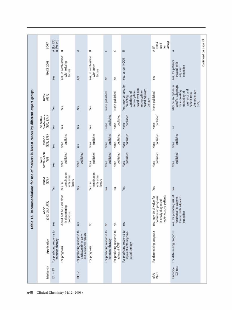
Tabl
e12
.Re
com
men
dati
ons
for
use
ofm
arke
rsin
brea
stca
ncer
bydi
ffer
ent
expe
rtgr
oups
.
Mar
ker(
s)A
pp
licat
ion
ASC
O(2
42,2
43,3
75)
EGTM
(371
)
Join
tEG
TM/N
AC
B(1
5)
ESM
Oa
(372
,373
)
StG
alle
nC
on
fere
nce
(350
,374
)N
CC
N(6
21)
NA
CB
2008
SOR
b
ER�
PRFo
rpr
edic
ting
resp
onse
toho
rmon
eth
erap
yYe
sYe
sYe
sYe
sYe
sYe
sYe
sA
(for
ER)
B(fo
rPR
)
For
prog
nosi
sSh
ould
not
beus
edal
one
inde
term
inin
gpr
ogno
sis
Yes,
inco
mbi
natio
nw
ithot
her
fact
ors
Non
e publ
ishe
dN
one publ
ishe
dYe
sYe
sYe
s,in
com
bina
tion
with
exis
ting
fact
ors
B
HER-
2Fo
rpr
edic
ting
resp
onse
totr
astu
zum
abin
early
and
adva
nced
dise
ase
Yes
Yes
Non
e publ
ishe
dYe
sYe
sYe
sYe
sA
For
prog
nosi
sN
oYe
s,in
com
bina
tion
with
othe
rfa
ctor
s
Non
e publ
ishe
dN
one publ
ishe
dYe
sYe
sYe
s,in
com
bina
tion
with
othe
rfa
ctor
s
B
For
pred
ictin
gre
spon
seto
horm
one
ther
apy
No
No
Non
e publ
ishe
dN
one publ
ishe
dN
one publ
ishe
dN
one
publ
ishe
dN
oC
For
pred
ictin
gre
spon
seto
adju
vant
CMF
No
No
Non
e publ
ishe
dN
one publ
ishe
dN
one publ
ishe
dN
one
publ
ishe
dN
oC
For
pred
ictin
gre
spon
seto
adju
vant
anth
racy
clin
e-ba
sed
ther
apy
Yes
Yes
Non
e publ
ishe
dN
one publ
ishe
dN
one publ
ishe
dYe
s,m
aybe
used
for
pred
ictin
gsu
perio
rity
ofan
thra
cycl
ine-
base
dov
erno
n-an
thra
cycl
ine-
base
dad
juva
ntth
erap
y
Yes,
aspe
rN
CCN
B
uPA/
PAI-1
For
dete
rmin
ing
prog
nosi
sYe
s,m
aybe
ofva
lue
for
dete
rmin
ing
prog
nosi
sin
new
lydi
agno
sed
node
-neg
ativ
epa
tient
s
Yes
Non
e publ
ishe
dN
one publ
ishe
dN
one publ
ishe
dN
one
publ
ishe
dYe
sA
(if ELIS
Aus
edfo
ras
say)
Onc
otyp
eDX
test
For
dete
rmin
ing
prog
nosi
sYe
s,fo
rpr
edic
ting
risk
ofre
curr
ence
inpa
tient
str
eate
dw
ithad
juva
ntta
mox
ifen
No
Non
e publ
ishe
dN
one publ
ishe
dN
oM
aybe
anop
tion
insp
ecifi
csu
bgro
ups
for
estim
atin
gpr
obab
ility
ofre
curr
ence
and
bene
fitfro
mch
emot
hera
py(6
22)
Yes,
for
patie
nts
trea
ted
with
adju
vant
tam
oxife
n
A
Cont
inue
don
page
49
e48 Clinical Chemistry 54:12 (2008)
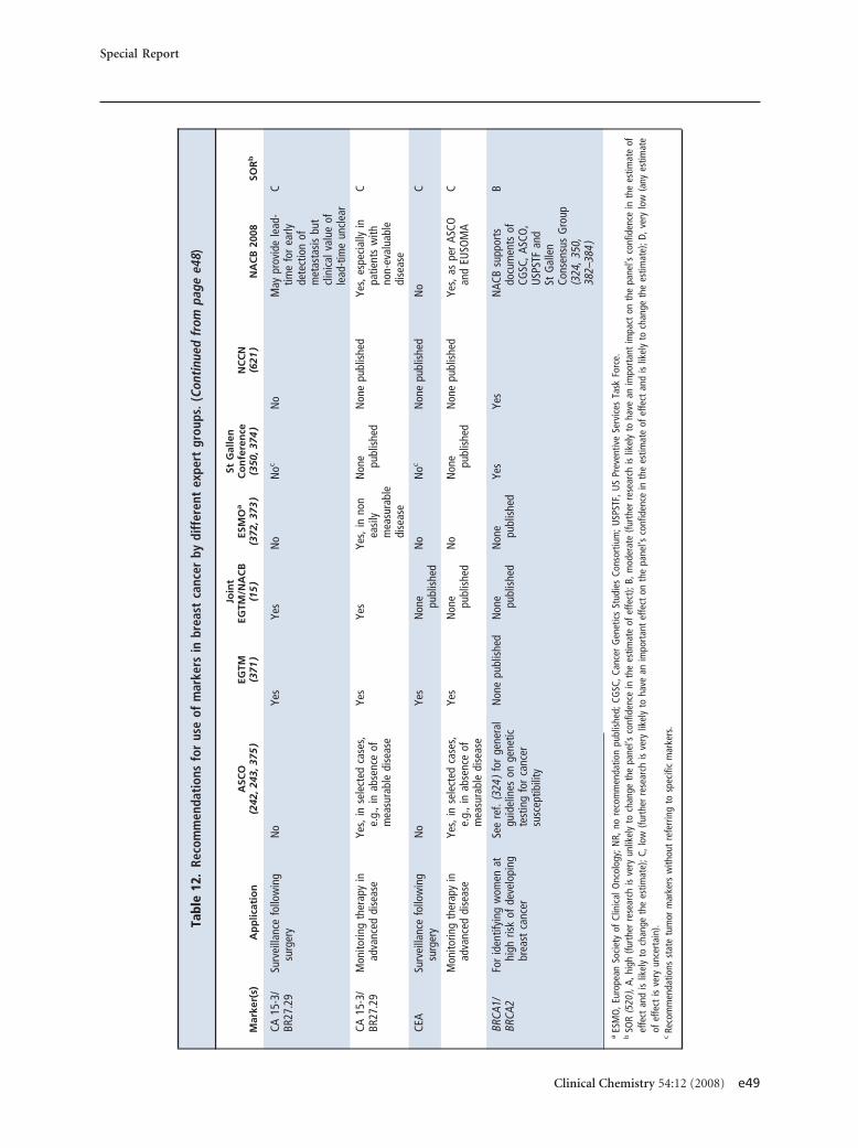
Tabl
e12
.Re
com
men
dati
ons
for
use
ofm
arke
rsin
brea
stca
ncer
bydi
ffer
ent
expe
rtgr
oups
.(Co
ntin
ued
from
page
e48)
Mar
ker(
s)A
pp
licat
ion
ASC
O(2
42,2
43,3
75)
EGTM
(371
)
Join
tEG
TM/N
AC
B(1
5)
ESM
Oa
(372
,373
)
StG
alle
nC
on
fere
nce
(350
,374
)N
CC
N(6
21)
NA
CB
2008
SOR
b
CA15
-3/
BR27
.29
Surv
eilla
nce
follo
win
gsu
rger
yN
oYe
sYe
sN
oN
ocN
oM
aypr
ovid
ele
ad-
time
for
early
dete
ctio
nof
met
asta
sis
but
clin
ical
valu
eof
lead
-tim
eun
clea
r
C
CA15
-3/
BR27
.29
Mon
itorin
gth
erap
yin
adva
nced
dise
ase
Yes,
inse
lect
edca
ses,
e.g.
,in
abse
nce
ofm
easu
rabl
edi
seas
e
Yes
Yes
Yes,
inno
nea
sily
mea
sura
ble
dise
ase
Non
e publ
ishe
dN
one
publ
ishe
dYe
s,es
peci
ally
inpa
tient
sw
ithno
n-ev
alua
ble
dise
ase
C
CEA
Surv
eilla
nce
follo
win
gsu
rger
yN
oYe
sN
one publ
ishe
dN
oN
ocN
one
publ
ishe
dN
oC
Mon
itorin
gth
erap
yin
adva
nced
dise
ase
Yes,
inse
lect
edca
ses,
e.g.
,in
abse
nce
ofm
easu
rabl
edi
seas
e
Yes
Non
e publ
ishe
dN
oN
one publ
ishe
dN
one
publ
ishe
dYe
s,as
per
ASCO
and
EUSO
MA
C
BRCA
1/BR
CA2
For
iden
tifyi
ngw
omen
athi
ghris
kof
deve
lopi
ngbr
east
canc
er
See
ref.
(324
)fo
rge
nera
lgu
idel
ines
onge
netic
test
ing
for
canc
ersu
scep
tibili
ty
Non
epu
blis
hed
Non
e publ
ishe
dN
one publ
ishe
dYe
sYe
sN
ACB
supp
orts
docu
men
tsof
CGSC
,ASC
O,
USP
STF
and
StG
alle
nCo
nsen
sus
Gro
up(3
24,3
50,
382–
384
)
B
aES
MO
,Eur
opea
nSo
ciet
yof
Clin
ical
Onc
olog
y;N
R,no
reco
mm
enda
tion
publ
ishe
d;CG
SC,C
ance
rG
enet
ics
Stud
ies
Cons
ortiu
m;U
SPST
F,U
SPr
even
tive
Serv
ices
Task
Forc
e.b
SOR
(520
),A,
high
(furt
her
rese
arch
isve
ryun
likel
yto
chan
geth
epa
nel’s
conf
iden
cein
the
estim
ate
ofef
fect
);B,
mod
erat
e(fu
rthe
rre
sear
chis
likel
yto
have
anim
port
ant
impa
cton
the
pane
l’sco
nfid
ence
inth
ees
timat
eof
effe
ctan
dis
likel
yto
chan
geth
ees
timat
e);C
,low
(furt
her
rese
arch
isve
rylik
ely
toha
vean
impo
rtan
tef
fect
onth
epa
nel’s
conf
iden
cein
the
estim
ate
ofef
fect
and
islik
ely
toch
ange
the
estim
ate)
;D,v
ery
low
(any
estim
ate
ofef
fect
isve
ryun
cert
ain)
.c
Reco
mm
enda
tions
stat
etu
mor
mar
kers
with
out
refe
rrin
gto
spec
ific
mar
kers
.
Special Report
Clinical Chemistry 54:12 (2008) e49
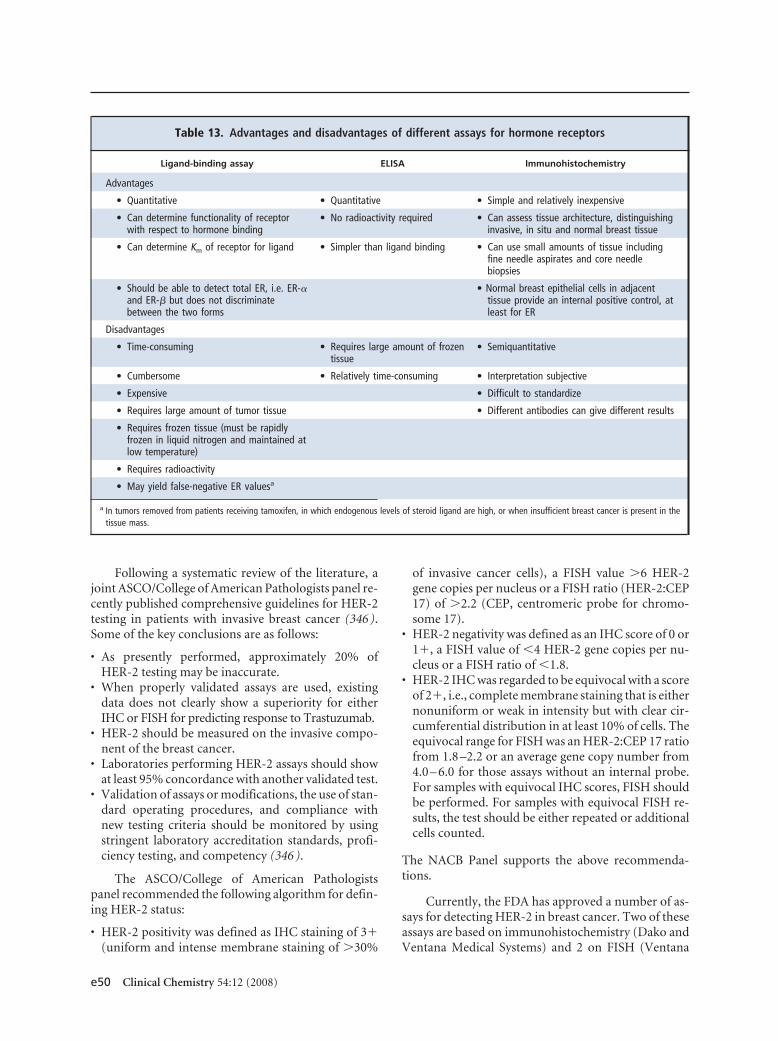
Following a systematic review of the literature, ajoint ASCO/College of American Pathologists panel re-cently published comprehensive guidelines for HER-2testing in patients with invasive breast cancer (346 ).Some of the key conclusions are as follows:
• As presently performed, approximately 20% ofHER-2 testing may be inaccurate.
• When properly validated assays are used, existingdata does not clearly show a superiority for eitherIHC or FISH for predicting response to Trastuzumab.
• HER-2 should be measured on the invasive compo-nent of the breast cancer.
• Laboratories performing HER-2 assays should showat least 95% concordance with another validated test.
• Validation of assays or modifications, the use of stan-dard operating procedures, and compliance withnew testing criteria should be monitored by usingstringent laboratory accreditation standards, profi-ciency testing, and competency (346 ).
The ASCO/College of American Pathologistspanel recommended the following algorithm for defin-ing HER-2 status:
• HER-2 positivity was defined as IHC staining of 3�(uniform and intense membrane staining of �30%
of invasive cancer cells), a FISH value �6 HER-2gene copies per nucleus or a FISH ratio (HER-2:CEP17) of �2.2 (CEP, centromeric probe for chromo-some 17).
• HER-2 negativity was defined as an IHC score of 0 or1�, a FISH value of �4 HER-2 gene copies per nu-cleus or a FISH ratio of �1.8.
• HER-2 IHC was regarded to be equivocal with a scoreof 2�, i.e., complete membrane staining that is eithernonuniform or weak in intensity but with clear cir-cumferential distribution in at least 10% of cells. Theequivocal range for FISH was an HER-2:CEP 17 ratiofrom 1.8 –2.2 or an average gene copy number from4.0 – 6.0 for those assays without an internal probe.For samples with equivocal IHC scores, FISH shouldbe performed. For samples with equivocal FISH re-sults, the test should be either repeated or additionalcells counted.
The NACB Panel supports the above recommenda-tions.
Currently, the FDA has approved a number of as-says for detecting HER-2 in breast cancer. Two of theseassays are based on immunohistochemistry (Dako andVentana Medical Systems) and 2 on FISH (Ventana
Table 13. Advantages and disadvantages of different assays for hormone receptors
Ligand-binding assay ELISA Immunohistochemistry
Advantages
• Quantitative • Quantitative • Simple and relatively inexpensive
• Can determine functionality of receptorwith respect to hormone binding
• No radioactivity required • Can assess tissue architecture, distinguishinginvasive, in situ and normal breast tissue
• Can determine Km of receptor for ligand • Simpler than ligand binding • Can use small amounts of tissue includingfine needle aspirates and core needlebiopsies
• Should be able to detect total ER, i.e. ER-�and ER-� but does not discriminatebetween the two forms
• Normal breast epithelial cells in adjacenttissue provide an internal positive control, atleast for ER
Disadvantages
• Time-consuming • Requires large amount of frozentissue
• Semiquantitative
• Cumbersome • Relatively time-consuming • Interpretation subjective
• Expensive • Difficult to standardize
• Requires large amount of tumor tissue • Different antibodies can give different results
• Requires frozen tissue (must be rapidlyfrozen in liquid nitrogen and maintained atlow temperature)
• Requires radioactivity
• May yield false-negative ER valuesa
a In tumors removed from patients receiving tamoxifen, in which endogenous levels of steroid ligand are high, or when insufficient breast cancer is present in thetissue mass.
e50 Clinical Chemistry 54:12 (2008)
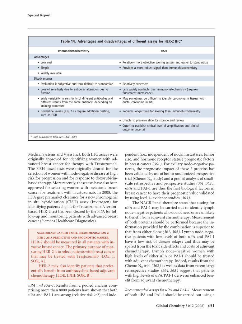
Medical Systems and Vysis Inc). Both IHC assays wereoriginally approved for identifying women with ad-vanced breast cancer for therapy with Trastuzumab.The FISH-based tests were originally cleared for theselection of women with node-negative disease at highrisk for progression and for response to doxorubicin-based therapy. More recently, these tests have also beenapproved for selecting women with metastatic breastcancer for treatment with Trastuzumab. In 2008, theFDA gave premarket clearance for a new chromogenicin situ hybridization (CISH) assay (Invitrogen) foridentifying patients eligible for Trastuzumab. A serum-based-HER-2 test has been cleared by the FDA for fol-low-up and monitoring patients with advanced breastcancer (Siemens Healthcare Diagnostics).
NACB BREAST CANCER PANEL RECOMMENDATION 2:
HER-2 AS A PREDICTIVE AND PROGNOSTIC MARKER
HER-2 should be measured in all patients with in-vasive breast cancer. The primary purpose of mea-suring HER-2 is to select patients with breast cancerthat may be treated with Trastuzumab [LOE, I;SOR, A].
HER-2 may also identify patients that prefer-entially benefit from anthracycline-based adjuvantchemotherapy [LOE, II/III; SOR, B].
uPA and PAI-1. Results from a pooled analysis com-prising more than 8000 patients have shown that bothuPA and PAI-1 are strong (relative risk �2) and inde-
pendent (i.e., independent of nodal metastases, tumorsize, and hormone receptor status) prognostic factorsin breast cancer (361 ). For axillary node–negative pa-tients, the prognostic impact of these 2 proteins hasbeen validated by use of both a randomized prospectivetrial (Chemo N0 study) and a pooled analysis of small-scale retrospective and prospective studies (361, 362 ).uPA and PAI-1 are thus the first biological factors inbreast cancer to have their prognostic value validatedby using level 1– evidence studies (363 ).
The NACB Panel therefore states that testing foruPA and PAI-1 may be carried out to identify lymphnode–negative patients who do not need or are unlikelyto benefit from adjuvant chemotherapy. Measurementof both proteins should be performed because the in-formation provided by the combination is superior tothat from either alone (361, 364 ). Lymph node–nega-tive patients with low levels of both uPA and PAI-1have a low risk of disease relapse and thus may bespared from the toxic side effects and costs of adjuvantchemotherapy. Lymph node–negative women withhigh levels of either uPA or PAI-1 should be treatedwith adjuvant chemotherapy. Indeed, results from theChemo N0 trial (362 ) as well as data from recent largeretrospective studies (364, 365 ) suggest that patientswith high levels of uPA/PAI-1 derive an enhanced ben-efit from adjuvant chemotherapy.
Recommended assays for uPA and PAI-1. Measurementof both uPA and PAI-1 should be carried out using a
Table 14. Advantages and disadvantages of different assays for HER-2 IHCa
Immunohistochemistry FISH
Advantages
• Low cost • Relatively more objective scoring system and easier to standardize
• Simple • Provides a more robust signal than immunohistochemistry
• Widely available
Disadvantages
• Evaluation is subjective and thus difficult to standardize • Relatively expensive
• Loss of sensitivity due to antigenic alteration due tofixation
• Less widely available than immunohistochemistry (requiresfluorescent microscope)
• Wide variability in sensitivity of different antibodies anddifferent results from the same antibody, depending onstaining procedure
• May sometimes be difficult to identify carcinoma in tissues withductal carcinoma in situ
• Borderline values (e.g. 2�) require additional testing,such as FISH
• Requires longer time for scoring than immunohistochemistry
• Unable to preserve slide for storage and review
• Cutoff to establish critical level of amplification and clinicaloutcome uncertain
a Data summarized from refs (354–360 ).
Special Report
Clinical Chemistry 54:12 (2008) e51
validated ELISA. A number of ELISAs have undergonetechnical validation (366 ), and some have also beenevaluated in an external quality assessment scheme(367 ). For determining prognosis in breast cancer, theNACB Panel recommends use of an ELISA that hasbeen both technically and clinically validated (e.g.,from American Diagnostic). Extraction of tumor tissuewith Triton X-100 is recommended (368 ). It is impor-tant to note that to perform an ELISA for uPA or PAI-1,a representative piece of fresh (i.e., not fixed in forma-lin) breast tumor (�200 –300 mg) must be storedin liquid nitrogen immediately after histologicaldiagnosis.
Recently, a microassay using as little as 100 mg oftumor tissue was described for the measurement ofuPA and PAI-1 (369, 370 ). This assay can also use ma-terial from 2 or 3 core biopsies or from 5–10 90-�m–thick cryosections. Although not yet clinically vali-dated, preliminary data showed that uPA and PAI-1levels in core biopsies correlated well with correspond-ing levels in surgically removed tissue. Because IHCdetermination of uPA/PAI-1 has not yet been clinicallyvalidated, this method cannot be recommended, atpresent, for the routine determination of these proteinsin breast cancer.
NACB BREAST CANCER PANEL RECOMMENDATION 3:
uPA AND PAI-1 FOR DETERMINING PROGNOSIS
uPA and PAI-1 may be used to identify lymph no-de–negative breast cancer patients who do not needor are unlikely to benefit from adjuvant chemother-apy. uPA and PAI-1 should be measured by a vali-dated ELISA using extracts of fresh or freshly frozentumor [LOE, I; SOR, A].
CA 15-3/BR 27.29. The CA 15-3 and BR 27.29 (alsoknown as CA 27.29) serum assays detect the same an-tigen, i.e., MUC1 protein, and provide similar clinicalinformation. CA 15-3 has, however, been more widelyinvestigated than BR 27.29. There are conflicting viewsabout the value of CA 15-3 and BR 27.29 in the post-operative surveillance of asymptomatic patients whohave undergone curative surgery for breast cancer(15, 242, 243, 371–375 ). Although increasing CA 15-3or BR 27.29 levels can preclinically detect distant met-astatic disease in approximately 70% of asymptomaticpatients, there is no high-level evidence study showingthat the early diagnosis of progressive disease followedby initiation of therapy positively impacts either pa-tient survival or quality of life. Furthermore, there is nouniversally accepted or clinically validated definition ofa clinically significant tumor marker increase. A con-
firmed increase of at least 25% however, is widely in-terpreted to signify a clinically significant increase.
Based on current evidence, the NACB Panel rec-ommends against routine CA 15-3 (or BR 27.29) test-ing in asymptomatic patients following diagnosis ofoperable breast cancer. The Panel, however, would liketo note that there are a number of small studies sug-gesting that the early initiation of therapy based on in-creasing serum markers levels can lead to an enhancedoutcome (376 –378 ). Although these studies do notprovide high-level evidence that early treatment basedon rising tumor marker levels positively impacts onpatient outcome, some doctors as well as some patientsmay wish to have serial levels of CA 15-3 (or BR 27.29)determined following primary surgery. The ultimatedecision about whether or not to use CA 15-3 (BR27.29) in this situation must be made by the doctor inconsultation with the patient.
According to both ASCO and NCCN, CA 15-3 (orBR 27.29) should not be used alone for monitoringtherapy in advanced disease (242, 243, 347, 375 ). TheEGTM Panel recommends that in patients with meta-static disease, markers should be determined beforeeach course of chemotherapy and at least every 3months for patients receiving hormone therapy (371 ).
The NACB Panel states that CA 15-3 or BR 27.29in combination with imaging and clinical examinationmay be used to monitor chemotherapy in patients withadvanced breast cancer. These markers may be partic-ularly helpful in patients with nonevaluable disease. Insuch patients, 2 successive increases (e.g., each �30%)are likely to indicate progressive disease and may resultin cessation of therapy, change in therapy, or entry ofthe patient into clinical trials evaluating new anticancertreatments. However, as with markers during postop-erative surveillance, there is no universally accepted orclinically validated definition of a clinically significantincrease in marker concentration during therapy of ad-vanced disease.
It is important to bear in mind that following theinitiation of chemotherapy, a transient increase in se-rum marker levels may occur (379, 380 ). Such tran-sient increases or spikes usually subside within 6 –12weeks after starting chemotherapy. Increases in markerlevels unrelated to tumor progression might also occuras a result of certain benign diseases (381 ). These in-creases may be transient or progressive depending onwhether the benign disease is short-lived or continuesto deteriorate.
Recommended assays for CA 15-3/BR 27.29. The FDAhas cleared a number of commercially available CA15-3 and BR 27.29 assays.
e52 Clinical Chemistry 54:12 (2008)
NACB BREAST CANCER PANEL RECOMMENDATION 4:
CA 15-3 AND BR 27.29 IN POSTOPERATIVE SURVEILLANCE
AND MONITORING THERAPY IN ADVANCED DISEASE
CA 15-3 and BR 27.29 should not be routinely usedfor the early detection of recurrences/metastases inasymptomatic patients with diagnosed breast can-cer. However, because some patients, as well assome doctors, may wish to have these measure-ments, the ultimate decision on whether or notto use CA 15-3 or BR 27.29 must be made by thedoctor in consultation with the patient [LOE, III;SOR, B].
In combination with radiology and clinical ex-amination, CA 15-3 or BR 27.29 may be used tomonitor chemotherapy in patients with advancedbreast cancer. For patients with nonevaluable dis-ease, sustained increases in marker concentrationssuggest progressive disease [LOE, III; SOR, B].
Carcinoembryonic antigen. As for CA 15-3 and BR27.29, the NACB Panel does not recommend routineuse of CEA in the surveillance of patients with diag-nosed breast cancer. For monitoring patients with ad-vanced disease, CEA should not be used alone. Formonitoring patients with nonevaluable disease, CEAmay occasionally be informative when CA 15-3/BR27.29 is not. As a marker for breast cancer, CEA is gen-erally less sensitive than CA 15-3/BR 27.29, but on oc-casion, CEA can be informative when levels of MUC-1-related markers remain below the cutoff point.
Recommended assay for CEA. The FDA has cleared anumber of commercially available CEA assays.
NACB BREAST CANCER PANEL RECOMMENDATION 5:
CEA IN POSTOPERATIVE SURVEILLANCE AND MONITORING
OF THERAPY IN ADVANCED DISEASE
CEA should not be routinely used for the early de-tection of recurrences/metastases in patients withdiagnosed breast cancer. However, because somepatients as well as some doctors may wish to havethese measurements, the ultimate decision onwhether or not to use CEA must be made by thedoctor in consultation with the patient [LOE, III;SOR, B].
In conjunction with radiology and clinical ex-amination, CEA may be used to monitor chemo-therapy in patients with advanced breast cancer. Inpatients with nonevaluable disease, sustained in-creases in CEA concentrations suggest progressivedisease [LOE, III; SOR, B].
BRCA1 and BRCA2 genes. According to the task forceof the Cancer Genetics Studies Consortium, earlybreast and ovarian cancer screening are recommendedfor individuals with breast cancer 1, early onset(BRCA1) mutations and early breast cancer screeningfor those with breast cancer 2, early onset (BRCA2)mutations (382 ). No recommendation, however, wasmade for or against prophylactic surgery (e.g., mastec-tomy or oophorectomy). The guidelines further statedthat “these surgeries are an option for mutation carri-ers, but evidence of benefit is lacking, and case reportshave documented the occurrence of cancer followingprophylactic surgery. It is recommended that individ-uals considering genetic testing be counselled regard-ing the unknown efficacy of measures to reduce riskand that care for individuals with cancer-predisposingmutations be provided whenever possible within thecontext of research protocols designed to evaluate clin-ical outcome” (382 ). It is important to point out thatthese guidelines were based on expert opinion only.
In 2003, an ASCO Panel published a detailed pol-icy statement regarding genetic testing for cancer sus-ceptibility (324 ). This statement included recommen-dations in the following areas: indications for genetictesting, regulation of testing, insurance reimburse-ment, protection from discrimination, confidentialityissues associated with genetic testing, and continuingeducational challenges and special research issues sur-rounding genetic testing of human tissues.
According to the 2005 Consensus Panel of the 8thSt Gallen Conference, treatment decisions for womenwith mutations in BRCA1 or BRCA2 genes “need toinclude consideration of bilateral mastectomy with plasticsurgical reconstruction, prophylactic oophorectomy,chemoprevention and intensified surveillance” (350).
The NACB Panel supports the statements pub-lished by the Cancer Genetics Studies Consortium,ASCO, US Preventive Services Task Force, and the StGallen Consensus Panel (324, 350, 382–384 ).
NACB BREAST CANCER PANEL RECOMMENDATION 6:
BRCA1 AND BRCA2 MUTATION TESTING FOR IDENTIFYING
WOMEN AT HIGH RISK OF DEVELOPING BREAST CANCER
BRCA1 and BRCA2 mutation testing may be usedfor identifying women who are at high risk of de-veloping breast or ovarian cancer in high-risk fam-ilies. For those with such mutations, screeningshould begin at 25–30 years of age. Insufficient dataexist, however, to recommend a specific surveil-lance/screening strategy for young women at highrisk. Appropriate counselling should be given toany individual considering BRCA1/2 testing [LOE,expert opinion; SOR, B].
Special Report
Clinical Chemistry 54:12 (2008) e53
MULTIGENE GENE SIGNATURES
Gene expression profiling. Gene expression profilinguses microarray technology to measure the simulta-neous expression of thousands of genes. At least 8 genesignatures have been described for predicting outcomein patients with breast cancer [for review, see ref(385 )]. Although these signatures contain few genesthat overlap, most give similar prognostic information(386 ).
In one of the first clinical microarray studies, van’tVeer et al. (387 ) described a 70-gene signature thatcorrectly predicted the later appearance of distant me-tastasis in 65 of 78 patients with newly diagnosedlymph node–negative breast cancer patients youngerthan 55 years who had not received systemic treatment.Application of this signature to an independent set of19 breast cancers resulted in only 2 incorrect classifica-tions. This 70-gene signature was subsequently bothinternally (388 ) and externally validated (389 ). In boththe internal and external validations studies, the prog-nostic impact of the gene signature was independent ofthe conventional prognostic factors for breast cancer.
Currently this 70-gene signature is undergoingprospective validation as part of the Microarrayfor Node-Negative Disease Avoids Chemotherapy(MINDACT) trial (390 ). The primary objective of thistrial is to establish if lymph node–negative breast cancerpatients with low risk of recurrence based on the abovegene signature but at high risk of recurrence based onclinicopathological factors can be safely spared adjuvantchemotherapy without affecting distant metastasis-freesurvival.
NACB BREAST CANCER PANEL RECOMMENDATION 7:
GENE EXPRESSION PROFILING, AS DETERMINED BY
MICROARRAY, FOR PREDICTING OUTCOME
None of the microarray-based gene signatures cur-rently available should be routinely used for pre-dicting patient outcome [LOE, III; SOR, B].
Oncotype DX™ test. Oncotype DX™ is a multigene as-say that quantifies the likelihood of breast cancer recur-rence in women with newly diagnosed, early-stagebreast cancer (for review, see (391 ). Rather than usingmicroarray technology, this test uses reverse-transcrip-tion PCR to measure the expression of 21 genes (16cancer-associated and 5 control genes). Based on theexpression of these genes, a recurrence score (RS) wascalculated that predicted low, intermediate, and highrisk of distant metastasis for ER-positive patientstreated with adjuvant tamoxifen (392 ). The RS wasprospectively validated in an independent populationof lymph node–negative ER-positive patients treatedwith adjuvant tamoxifen, as part of the National Surgi-
cal Adjuvant Breast and Bowel Project trial B14 (392 ).In this validation study, the RS was an independentpredictor of patient outcome. The independent prog-nostic impact of the RS was later confirmed in a popu-lation-based case-control study (393 ). Although a lowRS predicted good outcome in patients treated withadjuvant tamoxifen, a high RS was found to be associ-ated with favorable outcome in patients treated witheither neoadjuvant or adjuvant chemotherapy (394,395 ). A particular advantage of this test is that it may becarried out on formal-fixed paraffin embedded tissue.
Currently, the RS is undergoing prospective vali-dation as part of the Trial Assigning Individualized Op-tions for Treatment (TAILORx) trial (396 ). In thistrial, patients with intermediate RS are being random-ized to receive hormonal therapy alone or hormonetherapy plus chemotherapy. The aim is to establish ifadjuvant chemotherapy improves survival in the groupof patients with the intermediate score. Also, in thistrial, patients with low RS after tamoxifen therapy willreceive endocrine treatment, whereas those with highRS will be given chemotherapy and hormone therapy.
NACB BREAST CANCER PANEL RECOMMENDATION 8:
ONCOTYPE DX TEST FOR PREDICTING OUTCOME
The Oncotype DX test may be used for predictingrecurrence in lymph node–negative, ER-positivepatients receiving adjuvant tamoxifen. Patientspredicted to have a good outcome may be able toavoid having to undergo treatment with adjuvantchemotherapy [LOE, I/II; SOR, A].
The Oncotype DX test may also be used topredict benefit from adjuvant chemotherapy(cyclophosphamide-methotrexate-5-fluorouracil ormethotrexate-5-fluorouracil) in node-negative,ER-positive patients, i.e., patients with a high recur-rence score appear to derive greater benefit fromchemotherapy than those with low scores [LOE, III;SOR, B].
KEY POINTS: TUMOR MARKERS IN BREAST CANCER
The best-validated markers in breast cancer are all tis-sue based and include ER, PR, HER-2, uPA, and PAI-1.Assay of ER, PR, and HER-2 is now mandatory for allnewly diagnosed breast cancer patients. The measure-ment of uPA and PAI-1, although technically and clin-ically validated (361–363, 366, 367 ), is not presently inwidespread clinical use, mainly due to the requirementof a minimum amount of fresh or freshly frozen tissue.Assay of these proteins however, may be used to aid inthe selection of lymph node–negative breast cancer pa-tients who do not need adjuvant chemotherapy. Simi-larly, the Oncotype DX test may be used for predictingrecurrence in lymph node–negative, ER-positive pa-
e54 Clinical Chemistry 54:12 (2008)
tients receiving adjuvant tamoxifen. Although widelyused in postoperative surveillance and monitoringtherapy in advanced disease, the clinical value of CA15-3 and other serum markers has not yet been vali-dated by a level I evidence study.
Tumor Markers in Ovarian Cancer37,38
BACKGROUND
In the US, ovarian cancer is among the top 4 most lethalmalignant diseases in women, who have a lifetimeprobability of developing the disease of 1 in 59 (397 ).Worldwide, the incidence of ovarian cancer was esti-mated as 204 499 cases per year with a corresponding124 860 deaths (398 ).
The overall mortality of ovarian cancer is still highdespite new chemotherapeutic agents, which have sig-nificantly improved the 5-year survival rate (118 ). Themain reason for high mortality is lack of success indiagnosing ovarian cancer at an early stage, because thegreat majority of patients with advanced stage ovariancarcinoma die of the disease. In contrast, if ovariancancer is detected early, 90% of those with well-differ-entiated disease confined to the ovary survive. Further-more, biomarkers that can reliably predict clinical be-havior and response to treatment are generally lacking.The search for tumor markers for the early detectionand outcome prediction of ovarian carcinoma is there-fore of profound importance and is a subject of criticalimportance in the study of ovarian cancer.
Although ovarian cancer is often considered to bea single disease, it is composed of several related butdistinct tumor categories including surface epithelialtumors, sex-cord stromal tumors, and germ cell tu-mors (399 ). Within each category are several histolog-ical subtypes. Of these, epithelial tumors (carcinomas)are the most common and are divided, according to theInternational Federation of Gynecology and Obstetrics(FIGO) and WHO classifications, into 5 histologictypes: serous, mucinous, endometrioid, clear cell, andtransitional (400 ). The different types of ovarian can-cers are not only histologically distinct but are charac-terized by different clinical behavior, tumorigenesis,and pattern of gene expression. Based on prevalenceand mortality, serous ovarian carcinoma is the mostimportant and represents the majority of all primaryovarian carcinomas (401 ). Therefore, unless otherwisespecified, serous carcinoma is what is generally thoughtof as “ovarian cancer.”
The search for more effective biomarkers dependson a better understanding of the pathogenesis of ovar-ian cancer, i.e., the molecular events in its develop-ment. Based on a review of recent clinicopathologicaland molecular studies, a model for the developmentof ovarian carcinomas has been proposed (402 ). Inthis model, surface epithelial tumors are dividedinto 2 broad categories designated type I and type IItumors, which correspond to 2 main pathways oftumorigenesis.
Type I tumors tend to be low-grade neoplasmsthat arise in a stepwise fashion from borderline tumors,whereas type II tumors are high-grade neoplasms forwhich morphologically recognizable precursor lesionshave not been identified, so-called de novo develop-ment. Because serous tumors are the most commonsurface epithelial tumors, low-grade serous carcinomais the prototypic type I tumor, and high-grade serouscarcinoma is the prototypic type II tumor.
In addition to low-grade serous carcinomas, type Itumors are composed of mucinous carcinomas, endo-metrioid carcinomas, malignant Brenner tumors, andclear cell carcinomas. Type I tumors are associated withdistinct molecular changes that are rarely found in typeII tumors, such as BRAF39 and KRAS mutations forserous tumors, KRAS mutations for mucinous tumors,and �-catenin and PTEN mutations and MSI for endo-metrioid tumors.
Type II tumors include high-grade serous carci-noma, malignant mixed mesodermal tumors (carcino-sarcoma), and undifferentiated carcinoma. There arevery limited data on the molecular alterations associ-ated with type II tumors, except frequent p53 muta-tions in high-grade serous carcinomas and malignantmixed mesodermal tumors (carcinosarcomas). Thismodel of carcinogenesis provides a molecular platformfor the discovery of new ovarian cancer markers.
To prepare these guidelines, the literature relevantto the use of tumor markers in ovarian cancer was re-viewed. Particular attention was given to reviews in-cluding systematic reviews, prospective randomizedtrials that included the use of markers, and guidelinesissued by expert panels. Where possible, the consensusrecommendations of the NACB Panel were based onavailable evidence, i.e., were evidence-based.
CURRENTLY AVAILABLE MARKERS FOR OVARIAN CANCER
The most widely studied ovarian cancer body fluid–and tissue-based tumor markers for ovarian cancer arelisted in Table 15, which also summarizes the phase of
37 NACB Ovarian Cancer Sub-Committee Members: Daniel W. Chan, Chair; RobertC. Bast, Jr.; le-Ming Shih; Lori J. Sokoll; and Gyorgy Soletormos.
38 All comments received about the NACB Recommendations for Ovarian Cancerare included in the online Data Supplement. Professor Gordon Rustin, ProfessorBengt Tholander, and Professor M. Tuxen were invited expert reviewers.
39 BRAF, v-raf murine sarcoma viral oncogene homolog B1; KRAS, v-Ki-ras2Kirsten rat sarcoma viral oncogene homolog; PTEN, phosphatase and tensinhomolog.
Special Report
Clinical Chemistry 54:12 (2008) e55
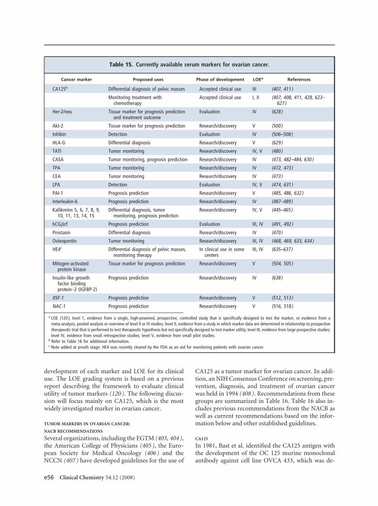
development of each marker and LOE for its clinicaluse. The LOE grading system is based on a previousreport describing the framework to evaluate clinicalutility of tumor markers (120 ). The following discus-sion will focus mainly on CA125, which is the mostwidely investigated marker in ovarian cancer.
TUMOR MARKERS IN OVARIAN CANCER:
NACB RECOMMENDATIONS
Several organizations, including the EGTM (403, 404 ),the American College of Physicians (405 ), the Euro-pean Society for Medical Oncology (406 ) and theNCCN (407 ) have developed guidelines for the use of
CA125 as a tumor marker for ovarian cancer. In addi-tion, an NIH Consensus Conference on screening, pre-vention, diagnosis, and treatment of ovarian cancerwas held in 1994 (408 ). Recommendations from thesegroups are summarized in Table 16. Table 16 also in-cludes previous recommendations from the NACB aswell as current recommendations based on the infor-mation below and other established guidelines.
CA125
In 1981, Bast et al. identified the CA125 antigen withthe development of the OC 125 murine monoclonalantibody against cell line OVCA 433, which was de-
Table 15. Currently available serum markers for ovarian cancer.
Cancer marker Proposed uses Phase of development LOEa References
CA125b Differential diagnosis of pelvic masses Accepted clinical use III (407, 411 )
Monitoring treatment withchemotherapy
Accepted clinical use I, II (407, 408, 411, 428, 623–627 )
Her-2/neu Tissue marker for prognosis predictionand treatment outcome
Evaluation IV (628 )
Akt-2 Tissue marker for prognosis prediction Research/discovery V (500 )
Inhibin Detection Evaluation IV (506–508 )
HLA-G Differential diagnosis Research/discovery V (629 )
TATI Tumor monitoring Research/discovery IV, V (480 )
CASA Tumor monitoring, prognosis prediction Research/discovery IV (473, 482–484, 630 )
TPA Tumor monitoring Research/discovery IV (472, 473 )
CEA Tumor monitoring Research/discovery IV (473 )
LPA Detection Evaluation IV, V (474, 631 )
PAI-1 Prognosis prediction Research/discovery V (485, 486, 632 )
Interleukin-6 Prognosis prediction Research/discovery IV (487–489 )
Kallikreins 5, 6, 7, 8, 9,10, 11, 13, 14, 15
Differential diagnosis, tumormonitoring, prognosis prediction
Research/discovery IV, V (445–465 )
hCG�cf Prognosis prediction Evaluation III, IV (491, 492 )
Prostasin Differential diagnosis Research/discovery IV (470 )
Osteopontin Tumor monitoring Research/discovery III, IV (468, 469, 633, 634 )
HE4c Differential diagnosis of pelvic masses,monitoring therapy
In clinical use in somecenters
III, IV (635–637 )
Mitogen-activatedprotein kinase
Tissue marker for prognosis prediction Research/discovery V (504, 505 )
Insulin-like growthfactor bindingprotein–2 (IGFBP-2)
Prognosis prediction Research/discovery IV (638 )
RSF-1 Prognosis prediction Research/discovery V (512, 513 )
NAC-1 Prognosis prediction Research/discovery V (516, 518 )
a LOE (120 ), level 1, evidence from a single, high-powered, prospective, controlled study that is specifically designed to test the marker, or evidence from ameta-analysis, pooled analysis or overview of level II or III studies; level II, evidence from a study in which marker data are determined in relationship to prospectivetherapeutic trial that is performed to test therapeutic hypothesis but not specifically designed to test marker utility; level III, evidence from large prospective studies;level IV, evidence from small retrospective studies; level V, evidence from small pilot studies.
b Refer to Table 16 for additional information.c Note added at proofs stage: HE4 was recently cleared by the FDA as an aid for monitoring patients with ovarian cancer.
e56 Clinical Chemistry 54:12 (2008)
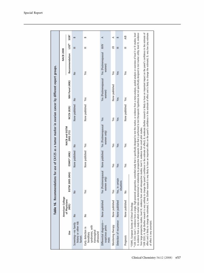
Tabl
e16
.Re
com
men
dati
ons
for
use
ofCA
125
asa
tum
orm
arke
rin
ovar
ian
canc
erby
diff
eren
tex
pert
grou
ps.
Use
Am
eric
anC
olle
ge
of
Phys
icia
ns
(405
)EG
TM20
05(4
04)
ESM
Oa
(406
)N
AC
Ban
dEG
TM20
02(1
5)
NC
CN
(639
)N
IHPa
nel
(408
)
NA
CB
2008
Rec
om
men
dat
ion
LOEb
SOR
c
Scre
enin
g—no
fam
ilyhi
stor
yor
othe
rris
kfa
ctor
s
No
No
Non
epu
blis
hed
No
Non
epu
blis
hed
No
No
IIIB
Early
dete
ctio
nin
here
dita
rysy
ndro
mes
,with
tran
svag
inal
ultr
asou
nd
No
Yes
Non
epu
blis
hed
Yes
Non
epu
blis
hed
Yes
Yes
IIIB
Diffe
rent
iald
iagn
osis
—su
spic
ious
pelv
icm
ass
Non
epu
blis
hed
Yes
(Pos
tmen
opau
sal
wom
enon
ly)
Non
epu
blis
hed
Yes
(Pos
tmen
opau
sal
wom
enon
ly)
Yes
Yes
(Pos
tmen
opau
sal
wom
en)
Yes
(Pos
tmen
opau
sal
wom
en)
III/IV
A
Mon
itorin
gth
erap
yN
one
publ
ishe
dYe
sYe
sYe
sYe
sN
one
publ
ishe
dYe
sI/I
IA
Dete
ctio
nof
recu
rren
ceN
one
publ
ishe
dYe
s,in
cert
ain
situ
atio
nsYe
sYe
sYe
sYe
sYe
sIII
B
Prog
nosi
sN
one
publ
ishe
dN
oYe
sYe
sN
one
publ
ishe
dYe
sYe
sIII
A/B
aES
MO
,Eur
opea
nSo
ciet
yof
Clin
ical
Onc
olog
y.b
LOE
(120
),le
vel1
,evi
denc
efro
ma
sing
le,h
igh-
pow
ered
,pro
spec
tive,
cont
rolle
dst
udy
that
issp
ecifi
cally
desi
gned
tote
stth
em
arke
r,or
evid
ence
from
am
eta-
anal
ysis
,poo
led
anal
ysis
orov
ervi
ewof
leve
lIIo
rIII
stud
ies;
leve
lII,
evid
ence
from
ast
udy
inw
hich
mar
ker
data
are
dete
rmin
edin
rela
tions
hip
topr
ospe
ctiv
eth
erap
eutic
tria
ltha
tis
perfo
rmed
tote
stth
erap
eutic
hypo
thes
isbu
tno
tsp
ecifi
cally
desi
gned
tote
stm
arke
rut
ility
;lev
elIII
,evi
denc
efro
mla
rge
pros
pect
ive
stud
ies;
leve
lIV,
evid
ence
from
smal
lret
rosp
ectiv
est
udie
s;le
velV
,evi
denc
efro
msm
allp
ilot
stud
ies.
cSO
R(5
20),
A,hi
gh(fu
rthe
rre
sear
chis
very
unlik
ely
toch
ange
the
pane
l’sco
nfid
ence
inth
ees
timat
eof
effe
ct);
B,m
oder
ate
(furt
her
rese
arch
islik
ely
toha
vean
impo
rtan
tim
pact
onth
epa
nel’s
conf
iden
cein
the
estim
ate
ofef
fect
and
islik
ely
toch
ange
the
estim
ate)
;C,l
ow(fu
rthe
rre
sear
chis
very
likel
yto
have
anim
port
ant
effe
cton
the
pane
l’sco
nfid
ence
inth
ees
timat
eof
effe
ctan
dis
likel
yto
chan
geth
ees
timat
e);D
,ver
ylo
w(a
nyes
timat
eof
effe
ctis
very
unce
rtai
n).
Special Report
Clinical Chemistry 54:12 (2008) e57
rived from a patient with ovarian serous carcinoma(409 ). The CA125 molecule has since been cloned byuse of a partial cDNA sequence originating from thepeptide core of the molecule identified (410 ). This newmucin molecule has been designated CA125/MUC16[mucin 16, cell surface associated (MUC16) gene] andconsists of a 156 –amino-acid tandem-repeat region inthe N-terminus and a possible transmembrane regionand tyrosine phosphorylation site in the C-terminus.
The first immunoassay for CA125, commercial-ized in 1983, used the OC 125 antibody for both cap-ture and detection (411, 412 ). A second-generationassay (CA125 II) was subsequently developed, incorpo-rating M11 and OC 125 antibodies, which have distinctnonoverlapping epitopes. Assays for CA125 have sincebeen adapted to automated platforms, and althoughthe majority of manufacturers quote a similar referenceinterval, concentrations of CA125 may vary amongmanufacturers owing to differences in calibration, as-say design, and reagent specificities. The lack of an In-ternational Standard for CA125 hampers progress inimproving between-method comparability, and theclinical and laboratory communities should work to-ward producing and adopting such a standard. For thepresent, values from different methods are not inter-changeable, and patients who are serially monitoredshould be rebaselined if there is a change in methodol-ogy (413 ). Manufacturers should specify the standardpreparation against which their method is calibrated,and laboratories should indicate the CA125 methodused on their clinical reports.
The cutoff of 35 kU/L for the CA125 and CA125IIassays was determined from the distribution of valuesin healthy individuals so as to include 99% of normals(414 ). Values tend to decline with menopause and ag-ing (415 ). It has recently been reported that CA125IIconcentrations vary 20%–50% by race in postmeno-pausal women, with concentrations in African andAsian women lower than in white women (415 ). Men-strual cycle variations can also be found (412 ). In-creased values may be found in 1%–2% of normalhealthy individuals, 5% of those with benign diseases,and 28% of those with nongynecologic cancers(15, 411, 412 ).
It is recommended that analysis be performedshortly after prompt centrifugation of the specimenand separation of serum from the clot, and that speci-mens be stored at either 4 °C (1–5 days) or �20 °C (2weeks–3 months) in the short term, or �70 °C in thelong term to ensure stability (15 ). Plasma is an accept-able specimen type for some assays, where indicated bythe manufacturer. As in other immunoassays, assay in-terferences may be observed if heterophilic antibodiesare present in the serum, particularly following thera-peutic or diagnostic use of monoclonal antibodies.
NACB OVARIAN CANCER PANEL RECOMMENDATION 1:
HANDLING OF SPECIMENS FOR CA125 DETERMINATION
Analysis should be performed shortly after promptcentrifugation of the specimen and separation ofserum from the clot, and specimens stored at either4 °C (1–5 days) or �20 °C (2 weeks–3 months) inthe short term or �70 °C in the long term [LOE,not applicable; SOR, A].
The recommendations of the current NACB paneland other groups with respect to the potential clinicalutility for CA125 are summarized in Table 16 and aredescribed below.
SCREENING/EARLY DETECTION
In women with epithelial ovarian cancer, 80% haveCA125 levels �35 kU/L, with elevations of 50%– 60%in clinically detected stage I disease, 90% in stage II, and�90% in stages III and IV (412, 416 ). Concentrationscorrelate with tumor burden and stage. Owing to thelack of sensitivity and specificity for a single determi-nation of the marker, CA125 is not recommended foruse in screening asymptomatic women by the NACBPanel or by other authoritative organizations(15, 403, 405– 408 ). An NIH Consensus DevelopmentPanel has concluded that evidence is not yet available toindicate that either CA125 or transvaginal ultrasonog-raphy effectively reduce mortality from ovarian cancer(408 ). However, the same panel did recommend an-nual CA125 determinations, in addition to pelvic andultrasound examinations, in women with a history ofhereditary ovarian cancer who have an estimated life-time risk of 40%, because early intervention may bebeneficial.
A number of approaches have been proposed toimprove the specificity of CA125 for early detection,because very high specificity (99.7%) is needed toachieve an acceptable positive predictive value of 10%with a prevalence of disease of 40 per 100 000 in womenolder than 50 years (417 ). Strategies have included se-quential or 2-stage strategies combining CA125 withultrasound, longitudinal measurements of CA125, andmeasurement of CA125 in combination with othermarkers such as OVX1, macrophage colony-stimulat-ing factor, or other new biomarkers discovered usingproteomic profiling approaches (411, 417– 419 ). Toevaluate the potential role for CA125 in screening forovarian cancer in asymptomatic populations, 2 majorprospective randomized trials are currently in progressin the US (420 ) and the United Kingdom (421 ). Intotal 200 000 women will be randomized to eitherscreening with ultrasound, screening with CA125 plusultrasound, or no screening. The studies are adequatelypowered to detect a significant improvement in sur-
e58 Clinical Chemistry 54:12 (2008)
vival among women screened with serial CA125 mea-surements and transvaginal sonography.
NACB OVARIAN CANCER PANEL RECOMMENDATION 2:
CA125 IN SCREENING
CA125 is not recommended for screening asymp-tomatic women [LOE, III; SOR, B].
CA125 is recommended, together with trans-vaginal ultrasound, for early detection of ovariancancer in women with hereditary syndromes be-cause early intervention may be beneficial in thesewomen [LOE, III; SOR B].
DISCRIMINATION OF PELVIC MASSES
In contrast to its use in early detection, CA125 is morewidely accepted as an adjunct in distinguishing benignfrom malignant disease in women, particularly in post-menopausal women presenting with ovarian masses(407, 408, 422 ), facilitating triage for operations by op-timally qualified surgeons. Benign conditions resultingin increased CA125 levels may be a confounding factorin premenopausal women. In the United KingdomCA125 measurement is an integral part of the RMI(risk of malignancy index), which forms the basis ofpatient pathway guidelines for the management of pel-vic masses and/or adnexal cysts (423 ). The RMI is cal-culated as a product of CA125 concentration multi-plied by menopausal status (1 for premenopausal and 3for postmenopausal) multiplied by ultrasound score(0, 1, or 3 depending on ultrasound features). A cutoffof 200 or 250 is frequently used, with patients withscores above this referred to specialist gynecology– on-cology teams. Sensitivities of 71%–78% and specifici-ties of 75%–94% have been reported in other studies(414 ). Increased concentrations of CA125 �95 kU/L inpostmenopausal women can discriminate malignantfrom benign pelvic masses with a positive predictivevalue of 95% (411 ). Therefore, based on current evi-dence, CA125 is recommended as an adjunct in distin-guishing benign from malignant pelvic masses, partic-ularly in postmenopausal women. When there is asuspicion of germ cell tumor, particularly in womenyounger than 40 years or in older women where scanfeatures suggest a germ cell tumor, AFP and hCG arealso important markers for triage, as for testicular germcell tumors.
NACB OVARIAN CANCER PANEL RECOMMENDATION 3:
CA125 IN DISCRIMINATION OF PELVIC MASSES
CA125 is recommended as an adjunct in distin-guishing benign from malignant suspicious pelvicmasses, particularly in postmenopausal women[LOE, III/IV; SOR, A].
MONITORING TREATMENT
Serial measurement of CA125 may also play a role inmonitoring response to chemotherapy. DecliningCA125 concentrations appear to correlate with treat-ment response even when disease is not detectable byeither palpation or imaging. In a meta-analysis, serialCA125 concentrations in 89% of 531 patients corre-lated with clinical outcome of disease (424 – 426 ).There is general consensus among current guidelines inrecommending that CA125 be used to monitor thera-peutic response, but there is no consensus as to howbest to define a CA125-based response (404, 427, 428 ).A response has been defined as a reduction of 50% ormore in pretreatment CA125 level that is maintainedfor at least 28 days (428 – 431 ). The pretreatment sam-ple must be at least twice the upper limit of the refer-ence range, which means that patients with pretreat-ment concentrations between the upper limit and twicethe upper limit are nonassessable by this criterion. Thefirst sample is recommended within 2 weeks beforetreatment, with subsequent samples at 2– 4 weeks dur-ing treatment and at intervals of 2–3 weeks during fol-low-up. The same assay method is required through-out, and patients who received immunotherapy(mouse antibodies) cannot be evaluated.
In addition to monitoring initial chemotherapeu-tic regiments, CA125 measurements may be useful inmonitoring salvage therapy, because a doubling of val-ues is associated with disease progression and treat-ment failure in more than 90% of cases (411 ). How-ever, disease progression may also occur without anincrease in CA125, and therefore the presence of tumorshould also be assessed by physical examination andimaging (15 ). Tuxen et al. (427 ) suggested that inter-pretation of changes in serial CA125 levels should bebased on a statistical estimation that takes account boththe analytical variation of the method used and of thenormal background intraindividual biological varia-tion of the marker (432, 433 ). The theoretical back-ground for this statistical procedure has recently beenreviewed in detail (434 ). Serial measurement of CA125to aid in monitoring response to therapy is a secondFDA-indicated use for the marker. Trials currently inprogress, including the UK Medical Research CouncilOV05 trial, have been designed to evaluate the benefitof early chemotherapy for recurrent ovarian cancer,based on a raised CA125 level alone, vs chemotherapybased on conventional clinical indicators (435 ). Pend-ing results of these trials, practice is likely to vary.
NACB OVARIAN CANCER PANEL RECOMMENDATION 4:
CA125 IN MONITORING TREATMENT
CA125 measurements may be used to monitor re-sponse to chemotherapeutic response. The first
Special Report
Clinical Chemistry 54:12 (2008) e59
sample should be taken within 2 weeks before treat-ment with subsequent samples at 2– 4 weeks duringtreatment and at intervals of 2–3 weeks during fol-low-up. The same assay method should be usedthroughout, and patients who received therapywith anti-CA125 antibodies cannot be evaluated[LOE, I/II; SOR, A].
CA125 MEASUREMENT POSTOPERATIVELY: SECOND-LOOK
OPERATION
Early studies on CA125 indicated that it was usefulpostoperatively in predicting the likelihood that tumorwould be found at a second-look operation, and CA125assays were initially cleared by the FDA for this indica-tion (412, 424 ). Elevations of CA125 � 35 kU/L afterdebulking surgery and chemotherapy indicate that re-sidual disease is likely (�95% accuracy) and that che-motherapy will be required (436 ). Second-look lapa-rotomy is now considered to be controversial andsuggested only for patients enrolled in clinical trials orin situations in which surgical findings would alterclinical management. Monitoring with CA125 testingin women with increased preoperative CA125 concen-trations, along with a routine history and physical andrectovaginal pelvic examination, has been advocatedinstead of surgery for asymptomatic women after pri-mary therapy (408 ).
CA125 MEASUREMENT POSTOPERATIVELY: DETECTION OF
RECURRENCE
Increased, rising, or doubling CA125 concentrationspredict relapse. However, it should be noted that post-operative CA125 levels below the cutoff concentrationdo not necessarily exclude disease presence.
The Gynecologic Cancer Intergroup (GCIG) isan organization consisting of representatives fromthirteen international groups performing clinical trialsin gynecologic cancer (437 ). The GCIG has definedcriteria progression using serial CA125 measurements(431 ) as:
• CA125 concentrations � twice the upper limit ofnormal on 2 occasions in patients with increasedCA125 levels pretreatment that normalize, or pa-tients with CA125 in the reference range or
• CA125 concentrations � the nadir value on 2 occa-sions in patients with increased CA125 levels pre-treatment that do not normalize.
The 2 measurements must be at least 1 week apart(431 ).
Although monitoring intervals are as yet undefined,current practice suggests following patients every 2 to 4months for 2 years and then less frequently (407 ). Ele-
vations in CA125 can precede clinical or radiologicalevidence of recurrence, with a median time of 2 to 6months, although there is no evidence to date that ini-tiating salvage chemotherapy before clinical recurrenceimproves survival (436 ). Early detection of recurrentdisease, however, permits the timely evaluation of themultiple drugs available for salvage therapy. Becauseonly a fraction of patients will respond to any singledrug and reliable predictive tests are not yet available,chemotherapeutic agents are generally used individu-ally and sequentially to identify those drugs that areactive against a particular patient’s cancer. Given themodest difference between time to recurrence andoverall survival, early detection of recurrence providestime in which to identify effective palliative therapy.Therefore, measurement of CA125 at follow-up visits isrecommended if values were initially increased. Lowpreoperative concentrations do not exclude the possi-bility that CA125 concentrations may increase abovethe cutoff before clinical relapse, and progressive in-creases in CA125 within the reference interval may bepredictive of recurrence (438 ).
NACB OVARIAN CANCER PANEL RECOMMENDATION 5:
CA125 IN MONITORING PATIENTS AFTER THERAPY
Measurement of CA125 at follow-up visits is rec-ommended if values were initially increased. Al-though monitoring intervals are as yet undefined,current practice suggests following patients every 2to 4 months for 2 years and then less frequently[LOE, III; SOR, B].
PROGNOSIS
CA125 is recommended during primary therapy as apotential prognostic marker because CA125 concen-trations, both preoperative and postoperative, may beof prognostic significance (439 – 442 ). After primarysurgery and chemotherapy, declines in CA125 concen-trations during chemotherapy have generally been ob-served to be independent prognostic factors, and insome studies the most important indicator. Persistentelevations indicate a poor prognosis. In patients whohad a preoperative CA125 concentration �65 kU/L,the 5-year survival rates were significantly lower andconferred a 6.37-fold risk of death compared to pa-tients who had values �65 kU/L (412, 426 ). In addi-tion to the measured level, the half-life of the CA125marker indicates prognosis after chemotherapy. A half-life of �20 days was associated with significantly im-proved survival (28 months vs 19 months) comparedto �20 days (411, 443 ). Improved survival also corre-lates with normalization of CA125 after 3 cycles ofcombination chemotherapy. These findings have beensupported by a recent study suggesting that CA125
e60 Clinical Chemistry 54:12 (2008)
half-life and CA125 nadir during induction chemo-therapy are independent predictors of epithelial ovar-ian cancer outcome (444 ).
NACB OVARIAN CANCER PANEL RECOMMENDATION 6:
CA125 IN PROGNOSIS
CA125 measurement during primary therapy isrecommended because CA125 concentrations,both preoperative and postoperative, may be ofprognostic significance. Persistent elevations indi-cate poor prognosis [LOE, III; SOR, A/B].
OTHER MARKERS FOR OVARIAN CANCER
Several other potential tumor-associated markers havebeen reported in body fluids and tissue specimens fromovarian cancer patients. Although these experimentalmarkers could represent promising new biomarkersfor future ovarian cancer screening, diagnosis, andmonitoring, it is uncertain whether they will becomeviable clinical tools, i.e., their clinical usefulness needsto be validated by assessing their sensitivity and speci-ficity in larger groups of patients with stage I disease.
The Kallikrein family. Kallikreins are a subgroup of theserine protease enzyme family that play an importantrole in the progression and metastasis of human can-cers (445 ). Kallikreins 4, 5, 6, 7, 8, 9, 10, 11, 13, 14, and15 in ovarian cancer have been shown to have value indetection, diagnosis, prognosis prediction, and moni-toring of ovarian cancer (446 – 463 ). Kallikrein 4, forexample, is expressed in the majority of serous carcino-mas but rarely in normal ovarian surface epithelium(449, 450 ). Kallikrein 4 expression is associated withhigher clinical stage and tumor grade in ovarian cancer:a univariate survival analysis revealed that patients withovarian tumors positive for kallikrein 4 expression hadan increased risk for relapse and death (450 ). Similarly,kallikrein 5 has been suggested to be a useful indepen-dent prognostic indicator in patients with stage I and IIdisease (451 ). Assessment of kallikrein 5 expressioncould help oncologists determine those patients athigher risk of relapse. Kallikrein 7 expression in ovariancancer tissue is associated with poorer prognosis ofovarian cancer patients, especially those with lowergrade disease and those who have been optimally de-bulked (464 ). In contrast, kallikrein 8 (neuropsin orovasin) (452 ), kallikrein 9 (465 ), and kallikrein 11(462 ) are favorable prognostic markers in ovarian can-cer. Patients with higher kallikrein 8 expression in theirtumors have lower-grade disease, lower residual tu-mor, longer survival, and low rate of recurrence. In amultivariate analysis, higher kallikrein 8 expressionwas significantly associated with longer disease-freesurvival. As well as their roles as tissue markers, kal-
likrein 6, 10, and 11 can be detected in serum, and arepotential serological markers of the disease(446, 448, 466 ). A recent comprehensive and parallelanalysis of different secreted kallikreins in ovarian can-cer has demonstrated that kallikreins 6, 7, 8, and 10 arethe 4 most specific secreted kallikreins in ovarian can-cer effusions (467 ). These kallikreins may have clinicalapplications in the differential diagnosis of ovarian car-cinoma from benign controls and other cancer types.
Osteopontin. Osteopontin was first identified by acDNA microarray approach used to identify upregu-lated genes in ovarian cancer cells, and osteopontin hasbeen found to be a potential diagnostic biomarker forovarian cancer (468 ). In the original report, osteopon-tin expression was higher in invasive ovarian cancerthan in borderline ovarian tumors, benign ovarian tu-mors, and normal ovarian surface epithelium (468 ).Plasma levels of osteopontin were significantly higherin patients with epithelial ovarian cancer compared tohealthy controls, patients with benign ovarian disease,and patients with other gynecologic cancers. In a morerecent report (469 ), osteopontin has been shown to beless sensitive than CA125 in predicting clinical re-sponse to therapy. However, osteopontin increasedearlier than CA125 in 90% of the study patients whodeveloped recurrent disease, indicating that osteopon-tin may be a clinically useful adjunct to CA125 in de-tecting recurrent ovarian cancer.
Prostasin. Using gene expression profiling by cDNAmicroarrays, Mok et al. have identified an overex-pressed gene called prostasin that produces a secretoryproduct (470 ). Prostasin [alias for PRSS8 (protease,serine, 8)] was originally isolated from human seminalfluid and its highest levels are found in the prostategland (471 ). Prostasin was detected more strongly inovarian carcinoma than in normal ovarian tissue. Themean level of serum prostasin was 13.7 �g/mL in pa-tients with ovarian cancer and 7.5 �g/mL in controlsubjects. In a series of patients with nonmucinousovarian carcinoma, the combination of prostasin andCA125 gave a sensitivity of 92% and a specificity of94% for detecting ovarian cancer. Although the abovefinding is promising, prostasin should be investigatedfurther as a screening or tumor marker, both alone andin combination with CA125.
Tissue polypeptide antigen. Tissue polypeptide antigen(TPA) is a single-chain polypeptide that may be madeup of proteolytic fragments of the cytokeratins (472 ).Production of TPA may be associated with rapid cellturnover, and increased TPA levels in serum have beenreported in patients suffering from cancers and otherdisease (473 ). In ovarian cancers of serous and muci-nous type, TPA levels correlate with FIGO stage; 33%–
Special Report
Clinical Chemistry 54:12 (2008) e61
50% of patients with stage I–II disease, and 88%–96%of patients with stage III–IV disease presented with in-creased serum TPA. Serial TPA measurements corre-lated with the clinical course of ovarian cancer in 42%–79% of the matched events. These findings suggest thatTPA may be a potential marker for following ovariancancer in patients.
Lysophosphatidic acid. Lysophosphatidic acid (LPA)was first identified in ascites of ovarian cancer patientsand has since been demonstrated to play a biologicalrole in ovarian cancer cell growth (474 – 477 ). In a pre-liminary study in a small number of patients (474 ),plasma LPA concentrations were increased in 90% ofpatients with stage I disease and 100% of patients withadvanced and recurrent disease compared to controlswithout apparent diseases, although 80% of womenwith other gynecologic cancers also had increased lev-els. CA125 concentrations appeared to complementLPA levels.
Tumor-associated trypsin inhibitor. Tumor-associatedtrypsin inhibitor (TATI) was first identified from theurine of patients with ovarian cancer (478 ). The aminoacid sequence and biochemical properties of TATI areidentical to those of pancreatic secretory trypsin inhib-itor (479 ). Increased serum and urinary concentra-tions of TATI are frequently observed in postoperativepatients, in severe inflammatory diseases, and in vari-ous types of cancer, especially gynecological and pan-creatic cancer (473 ). Increased concentrations of TATIcan be observed in ovarian cancers, especially the mu-cinous types. The increased serum levels of TATI ap-pear to correlate with higher stages of disease. In onereport, the sensitivity was only 8% in patients withstage I-II and 62% of patients with stage III-IV disease(480 ). Several reports suggest that TATI is not a goodmarker for monitoring disease during therapy, becauseTATI has a lower sensitivity for residual tumor thanCA125, and �50% of the matched clinical events areobserved to correlate serum levels of TATI.
Carcinoembryonic antigen. CEA is an oncofetal antigen(473 ), and increased serum levels of CEA are fre-quently found in a variety of benign diseases and can-cers, including ovarian carcinoma. The frequency ofincreased concentration in ovarian carcinoma varieswith the histological type and disease stage, generallybeing higher in patients with mucinous ovarian cancersand with metastatic disease. The sensitivity of CEA as amarker to detect ovarian cancer is approximately 25%,and the positive predictive value of an increased CEAconcentration is only 14% (473 ). Although CEA is nota marker for early diagnosis owing to its low sensitivity,CEA can be useful in determining treatment responsein ovarian cancer patients.
Cancer-associated serum antigen. Cancer-associated se-rum antigen (CASA) was initially defined by a mono-clonal antibody that bound to an epitope on the poly-morphic epithelial mucin (481 ). Increased CASA levelsin serum were found in individuals in the later stage ofpregnancy, in the elderly, in smokers, and in patientswith cancers. CASA is expressed in all histological typesof ovarian cancer and appears to have a sensitivity of46%–73% in patients with ovarian cancer (473 ). Onlya few studies have indicated that CASA is a potentiallyuseful marker in monitoring ovarian cancer. Ward etal. reported that inclusion of CASA in a diagnostic tu-mor panel might improve the detection of residual dis-ease by increasing the sensitivity from 33% to 62% andthe negative predictive value from 66% to 78%(482, 483 ). One study has demonstrated that CASAcan detect more cases with small volume disease thanCA125, and that 50% of patients with microscopic dis-ease are detected by CASA alone (473 ). Another studyhas shown that the prognostic value of postoperativeserum CASA level is superior to CA125 and other pa-rameters including residual disease, histological type,tumor grade, and the cisplatin-based chemotherapy(484 ).
PAI-1 and PAI-2. Fibrinolytic markers include PAI-1and PAI-2, for which diagnostic and prognostic valueshave recently been reported in ovarian cancer (485 ). Inthis pilot study, PAI-1 appeared to be a poor prognosticfactor (486 ), because plasma levels of PAI-1 are signifi-cantly higher in patients with ovarian cancer, andtheir levels correlate with the diseases at higher clin-ical stages. Whether PAI-1 can be used clinically forscreening and/or monitoring ovarian cancer awaitsfurther studies, including correlation with clinicaltreatment events and comparison with CA125. In con-trast, expression of PAI-2 in tumors has been shown tobe a favorable prognostic factor in ovarian cancer pa-tients (485 ).
Interleukin-6. High levels of interleukin-6 have beendetected in the serum and ascites of ovarian cancer pa-tients (487 ). Interleukin-6 correlates with tumor bur-den, clinical disease status, and survival time of patientswith ovarian cancer, implying that this marker may beuseful in diagnosis. Based on a multivariate analysis,investigators have found serum levels of interleukin-6to be of prognostic value, but less sensitive than CA125(488, 489 ).
Human chorionic gonadotropin. hCG normally is pro-duced by the trophoblast, and clinically has been usedas a serum or urine marker for pregnancy and gesta-tional trophoblastic disease (490 ). Ectopic hCG pro-duction, however, has been detected in a variety of hu-man cancers. Recent studies have demonstrated that
e62 Clinical Chemistry 54:12 (2008)
the immunoreactivity of total hCG in serum and urineprovides a strong independent prognostic factor inovarian carcinoma, and its prognostic value is similarto that of grade and stage (491, 492 ). When serum hCGis normal, the 5-year survival rate can be as high as80%, but it is only 22% when hCG is increased (491 ).In patients with stage III or IV and minimal residualdisease, the 5-year survival is 75% if hCG is not detect-able compared to 0% if hCG is increased. Similarly,hCG �-core fragment (hCG�cf) can be detected inurine in 84% of ovarian cancer patients (492 ). The in-cidence of positive urinary hCG�cf correlates with dis-ease progression, with elevations observed in a higherproportion of patients in advanced clinical stages. Al-though the availability of this marker before surgerycould facilitate selection of treatment modalities, theclinical application of hCG and hCG� for screeningand diagnosis is limited. Since several different types oftumors can produce hCG�hCG�, and only a smallproportion of ovarian tumors express these, detectionof serum hCG�hCG� or urinary hCG�cf will not pro-vide a specific or sensitive tool for screening or diagno-sis in ovarian cancer.
HER-2/neu gene. The c-erbB-2 oncogene expresses atransmembrane protein, p185, with intrinsic tyrosinekinase activity, also known as HER2/neu. Amplifica-tion of Her2/neu has been found in several human can-cers, including ovarian carcinoma. In ovarian cancer,9% to 38% of patients have increased levels of p105, theshed extracellular domain of the HER-2/neu protein(493– 495 ). According to one report, measurement ofHer2/neu alone or in combination with CA125 is notuseful for differentiating benign from malignant ovar-ian tumors (495 ). However, elevation of p105 in serumor the overexpression immunohistochemically ofHer2/neu in tumors has correlated with an aggressivetumor type, advanced clinical stages, and poor clinicaloutcome (496 ). Screening for increased p105 levelsmight therefore make it possible to identify a subset ofhigh-risk patients (494 ). Furthermore, the test couldbe potentially useful for detecting recurrent disease.
AKT2 gene. The v-akt murine thymoma viral oncogenehomolog 2 (AKT2) gene is one of the human homologsof v-akt, the transduced oncogene of the AKT8 virus,which experimentally induces lymphomas in mice.AKT2, which codes for a serine-threonine protein ki-nase, is activated by growth factors and other onco-genes such as v-Ha-ras and v-src through phosphati-dylinositol 3-kinase in human ovarian cancer cells(497, 498 ). Studies have shown that the AKT2 gene isamplified and overexpressed in approximately 12–36%of ovarian carcinomas (499 –501 ). In contrast, AKT2
alteration was not detected in 24 benign or borderlinetumors.
Ovarian cancer patients with AKT2 alterations ap-pear to have a poor prognosis. Amplification of AKT2is more frequently found in histologically high-gradetumors or tumors at advanced stages (III or IV), sug-gesting that AKT2 gene overexpression, like c-erbB-2,may be associated with tumor aggressiveness (500 ).
Mitogen-activated protein kinase. Activation of mito-gen-activated protein kinase occurs in response to var-ious growth stimulating signals and as a result of acti-vating mutations of the upstream regulators, KRASand BRAF, which can be found in many types of hu-man cancer. Activation of mitogen-activated proteinkinase activates downstream cellular targets (502, 503 )including a variety of cellular and nuclear proteins.Two studies have reported that expression of active mi-togen-activated protein kinase in ovarian cancer tissueor ascites cells correlates with better prognosis in theadvanced stage ovarian cancer (504, 505 ).
Inhibin. Inhibin is a glycoprotein and member of thetransforming growth factor � family. Inhibins A and Bare heterodimers consisting of identical �-subunitsand either �A or �B subunits linked with disulfidebonds (506 –508 ). Inhibin is primarily produced by thegonads and functions as a regulator of follicle-stimu-lating hormone secretion. Inhibin is associated withgranulosa cell tumors and mucinous carcinomas as op-posed to CA125, which is associated with serous, endo-metrioid, and undifferentiated tumors. In addition the� subunit may function as an ovarian tumor suppres-sor. Using a total inhibin ELISA in combination withCA125 has been shown to detect the majority of ovar-ian cancer types with 95% sensitivity and specificity(507 ).
RSF-1 gene. The clinical significance of the remodelingand spacing factor 1 (RSF-1) gene in ovarian cancerwas first demonstrated by analyzing a new amplifiedchromosomal region, 11q13.5, in the ovarian cancergenome by use of digital karyotyping. The RSF-1 genebelongs to the SWI/SNF chromatin remodelling genefamily and Rsf-1 protein partners with hSNF2h to formthe chromatin remodelling complex, RSF (remodellingand spacing factor) (509 ). It has been shown that Rsf-1participates in chromatin remodelling (509 ) and tran-scriptional regulation (510, 511 ). Previous studieshave demonstrated that RSF-1 amplification and over-expression are associated with the most aggressive typeof ovarian cancer, and patients with RSF-1 gene ampli-fication in their carcinomas had a significantly shorteroverall survival (512–514 ). Further multiinstitutionalstudies are required to validate the clinical significanceof RSF-1 gene amplification for future clinical practice.
Special Report
Clinical Chemistry 54:12 (2008) e63
NAC-1 gene.40 The genes within the BTB/POZ familyparticipate in several cellular functions including pro-liferation, apoptosis, transcription control, and cellmorphology maintenance (515 ). The roles of BTB/POZ proteins in human cancer have been recently re-vealed as several of BTB/POZ proteins such as BCL-6are involved in cancer development. Based on analyz-ing gene expression levels in all 130 deduced humanBTB/POZ genes using the serial analysis of gene expres-sion data, Nakayama et al. have recently identifiedNAC-1 as a carcinoma-associated BTB/POZ gene(516 ). NAC-1 is a transcription repressor and is in-volved in self-renewal and maintaining pluripotency ofembryonic stem cells (517 ). In ovarian carcinomas,NAC-1 is significantly over expressed in high-gradecarcinoma but not in borderline tumors or benign cys-tadenomas. The levels of NAC-1 expression correlatewith tumor recurrence in ovarian serous carcinomasand intense NAC-1 immunoreactivity in primary ovar-ian tumors predicts early recurrence (516, 518 ). As theNAC-1 specific antibody is available to evaluate NAC-1protein levels in archival paraffin sections, the markeralone or in combination with other biomarkers mayhold promise for prognosis and prediction in ovariancarcinoma patients.
NACB OVARIAN CANCER PANEL RECOMMENDATION 7:
TUMOR MARKERS OTHER THAN CA125
CA125 is the only marker that can be recom-mended for use in serous ovarian malignancies.New ovarian cancer markers offer promise; how-ever, their contribution to the current standard ofcare is unknown and further investigations in prop-erly designed clinical trials are needed [LOE, notapplicable; SOR, B].
KEY POINTS: TUMOR MARKERS IN OVARIAN CANCER
The NACB Panel recommends CA125 as the onlymarker for clinical use in ovarian cancer for the follow-ing indications: early detection in combination withtransvaginal ultrasound in hereditary syndromes, dif-ferential diagnosis in suspicious pelvic mass, detectionof recurrence, monitoring of therapy, and prognosis.The NACB Panel does not recommend CA125 forscreening of ovarian cancer in asymptomatic women.
All other markers are either in the evaluation phase orin the research/discovery phase, therefore the NACBPanel does not recommend these biomarkers for clin-ical use in ovarian cancer.
Author Contributions: All authors confirmed they have contributed tothe intellectual content of this paper and have met the following 3 re-quirements: (a) significant contributions to the conception and design,acquisition of data, or analysis and interpretation of data; (b) draftingor revising the article for intellectual content; and (c) final approval ofthe published article.
Authors’ Disclosures of Potential Conflicts of Interest: Uponmanuscript submission, all authors completed the Disclosures of Poten-tial Conflict of Interest form. Potential conflicts of interest:
Employment or Leadership: N. Brunner, Liplasome A/S; B. Dowell,Abbott Laboratories; H.G. Rittenhouse, Gen-Probe; P. Sibley, Sie-mens Medical Solutions Diagnostics.Consultant or Advisory Role: N. Brunner, DAKO A/S, Exiqon A/S,and Cancer Marker; R.J. Babaian, Endocare and Gen-probe; N. Har-beck, Roche Diagnostics; R.M. Lamerz, Abbott; A. Semjonow, Astel-las and BioMerieux; D. Hayes, StemCapturer, American Biosciences,Abraxis, Cytogen Corp., AviaraDx, Precision Therapeutics, Inc.,Monogram Bioscience, Siemens Medical Solutions, Pfizer, and Pre-dictive Biosciences.Stock Ownership: N. Brunner, Exiqon; B. Dowell, Abbott Laborato-ries; H.G. Rittenhouse, Gen-Probe.Honoraria: C.M. Sturgeon, (speaker expenses and/or honoraria),Abbott Diagnostics, Bayer Diagnostics, Beckman Coulter Diagnos-tics, Becton Dickinson Diagnostics, DAKO Ltd, DPC Ltd, Roche Di-agnostics, Tosoh Biosciences, and Wallac Oy Ltd; N. Brunner,DAKO A/S, EXIQON A/S, LIPLASOME A/S, Cancer Marker, andCancer marker; R.M. Lamerz, Euro 2000; A. Semjonow, Astra Zen-eca, Beckman Coulter, and Takeda Pharma.Research Funding: M.J. Duffy, Randox Laboratories; D. W. Chan,Vermillion; D.F. Hayes Astra Zeneca, Pfizer, Novartis, and GSK; G.Klee, Beckman Coulter, Inc; H.J. Nielsen Abbott Corp., Cancer CoreR&D, Chicago, IL, USA; L. Sokoll, Abbott Diagnostics, BeckmanCoulter, Dade Behring, DPC, Gen-Probe Inc, and Roche Diagnos-tics; D. Hayes, Astra-Zeneca, Glaxo Smith-Kline, Pfizer, Novartis,and Wyeth Ayerst-Genetics Institute; D. Chan, Abbott, Beckman,Ciphergen, Roche, and Tosoh.Expert Testimony: A. Semjonow, Siemens Medical.Other: M.J. Duffy, Roche Diagnostics, DPC Ltd, and American Di-agnostica; H. Lilja holds patents for free PSA and hK2 assays; D.Hayes, Scientific Collaborator-Genomics Health, Inc., Immunicon,Inc.
Role of Sponsor: The funding organizations played no role in thedesign of study, choice of enrolled patients, review and interpretationof data, or preparation or approval of manuscript.
Acknowledgments: We would like to thank the numerous scientistsand clinicians who have contributed to this undertaking, HassimaOmar Ali for her excellent assistance, Dr. David Bruns and Dr. NaderRifai for agreeing to consider publishing these guidelines in ClinicalChemistry, and of course the National Academy of Clinical Biochem-istry and the American Association of Clinical Chemistry for theirmuch appreciated support and encouragement.
40 NAC-1, alias for NACC1 (nucleus accumbens associated 1, BEN and BTB (POZ)domain containing).
e64 Clinical Chemistry 54:12 (2008)
References
1. Field M, Lorh K, editors. Clinical practiceguidelines: directions for a new program. Wash-ington (DC): National Academy Press, 1990; 168 p.
2. Diamandis EP, Hoffman BR, Sturgeon CM. Na-tional Academy of Clinical Biochemistry Labora-tory Medicine Practice Guidelines for the Use ofTumor Markers. Clin Chem 2008;54:1935–1939.
3. Sturgeon CM, Hoffman BR, Chan DW, Ch’ng S-L,Hammond E, Hayes DF, et al. National Academyof Clinical Biochemistry Laboratory MedicinePractice Guidelines for Use of Tumor Markers inClinical Practice: Quality Requirements. ClinChem 2008;54:e1–e10 [Epub 2008 Jul 7].
4. Bosl GJ, Motzer RJ. Testicular germ-cell cancer.N Engl J Med 1997;337:242–53.
5. Mead GM, Stenning SP. Prognostic factors inmetastatic non-seminomatous germ celltumours: the Medical Research Council studies.Eur Urol 1993;23:196–200.
6. Looijenga LH, Gillis AJ, van Gurp RJ, Verkerk AJ,Oosterhuis JW. X inactivation in human testicu-lar tumors: XIST expression and androgen recep-tor methylation status. Am J Pathol 1997;151:581–90.
7. Looijenga LH, Oosterhuis JW. Pathobiology oftesticular germ cell tumors: views and news.Anal Quant Cytol Histol 2002;24:263–79.
8. Woodward PJ, Heidenreich A, Looijenga LHJ,Oosterhuis JW, McLeod DG, Møller H, et al.Testicular germ cell tumors. In: Eble JN, SauterG, Epstein JI, Sesterhann IA, eds. World HealthOrganization classification of tumours: pathol-ogy and genetics of tumours of the urinarysystem and male genital organs. Lyon: IARCPress; 2004. p 217–278.
9. Oosterhuis JW, Looijenga LH, van Echten J, deJong B. Chromosomal constitution and develop-mental potential of human germ cell tumors andteratomas. Cancer Genet Cytogenet 1997;95:96–102.
10. Huyghe E, Matsuda T, Thonneau P. Increasingincidence of testicular cancer worldwide: a re-view. J Urol 2003;170:5–11.
11. McGlynn KA, Devesa SS, Sigurdson AJ, BrownLM, Tsao L, Tarone RE. Trends in the incidenceof testicular germ cell tumors in the UnitedStates. Cancer 2003;97:63–70.
12. Oliver RT, Mason MD, Mead GM, von der MaaseH, Rustin GJ, Joffe JK, et al. Radiotherapy versussingle-dose carboplatin in adjuvant treatment ofstage I seminoma: a randomised trial. Lancet2005;366:293–300.
13. Tumour markers in germ cell cancer: EGTM rec-ommendations. Anticancer Res 1999;19:2795–8.
14. Laguna MP, Pizzocaro G, Klepp O, Algaba F,Kisbenedek L, Leiva O. EAU guidelines on tes-ticular cancer. Eur Urol 2001;40:102–10.
15. Fleisher M, Dnistrian A, Sturgeon C, Lamerz R,Witliff J. Practice guidelines and recommenda-tions for use of tumor markers in the clinic.Tumor Markers: physiology, pathobiology, tech-nology and clinical applications. Washington(DC): AACC Press, 2002;33–63.
16. Schmoll HJ, Souchon R, Krege S, Albers P, BeyerJ, Kollmannsberger C, et al. European consensuson diagnosis and treatment of germ cell cancer:a report of the European Germ Cell Cancer
Consensus Group (EGCCCG). Ann Oncol 2004;15:1377–99.
17. Testicular seminoma: ESMO clinical recommen-dations for diagnosis, treatment and follow-up.Ann Oncol 2007;18 Suppl 2:ii40–1.
18. NCCN [National Comprehensive Cancer Net-work] clinical practice guidelines in oncology.Testicular cancer. Version 1. 2007. http://www.nccn.org/professionals/physician_gls/PDF/testicular.pdf (Accessed June 2007).
19. Mixed or non-seminomatous germ-cell tumors:ESMO clinical recommendations for diagnosis,treatment and follow-up. Ann Oncol 2007;18Suppl 2:ii42–3.
20. Albers P, Albrecht W, Algaba F, Bokemeyer C,Cohn-Cedermark G, Horwich A, et al. Guide-lines on testicular cancer. Eur Urol 2005;48:885–94.
21. Huddart R, Kataja V. Testicular seminoma:ESMO clinical recommendations for diagnosis,treatment and follow-up. Ann Oncol 2008;19Suppl 2:ii49–51.
22. Mostofi FK, Sesterhenn IA, Davis CJ Jr. Devel-opments in histopathology of testicular germcell tumors. Semin Urol 1988;6:171–88.
23. Pugh R. Combined tumours: pathology of testis.Oxford: Blackwell:, 1976;245–58.
24. Mostofi FK, Sesterhenn IA, Davis CJ Jr. Immu-nopathology of germ cell tumors of the testis.Semin Diagn Pathol 1987;4:320–41.
25. Skakkebaek NE. Possible carcinoma-in-situ ofthe testis. Lancet 1972;2:516–7.
26. Gondos B, Hobel CJ. Ultrastructure of germ celldevelopment in the human fetal testis. Z Zell-forsch Mikrosk Anat 1971;119:1–20.
27. Oosterhuis JW, Kersemaekers AM, Jacobsen GK,Timmer A, Steyerberg EW, Molier M, et al.Morphology of testicular parenchyma adjacentto germ cell tumours: an interim report. APMIS2003;111:32–40.
28. Rajpert-De ME, Skakkebaek NE. Expression ofthe c-kit protein product in carcinoma-in-situand invasive testicular germ cell tumours. Int JAndrol 1994;17:85–92.
29. van Gurp RJ, Oosterhuis JW, Kalscheuer V, Mari-man EC, Looijenga LH. Biallelic expression of theH19 and IGF2 genes in human testicular germ celltumors. J Natl Cancer Inst 1994;86:1070–5.
30. de Gouveia Brazao CA, Pierik FH, OosterhuisJW, Dohle GR, Looijenga LH, Weber RF. Bilateraltesticular microlithiasis predicts the presence ofthe precursor of testicular germ cell tumors insubfertile men. J Urol 2004;171:158–60.
31. Dieckmann KP, Classen J, Loy V. Diagnosis andmanagement of testicular intraepithelial neopla-sia (carcinoma in situ): surgical aspects. APMIS2003;111:64–8.
32. Motzer RJ, Rodriguez E, Reuter VE, SamaniegoF, Dmitrovsky E, Bajorin DF, et al. Genetic anal-ysis as an aid in diagnosis for patients withmidline carcinomas of uncertain histologies.J Natl Cancer Inst 1991;83:341–6.
33. Rao PH, Houldsworth J, Palanisamy N, MurtyVV, Reuter VE, Motzer RJ, et al. Chromosomalamplification is associated with cisplatin resis-tance of human male germ cell tumors. CancerRes 1998;58:4260–3.
34. Roelofs H, Mostert MC, Pompe K, Zafarana G,van Oorschot M, van Gurp RJ, et al. Restricted12p amplification and RAS mutation in humangerm cell tumors of the adult testis. Am J Pathol2000;157:1155–66.
35. Zafarana G, Gillis AJ, van Gurp RJ, Olsson PG,Elstrodt F, Stoop H, et al. Coamplification ofDAD-R, SOX5, and EKI1 in human testicularseminomas, with specific overexpression ofDAD-R, correlates with reduced levels of apo-ptosis and earlier clinical manifestation. CancerRes 2002;62:1822–31.
36. Spierings DC, de Vries EG, Vellenga E, de Jong S.The attractive Achilles heel of germ celltumours: an inherent sensitivity to apoptosis-inducing stimuli. J Pathol 2003;200:137–48.
37. Masters JR, Koberle B. Curing metastatic cancer:lessons from testicular germ-cell tumours. NatRev Cancer 2003;3:517–25.
38. Mayer F, Honecker F, Looijenga LH, BokemeyerC. Towards an understanding of the biologicalbasis of response to cisplatin-based chemother-apy in germ-cell tumors. Ann Oncol 2003;14:825–32.
39. Mayer F, Gillis AJ, Dinjens W, Oosterhuis JW,Bokemeyer C, Looijenga LH. Microsatellite insta-bility of germ cell tumors is associated withresistance to systemic treatment. Cancer Res2002;62:2758–60.
40. Velasco A, Riquelme E, Schultz M, Wistuba, II,Villarroel L, Koh MS, Leach FS. Microsatelliteinstability and loss of heterozygosity have dis-tinct prognostic value for testicular germ celltumor recurrence. Cancer Biol Ther 2004;3:1152–8; discussion 1159–61.
41. Velasco A, Riquelme E, Schultz M, Wistuba, II,Villarroel L, Pizarro J, et al. Mismatch repairgene expression and genetic instability in tes-ticular germ cell tumor. Cancer Biol Ther 2004;3:977–82.
42. Mueller T, Voigt W, Simon H, Fruehauf A, Bu-lankin A, Grothey A, Schmoll HJ. Failure ofactivation of caspase-9 induces a higher thresh-old for apoptosis and cisplatin resistance intesticular cancer. Cancer Res 2003;63:513–21.
43. van Echten J, Oosterhuis JW, Looijenga LH, vande Pol M, Wiersema J, te Meerman GJ, et al. Norecurrent structural abnormalities apart fromi(12p) in primary germ cell tumors of the adulttestis. Genes Chromosomes Cancer 1995;14:133–44.
44. Kawakami T, Okamoto K, Ogawa O, Okada Y.XIST unmethylated DNA fragments in male-de-rived plasma as a tumour marker for testicularcancer. Lancet 2004;363:40–2.
45. Strohmeyer T, Reissmann P, Cordon-Cardo C,Hartmann M, Ackermann R, Slamon D. Correla-tion between retinoblastoma gene expressionand differentiation in human testicular tumors.Proc Natl Acad Sci U S A 1991;88:6662–6.
46. Fan S, Chang JK, Smith ML, Duba D, Fornace AJJr, O’Connor PM. Cells lacking CIP1/WAF1 genesexhibit preferential sensitivity to cisplatin andnitrogen mustard. Oncogene 1997;14:2127–36.
47. Bartkova J, Thullberg M, Rajpert-De Meyts E,Skakkebaek NE, Bartek J. Cell cycle regulators intesticular cancer: loss of p18INK4C marks pro-
Special Report
Clinical Chemistry 54:12 (2008) e65
gression from carcinoma in situ to invasive germcell tumours. Int J Cancer 2000;85:370–5.
48. Mayer F, Stoop H, Scheffer GL, Scheper R, Oost-erhuis JW, Looijenga LH, Bokemeyer C. Molec-ular determinants of treatment response in hu-man germ cell tumors. Clin Cancer Res 2003;9:767–73.
49. Steyerberg EW, Keizer HJ, Habbema JD. Predic-tion models for the histology of residual massesafter chemotherapy for metastatic testicularcancer. ReHiT Study Group. Int J Cancer 1999;83:856–9.
50. Suurmeijer AJ, Oosterhuis JW, Sleijfer DT, KoopsHS, Fleuren GJ. Non-seminomatous germ celltumors of the testis: morphology of retroperito-neal lymph node metastases after chemother-apy. Eur J Cancer Clin Oncol 1984;20:727–34.
51. Rajpert-De Meyts E, Bartkova J, Samson M,Hoei-Hansen CE, Frydelund-Larsen L, Bartek J,Skakkebaek NE. The emerging phenotype of thetesticular carcinoma in situ germ cell. APMIS2003;111:267–78; discussion 278–9.
52. Bartkova J, Thullberg M, Rajpert-De Meyts E,Skakkebaek NE, Bartek J. Lack of p19INK4d inhuman testicular germ-cell tumours contrastswith high expression during normal spermato-genesis. Oncogene 2000;19:4146–50.
53. Bartkova J, Rajpert-De Meyts E, Skakkebaek NE,Lukas J, Bartek J. Deregulation of the G1/S-phase control in human testicular germ celltumours. Apmis 2003;111:252–65; discussion265–6.
54. Freedman LS, Parkinson MC, Jones WG, OliverRT, Peckham MJ, Read G, et al. Histopathologyin the prediction of relapse of patients withstage I testicular teratoma treated by orchidec-tomy alone. Lancet 1987;2:294–8.
55. Vergouwe Y, Steyerberg EW, Eijkemans MJ, Al-bers P, Habbema JD. Predictors of occult metas-tasis in clinical stage I nonseminoma: a system-atic review. J Clin Oncol 2003;21:4092–9.
56. Albers P, Siener R, Kliesch S, Weissbach L, KregeS, Sparwasser C, et al. Risk factors for relapse inclinical stage I nonseminomatous testiculargerm cell tumors: results of the German Testic-ular Cancer Study Group Trial. J Clin Oncol2003;21:1505–12.
57. Mazumdar M, Bacik J, Tickoo SK, Dobrzynski D,Donadio A, Bajorin D, et al. Cluster analysis ofp53 and Ki67 expression, apoptosis, alpha-feto-protein, and human chorionic gonadotrophinindicates a favorable prognostic subgroupwithin the embryonal carcinoma germ cell tu-mor. J Clin Oncol 2003;21:2679–88.
58. Looijenga LH, Stoop H, de Leeuw HP, deGouveia Brazao CA, Gillis AJ, van RoozendaalKE, et al. POU5F1 (OCT3/4) identifies cells withpluripotent potential in human germ cell tu-mors. Cancer Res 2003;63:2244–50.
59. Cheng L, Thomas A, Roth LM, Zheng W, MichaelH, Karim FW. OCT4: a novel biomarker for dys-germinoma of the ovary. Am J Surg Pathol2004;28:1341–6.
60. Jones TD, Ulbright TM, Eble JN, Baldridge LA,Cheng L. OCT4 staining in testicular tumors: asensitive and specific marker for seminoma andembryonal carcinoma. Am J Surg Pathol 2004;28:935–40.
61. Rajpert-De ME, Hanstein R, Jorgensen N, GraemN, Vogt PH, Skakkebaek NE. Developmental
expression of POU5F1 (OCT-3/4) in normal anddysgenetic human gonads. Hum Reprod 2004;19:1338–44.
62. Scottish Intercollegiate Guidelines Network(SIGN). Management of adult testicular germcell tumours. http://www.sign.ac.uk/ (AccessedOctober 2007). (SIGN 28; 1998 Sep)
63. Germa-Lluch JR, Garcia del Muro X, Maroto P,Paz-Ares L, Arranz JA, Guma J, et al. Clinicalpattern and therapeutic results achieved in 1490patients with germ-cell tumours of the testis:the experience of the Spanish Germ-Cell CancerGroup (GG). Eur Urol 2002;42:553–62; discus-sion 562–3.
64. Stenman UH, Alfthan H, Diamandis EP, FritscheHA, Lilja H, Chan DW, Schwartz MK. Markers fortesticular cancer. In: Diamandis EP, Fritsche HA,Chan DW, Schwartz MK, editors. Tumor markersphysiology, pathobiology, technology, and clin-ical applications. Washington (DC): AACC Press,2002;351–9.
65. Bosl GJ, Lange PH, Fraley EE, Goldman A, No-chomovitz LE, Rosai J, et al. Human chorionicgonadotropin and alphafetoprotein in the stag-ing of nonseminomatous testicular cancer. Can-cer 1981;47:328–32.
66. International Germ Cell Consensus Classifi-cation: a prognostic factor-based staging systemfor metastatic germ cell cancers. InternationalGerm Cell Cancer Collaborative Group. J ClinOncol 1997;15:594–603.
67. Vogelzang NJ. Prognostic factors in metastatictesticular cancer. Int J Androl 1987;10:225–37.
68. Davis BE, Herr HW, Fair WR, Bosl GJ. The man-agement of patients with nonseminomatousgerm cell tumors of the testis with serologicdisease only after orchiectomy. J Urol 1994;152:111–3; discussion 114.
69. Rabbani F, Sheinfeld J, Farivar-Mohseni H, LeonA, Rentzepis MJ, Reuter VE, et al. Low-volumenodal metastases detected at retroperitoneallymphadenectomy for testicular cancer: patternand prognostic factors for relapse. J Clin Oncol2001;19:2020–5.
70. Toner GC, Geller NL, Tan C, Nisselbaum J, BoslGJ. Serum tumor marker half-life during chemo-therapy allows early prediction of complete re-sponse and survival in nonseminomatous germcell tumors. Cancer Res 1990;50:5904–10.
71. Coogan CL, Foster RS, Rowland RG, Bihrle R,Smith ER Jr, Einhorn LH, et al. Postchemo-therapy retroperitoneal lymph node dissection iseffective therapy in selected patients with ele-vated tumor markers after primary chemother-apy alone. Urology 1997;50:957–62.
72. Vogelzang NJ, Lange PH, Goldman A, VesselaRH, Fraley EE, Kennedy BJ. Acute changes ofalpha-fetoprotein and human chorionic gonad-otropin during induction chemotherapy of germcell tumors. Cancer Res 1982;42:4855–61.
73. Kohn J. The dynamics of serum alpha-fetopro-tein in the course of testicular teratoma. ScandJ Immunol 1978;8(Suppl 8):103.
74. Lange PH, Vogelzang NJ, Goldman A, KennedyBJ, Fraley EE. Marker half-life analysis as aprognostic tool in testicular cancer. J Urol 1982;128:708–11.
75. Mazumdar M, Bajorin DF, Bacik J, Higgins G,Motzer RJ, Bosl GJ. Predicting outcome to che-motherapy in patients with germ cell tumors:
the value of the rate of decline of human cho-rionic gonadotrophin and alpha-fetoprotein dur-ing therapy. J Clin Oncol 2001;19:2534–41.
76. Seckl MJ, Rustin GJ, Bagshawe KD. Frequency ofserum tumour marker monitoring in patientswith non-seminomatous germ cell tumours. Br JCancer 1990;61:916–8.
77. Abelev GI. Alpha-fetoprotein as a marker ofembryo-specific differentiations in normal andtumor tissues. Transplant Rev 1974;20:3–37.
78. Gitlin D, Boesman M. Serum alpha-fetoprotein,albumin, and gamma-G-globulin in the humanconceptus. J Clin Invest 1966;45:1826–38.
79. Brewer JA, Tank ES. Yolk sac tumors and alpha-fetoprotein in first year of life. Urology 1993;42:79–80.
80. Blohm ME, Vesterling-Horner D, Calaminus G,Gobel U. Alpha 1-fetoprotein (AFP) referencevalues in infants up to 2 years of age. PediatrHematol Oncol 1998;15:135–42.
81. Carroll WL, Kempson RL, Govan DE, Freiha FS,Shochat SJ, Link MP. Conservative managementof testicular endodermal sinus tumor in child-hood. J Urol 1985;133:1011–4.
82. Huddart SN, Mann JR, Gornall P, Pearson D,Barrett A, Raafat F, et al. The UK Children’sCancer Study Group: testicular malignant germ celltumours 1979–1988. J Pediatr Surg 1990;25:406–10.
83. Christiansen M, Hogdall CK, Andersen JR, Nor-gaard-Pedersen B. Alpha-fetoprotein in plasmaand serum of healthy adults: preanalytical, an-alytical and biological sources of variation andconstruction of age-dependent reference inter-vals. Scand J Clin Lab Invest 2001;61:205–15.
84. Beck SD, Patel MI, Sheinfeld J. Tumor markerlevels in post-chemotherapy cystic masses: clin-ical implications for patients with germ cell tu-mors. J Urol 2004;171:168–71.
85. Germa JR, Llanos M, Tabernero JM, Mora J.False elevations of alpha-fetoprotein associatedwith liver dysfunction in germ cell tumors. Can-cer 1993;72:2491–4.
86. Morris MJ, Bosl GJ. Recognizing abnormal markerresults that do not reflect disease in patients withgerm cell tumors. J Urol 2000;163:796–801.
87. Ruoslahti E, Adamson E. Alpha-fetoproteins pro-duced by the yolk sac and the liver are glyco-sylated differently. Biochem Biophys Res Com-mun 1978;85:1622–30.
88. de Takats PG, Jones SR, Penn R, Cullen MH.Alpha-foetoprotein heterogeneity: what is itsvalue in managing patients with germ cell tu-mours? Clin Oncol (R Coll Radiol) 1996;8:323–6.
89. Perlin E, Engeler JE, Jr., Edson M, Karp D, McIn-tire KR, Waldmann TA. The value of serial mea-surement of both human chorionic gonadotro-pin and alpha-fetoprotein for monitoringgerminal cell tumors. Cancer 1976;37:215–9.
90. Butler SA, Ikram MS, Mathieu S, Iles RK. Theincrease in bladder carcinoma cell populationinduced by the free beta subunit of humanchorionic gonadotrophin is a result of an anti-apoptosis effect and not cell proliferation. Br JCancer 2000;82:1553–6.
91. Birken S, Yershova O, Myers RV, Bernard MP,Moyle W. Analysis of human choriogonado-tropin core 2 o-glycan isoforms. Mol Cell Endo-crinol 2003;204:21–30.
e66 Clinical Chemistry 54:12 (2008)
92. Vaitukaitis JL. Human chorionic gonadotropinas a tumor marker. Ann Clin Lab Sci 1974;4:276–80.
93. Birken S, Berger P, Bidart JM, Weber M, BristowA, Norman R, et al. Preparation and character-ization of new WHO reference reagents for hu-man chorionic gonadotropin and metabolites.Clin Chem 2003;49:144–54.
94. Berger P, Sturgeon C, Bidart JM, Paus E, GerthR, Niang M, et al. The ISOBM TD-7 Workshop onhCG and related molecules. Towards user-ori-ented standardization of pregnancy and tumordiagnosis: assignment of epitopes to the 3-di-mensional structure of diagnostically and com-mercially relevant monoclonal antibodies di-rected against human chorionic gonadotropinand derivatives. Tumour Biol 2002;23:1–38.
95. Bristow A, Berger P, Bidart JM, Birken S, Nor-man R, Stenman UH, Sturgeon C. Establishment,value assignment, and characterization of newWHO reference reagents for six molecular formsof human chorionic gonadotropin. Clin Chem2005;51:177–82.
96. Mann K, Saller B, Hoermann R. Clinical use ofHCG and hCG beta determinations. Scand J ClinLab Invest Suppl 1993;216:97–104.
97. Stenman UH, Alfthan H, Ranta T, Vartiainen E,Jalkanen J, Seppala M. Serum levels of humanchorionic gonadotropin in nonpregnant womenand men are modulated by gonadotropin-re-leasing hormone and sex steroids. J Clin Endo-crinol Metab 1987;64:730–6.
98. Alfthan H, Haglund C, Dabek J, Stenman UH.Concentrations of human choriogonadotropin,its beta-subunit, and the core fragment of thebeta-subunit in serum and urine of men andnonpregnant women. Clin Chem 1992;38:1981–7.
99. Lempiainen A, Hotakainen K, Blomqvist C, Alf-than H, Stenman UH. Increased human chori-onic gonadotropin due to hypogonadism aftertreatment of a testicular seminoma. Clin Chem2007;53:1560–1.
100. Catalona WJ, Vaitukaitis JL, Fair WR. Falselypositive specific human chorionic gonadotropinassays in patients with testicular tumors: con-version to negative with testosterone adminis-tration. J Urol 1979;122:126–8.
101. Stenman UH, Alfthan H, Hotakainen K. Humanchorionic gonadotropin in cancer. Clin Biochem2004;37:549–61.
102. Cole LA, Rinne KM, Shahabi S, Omrani A. False-positive hCG assay results leading to unneces-sary surgery and chemotherapy and needlessoccurrences of diabetes and coma. Clin Chem1999;45:313–4.
103. Saller B, Clara R, Spottl G, Siddle K, Mann K.Testicular cancer secretes intact human cho-riogonadotropin (hCG) and its free beta-subunit:evidence that hCG (�hCG-beta) assays are themost reliable in diagnosis and follow-up. ClinChem 1990;36:234–9.
104. Summers J, Raggatt P, Pratt J, Williams MV.Experience of discordant beta hCG results bydifferent assays in the management of non-seminomatous germ cell tumours of the testis.Clin Oncol (R Coll Radiol) 1999;11:388–92.
105. Li SS, Luedemann M, Sharief FS, Takano T,Deaven LL. Mapping of human lactate dehydro-genase-A, -B, and -C genes and their related
sequences: the gene for LDHC is located withthat for LDHA on chromosome 11. CytogenetCell Genet 1988;48:16–8.
106. Looijenga LH, Zafarana G, Grygalewicz B, Sum-mersgill B, biec-Rychter M, Veltman J, et al. Roleof gain of 12p in germ cell tumour development.APMIS 2003;111:161–71.
107. Rosenberg C, van Gurp RJ, Geelen E, OosterhuisJW, Looijenga LH. Overrepresentation of theshort arm of chromosome 12 is related to inva-sive growth of human testicular seminomas andnonseminomas. Oncogene 2000;19:5858–62.
108. Summersgill B, Osin P, Lu YJ, Huddart R, ShipleyJ. Chromosomal imbalances associated withcarcinoma in situ and associated testicular germcell tumours of adolescents and adults. Br JCancer 2001;85:213–20.
109. von Eyben FE. A systematic review of lactatedehydrogenase isoenzyme 1 and germ cell tu-mors. Clin Biochem 2001;34:441–54.
110. Fishman WH. Perspectives on alkaline phospha-tase isoenzymes. Am J Med 1974;56:617–50.
111. Roelofs H, Manes T, Janszen T, Millan JL, Oos-terhuis JW, Looijenga LH. Heterogeneity in al-kaline phosphatase isozyme expression in hu-man testicular germ cell tumours: an enzyme-/immunohistochemical and molecular analysis.J Pathol 1999;189:236–44.
112. Lange PH, Millan JL, Stigbrand T, Vessella RL,Ruoslahti E, Fishman WH. Placental alkalinephosphatase as a tumor marker for seminoma.Cancer Res 1982;42:3244–7.
113. De Broe ME, Pollet DE. Multicenter evaluationof human placental alkaline phosphatase as apossible tumor-associated antigen in serum.Clin Chem 1988;34:1995–9.
114. Mosselman S, Looijenga LH, Gillis AJ, vanRooijen MA, Kraft HJ, van Zoelen EJ, OosterhuisJW. Aberrant platelet-derived growth factor al-pha-receptor transcript as a diagnostic markerfor early human germ cell tumors of the adulttestis. Proc Natl Acad Sci U S A 1996;93:2884–8.
115. de Bruijn HW, Sleijfer DT, Schraffordt Koops H,Suurmeijer AJ, Marrink J, Ockhuizen T. Signifi-cance of human chorionic gonadotropin, alpha-fetoprotein, and pregnancy-specific beta-1-gly-coprotein in the detection of tumor relapse andpartial remission in 126 patients with nonsemi-nomatous testicular germ cell tumors. Cancer1985;55:829–35.
116. Kuzmits R, Schernthaner G, Krisch K. Serumneuron-specific enolase: a marker for re-sponses to therapy in seminoma. Cancer 1987;60:1017–21.
117. Fossa SD, Klepp O, Paus E. Neuron-specificenolase–a serum tumour marker in seminoma?Br J Cancer 1992;65:297–9.
118. Jemal A, Siegel R, Ward E, Murray T, Xu J, ThunMJ. Cancer statistics, 2007. CA Cancer J Clin2007;57:43–66.
119. Sakr WA, Grignon DJ. Prostate cancer: indica-tors of aggressiveness. Eur Urol 1997;32 Suppl3:15–23.
120. Hayes DF, Bast RC, Desch CE, Fritsche H, Jr.,Kemeny NE, Jessup JM, et al. Tumor markerutility grading system: a framework to evaluateclinical utility of tumor markers. J Natl CancerInst 1996;88:1456–66.
121. National Institute for Health and Clinical Excel-
lence (NICE). Prostate cancer: diagnosis andtreatment.http://www.nice.org.uk/guidance/index.jsp?action�byID&o�11924 (Accessed May2008).
122. Makinen T, Tammela TL, Hakama M, StenmanUH, Rannikko S, Aro J, et al. Tumor character-istics in a population-based prostate cancerscreening trial with prostate-specific antigen.Clin Cancer Res 2003;9:2435–9.
123. Draisma G, Boer R, Otto SJ, van der Cruijsen IW,Damhuis RA, Schroder FH, de Koning HJ. Leadtimes and overdetection due to prostate-specificantigen screening: estimates from the EuropeanRandomized Study of Screening for ProstateCancer. J Natl Cancer Inst 2003;95:868–78.
124. Punglia RS, D’Amico AV, Catalona WJ, RoehlKA, Kuntz KM. Effect of verification bias onscreening for prostate cancer by measurementof prostate-specific antigen. N Engl J Med 2003;349:335–42.
125. Stamey TA, Caldwell M, McNeal JE, Nolley R,Hemenez M, Downs J. The prostate specificantigen era in the United States is over forprostate cancer: what happened in the last 20years? J Urol 2004;172:1297–301.
126. Stenman UH, Hakama M, Knekt P, Aromaa A,Teppo L, Leinonen J. Serum concentrations ofprostate specific antigen and its complex withalpha 1-antichymotrypsin before diagnosis ofprostate cancer. Lancet 1994;344:1594–8.
127. Gann PH, Hennekens CH, Stampfer MJ. A pro-spective evaluation of plasma prostate-specificantigen for detection of prostatic cancer. Jama1995;273:289–94.
128. Thompson IM, Pauler DK, Goodman PJ, TangenCM, Lucia MS, Parnes HL, et al. Prevalence ofprostate cancer among men with a prostate-specific antigen level � or �4.0 ng per millili-ter. N Engl J Med 2004;350:2239–46.
129. Whittemore AS, Cirillo PM, Feldman D, CohnBA. Prostate specific antigen levels in youngadulthood predict prostate cancer risk: resultsfrom a cohort of Black and White Americans.J Urol 2005;174:872–6; discussion 876.
130. Lilja H, Ulmert D, Bjork T, Becker C, Serio AM,Nilsson JA, et al. Long-term prediction of pros-tate cancer up to 25 years before diagnosis ofprostate cancer using prostate kallikreins mea-sured at age 44 to 50 years. J Clin Oncol 2007;25:431–6.
131. Wirth MP, Frohmuller HG. Prostate-specific an-tigen and prostate acid phosphatase in the de-tection of early prostate cancer and the predic-tion of regional lymph node metastases. EurUrol 1992;22:27–32.
132. Kontturi M. Is acid phosphatase (PAP) still jus-tified in the management of prostatic cancer?Acta Oncol 1991;30:169–70.
133. Hugosson J, Aus G, Lilja H, Lodding P, Pihl CG.Results of a randomized, population-based studyof biennial screening using serum prostate-specificantigen measurement to detect prostate carci-noma. Cancer 2004;100:1397–405.
134. Oesterling JE, Jacobsen SJ, Klee GG, PetterssonK, Piironen T, Abrahamsson PA, et al. Free,complexed and total serum prostate specificantigen: the establishment of appropriate refer-ence ranges for their concentrations and ratios.J Urol 1995;154:1090–5.
135. Aus G, Damber JE, Khatami A, Lilja H, Stranne J,
Special Report
Clinical Chemistry 54:12 (2008) e67
Hugosson J. Individualized screening interval forprostate cancer based on prostate-specific anti-gen level: results of a prospective, randomized,population-based study. Arch Intern Med 2005;165:1857–61.
136. Catalona WJ, Richie JP, Ahmann FR, HudsonMA, Scardino PT, Flanigan RC, et al. Comparisonof digital rectal examination and serum prostatespecific antigen in the early detection of prostatecancer: results of a multicenter clinical trial of6,630 men. J Urol 1994;151:1283–90.
137. Crawford ED, Leewansangtong S, Goktas S,Holthaus K, Baier M. Efficiency of prostate-spe-cific antigen and digital rectal examination inscreening, using 4.0 ng/ml and age-specific ref-erence range as a cutoff for abnormal values.Prostate 1999;38:296–302.
138. Smith RA, Cokkinides V, Eyre HJ. American Can-cer Society Guidelines for the Early Detection ofCancer, 2005. CA Cancer J Clin 2005;55:31–44.
139. Graham J, Baker M, Macbeth F, Titshall V. Di-agnosis and treatment of prostate cancer: sum-mary of NICE guidance. BMJ 2008;336:610–2.
140. Wilt TJ. Commentary: controversies in NICEguidance on prostate cancer. BMJ 2008;336:612–4.
141. Myrtle JF, Klimley PG, Ivor IP, Bruni JF. Clinicalutility of prostate specific antigen (PSA) in themanagement of prostate cancer. San Diego:Hybritech; 1986. p. 1–5.
142. Catalona WJ, Smith DS, Ornstein DK. Prostatecancer detection in men with serum PSA con-centrations of 2.6 to 4.0 ng/mL and benignprostate examination: enhancement of specific-ity with free PSA measurements. JAMA 1997;277:1452–5.
143. Lodding P, Aus G, Bergdahl S, Frosing R, Lilja H,Pihl CG, Hugosson J. Characteristics of screen-ing detected prostate cancer in men 50 to 66years old with 3 to 4 ng./ml. Prostate specificantigen. J Urol 1998;159:899–903.
144. Thompson IM, Ankerst DP, Chi C, Goodman PJ,Tangen CM, Lucia MS, et al. Assessing prostatecancer risk: results from the Prostate CancerPrevention Trial. J Natl Cancer Inst 2006;98:529–34.
145. Gann PH. Interpreting recent trends in prostatecancer incidence and mortality. Epidemiology1997;8:117–20.
146. Oesterling JE, Jacobsen SJ, Chute CG, Guess HA,Girman CJ, Panser LA, Lieber MM. Serum pros-tate-specific antigen in a community-based pop-ulation of healthy men: establishment of age-specific reference ranges. JAMA 1993;270:860–4.
147. Catalona WJ, Smith DS. Comparison of differentserum prostate specific antigen measures forearly prostate cancer detection. Cancer 1994;74:1516–8.
148. Tumour markers in prostate cancer: EGTM rec-ommendations. European Group on TumourMarkers. Anticancer Res 1999;19:2799–801.
149. Partin AW, Brawer MK, Bartsch G, Horninger W,Taneja SS, Lepor H, et al. Complexed prostatespecific antigen improves specificity for prostatecancer detection: results of a prospective multi-center clinical trial. J Urol 2003;170:1787–91.
150. Pettersson K, Piironen T, Seppala M, LiukkonenL, Christensson A, Matikainen MT, et al. Freeand complexed prostate-specific antigen (PSA):
in vitro stability, epitope map, and developmentof immunofluorometric assays for specific andsensitive detection of free PSA and PSA-alpha1-antichymotrypsin complex. Clin Chem 1995;41:1480–8.
151. Babaian RJ, Fritsche HA, Evans RB. Prostate-specific antigen and prostate gland volume: cor-relation and clinical application. J Clin Lab Anal1990;4:135–7.
152. Veneziano S, Pavlica P, Querze R, Nanni G,Lalanne MG, Vecchi F. Correlation betweenprostate-specific antigen and prostate volume,evaluated by transrectal ultrasonography: use-fulness in diagnosis of prostate cancer. Eur Urol1990;18:112–6.
153. Benson MC, Whang IS, Pantuck A, Ring K,Kaplan SA, Olsson CA, Cooner WH. Prostatespecific antigen density: a means of distinguish-ing benign prostatic hypertrophy and prostatecancer. J Urol 1992;147:815–6.
154. Carter HB, Pearson JD, Metter EJ, Brant LJ, ChanDW, Andres R, et al. Longitudinal evaluation ofprostate-specific antigen levels in men with andwithout prostate disease. JAMA 1992;267:2215–20.
155. Schmid HP, McNeal JE, Stamey TA. Observationson the doubling time of prostate cancer: the useof serial prostate-specific antigen in patientswith untreated disease as a measure of increas-ing cancer volume. Cancer 1993;71:2031–40.
156. Semjonow A, Schmid HP. The rise and fall ofPSA: clinical implications of prostate specificantigen kinetics. Urol Res 2002;30:85–8.
157. Lilja H, Christensson A, Dahlen U, MatikainenMT, Nilsson O, Pettersson K, Lovgren T. Pros-tate-specific antigen in serum occurs predomi-nantly in complex with alpha 1-antichymotryp-sin. Clin Chem 1991;37:1618–25.
158. Stenman UH, Leinonen J, Alfthan H, Rannikko S,Tuhkanen K, Alfthan O. A complex betweenprostate-specific antigen and alpha 1-antichy-motrypsin is the major form of prostate-specificantigen in serum of patients with prostaticcancer: assay of the complex improves clinicalsensitivity for cancer. Cancer Res 1991;51:222–6.
159. Catalona WJ, Partin AW, Finlay JA, Chan DW,Rittenhouse HG, Wolfert RL, Woodrum DL. Useof percentage of free prostate-specific antigento identify men at high risk of prostate cancerwhen PSA levels are 2.51 to 4 ng/mL and digitalrectal examination is not suspicious for prostatecancer: an alternative model. Urology 1999;54:220–4.
160. Catalona WJ, Partin AW, Slawin KM, BrawerMK, Flanigan RC, Patel A, et al. Use of thepercentage of free prostate-specific antigen toenhance differentiation of prostate cancer frombenign prostatic disease: a prospective multi-center clinical trial. JAMA 1998;279:1542–7.
161. Raaijmakers R, Blijenberg BG, Finlay JA, Ritten-house HG, Wildhagen MF, Roobol MJ, SchroderFH. Prostate cancer detection in the prostatespecific antigen range of 2.0 to 3.9 ng/ml: valueof percent free prostate specific antigen on tu-mor detection and tumor aggressiveness. J Urol2004;171:2245–9.
162. Stephan C, Lein M, Jung K, Schnorr D, LoeningSA. The influence of prostate volume on theratio of free to total prostate specific antigen in
serum of patients with prostate carcinoma andbenign prostate hyperplasia. Cancer 1997;79:104–9.
163. Roddam AW, Duffy MJ, Hamdy FC, Ward AM,Patnick J, Price CP, et al. Use of prostate-specificantigen (PSA) isoforms for the detection of pros-tate cancer in men with a PSA level of 2–10ng/ml: systematic review and meta-analysis. EurUrol 2005;48:386–98; discussion 398–9.
164. Lee R, Localio AR, Armstrong K, Malkowicz SB,Schwartz JS. A meta-analysis of the perfor-mance characteristics of the free prostate-spe-cific antigen test. Urology 2006;67:762–8.
165. Oberpenning F, Weining C, Brandt B, De AG,Heinecke A, Hamm M, et al. Combining free andtotal prostate specific antigen assays from dif-ferent manufacturers: the pitfalls. Eur Urol2002;42:577–82.
166. Lilja H, Haese A, Bjork T, Friedrich MG, PiironenT, Pettersson K, et al. Significance and metab-olism of complexed and noncomplexed prostatespecific antigen forms, and human glandularkallikrein 2 in clinically localized prostate cancerbefore and after radical prostatectomy. J Urol1999;162:2029–34.
167. Allard WJ, Zhou Z, Yeung KK. Novel immunoas-say for the measurement of complexed pros-tate-specific antigen in serum. Clin Chem 1998;44:1216–23.
168. Finne P, Zhang WM, Auvinen A, Leinonen J,Maattanen L, Rannikko S, et al. Use of thecomplex between prostate specific antigen andalpha 1-protease inhibitor for screening prostatecancer. J Urol 2000;164:1956–60.
169. Lein M, Jung K, Hammerer P, Graefen M, Sem-jonow A, Stieber P, et al. A multicenter clinicaltrial on the use of alpha1-antichymotrypsin-prostate-specific antigen in prostate cancer di-agnosis. Prostate 2001;47:77–84.
170. Lein M, Kwiatkowski M, Semjonow A, LuboldtHJ, Hammerer P, Stephan C, et al. A multicenterclinical trial on the use of complexed prostatespecific antigen in low prostate specific antigenconcentrations. J Urol 2003;170:1175–9.
171. Okihara K, Fritsche HA, Ayala A, Johnston DA,Allard WJ, Babaian RJ. Can complexed prostatespecific antigen and prostatic volume enhanceprostate cancer detection in men with totalprostate specific antigen between 2.5 and 4.0ng./ml. J Urol 2001;165:1930–6.
172. Keller T, Butz H, Lein M, Kwiatkowski M, Sem-jonow A, Luboldt HJ, et al. Discordance analysischaracteristics as a new method to compare thediagnostic accuracy of tests: example of com-plexed versus total prostate-specific antigen.Clin Chem 2005;51:532–9.
173. Moul JW, Sesterhenn IA, Connelly RR, DouglasT, Srivastava S, Mostofi FK, McLeod DG. Pros-tate-specific antigen values at the time of pros-tate cancer diagnosis in African-American men.JAMA 1995;274:1277–81.
174. National Health Service (NHS). NHS cancerscreening programmes: prostate cancer riskmanagement. http://www.cancerscreening.nhs.uk/prostate/index.html (Accessed October 2007).
175. Evans R, Edwards A, Brett J, Bradburn M,Watson E, Austoker J, Elwyn G. Reduction inuptake of PSA tests following decision aids:systematic review of current aids and their eval-uations. Patient Educ Couns 2005;58:13–26.
e68 Clinical Chemistry 54:12 (2008)
176. Sturgeon C. Practice guidelines for tumor markeruse in the clinic. Clin Chem 2002;48:1151–9.
177. Zoorob R, Anderson R, Cefalu C, Sidani M.Cancer screening guidelines. Am Fam Physician2001;63:1101–12.
178. Harris R, Lohr KN. Screening for prostate cancer:an update of the evidence for the U.S. Preven-tive Services Task Force. Ann Intern Med 2002;137:917–29.
179. Bangma CH, Roemeling S, Schroder FH. Overdi-agnosis and overtreatment of early detectedprostate cancer. World J Urol 2007;25:3–9.
180. Schroder FH, Denis LJ, Roobol M, Nelen V, Au-vinen A, Tammela T, et al. The story of theEuropean Randomized Study of Screening forProstate Cancer. BJU Int 2003;92 Suppl 2:1–13.
181. de Koning HJ, Liem MK, Baan CA, Boer R,Schroder FH, Alexander FE. Prostate cancer mor-tality reduction by screening: power and timeframe with complete enrollment in the Euro-pean Randomised Screening for Prostate Cancer(ERSPC) trial. Int J Cancer 2002;98:268–73.
182. Andriole GL, Levin DL, Crawford ED, GelmannEP, Pinsky PF, Chia D, et al. Prostate CancerScreening in the Prostate, Lung, Colorectal andOvarian (PLCO) Cancer Screening Trial: findingsfrom the initial screening round of a randomizedtrial. J Natl Cancer Inst 2005;97:433–8.
183. Bartsch G, Horninger W, Klocker H, Reissigl A,Oberaigner W, Schonitzer D, et al. Prostate can-cer mortality after introduction of prostate-spe-cific antigen mass screening in the Federal Stateof Tyrol, Austria. Urology 2001;58:417–24.
184. Oberaigner W, Horninger W, Klocker H,Schonitzer D, Stuhlinger W, Bartsch G. Reduc-tion of prostate cancer mortality in Tyrol, Aus-tria, after introduction of prostate-specific anti-gen testing. Am J Epidemiol 2006;164:376–84.
185. Xia Z, Jacobsen SJ, Bergstralh EJ, Chute CG,Katusic SK, Lieber MM. Secular changes in rad-ical prostatectomy utilization rates in OlmstedCounty, Minnesota 1980 to 1995. J Urol 1998;159:904–8.
186. Collin SM, Martin RM, Metcalfe C, Gunnell D,Albertsen PC, Neal D, et al. Prostate-cancermortality in the USA and UK in 1975–2004: anecological study. Lancet Oncol 2008;9:445–52.
187. Hugosson J, Aus G, Becker C, Carlsson S, Eriks-son H, Lilja H, et al. Would prostate cancerdetected by screening with prostate-specific an-tigen develop into clinical cancer if left undiag-nosed? A comparison of two population-basedstudies in Sweden. BJU Int 2000;85:1078–84.
188. Ilic D, O’Connor D, Green S, Wilt T.. Screeningfor prostate cancer: a Cochrane systematic re-view. Cancer Causes Control 2006;18:279–285.
189. Hammerer P, Graefen M, Henke RP, Haese A,Palisaar J, Huland E, Huland H. Analysis ofmolecular isoforms of PSA and their ratios inmen with PSA-relapse after radical prostatec-tomy. Anticancer Res 2000;20:5253–5.
190. Hsu CY, Joniau S, Oyen R, Roskams T, VanPoppel H. Outcome of surgery for clinical uni-lateral T3a prostate cancer: a single-institutionexperience. Eur Urol 2007;51:121–8; discussion128–9.
191. Narayan P, Gajendran V, Taylor SP, Tewari A,Presti JC Jr, Leidich R, et al. The role of trans-rectal ultrasound-guided biopsy-based staging,preoperative serum prostate-specific antigen,
and biopsy Gleason score in prediction of finalpathologic diagnosis in prostate cancer. Urology1995;46:205–12.
192. Kattan MW, Stapleton AM, Wheeler TM, ScardinoPT. Evaluation of a nomogram used to predict thepathologic stage of clinically localized prostatecarcinoma. Cancer 1997;79:528–37.
193. Partin AW, Kattan MW, Subong EN, Walsh PC,Wojno KJ, Oesterling JE, et al. Combination ofprostate-specific antigen, clinical stage, andGleason score to predict pathological stage oflocalized prostate cancer. a multi-institutionalupdate. JAMA 1997;277:1445–51.
194. Ohori M, Kattan MW, Koh H, Maru N, SlawinKM, Shariat S, et al. Predicting the presence andside of extracapsular extension: a nomogram forstaging prostate cancer. J Urol 2004;171:1844–9.
195. D’Amico AV, Chen MH, Roehl KA, Catalona WJ.Preoperative PSA velocity and the risk of deathfrom prostate cancer after radical prostatec-tomy. N Engl J Med 2004;351:125–35.
196. D’Amico AV, Manola J, Loffredo M, RenshawAA, DellaCroce A, Kantoff PW. 6-month andro-gen suppression plus radiation therapy vs radi-ation therapy alone for patients with clinicallylocalized prostate cancer: a randomized con-trolled trial. Jama 2004;292:821–7.
197. Carter HB, Ferrucci L, Kettermann A, Landis P,Wright EJ, Epstein JI, et al. Detection of life-threatening prostate cancer with prostate-spe-cific antigen velocity during a window of cur-ability. J Natl Cancer Inst 2006;98:1521–7.
198. Pontes JE, Jabalameli P, Montie J, Foemmel R,Howard PD, Boyett J. Prognostic implications ofdisappearance rate of biologic markers follow-ing radical prostatectomy. Urology 1990;36:415–9.
199. Semjonow A, Hamm M, Rathert P. Half-life ofprostate-specific antigen after radical prosta-tectomy: the decisive predictor of curative treat-ment? Eur Urol 1992;21:200–5.
200. Amling CL, Bergstralh EJ, Blute ML, Slezak JM,Zincke H. Defining prostate specific antigen pro-gression after radical prostatectomy: what is themost appropriate cut point? J Urol 2001;165:1146–51.
201. Pound CR, Partin AW, Eisenberger MA, ChanDW, Pearson JD, Walsh PC. Natural history ofprogression after PSA elevation following radi-cal prostatectomy. JAMA 1999;281:1591–7.
202. Semjonow A, De Angelis G. “Ultrasensitive”prostate specific antigen (PSA) assays: how lowdo we want to go?“ J Lab Med 2003;27:16–9.
203. Stephenson AJ, Scardino PT, Bianco FJ, Jr., Eas-tham JA. Salvage therapy for locally recurrentprostate cancer after external beam radiother-apy. Curr Treat Options Oncol 2004;5:357–65.
204. Coquard R, Bachaud J. Report of the 38th meet-ing of the American Society for TherapeuticRadiology and Oncology (ASTRO). Cancer Ra-diother 1997;1:88–93.
205. Kattan MW, Eastham JA, Stapleton AM,Wheeler TM, Scardino PT. A preoperative nomo-gram for disease recurrence following radicalprostatectomy for prostate cancer. J Natl CancerInst 1998;90:766–71.
206. Kattan MW, Wheeler TM, Scardino PT. Postop-erative nomogram for disease recurrence afterradical prostatectomy for prostate cancer. J ClinOncol 1999;17:1499–507.
207. Price CP, Allard J, Davies G, Dawnay A, DuffyMJ, France M, et al. Pre-and post-analyticalfactors that may influence use of serum prostatespecific antigen and its isoforms in a screeningprogramme for prostate cancer. Ann Clin Bio-chem 2001;38:188–216.
208. Oberpenning F, Schmid HP, Fuchs-Surdel W,Hertle L, Semjonow A. The impact of intraoper-ative manipulation of the prostate on total andfree prostate-specific antigen. Int J Biol Markers2002;17:154–60.
209. Bjork T, Ljungberg B, Piironen T, AbrahamssonPA, Pettersson K, Cockett AT, Lilja H. Rapidexponential elimination of free prostate-specificantigen contrasts the slow, capacity-limitedelimination of PSA complexed to alpha 1-anti-chymotrypsin from serum. Urology 1998;51:57–62.
210. Piironen T, Pettersson K, Suonpaa M, StenmanUH, Oesterling JE, Lovgren T, Lilja H. In vitrostability of free prostate-specific antigen (PSA)and prostate-specific antigen (PSA) complexedto alpha 1-antichymotrypsin in blood samples.Urology 1996;48:81–87.
211. Woodrum D, French C, Shamel LB. Stability offree prostate-specific antigen in serum samplesunder a variety of sample collection and samplestorage conditions. Urology 1996;48:33–39.
212. Sturgeon CM, Ellis AR. Improving the compara-bility of immunoassays for prostate-specific an-tigen (PSA): progress and problems. Clin ChimActa 2007;381:85–92.
213. Graves HC, Kamarei M, Stamey TA. Identity ofprostate specific antigen and the semen proteinP30 purified by a rapid chromatography tech-nique. J Urol 1990;144:1510–5.
214. Vessella RL, Noteboom J, Lange PH. Evaluationof the Abbott IMx automated immunoassay ofprostate-specific antigen. Clin Chem 1992;38:2044–54.
215. Stamey TA, Teplow DB, Graves HC. Identity ofPSA purified from seminal fluid by differentmethods: comparison by amino acid analysisand assigned extinction coefficients. Prostate1995;27:198–203.
216. Rafferty B, Rigsby P, Rose M, Stamey T, GainesDR. Reference reagents for prostate-specific an-tigen (PSA): establishment of the first interna-tional standards for free PSA and PSA (90:10).Clin Chem 2000;46:1310–7.
217. Chan DW, Sokoll LJ. WHO first internationalstandards for prostate-specific antigen: the be-ginning of the end for assay discrepancies? ClinChem 2000;46:1291–2.
218. Roddam AW, Rimmer J, Nickerson C, Ward AM.Prostate-specific antigen: bias and molarity ofcommercial assays for PSA in use in England.Ann Clin Biochem 2006;43:35–48.
219. Kort SA, Martens F, Vanpoucke H, van Duijn-hoven HL, Blankenstein MA. Comparison of 6automated assays for total and free prostate-specific antigen with special reference to theirreactivity toward the WHO 96/670 referencepreparation. Clin Chem 2006;52:1568–74.
220. Stephan C, Klaas M, Muller C, Schnorr D, Loe-ning SA, Jung K. Interchangeability of measure-ments of total and free prostate-specific antigen inserum with 5 frequently used assay combinations:an update. Clin Chem 2006;52:59–64.
221. Singh R, Cahill D, Popert R, O’Brien TS. Repeat-
Special Report
Clinical Chemistry 54:12 (2008) e69
ing the measurement of prostate-specific anti-gen in symptomatic men can avoid unnecessaryprostatic biopsy. BJU Int 2003;92:932–5.
222. Eastham JA, Riedel E, Scardino PT, Shike M,Fleisher M, Schatzkin A, et al. Variation of se-rum prostate-specific antigen levels: an evalua-tion of year-to-year fluctuations. JAMA 2003;289:2695–700.
223. Soletormos G, Semjonow A, Sibley PE, LamerzR, Petersen PH, Albrecht W, et al. Biologicalvariation of total prostate-specific antigen: a sur-vey of published estimates and consequences forclinical practice. Clin Chem 2005;51:1342–51.
224. Bruun L, Becker C, Hugosson J, Lilja H, Chris-tensson A. Assessment of intra-individual vari-ation in prostate-specific antigen levels in abiennial randomized prostate cancer screeningprogram in Sweden. Prostate 2005;65:216–21.
225. Bunting PS, DeBoer G, Choo R, Danjoux C, KlotzL, Fleshner N. Intraindividual variation of PSA,free PSA and complexed PSA in a cohort ofpatients with prostate cancer managed withwatchful observation. Clin Biochem 2002;35:471–5.
226. Cristofanilli M, Budd GT, Ellis MJ, Stopeck A,Matera J, Miller MC, et al. Circulating tumorcells, disease progression, and survival in met-astatic breast cancer. N Engl J Med 2004;351:781–91.
227. Allard WJ, Matera J, Miller MC, Repollet M,Connelly MC, Rao C, et al. Tumor cells circulatein the peripheral blood of all major carcinomasbut not in healthy subjects or patients withnonmalignant diseases. Clin Cancer Res 2004;10:6897–904.
228. Moreno JG, Miller MC, Gross S, Allard WJ, Go-mella LG, Terstappen LW. Circulating tumorcells predict survival in patients with metastaticprostate cancer. Urology 2005;65:713–8.
229. Rissanen M, Helo P, Vaananen RM, Wahlroos V,Lilja H, Nurmi M, et al. Novel homogenoustime-resolved fluorometric RT-PCR assays forquantification of PSA and hK2 mRNAs in blood.Clin Biochem 2007;40:111–8.
230. Parkin DM, Bray F, Ferlay J, Pisani P. Globalcancer statistics, 2002. CA Cancer J Clin 2005;55:74–108.
231. Davies RJ, Miller R, Coleman N. Colorectal can-cer screening: prospects for molecular stoolanalysis. Nat Rev Cancer 2005;5:199–209.
232. Sobin LH, Wittekind C, eds. TNM: classificationof malignant tumors. 6th. ed. New York: Wiley-Liss; 2002. 239 p.
233. Greene FL, Page DL, Fleming ID, Fritz A, BalchCM, Haller DG, Morrow M, eds. AJCC cancerstaging manual. New York: Springer-Verlag;2002. 421 p.
234. Compton C, Fenoglio-Preiser CM, Pettigrew N,Fielding LP. American Joint Committee on Can-cer Prognostic Factors Consensus Conference:Colorectal Working Group. Cancer 2000;88:1739–57.
235. NIH Consensus Conference. Adjuvant therapyfor patients with colon and rectal cancer. JAMA1990;264:1444–50.
236. Gill S, Loprinzi CL, Sargent DJ, Thome SD, Al-berts SR, Haller DG, et al. Pooled analysis offluorouracil-based adjuvant therapy for stage IIand III colon cancer: who benefits and by howmuch? J Clin Oncol 2004;22:1797–806.
237. Benson AB 3rd, Schrag D, Somerfield MR, Co-hen AM, Figueredo AT, Flynn PJ, et al. AmericanSociety of Clinical Oncology recommendationson adjuvant chemotherapy for stage II coloncancer. J Clin Oncol 2004;22:3408–19.
238. Kievit J. Follow-up of patients with colorectalcancer: numbers needed to test and treat. Eur JCancer 2002;38:986–99.
239. Fletcher RH. Carcinoembryonic antigen. Ann In-tern Med 1986;104:66–73.
240. Duffy MJ. Carcinoembryonic antigen as amarker for colorectal cancer: is it clinically use-ful? Clin Chem 2001;47:624–30.
241. Goldstein MJ, Mitchell EP. Carcinoembryonicantigen in the staging and follow-up of patientswith colorectal cancer. Cancer Invest 2005;23:338–51.
242. Clinical practice guidelines for the use of tumormarkers in breast and colorectal cancer.Adopted on May 17, 1996 by the AmericanSociety of Clinical Oncology. J Clin Oncol 1996;14:2843–77.
243. Bast RC, Jr., Ravdin P, Hayes DF, Bates S,Fritsche H Jr, Jessup JM, et al. 2000 update ofrecommendations for the use of tumor markersin breast and colorectal cancer: clinical practiceguidelines of the American Society of ClinicalOncology. J Clin Oncol 2001;19:1865–78.
244. Locker GY, Hamilton S, Harris J, Jessup JM,Kemeny N, Macdonald JS, et al. ASCO 2006update of recommendations for the use of tu-mor markers in gastrointestinal cancer. J ClinOncol 2006;24:5313–27.
245. Klapdor R, Aronsson AC, Duffy MJ, Hansson LO,Khalifa R, Lamerz R, et al. Tumor markers ingastrointestinal cancers: EGTM recommenda-tions. Anticancer Res 1999;119:2811–15.
246. Duffy MJ, van Dalen A, Haglund C, Hansson L,Klapdor R, Lamerz R, et al. Clinical utility ofbiochemical markers in colorectal cancer: Euro-pean Group on Tumour Markers (EGTM) guide-lines. Eur J Cancer 2003;39:718–27.
247. Grem J. The prognostic importance of tumormarkers in adenocarcinomas of the gastrointes-tinal tract. Curr Opin Oncol 1997;9:380–7.
248. Watine J, Miedouge M, Friedberg B. Carcinoem-bryonic antigen as an independent prognosticfactor of recurrence and survival in patientsresected for colorectal liver metastases: a sys-tematic review. Dis Colon Rectum 2001;44:1791–9.
249. Watine J, Friedberg B. Laboratory variables andstratification of metastatic colorectal cancerpatients: recommendations for therapeutic trialsand for clinical practice guidelines. Clin ChimActa 2004;345:1–15.
250. Compton CC, Fielding LP, Burgart LJ, Conley B,Cooper HS, Hamilton SR, et al. Prognostic fac-tors in colorectal cancer. College of AmericanPathologists Consensus Statement 1999. ArchPathol Lab Med 2000;124:979–94.
251. Bruinvels DJ, Stiggelbout AM, Kievit J, van Hou-welingen HC, Habbema JD, van de Velde CH.Follow-up of patients with colorectal cancer. ameta-analysis. Ann Surg 1994;219:174–82.
252. Rosen M, Chan L, Beart RW, Jr., Vukasin P, An-thone G. Follow-up of colorectal cancer: a meta-analysis. Dis Colon Rectum 1998;41:1116–26.
253. Renehan AG, Egger M, Saunders MP, O’DwyerST. Impact on survival of intensive follow up
after curative resection for colorectal cancer:systematic review and meta-analysis of random-ised trials. BMJ 2002;324:813–20.
254. Figueredo A, Rumble RB, Maroun J, Earle CC,Cummings B, McLeod R, et al. Follow-up ofpatients with curatively resected colorectalcancer: a practice guideline. BMC Cancer 2003;3:26–38.
255. Jeffery M, Hickey BE, Hider PN. Follow-up strat-egies for patients treated for nonmetastaticcolorectal cancer. Cochrane Database Syst Rev2007;(1):CD002200. DOI: 10.1002/14651858.CD002200.pub2. Update of Cochrane DatabaseSyst Rev 2002;(1):CD002200.
256. Tjandra JJ, Chan MK. Follow-up after curativeresection of colorectal cancer: a meta-analysis.Dis Colon Rectum 2007;50:1783–99.
257. Desch CE, Benson AB 3rd, Somerfield MR, FlynnPJ, Krause C, Loprinzi CL, et al. Colorectal can-cer surveillance: 2005 update of an AmericanSociety of Clinical Oncology practice guideline.J Clin Oncol 2005;23:8512–9.
258. Thirion P, Michiels S, Pignon JP, Buyse M, BraudAC, Carlson RW, et al. Modulation of fluorou-racil by leucovorin in patients with advancedcolorectal cancer: an updated meta-analysis.J Clin Oncol 2004;22:3766–75.
259. Meyerhardt JA, Mayer RJ. Systemic therapy forcolorectal cancer. N Engl J Med 2005;352:476–87.
260. Golfinopoulos V, Salanti G, Pavlidis N, IoannidisJP. Survival and disease-progression benefitswith treatment regimens for advanced colorec-tal cancer: a meta-analysis. Lancet Oncol 2007;8:898–911.
261. Duffy MJ. CA. 19–9 as a marker for gastroin-testinal cancers: a review. Ann Clin Biochem1998;35(Pt 3):364–70,.
262. Carpelan-Holmstrom M, Louhimo J, StenmanUH, Alfthan H, Haglund C. CEA, CA 19–9 andCA 72–4 improve the diagnostic accuracy ingastrointestinal cancers. Anticancer Res 2002;22:2311–6.
263. Carpelan-Holmstrom M, Louhimo J, StenmanUH, Alfthan H, Jarvinen H, Haglund C. CEA, CA242, CA 19–9, CA 72–4 and hCGbeta in thediagnosis of recurrent colorectal cancer. TumourBiol 2004;25:228–34.
264. Lindmark G, Bergstrom R, Pahlman L, Glime-lius B. The association of preoperative serumtumour markers with Dukes’ stage and sur-vival in colorectal cancer. Br J Cancer 1995;71:1090 – 4.
265. Nakayama T, Watanabe M, Teramoto T, Kita-jima M. CA19–9 as a predictor of recurrence inpatients with colorectal cancer. J Surg Oncol1997;66:238–43.
266. Reiter W, Stieber P, Reuter C, Nagel D, Lau-Werner U, Lamerz R. Multivariate analysis of theprognostic value of CEA and CA 19–9 serumlevels in colorectal cancer. Anticancer Res 2000;20:5195–8.
267. Behbehani AI, Al-Sayer H, Farghaly M, KanawatiN, Mathew A, al-Bader A, van DA. Prognosticsignificance of CEA and CA 19–9 in colorectalcancer in Kuwait. Int J Biol Markers 2000;15:51–5.
268. Filella X, Molina R, Pique JM, Garcia-ValdecasasJC, Grau JJ, Novell F, et al. Use of CA 19–9 inthe early detection of recurrences in colorectal
e70 Clinical Chemistry 54:12 (2008)
cancer: comparison with CEA. Tumour Biol1994;15:1–6.
269. Nilsson O, Johansson C, Glimelius B, Persson B,Norgaard-Pedersen B, Andren-Sandberg A,Lindholm L. Sensitivity and specificity of CA242in gastro-intestinal cancer. A comparison withCEA, CA50 and CA 19–9. Br J Cancer 1992;65:215–21.
270. Carpelan-Holmstrom M, Haglund C, Lundin J,Alfthan H, Stenman UH, Roberts PJ. Indepen-dent prognostic value of preoperative serummarkers CA 242, specific tissue polypeptide an-tigen and human chorionic gonadotrophin beta,but not of carcinoembryonic antigen or tissuepolypeptide antigen in colorectal cancer. Br JCancer 1996;74:925–9.
271. Carpelan-Holmstrom M, Haglund C, Lundin J,Jarvinen H, Roberts P. Pre-operative serumlevels of CA 242 and CEA predict outcomein colorectal cancer. Eur J Cancer 1996;32A:1156–61.
272. Holten-Andersen MN, Christensen IJ, Nielsen HJ,Stephens RW, Jensen V, Nielsen OH, et al. Totallevels of tissue inhibitor of metalloproteinases 1in plasma yield high diagnostic sensitivity andspecificity in patients with colon cancer. ClinCancer Res 2002;8:156–64.
273. Holten-Andersen MN, Fenger C, Nielsen HJ, Ras-mussen AS, Christensen IJ, Brunner N, KronborgO. Plasma TIMP-1 in patients with colorectaladenomas: a prospective study. Eur J Cancer2004;40:2159–64.
274. Holten-Andersen MN, Stephens RW, Nielsen HJ,Murphy G, Christensen IJ, Stetler-Stevenson W,Brunner N. High preoperative plasma tissue in-hibitor of metalloproteinase-1 levels are associ-ated with short survival of patients with colo-rectal cancer. Clin Cancer Res 2000;6:4292–9.
275. Holten-Andersen M, Christensen IJ, Nilbert M,Bendahl PO, Nielsen HJ, Brunner N, Fernebro E.Association between preoperative plasma levelsof tissue inhibitor of metalloproteinases 1 andrectal cancer patient survival. a validation study.Eur J Cancer 2004;40:64–72.
276. Johnston PG, Benson AB, III, Catalano P, RaoMS, O’Dwyer PJ, Allegra CJ. Thymidylate syn-thase protein expression in primary colorectalcancer: lack of correlation with outcome andresponse to fluorouracil in metastatic diseasesites. J Clin Oncol 2003;21:815–9.
277. Edler D, Glimelius B, Hallstrom M, Jakobsen A,Johnston PG, Magnusson I, et al. Thymidylatesynthase expression in colorectal cancer: a prog-nostic and predictive marker of benefit fromadjuvant fluorouracil-based chemotherapy.J Clin Oncol 2002;20:1721–8.
278. Allegra C. Thymidylate synthase levels: prognos-tic, predictive, or both? J Clin Oncol 2002;20:1711–3.
279. Popat S, Matakidou A, Houlston RS. Thymidy-late synthase expression and prognosis in colo-rectal cancer: a systematic review and meta-analysis. J Clin Oncol 2004;22:529–36.
280. Aschele C, Lonardi S, Monfardini S. Thymidylatesynthase expression as a predictor of clinicalresponse to fluoropyrimidine-based chemother-apy in advanced colorectal cancer. Cancer TreatRev 2002;28:27–47.
281. Samowitz WS, Curtin K, Ma KN, Schaffer D,Coleman LW, Leppert M, Slattery ML. Microsat-
ellite instability in sporadic colon cancer is as-sociated with an improved prognosis at thepopulation level. Cancer Epidemiol BiomarkersPrev 2001;10:917–23.
282. Kohonen-Corish MR, Daniel JJ, Chan C, Lin BP,Kwun SY, Dent OF, et al. Low microsatelliteinstability is associated with poor prognosis instage C colon cancer. J Clin Oncol 2005;23:2318–24.
283. Popat S, Hubner R, Houlston RS. Systematicreview of microsatellite instability and colorectalcancer prognosis. J Clin Oncol 2005;23:609–18.
284. Elsaleh H, Powell B, McCaul K, Grieu F, Grant R,Joseph D, Iacopetta B. P53 alteration and mic-rosatellite instability have predictive value forsurvival benefit from chemotherapy in stage IIIcolorectal carcinoma. Clin Cancer Res 2001;7:1343–9.
285. Ribic CM, Sargent DJ, Moore MJ, Thibodeau SN,French AJ, Goldberg RM, et al. Tumor microsat-ellite-instability status as a predictor of benefitfrom fluorouracil-based adjuvant chemotherapyfor colon cancer. N Engl J Med 2003;349:247–57.
286. Shibata D, Reale MA, Lavin P, Silverman M,Fearon ER, Steele G, Jr., et al. The DCC proteinand prognosis in colorectal cancer. N Engl J Med1996;335:1727–32.
287. Aschele C, Debernardis D, Lonardi S, BandelloniR, Casazza S, Monfardini S, Gallo L. Deleted incolon cancer protein expression in colorectalcancer metastases: a major predictor of survivalin patients with unresectable metastatic diseasereceiving palliative fluorouracil-based chemo-therapy. J Clin Oncol 2004;22:3758–65.
288. Popat S, Houlston RS. A systematic review andmeta-analysis of the relationship between chro-mosome 18q genotype, DCC status and colorec-tal cancer prognosis. Eur J Cancer 2005;41:2060–70.
289. Skelly MM, Troy A, Duffy MJ, Mulcahy HE,Duggan C, Connell TG, et al. Urokinase-typeplasminogen activator in colorectal cancer: re-lationship with clinicopathological featuresand patient outcome. Clin Cancer Res 1997;3:1837–40.
290. Mulcahy HE, Duffy MJ, Gibbons D, McCarthy P,Parfrey NA, O’Donoghue DP, Sheahan K. Uroki-nase-type plasminogen activator and outcomein Dukes’ B colorectal cancer. Lancet 1994;344:583–4.
291. Yang JL, Seetoo D, Wang Y, Ranson M, BerneyCR, Ham JM, et al. Urokinase-type plasminogenactivator and its receptor in colorectal cancer:independent prognostic factors of metastasisand cancer-specific survival and potential ther-apeutic targets. Int J Cancer 2000;89:431–9.
292. Andreyev HJ, Norman AR, Cunningham D, OatesJ, Dix BR, Iacopetta BJ, et al. Kirsten ras muta-tions in patients with colorectal cancer: the‘RASCAL II’ study. Br J Cancer 2001;85:692–6.
293. Munro AJ, Lain S, Lane DP. P53 abnormalitiesand outcomes in colorectal cancer: a systematicreview. Br J Cancer 2005;92:434–44.
294. Lievre A, Bachet JB, Boige V, Cayre A, Le CorreD, Buc E, et al. KRAS mutations as an indepen-dent prognostic factor in patients with ad-vanced colorectal cancer treated with cetux-imab. J Clin Oncol 2008;26:374–9.
295. De Roock W, Piessevaux H, De Schutter J, Jans-
sens M, De Hertogh G, Personeni N, et al. KRASwild-type state predicts survival and is associ-ated to early radiological response in metastaticcolorectal cancer treated with cetuximab. AnnOncol 2008;19:508–15.
296. Lievre A, Bachet JB, Le Corre D, Boige V, LandiB, Emile JF, et al. KRAS mutation status ispredictive of response to cetuximab therapy incolorectal cancer. Cancer Res 2006;66:3992–5.
297. Amado RG, Wolf M, Peeters M, Van Cutsem E,Siena S, Freeman DJ, et al. Wild-type KRAS isrequired for panitumumab efficacy in patientswith metastatic colorectal cancer. J Clin Oncol2008;26:1626–34.
298. Allison JE, Lawson M. Screening tests for colo-rectal cancer: a menu of options remains rele-vant. Curr Oncol Rep 2006;8:492–8.
299. Imperiale TF, Jemal A, Siegel R, Ward E, MurrayT, Xu J, Thun MJ. Quantitative immunochemicalfecal occult blood tests: is it time to go back tothe future? Ann Intern Med 2007;146:309–11.
300. Allison JE, Tekawa IS, Ransom LJ, Adrain AL. Acomparison of fecal occult-blood tests for colo-rectal-cancer screening. N Engl J Med 1996;334:155–9.
301. Allison JE, Sakoda LC, Levin TR, Tucker JP,Tekawa IS, Cuff T, et al. Screening for colorectalneoplasms with new fecal occult blood tests:update on performance characteristics. J NatlCancer Inst 2007;99:1462–70.
302. Mandel JS, Bond JH, Church TR, Snover DC,Bradley GM, Schuman LM, Ederer F. Reducingmortality from colorectal cancer by screening forfecal occult blood. Minnesota Colon CancerControl Study. N Engl J Med 1993;328:1365–71.
303. Kronborg O, Fenger C, Olsen J, Jorgensen OD,Sondergaard O. Randomised study of screeningfor colorectal cancer with faecal-occult-bloodtest. Lancet 1996;348:1467–71.
304. Mandel JS, Church TR, Bond JH, Ederer F, Gei-sser MS, Mongin SJ, et al. The effect of fecaloccult-blood screening on the incidence of colo-rectal cancer. N Engl J Med 2000;343:1603–7.
305. Hardcastle JD, Chamberlain JO, Robinson MH,Moss SM, Amar SS, Balfour TW, et al. Random-ised controlled trial of faecal-occult-bloodscreening for colorectal cancer. Lancet 1996;348:1472–7.
306. Towler BP IL, Glasziou P, Weller D, Kewenter J.Screening for colorectal cancer using the fecaloccult blood test Haemoccult. Cochrane Data-base Sys 1998;Rev 2 CD001216.
307. Allison JE. Colon Cancer Screening Guidelines2005: the fecal occult blood test option hasbecome a better FIT. Gastroenterology 2005;129:745–8.
308. NCCN [National Comprehensive Cancer Net-work] clinical practice guidelines in oncology.Colorectal cancer screening. Version 1. http://www.nccn.org/physician_gls?PDF/colorectal_screening.pdf (Accessed October 2006).
309. Smith RA, von Eschenbach AC, Wender R, LevinB, Byers T, Rothenberger D, et al. American CancerSociety guidelines for the early detection of cancer:update of early detection guidelines for prostate,colorectal, and endometrial cancers. Also: update2001–testing for early lung cancer detection. CACancer J Clin 2001;51:38–75.
310. US Preventive Services Task Force. Screening forcolorectal cancer: recommendation and ratio-
Special Report
Clinical Chemistry 54:12 (2008) e71
nale. Ann Intern Med 2002;137:129–31.311. Smith RA, Cokkinides V, Eyre HJ. American Can-
cer Society guidelines for the early detection ofcancer, 2006. CA Cancer J Clin 56:11–25, 2006;quiz 49–50.
312. Hewitson P, Glasziou P, Irwig L, Towler B,Watson E. Screening for colorectal cancer usingthe faecal occult blood test, Hemoccult. Coch-rane Database Syst Rev 2007;CD001216.
313. Sidransky D, Tokino T, Hamilton SR, Kinzler KW,Levin B, Frost P, Vogelstein B. Identification ofras oncogene mutations in the stool of patientswith curable colorectal tumors. Science (WashDC) 1992;256:102–5.
314. Ahlquist DA, Skoletsky JE, Boynton KA, HarringtonJJ, Mahoney DW, Pierceall WE, et al. Colorectalcancer screening by detection of altered humanDNA in stool: feasibility of a multitarget assaypanel. Gastroenterology 2000;119:1219–27.
315. Dong SM, Traverso G, Johnson C, Geng L, FavisR, Boynton K, et al. Detecting colorectal cancerin stool with the use of multiple genetic targets.J Natl Cancer Inst 2001;93:858–65.
316. Ahlquist DA, Shuber AP. Stool screening forcolorectal cancer: evolution from occult blood tomolecular markers. Clin Chim Acta 2002;315:157–68.
317. Imperiale TF, Ransohoff DF, Itzkowitz SH, Turn-bull BA, Ross ME. Fecal DNA versus fecal occultblood for colorectal-cancer screening in an av-erage-risk population. N Engl J Med 2004;351:2704–14.
318. Song K, Fendrick AM, Ladabaum U. Fecal DNAtesting compared with conventional colorectalcancer screening methods: a decision analysis.Gastroenterology 2004;126:1270–9.
319. Loganayagam A. Faecal screening of colorectalcancer. Int J Clin Pract 2008;62:454–9.
320. Levin B, Lieberman DA, McFarland B, AndrewsKS, Brooks D, Bond J, et al. Screening andsurveillance for the early detection of colorectalcancer and adenomatous polyps, 2008: a jointguideline from the American Cancer Society, theUS Multi-Society Task Force on Colorectal Can-cer, and the American College of Radiology.Gastroenterology 2008;134:1570–95.
321. Levin B, Lieberman DA, McFarland B, Smith RA,Brooks D, Andrews KS, et al. Screening andSurveillance for the Early Detection of ColorectalCancer and Adenomatous Polyps, 2008: a JointGuideline from the American Cancer Society,the US Multi-Society Task Force on ColorectalCancer, and the American College of Radiology.CA Cancer J Clin 2008;58:130–60.
322. American Gastroenterological Association Med-ical Position statement: hereditary colorectalcancer and genetic testing. Gastroenterology2001;121:195–7.
323. Giardiello FM, Brensinger JD, Petersen GM. AGAtechnical review on hereditary colorectal cancerand genetic testing. Gastroenterology 2001;121:198–213.
324. American Society of Clinical Oncology policystatement update: genetic testing for cancersusceptibility. J Clin Oncol 2003;21:2397–406.
325. Guillem JG, Wood WC, Moley JF, Berchuck A,Karlan BY, Mutch DG, et al. ASCO/SSO review ofcurrent role of risk-reducing surgery in commonhereditary cancer syndromes. J Clin Oncol 2006;24:4642–60.
326. Lynch HT, Boland CR, Rodriguez-Bigas MA,Amos C, Lynch JF, Lynch PM. Who shouldbe sent for genetic testing in hereditary colo-rectal cancer syndromes? J Clin Oncol 2007;25:3534–42.
327. Pisani P, Bray F, Parkin DM. Estimates of theworld-wide prevalence of cancer for 25 sites inthe adult population. Int J Cancer 2002;97:72–81.
328. Hewitt M, Breen N, Devesa S. Cancer prevalenceand survivorship issues: analyses of the 1992National Health Interview Survey. J Natl CancerInst 1999;91:1480–6.
329. Peto R, Boreham J, Clarke M, Davies C, Beral V.UK and USA breast cancer deaths down 25% inyear 2000 at ages 20–69 years. Lancet 2000;355:1822.
330. Early Breast Cancer Trialists’ CollaborativeGroup (EBCTCG). Effects of chemotherapy andhormonal therapy for early breast cancer onrecurrence and 15-year survival: an overviewof the randomised trials. Lancet 2005;365:1687–717.
331. Early Breast Cancer Trialists’ CollaborativeGroup. Tamoxifen for early breast cancer: anoverview of the randomised trials. Lancet 1998;351:1451–67.
332. Morris AD, Morris RD, Wilson JF, White J, Stein-berg S, Okunieff P, et al. Breast-conserving ther-apy vs mastectomy in early-stage breast cancer:a meta-analysis of 10-year survival. Cancer J SciAm 1997;3:6–12.
333. Khatcheressian JL, Wolff AC, Smith TJ, GrunfeldE, Muss HB, Vogel VG, et al. American Societyof Clinical Oncology 2006 update of the breastcancer follow-up and management guidelines inthe adjuvant setting. J Clin Oncol 2006;24:5091–7.
334. Bernard-Marty C, Cardoso F, Piccart MJ. Factsand controversies in systemic treatment ofmetastatic breast cancer. Oncologist 2004;9:617–32.
335. Colozza M, de Azambuja E, Cardoso F, BernardC, Piccart MJ. Breast cancer: achievements inadjuvant systemic therapies in the pre-genomicera. Oncologist 2006;11:111–25.
336. Harvey JM, Clark GM, Osborne CK, Allred DC.Estrogen receptor status by immunohistochem-istry is superior to the ligand-binding assay forpredicting response to adjuvant endocrine ther-apy in breast cancer. J Clin Oncol 1999;17:1474–81.
337. Elledge RM, Green S, Pugh R, Allred DC, ClarkGM, Hill J, et al. Estrogen receptor (ER) andprogesterone receptor (PgR), by ligand-bindingassay compared with ER, PgR and pS2, by im-muno-histochemistry in predicting response totamoxifen in metastatic breast cancer: a South-west Oncology Group Study. Int J Cancer 2000;89:111–7.
338. McCarty KS, Jr., Miller LS, Cox EB, Konrath J,McCarty KS, Sr. Estrogen receptor analyses. Cor-relation of biochemical and immunohistochem-ical methods using monoclonal antireceptor an-tibodies. Arch Pathol Lab Med 1985;109:716–21.
339. Barnes DM, Harris WH, Smith P, Millis RR,Rubens RD. Immunohistochemical determina-tion of oestrogen receptor: comparison of dif-ferent methods of assessment of staining and
correlation with clinical outcome of breast can-cer patients. Br J Cancer 1996;74:1445–51.
340. Pertschuk LP, Feldman JG, Kim YD, BraithwaiteL, Schneider F, Braverman AS, Axiotis C. Estro-gen receptor immunocytochemistry in paraffinembedded tissues with ER1D5 predicts breastcancer endocrine response more accurately thanH222Sp gamma in frozen sections or cytosol-based ligand-binding assays. Cancer 1996;77:2514–9.
341. Fisher ER, Anderson S, Dean S, Dabbs D, FisherB, Siderits R, et al. Solving the dilemma of theimmunohistochemical and other methods usedfor scoring estrogen receptor and progesteronereceptor in patients with invasive breast carci-noma. Cancer 2005;103:164–73.
342. Chebil G, Bendahl PO, Ferno M. Estrogen andprogesterone receptor assay in paraffin-embed-ded breast cancer–reproducibility of assess-ment. Acta Oncol 2003;42:43–7.
343. Mohsin SK, Weiss H, Havighurst T, Clark GM,Berardo M, Roanh lD, et al. Progesterone recep-tor by immunohistochemistry and clinical out-come in breast cancer: a validation study. ModPathol 2004;17:1545–54.
344. Leake R, Barnes D, Pinder S, Ellis I, Anderson L,Anderson T, et al. Immunohistochemical detec-tion of steroid receptors in breast cancer: aworking protocol. UK Receptor Group, UKNEQAS, the Scottish Breast Cancer PathologyGroup, and the Receptor and Biomarker StudyGroup of the EORTC. J Clin Pathol 2000;53:634–5.
345. Fitzgibbons PL, Page DL, Weaver D, Thor AD,Allred DC, Clark GM, et al. Prognostic factors inbreast cancer. College of American PathologistsConsensus Statement 1999. Arch Pathol LabMed 2000;124:966–78.
346. Wolff AC, Hammond ME, Schwartz JN, HagertyKL, Allred DC, Cote RJ, et al. American Societyof Clinical Oncology/College of American Pa-thologists guideline recommendations for hu-man epidermal growth factor receptor 2 testingin breast cancer. J Clin Oncol 2007;25:118–45.
347. NCCN [National Comprehensive Cancer Net-work] clinical practice guidelines in oncology.Breast cancer. Version 2, 2007. http://www.nccn.org/patients/patient_gls/_english/_breast/contents.asp (Accessed April 2007).
348. Yamauchi H, Stearns V, Hayes DF. When is atumor marker ready for prime time? A casestudy of c-erbB-2 as a predictive factor in breastcancer. J Clin Oncol 2001;19:2334–56.
349. Duffy MJ. Predictive markers in breast and othercancers: a review. Clin Chem 2005;51:494–503.
350. Goldhirsch A, Wood WC, Gelber RD, Coates AS,Thurlimann B, Senn HJ. Meeting highlights: up-dated international expert consensus on theprimary therapy of early breast cancer. J ClinOncol 2003;21:3357–65.
351. Gennari A, Sormani MP, Pronzato P, Puntoni M,Colozza M, Pfeffer U, Bruzzi P. HER2 status andefficacy of adjuvant anthracyclines in earlybreast cancer: a pooled analysis of randomizedtrials. J Natl Cancer Inst 2008;100:14–20.
352. Pritchard KI, Shepherd LE, O’Malley FP, AndrulisIL, Tu D, Bramwell VH, Levine MN. HER2 andresponsiveness of breast cancer to adjuvantchemotherapy. N Engl J Med 2006;354:2103–11.
e72 Clinical Chemistry 54:12 (2008)
353. Esteva FJ, Cheli CD, Fritsche H, Fornier M,Slamon D, Thiel RP, et al. Clinical utility ofserum HER2/neu in monitoring and prediction ofprogression-free survival in metastatic breastcancer patients treated with trastuzumab-basedtherapies. Breast Cancer Res 2005;7:R436–43.
354. Bartlett J, Mallon E, Cooke T. The clinical eval-uation of HER-2 status: which test to use?J Pathol 2003;199:411–7.
355. Pauletti G, Dandekar S, Rong H, Ramos L, PengH, Seshadri R, Slamon DJ. Assessment of meth-ods for tissue-based detection of the HER-2/neualteration in human breast cancer: a direct com-parison of fluorescence in situ hybridization andimmunohistochemistry. J Clin Oncol 2000;18:3651–64.
356. Press MF, Hung G, Godolphin W, Slamon DJ.Sensitivity of HER-2/neu antibodies in archivaltissue samples: potential source of error in im-munohistochemical studies of oncogene expres-sion. Cancer Res 1994;54:2771–7.
357. Press MF, Slamon DJ, Flom KJ, Park J, Zhou JY,Bernstein L. Evaluation of HER-2/neu gene am-plification and overexpression: comparison offrequently used assay methods in a molecularlycharacterized cohort of breast cancer speci-mens. J Clin Oncol 2002;20:3095–105.
358. Bartlett JMS, Going JJ, Mallon EA, Watters AD,Reeves JR, Stanton P, et al. Evaluating HER-2amplification and over expression in breast can-cer. J Pathol 2001;195:422–8.
359. Birner P, Oberhuber G, Stani J, Reithofer C,Samonigg H, Hausmaninger H, et al. Evaluationof the United States Food and Drug Administra-tion-approved scoring and test system of HER-2protein expression in breast cancer. Clin CancerRes 2001;7:1669–75.
360. Yaziji H, Goldstein LC, Barry TS, Werling R,Hwang H, Ellis GK, et al. HER-2 testing in breastcancer using parallel tissue-based methods.JAMA 2004;291:1972–7.
361. Look MP, van Putten WL, Duffy MJ, Harbeck N,Christensen IJ, Thomssen C, et al. Pooled anal-ysis of prognostic impact of urokinase-type plas-minogen activator and its inhibitor PAI-1 in8377 breast cancer patients. J Natl Cancer Inst2002;94:116–28.
362. Janicke F, Prechtl A, Thomssen C, Harbeck N,Meisner C, Untch M, et al. Randomized adju-vant chemotherapy trial in high-risk, lymphnode-negative breast cancer patients identifiedby urokinase-type plasminogen activator andplasminogen activator inhibitor type 1. J NatlCancer Inst 2001;93:913–20.
363. Duffy MJ. Urokinase plasminogen activator andits inhibitor, PAI-1, as prognostic markers inbreast cancer: from pilot to level 1 evidencestudies. Clin Chem 2002;48:1194–7.
364. Harbeck N, Kates RE, Schmitt M. Clinical rele-vance of invasion factors urokinase-type plas-minogen activator and plasminogen activatorinhibitor type 1 for individualized therapy deci-sions in primary breast cancer is greatest whenused in combination. J Clin Oncol 2002;20:1000–7.
365. Harbeck N, Kates RE, Look MP, Meijer-vanGelder ME, Klijn JG, Kruger A, et al. Enhancedbenefit from adjuvant chemotherapy in breastcancer patients classified high-risk according tourokinase-type plasminogen activator (uPA) and
plasminogen activator inhibitor type 1 (n �3 424). Cancer Res 2002;62:4617–22.
366. Benraad TJ, Geurts-Moespot J, Grondahl-Han-sen J, Schmitt M, Heuvel JJ, de Witte JH, et al.Immunoassays (ELISA) of urokinase-type plas-minogen activator (uPA): report of an EORTC/BIOMED-1 workshop. Eur J Cancer 1996;32A:1371–81.
367. Sweep CG, Geurts-Moespot J, Grebenschikov N,de Witte JH, Heuvel JJ, Schmitt M, et al. Externalquality assessment of trans-European multicen-tre antigen determinations (enzyme-linked im-munosorbent assay) of urokinase-type plasmin-ogen activator (uPA) and its type 1 inhibitor(PAI-1) in human breast cancer tissue extracts.Br J Cancer 1998;78:1434–41.
368. Janicke F, Pache L, Schmitt M, Ulm K, ThomssenC, Prechtl A, Graeff H. Both the cytosols anddetergent extracts of breast cancer tissues aresuited to evaluate the prognostic impact of theurokinase-type plasminogen activator and itsinhibitor, plasminogen activator inhibitor type 1.Cancer Res 1994;54:2527–30.
369. Abraha RS, Thomssen C, Harbeck N, Mueller V,Baack K, Schmitt M, et al. Micromethod fordetermination of uPA and PAI-1 from preoper-ative core-needle biopsies in breast cancer.Breast Cancer Res Treat 2003;82:S144.
370. Schmitt M, Sturmheit AS, Welk A, SchnelldorferC, Harbeck N. Procedures for the quantitativeprotein determination of urokinase and its in-hibitor PAI-1 in human breast cancer tissueextracts using ELISA. In: Brooks SA, Harris AL,eds. Breast cancer research protocols. Totowa(NJ): Humana Press Inc.; 2006. p 245–265.(Methods in molecular medicine, Vol. 120)
371. Molina R, Barak V, van DA, Duffy MJ, EinarssonR, Gion M, et al. Tumor markers in breastcancer: European Group on Tumor Markers rec-ommendations. Tumour Biol 2005;26:281–93.
372. Pestalozzi BC, Luporsi-Gely E, Jost LM, Bergh J.ESMO Minimum Clinical Recommendations fordiagnosis, adjuvant treatment and follow-up ofprimary breast cancer. Ann Oncol 2005;16 Suppl1:i7–9.
373. Kataja VV, Colleoni M, Bergh J. ESMO MinimumClinical Recommendations for diagnosis, treat-ment and follow-up of locally recurrent or met-astatic breast cancer (MBC). Ann Oncol 2005;16Suppl 1:i10–2.
374. Goldhirsch A, Wood WC, Gelber RD, Coates AS,Thurlimann B, Senn HJ. Progress and promise:highlights of the international expert consensuson the primary therapy of early breast cancer2007. Ann Oncol 2007;18:1133–44.
375. Harris L, Fritsche H, Mennel R, Norton L, RavdinP, Taube S, et al. American Society of ClinicalOncology 2007 Update of Recommendations forthe Use of Tumor Markers in Breast Cancer.J Clin Oncol 2007;25:5287–312.
376. Jager W. The early detection of disseminated(metastasized) breast cancer by serial tumourmarker measurements. Eur J Cancer Prev 1993;2Suppl 3:133–9.
377. Nicolini A, Anselmi L, Michelassi C, Carpi A.Prolonged survival by ‘early’ salvage treatmentof breast cancer patients: a retrospective 6-yearstudy. Br J Cancer 1997;76:1106–11.
378. Kovner F, Merimsky O, Hareuveni M, Wigler N,Chaitchik S. Treatment of disease-negative but
mucin-like carcinoma-associated antigen-posi-tive breast cancer patients with tamoxifen: pre-liminary results of a prospective controlled ran-domized trial. Cancer Chemother Pharmacol1994;35:80–3.
379. Hayes DF, Kiang DT, Korzum AH, Tondini C,Wood WC, Kufe DW. CA15-3 and CEA spikesduring chemotherapy for metastatic breast can-cer. Proc Am Soc Clin Oncol 1998;7:38.
380. Yasasever V, Dincer M, Camlica H, Karaloglu D,Dalay N. Utility of CA 15-3 and CEA in moni-toring breast cancer patients with bonemetastases: special emphasis on ”spiking“ phe-nomena. Clin Biochem 1997;30:53–6.
381. Duffy MJ. Serum tumor markers in breastcancer: are they of clinical value? Clin Chem2006;52:345–51.
382. Burke W, Daly M, Garber J, Botkin J, Kahn MJ,Lynch P, et al. Recommendations for follow-upcare of individuals with an inherited predispo-sition to cancer. II. BRCA1 and BRCA2. CancerGenetics Studies Consortium. JAMA 1997;277:997–1003.
383. Nelson HD, Huffman LH, Fu R, Harris EL. Geneticrisk assessment and BRCA mutation testing forbreast and ovarian cancer susceptibility: system-atic evidence review for the U.S. PreventiveServices Task Force. Ann Intern Med 2005;143:362–79.
384. US Preventive Services Task Force. Genetic riskassessment and BRCA mutation testing forbreast and ovarian cancer susceptibility: Recom-mendation statement. Ann Intern Med 2005;143:355–61.
385. Sotiriou C, Piccart MJ. Taking gene-expressionprofiling to the clinic: when will molecular sig-natures become relevant to patient care? NatRev Cancer 2007;7:545–53.
386. Fan C, Oh DS, Wessels L, Weigelt B, Nuyten DS,Nobel AB, et al. Concordance among gene-expression-based predictors for breast cancer.N Engl J Med 2006;355:560–9.
387. van’t Veer LJ, Dai H, van dV, M.J., He YD, HartAA, Mao M, et al. Gene expression profilingpredicts clinical outcome of breast cancer. Na-ture (Lond) 2002;415:530–6.
388. van de Vijver MJ, He YD, van’t Veer LJ, Dai H,Hart AA, Voskuil DW, et al. A gene-expressionsignature as a predictor of survival in breastcancer. N Engl J Med 2002;347:1999–2009.
389. Buyse M, Loi S, van’t Veer L, Viale G, DelorenziM, Glas AM, et al. Validation and clinical utilityof a 70-gene prognostic signature for womenwith node-negative breast cancer. J Natl CancerInst 2006;98:1183–92.
390. Cardoso F, Van’t Veer L, Rutgers E, Loi S, MookS, Piccart-Gebhart MJ. Clinical application of the70-gene profile: the MINDACT trial. J Clin Oncol2008;26:729–35.
391. Paik S. Development and clinical utility of a21-gene recurrence score prognostic assay inpatients with early breast cancer treated withtamoxifen. Oncologist 2007;12:631–5.
392. Paik S, Shak S, Tang G, Kim C, Baker J, CroninM, et al. A multi-gene assay to predict recur-rence of tamoxifen-treated node-negativebreast cancer. N Engl J Med 2005;347:2817–26.
393. Habel LA, Shak S, Jacobs MK, Capra A, Alex-ander C, Pho M, et al. A population-based studyof tumor gene expression and risk of breast
Special Report
Clinical Chemistry 54:12 (2008) e73

cancer death among lymph node-negative pa-tients. Breast Cancer Res 2006;8:R25.
394. Gianni L, Zambetti M, Clark K, Baker J, CroninM, Wu J, et al. Gene expression profiles inparaffin-embedded core biopsy tissue predictresponse to chemotherapy in women with lo-cally advanced breast cancer. J Clin Oncol 2005;23:7265–77.
395. Paik S, Tang G, Shak S, Kim C, Baker J, Kim W,et al. Gene expression and benefit of chemo-therapy in women with node-negative, estrogenreceptor-positive breast cancer. J Clin Oncol2006;24:3726–34.
396. Sparano JA, Paik S. Development of the 21-geneassay and its application in clinical practice andclinical trials. J Clin Oncol 2008;26:721–8.
397. American Cancer Society (ACS). American Can-cer Society statistics for 2006. http: // www.cancer.org / docroot / STT / stt_0.asp (AccessedMay 2007).
398. International Agency for Research on Cancer(IARC). Cancer mondial. http://www-dep.iarc.fr/(Accessed May 2007).
399. Young RH, Clement PB, Scully RE, Sternberg SS.The ovary: diagnostic surgical pathology. Vol. 3.Philadelphia: Lippincott Williams & Wilkins;1999. p. 2307–94.
400. Scully RE, ed. Histological typing of ovariantumours. Sobin LH and pathologists in 5 coun-tries, collaborators. 2nd ed. New York: Springer;1999. 136 p.
401. Seidman JD, Horkayne-Szakaly I, Haiba M, BoiceCR, Kurman RJ, Ronnett BM. The histologic typeand stage distribution of ovarian carcinomas ofsurface epithelial origin. Int J Gynecol Pathol2004;23:41–4.
402. Shih I, Kurman RJ. Ovarian tumorigenesis: aproposed model based on morphological andmolecular genetic analysis. Am J Pathol 2004;164:1511–8.
403. Bonfrer JMG, Duffy MJ, Radtke M, Segurado O,Torre GC, Van Dalen A, et al. Tumour markers ingynaecological cancers: EGTM recommenda-tions. Anticancer Res 1999;19:2807–10.
404. Duffy MJ, Bonfrer JM, Kulpa J, Rustin GJ, Sole-tormos G, Torre GC, et al. CA125 in ovariancancer: European Group on Tumor Markersguidelines for clinical use. Int J Gynecol Cancer2005;15:679–91.
405. American College of Physicians. Screening forovarian cancer: recommendations and rationale.Ann Intern Med 1994;121:141–2.
406. Vasey PA, Herrstedt J, Jelic S. ESMO minimumclinical recommendations for diagnosis, treat-ment and follow-up of epithelial ovarian carci-noma. Ann Oncol 2005;16 Suppl 1:i13–5.
407. NCCN [National Comprehensive Cancer Net-work] clinical practice guidelines in oncology.Ovarian cancer. Version 1.2008. http://www.nccn.org/professionals/physician_gls/PDF/ovarian.pdf.(Accessed November 2008).
408. NIH consensus conference. Ovarian cancer:screening, treatment, and follow-up. NIH Con-sensus Development Panel on Ovarian Cancer.JAMA 1995;273:491–7.
409. Bast RC Jr, Feeney M, Lazarus H, Nadler LM,Colvin RB, Knapp RC. Reactivity of a monoclonalantibody with human ovarian carcinoma. J ClinInvest 1981;68:1331–7.
410. Yin BW, Lloyd KO. Molecular cloning of the
CA125 ovarian cancer antigen: identification asa new mucin, MUC16. J Biol Chem 2001;276:27371–5.
411. Bast RC, Jr., Xu FJ, Yu YH, Barnhill S, Zhang Z,Mills GB. CA 125: the past and the future. IntJ Biol Markers 1998;13:179–87.
412. Jacobs I, Bast RC, Jr. The CA 125 tumour-associated antigen: a review of the literature.Hum Reprod 1989;4:1–12.
413. Davelaar EM, van Kamp GJ, Verstraeten RA,Kenemans P. Comparison of seven immunoas-says for the quantification of CA 125 antigen inserum. Clin Chem 1998;44:1417–22.
414. Shih Ie M, Sokoll L, Chan DW. Ovarian cancer.In: Diamandis EP, Fritsche HA, Lilja H, Chan DW,Schwartz MK, editors. Tumor markers: physiol-ogy, pathobiology, technology and clinical ap-plications. Washington DC: AACC Press; 2002.p. 239–52.
415. Pauler DK, Menon U, McIntosh M, Symecko HL,Skates SJ, Jacobs IJ. Factors influencing serumCA125II levels in healthy postmenopausalwomen. Cancer Epidemiol Biomarkers Prev2001;10:489–93.
416. Munkarah A, Chatterjee M, Tainsky MA. Updateon ovarian cancer screening. Curr Opin ObstetGynecol 2007;19:22–6.
417. Bast RC, Jr., Urban N, Shridhar V, Smith D,Zhang Z, Skates S, et al. Early detection ofovarian cancer: promise and reality. CancerTreat Res 2002;107:61–97.
418. Skates SJ, Xu FJ, Yu YH, Sjovall K, Einhorn N,Chang Y, et al. Toward an optimal algorithm forovarian cancer screening with longitudinal tu-mor markers. Cancer 1995;76:2004–10.
419. Zhang Z, Bast RC, Jr., Yu Y, Li J, Sokoll LJ, RaiAJ, et al. Three biomarkers identified from se-rum proteomic analysis for the detection of earlystage ovarian cancer. Cancer Res 2004;64:5882–90.
420. Simpson NK, Johnson CC, Ogden SL, Gamito E,Trocky N, McGuire C, et al. Recruitment strate-gies in the Prostate, Lung, Colorectal and Ovar-ian (PLCO) Cancer Screening Trial: the first sixyears. Control Clin Trials 2000;21:356S–78S.
421. Menon U, Jacobs I. Screening for ovarian can-cer. Best Pract Res Clin Obstet Gynaecol 2002;16:469–82.
422. Tholander B, Taube A, Lindgren A, Sjoberg O,Stendahl U, Kiviranta A, et al. Pretreatmentserum levels of CA-125, carcinoembryonic anti-gen, tissue polypeptide antigen, and placentalalkaline phosphatase, in patients with ovariancarcinoma, borderline tumors, or benign ad-nexal masses: relevance for differential diagno-sis. Gynecol Oncol 1990;39:16–25.
423. Scottish Intercollegiate Guidelines Network(SIGN). Epithelial ovarian cancer: a national clini-cal guideline. http://www.sign.ac.uk/guidelines/fulltext/75/index.html (Accessed November2008). (SIGN 75; 2003 Oct)
424. Fritsche HA, Bast RC. CA 125 in ovarian cancer:advances and controversy. Clin Chem 1998;44:1379–80.
425. Duffy MJ. Clinical uses of tumor markers: acritical review. Crit Rev Clin Lab Sci 2001;38:225–62.
426. Meyer T, Rustin GJ. Role of tumour markers inmonitoring epithelial ovarian cancer. Br J Cancer2000;82:1535–8.
427. Tuxen M. CA125 in ovarian cancer. J TumorMarker Oncol 2001;16:49–68.
428. Gronlund B, Hogdall C, Hilden J, Engelholm SA,Hogdall EV, Hansen HH. Should CA-125 responsecriteria be preferred to response evaluation criteriain solid tumors (RECIST) for prognostication duringsecond-line chemotherapy of ovarian carcinoma?J Clin Oncol 2004;22:4051–8.
429. Rustin GJ, Quinn M, Thigpen T, du BA, Pujade-Lauraine E, Jakobsen A, et al. Re: New guide-lines to evaluate the response to treatment insolid tumors (ovarian cancer). J Natl Cancer Inst2004;96:487–8.
430. Rustin GJ. Can we now agree to use the samedefinition to measure response according toCA-125? J Clin Oncol 2004;22:4035–6.
431. National Cancer Institute. CA125 definitionsagreed by GCIG November 2005. http://ctep.cancer.gov/resources/gcig/respdef.html (AccessedMay 2007).
432. Tuxen MK, Soletormos G, Petersen PH, SchiolerV, Dombernowsky P. Assessment of biologicalvariation and analytical imprecision of CA 125,CEA, and TPA in relation to monitoring of ovar-ian cancer. Gynecol Oncol 1999;74:12–22.
433. Tuxen MK, Soletormos G, Dombernowsky P.Serum tumour marker CA 125 in monitoring ofovarian cancer during first-line chemotherapy.Br J Cancer 2001;84:1301–7.
434. Fraser C. Biological variation: from principles topractice. Washington (DC): AACC Press; 2001.p. 67–90.
435. MRC Clinical Trials Unit. OV05: Second-linetreatment based on CA125 or clinical progres-sion. http://www.ctu.mrc.ac.uk/studies/OV05.asp(Accessed November 2008).
436. Partridge EE, Barnes MN. Epithelial ovariancancer: prevention, diagnosis, and treatment.CA Cancer J Clin 1999;49:297–320.
437. Vermorken JB, Avall-Lundqvist E, Pfisterer J,Bacon M. The Gynecologic Cancer Intergroup(GCIG): history and current status. Ann Oncol2005;16 Suppl 8:viii39-viii42.
438. Santillan A, Garg R, Zahurak ML, Gardner GJ,Giuntoli RL, 2nd, Armstrong DK, Bristow RE.Risk of epithelial ovarian cancer recurrencein patients with rising serum CA-125 levelswithin the normal range. J Clin Oncol 2005;23:9338–43.
439. Cooper BC, Sood AK, Davis CS, Ritchie JM,Sorosky JI, Anderson B, Buller RE. PreoperativeCA 125 levels: an independent prognostic factorfor epithelial ovarian cancer. Obstet Gynecol2002;100:59–64.
440. Gadducci A, Cosio S, Fanucchi A, Negri S, Cris-tofani R, Genazzani AR. The predictive andprognostic value of serum CA 125 half-life dur-ing paclitaxel/platinum-based chemotherapy inpatients with advanced ovarian carcinoma. Gy-necol Oncol 2004;93:131–6.
441. Rustin GJ. The clinical value of tumour markersin the management of ovarian cancer. Ann ClinBiochem 1996;33(Pt 4):284–9.
442. Fayers PM, Rustin G, Wood R, Nelstrop A, Leo-nard RC, Wilkinson P, et al. The prognosticvalue of serum CA 125 in patients with ad-vanced ovarian carcinoma: an analysis of 573patients by the Medical Research Council Work-ing Party on Gynaecological Cancer. Int J Gy-necol Cancer 1993;3:285–92.
e74 Clinical Chemistry 54:12 (2008)
443. Verheijen RH, von Mensdorff-Pouilly S, vanKamp GJ, Kenemans P. CA 125: fundamentaland clinical aspects. Semin Cancer Biol 1999;9:117–24.
444. Riedinger JM, Wafflart J, Ricolleau G, Eche N,Larbre H, Basuyau JP, et al. CA 125 half-life andCA 125 nadir during induction chemotherapyare independent predictors of epithelial ovariancancer outcome: results of a French multicentricstudy. Ann Oncol 2006;17:1234–8.
445. Diamandis EP, Yousef GM. Human tissuekallikreins: a family of new cancer biomarkers.Clin Chem 2002;48:1198–205.
446. Diamandis EP, Yousef GM, Soosaipillai AR,Bunting P. Human kallikrein 6 (zyme/proteaseM/neurosin): a new serum biomarker of ovariancarcinoma. Clin Biochem 2000;33:579–83.
447. Hoffman BR, Katsaros D, Scorilas A, DiamandisP, Fracchioli S, Rigault de la Longrais IA, et al.Immunofluorometric quantitation and histo-chemical localisation of kallikrein 6 protein inovarian cancer tissue: a new independent un-favourable prognostic biomarker. Br J Cancer2002;87:763–71.
448. Diamandis EP, Okui A, Mitsui S, Luo LY,Soosaipillai A, Grass L, et al. Human kallikrein11: a new biomarker of prostate and ovariancarcinoma. Cancer Res 2002;62:295–300.
449. Dong Y, Kaushal A, Bui L, Chu S, Fuller PJ,Nicklin J, et al. Human kallikrein 4 (KLK4) ishighly expressed in serous ovarian carcinomas.Clin Cancer Res 2001;7:2363–71.
450. Obiezu CV, Scorilas A, Katsaros D, Massobrio M,Yousef GM, Fracchioli S, et al. Higher humankallikrein gene 4 (KLK4) expression indicatespoor prognosis of ovarian cancer patients. ClinCancer Res 2001;7:2380–6.
451. Kim H, Scorilas A, Katsaros D, Yousef GM, Mas-sobrio M, Fracchioli S, et al. Human kallikreingene 5 (KLK5) expression is an indicator of poorprognosis in ovarian cancer. Br J Cancer 2001;84:643–50.
452. Magklara A, Scorilas A, Katsaros D, Massobrio M,Yousef GM, Fracchioli S, et al. The human KLK8(neuropsin/ovasin) gene: identification of twonovel splice variants and its prognostic value inovarian cancer. Clin Cancer Res 2001;7:806–11.
453. Chang A, Yousef GM, Jung K, Rajpert-De ME,Diamandis EP. Identification and molecularcharacterization of five novel kallikrein gene 13(KLK13; KLK-L4) splice variants: differential ex-pression in the human testis and testicular can-cer. Anticancer Res 2001;21:3147–52.
454. Yousef GM, Scorilas A, Katsaros D, FracchioliS, Iskander L, Borgono C, et al. Prognosticvalue of the human kallikrein gene 15 expres-sion in ovarian cancer. J Clin Oncol 2003;21:3119 –26.
455. Borgono CA, Grass L, Soosaipillai A, Yousef GM,Petraki CD, Howarth DH, et al. Human kallikrein14: a new potential biomarker for ovarian andbreast cancer. Cancer Res 2003;63:9032–41.
456. Yousef GM, Polymeris ME, Grass L, SoosaipillaiA, Chan PC, Scorilas A, et al. Human kallikrein 5:a potential novel serum biomarker for breast andovarian cancer. Cancer Res 2003;63:3958–65.
457. Kishi T, Grass L, Soosaipillai A, Scorilas A, Har-beck N, Schmalfeldt B, et al. Human kallikrein 8,a novel biomarker for ovarian carcinoma. Can-cer Res 2003;63:2771–4.
458. Diamandis EP, Scorilas A, Fracchioli S, Van GM,De BH, Henrik A, et al. Human kallikrein 6 (hK6):a new potential serum biomarker for diagnosisand prognosis of ovarian carcinoma. J Clin On-col 2003;21:1035–43.
459. Luo LY, Katsaros D, Scorilas A, Fracchioli S,Bellino R, Van GM, et al. The serum concentra-tion of human kallikrein 10 represents a novelbiomarker for ovarian cancer diagnosis andprognosis. Cancer Res 2003;63:807–11.
460. Yousef GM, Polymeris ME, Yacoub GM, ScorilasA, Soosaipillai A, Popalis C, et al. Parallel over-expression of seven kallikrein genes in ovariancancer. Cancer Res 2003;63:2223–7.
461. Kurlender L, Yousef GM, Memari N, Robb JD,Michael IP, Borgono C, et al. Differential expres-sion of a human kallikrein 5 (KLK5) splice vari-ant in ovarian and prostate cancer. Tumour Biol2004;25:149–56.
462. Diamandis EP, Borgono CA, Scorilas A, HarbeckN, Dorn J, Schmitt M. Human kallikrein 11: anindicator of favorable prognosis in ovarian can-cer patients. Clin Biochem 2004;37:823–9.
463. Scorilas A, Borgono CA, Harbeck N, Dorn J,Schmalfeldt B, Schmitt M, Diamandis EP. Hu-man kallikrein 13 protein in ovarian cancercytosols: a new favorable prognostic marker.J Clin Oncol 2004;22:678–85.
464. Kyriakopoulou LG, Yousef GM, Scorilas A, Kat-saros D, Massobrio M, Fracchioli S, DiamandisEP. Prognostic value of quantitatively assessedKLK7 expression in ovarian cancer. Clin Biochem2003;36:135–43.
465. Yousef GM, Kyriakopoulou LG, Scorilas A, Frac-chioli S, Ghiringhello B, Zarghooni M, et al.Quantitative expression of the human kallikreingene 9 (KLK9) in ovarian cancer: a new inde-pendent and favorable prognostic marker. Can-cer Res 2001;61:7811–8.
466. Luo LY, Bunting P, Scorilas A, Diamandis EP.Human kallikrein 10: a novel tumor marker forovarian carcinoma? Clin Chim Acta 2001;306:111–8.
467. Shih Ie M, Salani R, Fiegl M, Wang TL, Soosaipil-lai A, Marth C, et al. Ovarian cancer specifickallikrein profile in effusions. Gynecol Oncol2007;105:501–7.
468. Kim JH, Skates SJ, Uede T, Wong KK, SchorgeJO, Feltmate CM, et al. Osteopontin as a poten-tial diagnostic biomarker for ovarian cancer.JAMA 2002;287:1671–9.
469. Schorge JO, Drake RD, Lee H, Skates SJ, Rajan-babu R, Miller DS, et al. Osteopontin as anadjunct to CA125 in detecting recurrent ovariancancer. Clin Cancer Res 2004;10:3474–8.
470. Mok SC, Chao J, Skates S, Wong K, Yiu GK,Muto MG, et al. Prostasin, a potential serummarker for ovarian cancer: identificationthrough microarray technology. J Natl CancerInst 2001;93:1458–64.
471. Yu JX, Chao L, Chao J. Prostasin is a novelhuman serine proteinase from seminal fluid.Purification, tissue distribution, and localizationin prostate gland. J Biol Chem 1994;269:18843–8.
472. Sundstrom BE, Stigbrand TI. Cytokeratins andtissue polypeptide antigen. Int J Biol Markers1994;9:102–8.
473. Tuxen MK, Soletormos G, Dombernowsky P.Tumor markers in the management of patients
with ovarian cancer. Cancer Treat Rev 1995;21:215–45.
474. Xu Y, Shen Z, Wiper DW, Wu M, Morton RE,Elson P, et al. Lysophosphatidic acid as a po-tential biomarker for ovarian and other gyneco-logic cancers. JAMA 1998;280:719–23.
475. Pustilnik TB, Estrella V, Wiener JR, Mao M, EderA, Watt MA, et al. Lysophosphatidic acid in-duces urokinase secretion by ovarian cancercells. Clin Cancer Res 1999;5:3704–10.
476. Hu YL, Albanese C, Pestell RG, Jaffe RB. Dualmechanisms for lysophosphatidic acid stimula-tion of human ovarian carcinoma cells. J NatlCancer Inst 2003;95:733–40.
477. Fang X, Yu S, Bast RC, Liu S, Xu HJ, Hu SX, et al.Mechanisms for lysophosphatidic acid-inducedcytokine production in ovarian cancer cells.J Biol Chem 2004;279:9653–61.
478. Stenman UH, Huhtala ML, Koistinen R, SeppalaM. Immunochemical demonstration of an ovar-ian cancer-associated urinary peptide. Int J Can-cer 1982;30:53–7.
479. Huhtala ML, Pesonen K, Kalkkinen N, StenmanUH. Purification and characterization of a tu-mor-associated trypsin inhibitor from the urineof a patient with ovarian cancer. J Biol Chem1982;257:13713–6.
480. Gadducci A, Ferdeghini M, Rispoli G, Prontera C,Bianchi R, Fioretti P. Comparison of tumor-as-sociated trypsin inhibitor (TATI) with CA125 as amarker for diagnosis and monitoring of epithe-lial ovarian cancer. Scand J Clin Lab Invest Suppl1991;207:19–24.
481. Zotter S, Hageman PC, Lossnitzer A, Moo WJ,Hilgers J. Tissue and tumor distribution of hu-man polymorphic epithelial mucin. Cancer Rev1988;11:55–101.
482. Ward BG, McGuckin MA. Are CASA and CA125concentrations in peripheral blood sourced fromperitoneal fluid in women with pelvic masses?Cancer 1994;73:1699–703.
483. Ward BG, McGuckin MA, Ramm L, Forbes KL.Expression of tumour markers CA125, CASA andOSA in minimal/mild endometriosis. Aust N Z JObstet Gynaecol 1991;31:273–5.
484. Ward BG, McGuckin MA, Ramm LE, Coglan M,Sanderson B, Tripcony L, Free KE. The manage-ment of ovarian carcinoma is improved by theuse of cancer-associated serum antigen and CA125 assays. Cancer 1993;71:430–8.
485. Chambers SK, Ivins CM, Carcangiu ML. Expres-sion of plasminogen activator inhibitor-2 in ep-ithelial ovarian cancer: a favorable prognosticfactor related to the actions of CSF-1. Int JCancer 1997;74:571–5.
486. Chambers SK, Ivins CM, Carcangiu ML. Plasmin-ogen activator inhibitor-1 is an independentpoor prognostic factor for survival in advancedstage epithelial ovarian cancer patients. Int JCancer 1998;79:449–54.
487. Gastl G, Plante M, Bartlett JMS. Bioactive inter-leukin-6 levels in serum and ascites as a prognos-tic factor in patients with epithelial ovarian cancer.Methods in molecular medicine-ovarian cancer,Vol. Totowa: Humana Press Inc., 2000;121–3.
488. Foti E, Ferrandina G, Martucci R, Romanini ME,Benedetti PP, Testa U, et al. IL-6, M-CSF and IAPcytokines in ovarian cancer: simultaneous as-sessment of serum levels. Oncology 1999;57:211–5.
Special Report
Clinical Chemistry 54:12 (2008) e75
489. Scambia G, Testa U, Benedetti PP, Foti E, Mar-tucci R, Gadducci A, et al. Prognostic signifi-cance of interleukin 6 serum levels in patientswith ovarian cancer. Br J Cancer 1995;71:354–6.
490. Cole LA. hCG, its free subunits and itsmetabolites: roles in pregnancy and trophoblas-tic disease. J Reprod Med 1998;43:3–10.
491. Vartiainen J, Lehtovirta P, Finne P, Stenman UH,Alfthan H. Preoperative serum concentration ofhCGbeta as a prognostic factor in ovarian can-cer. Int J Cancer 2001;95:313–6.
492. Nishimura R, Koizumi T, Das H, Takemori M, Ha-segawa K, Bartlett JMS. Enzyme immunoassay ofurinary b-core fragment of human chorionic go-nadotropin as a tumor marker for ovarian cancer:methods in molecular medicine-ovarian cancer.Totowa: Humana Press; 2000. p. 135–41.
493. Meden H, Fattahi-Meibodi A, Marx D, BartlettJMS. ELISA-based quantification of p105 (c-erbB-2, HER2/neu) in serum of ovarian carci-noma. Methods in molecular medicine - ovariancancer. Totowa: Humana Press; 2000. p. 125–33.
494. Meden H, Kuhn W. Overexpression of the on-cogene c-erbB-2 (HER2/neu) in ovarian cancer: anew prognostic factor. Eur J Obstet GynecolReprod Biol 1997;71:173–9.
495. Cheung TH, Wong YF, Chung TK, Maimonis P,Chang AM. Clinical use of serum c-erbB-2 inpatients with ovarian masses. Gynecol ObstetInvest 1999;48:133–7.
496. Hellstrom I, Goodman G, Pullman J, Yang Y,Hellstrom KE. Overexpression of HER-2 in ovar-ian carcinomas. Cancer Res 2001;61:2420–3.
497. Liu AX, Testa JR, Hamilton TC, Jove R, NicosiaSV, Cheng JQ. AKT2, a member of the proteinkinase B family, is activated by growth factors,v-Ha-ras, and v-src through phosphatidylinositol3-kinase in human ovarian epithelial cancercells. Cancer Res 1998;58:2973–7.
498. Shayesteh L, Lu Y, Kuo WL, Baldocchi R, GodfreyT, Collins C, et al. PIK3CA is implicated as anoncogene in ovarian cancer. Nat Genet 1999;21:99–102.
499. Cheng JQ, Godwin AK, Bellacosa A, Taguchi T,Franke TF, Hamilton TC, et al. AKT2, a putativeoncogene encoding a member of a subfamily ofprotein-serine/threonine kinases, is amplified inhuman ovarian carcinomas. Proc Natl Acad SciU S A 1992;89:9267–71.
500. Bellacosa A, de FD, Godwin AK, Bell DW, ChengJQ, Altomare DA, et al. Molecular alterations ofthe AKT2 oncogene in ovarian and breast car-cinomas. Int J Cancer 1995;64:280–5.
501. Yuan ZQ, Sun M, Feldman RI, Wang G, Ma X,Jiang C, et al. Frequent activation of AKT2 andinduction of apoptosis by inhibition of phospho-inositide-3-OH kinase/Akt pathway in humanovarian cancer. Oncogene 2000;19:2324–30.
502. Peyssonnaux C, Eychene A. The Raf/MEK/ERKpathway: new concepts of activation. Biol Cell2001;93:53–62.
503. Allen LF, Sebolt-Leopold J, Meyer MB. CI-1040(PD184352), a targeted signal transduction in-hibitor of MEK (MAPKK). Semin Oncol 2003;30:105–6.
504. Hsu CY, Bristow R, Cha MS, Wang BG, Ho CL,Kurman RJ, et al. Characterization of active mito-gen-activated protein kinase in ovarian serous car-cinomas. Clin Cancer Res 2004;10:6432–6.
505. Givant-Horwitz V, Davidson B, Lazarovici P,
Schaefer E, Nesland JM, Trope CG, Reich R.Mitogen-activated protein kinases (MAPK) aspredictors of clinical outcome in serous ovariancarcinoma in effusions. Gynecol Oncol 2003;91:160–72.
506. Robertson DM, Stephenson T, Pruysers E, Mc-Cloud P, Tsigos A, Groome N, et al. Character-ization of inhibin forms and their measurementby an inhibin alpha-subunit ELISA in serum frompostmenopausal women with ovarian cancer.J Clin Endocrinol Metab 2002;87:816–24.
507. Robertson DM, Pruysers E, Burger HG, Jobling T,McNeilage J, Healy D. Inhibins and ovarian can-cer. Mol Cell Endocrinol 2004;225:65–71.
508. Robertson DM, Burger HG, Fuller PJ. Inhibin/activin and ovarian cancer. Endocr Relat Cancer2004;11:35–49.
509. Loyola A, Huang JY, LeRoy G, Hu S, Wang YH,Donnelly RJ, et al. Functional analysis of thesubunits of the chromatin assembly factor RSF.Mol Cell Biol 2003;23:6759–68.
510. Shamay M, Barak O, Doitsh G, Ben-Dor I, ShaulY. Hepatitis B virus pX interacts with HBXAP, aPHD finger protein to coactivate transcription.J Biol Chem 2002;277:9982–8.
511. Shamay M, Barak O, Shaul Y. HBXAP, a novelPHD-finger protein, possesses transcription re-pression activity. Genomics 2002;79:523–9.
512. Shih Ie M, Sheu JJ, Santillan A, Nakayama K,Yen MJ, Bristow RE, et al. Amplification of achromatin remodeling gene, Rsf-1/HBXAP, inovarian carcinoma. Proc Natl Acad Sci U S A2005;102:14004–9.
513. Davidson B, Trope CG, Wang TL, Shih Ie M.Expression of the chromatin remodeling factorRsf-1 is upregulated in ovarian carcinoma effu-sions and predicts poor survival. Gynecol Oncol2006;103:814–9.
514. Mao TL, Hsu CY, Yen MJ, Gilks B, Sheu JJ,Gabrielson E, et al. Expression of Rsf-1, a chro-matin-remodeling gene, in ovarian and breastcarcinoma. Hum Pathol 2006;37:1169–75.
515. Stogios PJ, Downs GS, Jauhal JJ, Nandra SK,Prive GG. Sequence and structural analysis ofBTB domain proteins. Genome Biol 2005;6:R82.
516. Nakayama K, Nakayama N, Davidson B, Sheu JJ,Jinawath N, Santillan A, et al. A BTB/POZ pro-tein, NAC-1, is related to tumor recurrence andis essential for tumor growth and survival. ProcNatl Acad Sci U S A 2006;103:18739–44.
517. Wang J, Rao S, Chu J, Shen X, Levasseur DN,Theunissen TW, Orkin SH. A protein interactionnetwork for pluripotency of embryonic stemcells. Nature (Lond) 2006;444:364–8.
518. Davidson B, Berner A, Trope CG, Wang TL, ShihIM. Expression and clinical role of the bric-a-brac tramtrack broad complex/poxvirus and zincprotein NAC-1 in ovarian carcinoma effusions.Hum Pathol 2007;38:1030–6.
519. Cheng L. Establishing a germ cell origin formetastatic tumors using OCT4 immunohisto-chemistry. Cancer 2004;101:2006–10.
520. Atkins D, Best D, Briss PA, Eccles M, Falck-YtterY, Flottorp S, et al. Grading quality of evidenceand strength of recommendations. BMJ 2004;328:1490.
521. Cooner WH, Mosley BR, Rutherford CL, Jr.,Beard JH, Pond HS, Terry WJ, et al. Prostatecancer detection in a clinical urological practiceby ultrasonography, digital rectal examination
and prostate specific antigen. J Urol 1990;143:1146–52.
522. Babaian RJ, Johnston DA, Naccarato W, AyalaA, Bhadkamkar VA, Fritsche HH Jr. The inci-dence of prostate cancer in a screening popu-lation with a serum prostate specific antigenbetween 2.5 and 4.0 ng/ml: relation to biopsystrategy. J Urol 2001;165:757–60.
523. Partin AW, Mangold LA, Lamm DM, Walsh PC,Epstein JI, Pearson JD. Contemporary update ofprostate cancer staging nomograms (Partin Ta-bles) for the new millennium. Urology 2001;58:843–8.
524. Blackledge GR, Lowery K. Role of prostate-spe-cific antigen as a predictor of outcome in pros-tate cancer. Prostate Suppl 1994;5:34–8.
525. Trapasso JG, deKernion JB, Smith RB, Dorey F.The incidence and significance of detectablelevels of serum prostate specific antigen afterradical prostatectomy. J Urol 1994;152:1821–5.
526. Bjork T, Lilja H, Christensson A. The prognosticvalue of different forms of prostate specific an-tigen and their ratios in patients with prostatecancer. BJU Int 1999;84:1021–7.
527. Consensus statement: guidlines for PSA followingradiation therapy. American Society for Therapeu-tic Radiology and Oncology Consensus Panel. Int JRadiat Oncol Biol Phys 1997;37:1035–41.
528. American Urological Association (AUA). Pros-tate-specific antigen (PSA) best practice policy.Oncology (Williston Park) 2000;14:267–8; 280.
529. Partin AW, Brawer MK, Subong EN, Kelley CA,Cox JL, Bruzek DJ, et al. Prospective evaluationof percent free-PSA and complexed-PSA forearly detection of prostate cancer. Prostate Can-cer Prostatic Dis 1998;1:197–203.
530. Screening for prostate cancer. American Collegeof Physicians. Ann Intern Med 1997;126:480–4.
531. Aus G, Abbou CC, Pacik D, Schmid HP, Van PH,Wolff JM, Zattoni F. EAU guidelines on prostatecancer. Eur Urol 2001;40:97–101.
532. Prostate cancer: ESMO Clinical Recommenda-tions for diagnosis, treatment and follow-up.Ann Oncol 2007;18 Suppl 2:ii36-ii37.
533. NCCN [National Comprehensive Cancer Net-work] clinical practice guidelines in oncology.Prostate cancer. Version 2. http://www.nccn.org/professionals/physician_gls/f_guidelines.asp(Accessed May 2007).
534. Screening for prostate cancer: recommendationand rationale. U.S. Preventative Services TaskForce. Ann Intern Med 2002;915–6.
535. Screening for prostate cancer: U.S. PreventiveServices Task Force recommendation statement.Ann Intern Med 2008;149:185–91.
536. Gao CL, Rawal SK, Sun L, Ali A, Connelly RR,Banez LL, et al. Diagnostic potential of prostate-specific antigen expressing epithelial cells inblood of prostate cancer patients. Clin CancerRes 2003;9:2545–50.
537. Khan MA, Sokoll LJ, Chan DW, Mangold LA,Mohr P, Mikolajczyk SD, et al. Clinical utility ofproPSA and “benign” PSA when percent freePSA is less than 15%. Urology 2004;64:1160–4.
538. Steuber T, Niemela P, Haese A, Pettersson K,Erbersdobler A, Felix Chun KH, et al. Associationof free-prostate specific antigen subfractionsand human glandular kallikrein 2 with volumeof benign and malignant prostatic tissue. Pros-tate 2005;63:13–8.
e76 Clinical Chemistry 54:12 (2008)
539. Haese A, Vaisanen V, Lilja H, Kattan MW, Rit-tenhouse HG, Pettersson K, et al. Comparison ofpredictive accuracy for pathologically organconfined clinical stage T1c prostate cancer usinghuman glandular kallikrein 2 and prostate spe-cific antigen combined with clinical stage andGleason grade. J Urol 2005;173:752–6.
540. Renehan AG, Zwahlen M, Minder C, O’DwyerST, Shalet SM, Egger M. Insulin-like growthfactor (IGF)-I, IGF binding protein-3, and cancerrisk: systematic review and meta-regressionanalysis. Lancet 2004;363:1346–53.
541. Wolk A. The growth hormone and insulin-likegrowth factor I axis, and cancer. Lancet 2004;363:1336–7.
542. Fradet Y, Saad F, Aprikian A, Dessureault J,Elhilali M, Trudel C, et al. uPM3, a new molec-ular urine test for the detection of prostatecancer. Urology 2004;64:311–5.
543. Hessels D, Klein Gunnewiek JM, van OI,Karthaus HF, van Leenders GJ, van BB, et al.DD3(PCA3)-based molecular urine analysis forthe diagnosis of prostate cancer. Eur Urol 2003;44:8–15.
544. Browne TJ, Hirsch MS, Brodsky G, Welch WR,Loda MF, Rubin MA. Prospective evaluation ofAMACR (P504S) and basal cell markers in theassessment of routine prostate needle biopsyspecimens. Hum Pathol 2004;35:1462–8.
545. Jiang Z, Wu CL, Woda BA, Iczkowski KA, ChuPG, Tretiakova MS, et al. Alpha-methylacyl-CoAracemase: a multi-institutional study of a newprostate cancer marker. Histopathology 2004;45:218–25.
546. Kumar-Sinha C, Shah RB, Laxman B, Tomlins SA,Harwood J, Schmitz W, et al. Elevated alpha-methylacyl-CoA racemase enzymatic activity inprostate cancer. Am J Pathol 2004;164:787–93.
547. Magi-Galluzzi C, Luo J, Isaacs WB, Hicks JL, deMarzo AM, Epstein JI. Alpha-methylacyl-CoAracemase: a variably sensitive immunohisto-chemical marker for the diagnosis of small pros-tate cancer foci on needle biopsy. Am J SurgPathol 2003;27:1128–33.
548. Rubin MA, Zerkowski MP, Camp RL, Kuefer R,Hofer MD, Chinnaiyan AM, Rimm DL. Quantita-tive determination of expression of the prostatecancer protein alpha-methylacyl-CoA racemaseusing automated quantitative analysis (AQUA):a novel paradigm for automated and continuousbiomarker measurements. Am J Pathol 2004;164:831–40.
549. Goessl C, Muller M, Heicappell R, Krause H,Straub B, Schrader M, Miller K. DNA-based de-tection of prostate cancer in urine after prostaticmassage. Urology 2001;58:335–8.
550. Ntais C, Polycarpou A, Ioannidis JP. Associationof GSTM1, GSTT1, and GSTP1 gene polymor-phisms with the risk of prostate cancer: a meta-analysis. Cancer Epidemiol Biomarkers Prev2005;14:176–81.
551. Maruyama R, Toyooka S, Toyooka KO, VirmaniAK, Zochbauer-Muller S, Farinas AJ, et al. Ab-errant promoter methylation profile of prostatecancers and its relationship to clinicopathologi-cal features. Clin Cancer Res 2002;8:514–9.
552. Tokumaru Y, Harden SV, Sun DI, Yamashita K,Epstein JI, Sidransky D. Optimal use of a panelof methylation markers with GSTP1 hypermeth-ylation in the diagnosis of prostate adenocarci-
noma. Clin Cancer Res 2004;10:5518–22.553. Straub B, Muller M, Krause H, Goessl C,
Schrader M, Heicappell R, Miller K. Molecularstaging of pelvic surgical margins after radicalprostatectomy: comparison of RT-PCR for pros-tate-specific antigen and telomerase activity.Oncol Rep 2002;9:545–9.
554. Vicentini C, Gravina GL, Angelucci A, Pascale E,D’Ambrosio E, Muzi P, et al. Detection of telom-erase activity in prostate massage samples im-proves differentiating prostate cancer from be-nign prostatic hyperplasia. J Cancer Res ClinOncol 2004;130:217–21.
555. Ylikoski A, Pettersson K, Nurmi J, Irjala K, KarpM, Lilja H, et al. Simultaneous quantification ofprostate-specific antigen and human glandularkallikrein 2 mRNA in blood samples from pa-tients with prostate cancer and benign disease.Clin Chem 2002;48:1265–71.
556. Thomas GV, Horvath S, Smith BL, Crosby K,Lebel LA, Schrage M, et al. Antibody-based pro-filing of the phosphoinositide 3-kinase pathwayin clinical prostate cancer. Clin Cancer Res 2004;10:8351–6.
557. Trotman LC, Niki M, Dotan ZA, Koutcher JA, DiCA, Xiao A, et al. Pten dose dictates cancerprogression in the prostate. PLoS Biol 2003;1:E59.
558. Shaffer DR, Viale A, Ishiwata R, Leversha M,Olgac S, Manova K, et al. Evidence for a p27tumor suppressive function independent of itsrole regulating cell proliferation in the prostate.Proc Natl Acad Sci U S A 2005;102:210–5.
559. Gao H, Ouyang X, Banach-Petrosky W,Borowsky AD, Lin Y, Kim M, et al. A critical rolefor p27kip1 gene dosage in a mouse model ofprostate carcinogenesis. Proc Natl Acad Sci U SA 2004;101:17204–9.
560. Pollack A, DeSilvio M, Khor LY, Li R, Al-SaleemTI, Hammond ME, et al. Ki-67 staining is astrong predictor of distant metastasis and mor-tality for men with prostate cancer treated withradiotherapy plus androgen deprivation: Radia-tion Therapy Oncology Group Trial 92–02. J ClinOncol 2004;22:2133–40.
561. Qian J, Hirasawa K, Bostwick DG, Bergstralh EJ,Slezak JM, Anderl KL, et al. Loss of p53 andc-myc overrepresentation in stage T(2–3)N(1–3)M(0) prostate cancer are potential markers forcancer progression. Mod Pathol 2002;15:35–44.
562. Han KR, Seligson DB, Liu X, Horvath S, ShintakuPI, Thomas GV, et al. Prostate stem cell antigenexpression is associated with gleason score,seminal vesicle invasion and capsular invasionin prostate cancer. J Urol 2004;171:1117–21.
563. Prostate Cancer Foundation (PCF). Report tothe nation on prostate cancer. http://www.prostatecancerfoundation.org/guide/ (AccessedNovember 2008).
564. Johnston PG, Fisher ER, Rockette HE, Fisher B,Wolmark N, Drake JC, et al. The role of thymi-dylate synthase expression in prognosis andoutcome of adjuvant chemotherapy in pati-ents with rectal cancer. J Clin Oncol 1994;12:2640–7.
565. Boland CR. Clinical uses of microsatellite insta-bility testing in colorectal cancer: an ongoingchallenge. J Clin Oncol 2007;25:754–6.
566. Kim GP, Colangelo LH, Wieand HS, Paik S,
Kirsch IR, Wolmark N, Allegra CJ. Prognosticand predictive roles of high-degree microsatel-lite instability in colon cancer: a National CancerInstitute-National Surgical Adjuvant Breast andBowel Project Collaborative Study. J Clin Oncol2007;25:767–72.
567. Lynch HT, de la Chapelle A. Hereditary colorec-tal cancer. N Engl J Med 2003;348:919–32.
568. Rowley PT. Inherited susceptibility to colorectalcancer. Annu Rev Med 2005;56:539–54.
569. Hendriks YM, de Jong AE, Morreau H, Tops CM,Vasen HF, Wijnen JT, et al. Diagnostic approachand management of Lynch syndrome (hereditarynonpolyposis colorectal carcinoma): a guide forclinicians. CA Cancer J Clin 2006;56:213–25.
570. Duffy MJ, van Dalen A, Haglund C, Hansson L,Holinski-Feder E, Klapdor R, et al. Tumour mark-ers in colorectal cancer: European Group onTumour Markers (EGTM) guidelines for clinicaluse. Eur J Cancer 2007;43:1348–60.
571. Van Cutsem EJDKW. ESMO minimum clinicalrecommendations for diagnosis, adjuvant treat-ment and follow-up of primary colon cancer.Ann Oncol 2005;16:i16–7.
572. Tveit KM, Kataja VV. ESMO Minimum ClinicalRecommendations for diagnosis, treatment andfollow-up of rectal cancer. Ann Oncol 2005;16(Suppl 1):i20–1.
573. Van Cutsem EJ. Colon cancer: ESMO clinicalrecommendations for diagnosis, adjuvant treat-ment of follow-up of primary colon cancer. AnnOncol 2007;18(Suppl 2):ii21–2.
574. Van Cutsem EJ. Advanced colorectal cancer:ESMO clinical recommendations for diagnosis,treatment and follow-up of rectal cancer. AnnOncol 2007;18(Suppl 2):ii25–6.
575. NCCN [National Comprehensive Cancer Net-work] clinical practice guidelines in oncology.Colon cancer. Version 1. 2007. http://www.nccn.org/professionals/physician_gls/PDF/colon.pdf (Accessed April 2007).
576. Duffy MJ. Estrogen receptors: role in breastcancer. Crit Rev Clin Lab Sci 2006;43:325–47.
577. Mirza AN, Mirza NQ, Vlastos G, Singletary SE.Prognostic factors in node-negative breastcancer: a review of studies with sample sizemore than 200 and follow-up more than 5years. Ann Surg 2002;235:10–26.
578. Ravdin PM, Green S, Dorr TM, McGuire WL,Fabian C, Pugh RP, et al. Prognostic significanceof progesterone receptor levels in estrogen re-ceptor-positive patients with metastatic breastcancer treated with tamoxifen: results of a pro-spective Southwest Oncology Group study.J Clin Oncol 1992;10:1284–91.
579. Bardou VJ, Arpino G, Elledge RM, Osborne CK,Clark GM. Progesterone receptor status signifi-cantly improves outcome prediction over estro-gen receptor status alone for adjuvant endo-crine therapy in two large breast cancerdatabases. J Clin Oncol 2003;21:1973–9.
580. Ross JS, Fletcher JA, Linette GP, Stec J, Clark E,Ayers M, et al. The Her-2/neu gene and proteinin breast cancer 2003: biomarker and target oftherapy. Oncologist 2003;8:307–25.
581. Slamon DJ, Leyland-Jones B, Shak S, Fuchs H,Paton V, Bajamonde A, et al. Use of chemother-apy plus a monoclonal antibody against HER2for metastatic breast cancer that overexpressesHER2. N Engl J Med 2001;344:783–92.
Special Report
Clinical Chemistry 54:12 (2008) e77
582. Romond EH, Perez EA, Bryant J, Suman VJ,Geyer CE Jr, Davidson NE, et al. Trastuzumabplus adjuvant chemotherapy for operable HER2-positive breast cancer. N Engl J Med 2005;353:1673–84.
583. Piccart-Gebhart MJ, Procter M, Leyland-Jones B,Goldhirsch A, Untch M, Smith I, et al. Trastu-zumab after adjuvant chemotherapy in HER2-positive breast cancer. N Engl J Med 2005;353:1659–72.
584. Foekens JA, Look MP, Peters HA, van PuttenWL, Portengen H, Klijn JG. Urokinase-type plas-minogen activator and its inhibitor PAI-1: pre-dictors of poor response to tamoxifen therapy inrecurrent breast cancer. J Natl Cancer Inst 1995;87:751–6.
585. Meijer-van Gelder ME, Look MP, Peters HA,Schmitt M, Brunner N, Harbeck N, et al. Uroki-nase-type plasminogen activator system inbreast cancer: association with tamoxifen ther-apy in recurrent disease. Cancer Res 2004;64:4563–8.
586. Harbeck N, Kates RE, Look MP, Foekens JA.Pooled analysis (n�8,377) evaluates predictiveimpact of uPA and PAI-1 for response to adju-vant therapy in breast cancer. In: Journal ofClinical Oncology, 2004 ASCO Annual Meetingproceedings (post-meeting edition). 2004 Jul 15;22(14S):(Abstract 523).
587. Foekens JA, Look MP, Bolt-de VJ, Meijer-vanGelder ME, van Putten WL, Klijn JG. Cathep-sin-D in primary breast cancer: prognostic eval-uation involving 2810 patients. Br J Cancer1999;79:300–7.
588. Ferrandina G, Scambia G, Bardelli F, BenedettiPP, Mancuso S, Messori A. Relationship be-tween cathepsin-D content and disease-free sur-vival in node-negative breast cancer patients: ameta-analysis. Br J Cancer 1997;76:661–6.
589. Ravdin PM, Tandon AK, Allred DC, Clark GM,Fuqua SA, Hilsenbeck SH, et al. Cathepsin D bywestern blotting and immunohistochemistry:failure to confirm correlations with prognosis innode-negative breast cancer. J Clin Oncol 1994;12:467–74.
590. Elledge RM, Allred DC. Prognostic and predic-tive value of p53 and p21 in breast cancer.Breast Cancer Res Treat 1998;52:79–98.
591. Pharoah PD, Day NE, Caldas C. Somatic muta-tions in the p53 gene and prognosis in breastcancer: a meta-analysis. Br J Cancer 1999;80:1968–73.
592. Soussi T, Beroud C. Assessing TP53 status inhuman tumours to evaluate clinical outcome.Nat Rev Cancer 2001;1:233–40.
593. Colozza M, Azambuja E, Cardoso F, Sotiriou C,Larsimont D, Piccart MJ. Proliferative markers asprognostic and predictive tools in early breastcancer: where are we now? Ann Oncol 2005;16:1723–39.
594. Michels JJ, Marnay J, Delozier T, Denoux Y,Chasle J. Proliferative activity in primary breastcarcinomas is a salient prognostic factor. Cancer2004;100:455–64.
595. Cheung KL, Graves CR, Robertson JF. Tumourmarker measurements in the diagnosis andmonitoring of breast cancer. Cancer Treat Rev2000;26:91–102.
596. Molina R, Filella X, Alicarte J, Zanon G, Pahisa J,Munoz M, et al. Prospective evaluation of CEA
and CA 15.3 in patients with locoregional breastcancer. Anticancer Res 2003;23:1035–41.
597. Gion M, Boracchi P, Dittadi R, Biganzoli E, Pe-loso L, Mione R, et al. Prognostic role of serumCA15.3 in 362 node-negative breast cancers. Anold player for a new game. Eur J Cancer 2002;38:1181–8.
598. Ebeling FG, Stieber P, Untch M, Nagel D,Konecny GE, Schmitt UM, et al. Serum CEA andCA 15-3 as prognostic factors in primary breastcancer. Br J Cancer 2002;86:1217–22.
599. Duffy MJ, Duggan C, Keane R, Hill AD, McDer-mott E, Crown J, O’Higgins N. High preoperativeCA 15-3 concentrations predict adverse out-come in node-negative and node-positive breastcancer: study of 600 patients with histologicallyconfirmed breast cancer. Clin Chem 2004;50:559–63.
600. Dnistrian AM, Schwartz MK, Greenberg EJ,Schwartz DC. BR29.29 as a marker in breastcancer. J Tumor Marker Oncol 1995;10:91–7.
601. Chan DW, Beveridge RA, Muss H, Fritsche HA,Hortobagyi G, Theriault R, et al. Use of TruquantBR radioimmunoassay for early detection ofbreast cancer recurrence in patients with stageII and stage III disease. J Clin Oncol 1997;15:2322–8.
602. Molina R, Zanon G, Filella X, Moreno F, Jo J,Daniels M, et al. Use of serial carcinoembryonicantigen and CA 15.3 assays in detecting re-lapses in breast cancer patients. Breast CancerRes Treat 1995;36:41–8.
603. Soletormos G, Nielsen D, Schioler V, MouridsenH, Dombernowsky P. Monitoring differentstages of breast cancer using tumour markersCA 15-3, CEA and TPA. Eur J Cancer 2004;40:481–6.
604. Molina R, Jo J, Filella X, Zanon G, Pahisa J,Munoz M, et al. c-erbB-2 oncoprotein, CEA, andCA 15.3 in patients with breast cancer: prog-nostic value. Breast Cancer Res Treat 1998;51:109–19.
605. van Dalen A. TPS in breast cancer–a compara-tive study with carcinoembryonic antigen andCA 15-3. Tumour Biol 1992;13:10–7.
606. van Dalen A, Heering KJ, Barak V, Peretz T,Cremaschi A, Geroni P, et al. Treatment re-sponse in metastatic breast cancer: A multi-center study comparing UICC criteria and tumormarker changes. Breast 1996;5:82–8.
607. Carney WP, Leitzel K, Ali S, Neumann R, LiptonA. HER-2/neu diagnostics in breast cancer.Breast Cancer Res 2007;9:207.
608. Li J, Zhang Z, Rosenzweig J, Wang YY, ChanDW. Proteomics and bioinformatics approachesfor identification of serum biomarkers to detectbreast cancer. Clin Chem 2002;48:1296–304.
609. Vlahou A, Laronga C, Wilson L, Gregory B,Fournier K, McGaughey D, et al. A novel ap-proach toward development of a rapid bloodtest for breast cancer. Clin Breast Cancer 2003;4:203–9.
610. Braun S, Pantel K, Muller P, Janni W, Hepp F,Kentenich CR, et al. Cytokeratin-positive cells inthe bone marrow and survival of patients withstage I, II, or III breast cancer. N Engl J Med2000;342:525–33.
611. Wiedswang G, Borgen E, Karesen R, KvalheimG, Nesland JM, Qvist H, et al. Detection ofisolated tumor cells in bone marrow is an inde-
pendent prognostic factor in breast cancer.J Clin Oncol 2003;21:3469–78.
612. Braun S, Vogl FD, Naume B, Janni W, OsborneMP, Coombes RC, et al. A pooled analysis ofbone marrow micrometastasis in breast cancer.N Engl J Med 2005;353:793–802.
613. Chen SL, Hoehne FM, Giuliano AE. The prog-nostic significance of micrometastases in breastcancer: A SEER population-based analysis. AnnSurg Oncol 2007;14:3378–84.
614. International (Ludwig) Breast Cancer StudyGroup. Prognostic importance of occult axillarylymph node micrometastases from breast can-cers. Lancet 1990;335:1565–8.
615. Cserni G, Amendoeira I, Apostolikas N, BellocqJP, Bianchi S, Bussolati G, et al. European Work-ing Group for Breast Cancer Screening Pathol-ogy. Pathological work-up of sentinel lymphnodes in breast cancer: review of current data tobe considered for the formulation of guidelines.Eur J Cancer 2003;39:1654–67.
616. Wilke LG, Giuliano A. Sentinel lymph node bi-opsy in patients with early-stage breast cancer:status of the National Clinical Trials. Surg ClinNorth Am 2003;83:901–10.
617. Cristofanilli M, Hayes DF, Budd GT, Ellis M,Stopeck A, Reuben JM, et al. Circulating tumorcells: a novel prognostic factor for newly diag-nosed metastatic breast cancer. N Engl J Med2005;23:1420–30.
618. Smerage JB, Hayes DF. The measurement andtherapeutic implications of circulating tumourcells in breast cancer. Br J Cancer 2006;94:8–12.
619. Tanner M, Isola J, Wiklund T, Erikstein B, Kello-kumpu-Lehtinen P, Malmstrom P, et al. Topo-isomerase IIalpha gene amplification predictsfavorable treatment response to tailored anddose-escalated anthracycline-based adjuvantchemotherapy in HER-2/neu-amplified breastcancer: Scandinavian Breast Group Trial 9401.J Clin Oncol 2006;24:2428–36.
620. Mano MS, Rosa DD, De Azambuja E, Ismael GF,Durbecq V. The 17q12–q21 amplicon: Her2 andtopoisomerase-IIalpha and their importance tothe biology of solid tumours. Cancer Treat Rev2007;33:64–77.
621. NCCN [National Comprehensive Cancer Net-work] clinical practice guidelines in oncology.Genetic/familial high-risk assessment: breastand ovarian cancer. Version 1.2008. http://www.nccn.org/professionals/physician_gls/PDF/genetics_screening.pdf (Accessed November 2008).
622. NCCN [National Comprehensive Cancer Net-work] clinical practice guidelines in oncology.Breast cancer. Version 2, 2008. http://www.nccn.org/patients/patient_gls/_english/_breast/contents.asp (Accessed August 2008).
623. Fisken J, Leonard RC, Roulston JE. Immunoassayof CA125 in ovarian cancer: a comparison ofthree assays for use in diagnosis and monitor-ing. Dis Markers 1989;7:61–7.
624. Bridgewater JA, Nelstrop AE, Rustin GJ, GoreME, McGuire WP, Hoskins WJ. Comparison ofstandard and CA-125 response criteria in pa-tients with epithelial ovarian cancer treatedwith platinum or paclitaxel. J Clin Oncol 1999;17:501–8.
625. van der Burg ME, Lammes FB, Verweij J. Therole of CA 125 in the early diagnosis of progres-
e78 Clinical Chemistry 54:12 (2008)
sive disease in ovarian cancer. Ann Oncol 1990;1:301–2.
626. Rustin GJ, Nelstrop AE, McClean P, Brady MF,McGuire WP, Hoskins WJ, et al. Defining re-sponse of ovarian carcinoma to initial chemo-therapy according to serum CA 125. J Clin Oncol1996;14:1545–51.
627. Rustin GJ, Nelstrop AE, Tuxen MK, Lambert HE.Defining progression of ovarian carcinoma duringfollow-up according to CA 125: a North ThamesOvary Group Study. Ann Oncol 1996;7:361–4.
628. Camilleri-Broet S, Hardy-Bessard AC, Le TA,Paraiso D, Levrel O, Leduc B, et al. HER-2 over-expression is an independent marker of poorprognosis of advanced primary ovariancarcinoma: a multicenter study of the GINECOgroup. Ann Oncol 2004;15:104–12.
629. Singer G, Rebmann V, Chen YC, Liu HT, Ali SZ,Reinsberg J, et al. HLA-G is a potential tumormarker in malignant ascites. Clin Cancer Res2003;9:4460–4.
630. Sehouli J, Akdogan Z, Heinze T, Konsgen D,Stengel D, Mustea A, Lichtenegger W. Preoper-
ative determination of CASA (Cancer AssociatedSerum Antigen) and CA-125 for the discrimina-tion between benign and malignant pelvic tu-mor mass: a prospective study. Anticancer Res2003;23:1115–8.
631. Sutphen R, Xu Y, Wilbanks GD, Fiorica J, GrendysEC Jr, LaPolla JP, et al. Lysophospholipids arepotential biomarkers of ovarian cancer. CancerEpidemiol Biomarkers Prev 2004;13:1185–91.
632. Chambers SK, Gertz RE, Jr., Ivins CM, KacinskiBM. The significance of urokinase- type plas-minogen activator, its inhibitors, and its recep-tor in ascites of patients with epithelial ovariancancer. Cancer 1995;75:1627–33.
633. Coppola D, Szabo M, Boulware D, Muraca P,Alsarraj M, Chambers AF, Yeatman TJ. Correla-tion of osteopontin protein expression and patho-logical stage across a wide variety of tumor his-tologies. Clin Cancer Res 2004;10:184–90.
634. Brakora KA, Lee H, Yusuf R, Sullivan L, Harris A,Colella T, Seiden MV. Utility of osteopontin as abiomarker in recurrent epithelial ovarian cancer.Gynecol Oncol 2004;93:361–5.
635. Hellstrom I, Raycraft J, Hayden-Ledbetter M, et al.The HE4 (WFDC2) protein is a biomarker for ovar-ian carcinoma. Cancer Res 2003;63:3695–700.
636. Scholler N, Crawford M, Sato A, Drescher CW,O’Briant KC, Kiviat N, et al. Bead-based ELISAfor validation of ovarian cancer early detectionmarkers. Clin Cancer Res 2006;12:2117–24.
637. Moore RG, Brown AK, Miller MC, Badgwell D,Lu Z, Allard WJ, et al. Utility of a novel serumtumor biomarker HE4 in patients with endo-metrioid adenocarcinoma of the uterus. GynecolOncol 2008;108:402–8.
638. Baron-Hay S, Boyle F, Ferrier A, Scott C. Ele-vated serum insulin-like growth factor bindingprotein-2 as a prognostic marker in patientswith ovarian cancer. Clin Cancer Res 2004;10:1796–806.
639. NCCN [National Comprehensive Cancer Net-work] clinical practice guidelines in oncology.Ovarian cancer. Version 1, 2007. http://www.nccn.org/professionals/physician_gls /f_guidelines.asp (Accessed June 2007).
Special Report
Clinical Chemistry 54:12 (2008) e79
Related Documents