Virchows Arch [Cell Pathol] (1982) 40:163-169 V' Arc/dv B Springer-Verlag 1982 Nasopharyngeal Lymphoepithelioma Histological Diagnosis as Aided by Immunohistochemical Demonstration of Keratin Markku Miettinen, Veli-Pekka Lehto, and Ismo Virtanen Department of Pathology, University of Helsinki, Haartmaninkatu 3, SF-00290 Helsinki 29, Finland Summary. Eight cases of primary and metastatic nasopharyngeal lym- phoepithelioma and four cases of malignant lymphoma of the pharyn- geal region were studied for the presence of keratin by indirect immuno- fluorescence microscopy. The results showed that all the cases of primary and metastatic lymphoepithelioma contained keratin-positive cells, whereas all the lymphomas were negative for keratin. Anti-keratin anti- body thus seems to be a valuable aid in the differential diagnosis between lymphoepithelioma and lymphoma. Key words: Immunofluorescence - Keratin - Lymphoepithelioma - Lym- phoma - Nasopharynx Introduction Undifferentiated nasopharyngeal carcinoma, often referred to "lympho- epithelioma" or "Schmincke's tumor", may be a histopathological problem. This tumor consists of undifferentiated large cells mixed with lymphatic cells. Thus, the differential diagnosis between lymphoepithelioma and malig- nant lymphoma may be difficult at the light microscopic level (Lennert 1981; Rosai 1981), but it can be made easily at the ultrastructural level, where lymphoepithelioma shows tonofilaments and desmosomes not present in lymphomas (Rosai and Rodriguez 1968). In this study, we show that the immunohistochemical demonstration of keratin tonofilaments is an even simpler way of making the differential diagnosis between undifferentiated nasopharyngeal carcinoma and malignant lymphoma. Materials and Methods Paraffin blocks from four cases of undifferentiated nasopharyngeal carcinoma (lymphoepithe- lioma), four lymph node metastases of lymphoepithelioma and four cases of large cell non- Offprint requests to: M. Miettinen at the above address 0340-6075/82/0040/0163/$ 01.40

Welcome message from author
This document is posted to help you gain knowledge. Please leave a comment to let me know what you think about it! Share it to your friends and learn new things together.
Transcript
Virchows Arch [Cell Pathol] (1982) 40:163-169 V' Arc/dv B �9 Springer-Verlag 1982
Nasopharyngeal Lymphoepithelioma Histological Diagnosis as Aided by Immunohistochemical Demonstration of Keratin
Markku Miettinen, Veli-Pekka Lehto, and Ismo Virtanen Department of Pathology, University of Helsinki, Haartmaninkatu 3, SF-00290 Helsinki 29, Finland
Summary. Eight cases of primary and metastatic nasopharyngeal lym- phoepithelioma and four cases of malignant lymphoma of the pharyn- geal region were studied for the presence of keratin by indirect immuno- fluorescence microscopy. The results showed that all the cases of primary and metastatic lymphoepithelioma contained keratin-positive cells, whereas all the lymphomas were negative for keratin. Anti-keratin anti- body thus seems to be a valuable aid in the differential diagnosis between lymphoepithelioma and lymphoma.
Key words: Immunofluorescence - Keratin - Lymphoepithelioma - Lym- phoma - Nasopharynx
Introduction
Undifferentiated nasopharyngeal carcinoma, often referred to "lympho- epithelioma" or "Schmincke's tumor", may be a histopathological problem. This tumor consists of undifferentiated large cells mixed with lymphatic cells. Thus, the differential diagnosis between lymphoepithelioma and malig- nant lymphoma may be difficult at the light microscopic level (Lennert 1981; Rosai 1981), but it can be made easily at the ultrastructural level, where lymphoepithelioma shows tonofilaments and desmosomes not present in lymphomas (Rosai and Rodriguez 1968). In this study, we show that the immunohistochemical demonstration of keratin tonofilaments is an even simpler way of making the differential diagnosis between undifferentiated nasopharyngeal carcinoma and malignant lymphoma.
Materials and Methods
Paraffin blocks from four cases of undifferentiated nasopharyngeal carcinoma (lymphoepithe- lioma), four lymph node metastases of lymphoepithelioma and four cases of large cell non-
Offprint requests to: M. Miettinen at the above address
0340-6075/82/0040/0163/$ 01.40
164 M. Miettinen et al.
Hodgkin lymphoma of the pharyngeal region were obtained from the files of the Department of Pathology, University of Helsinki. The sections (cut at 3--5 lam) were stained with hematoxy- lin and eosin for routine histology, and for immunohistological studies they were deparaffinized and treated with 0.4% pepsin (Merck & Co, Darmstadt, FRG) in 0.01 N HCI at 37 ~ C for 2 h (Brozman 1978) to enhance the antigenic activity of keratin. Keratin was purified from human plantar callus, and antibodies were raised in rabbits and purified by affinity chromatog- raphy (Virtanen et al. 1981 a, b). The antibody decorated various epithelial cells, but not fibro- blasts or other mesenchymal cells (Virtanen et al. 1981 a, b). Anti-keratin antibody was used at a protein content of 25 I~g/ml. The staining procedure was as follows : the pepsin-pretreated sections were exposed sequentially to anti-keratin antibody and FITC-conjuged goat anti- rabbit IgG (Cappel laboratories, Cohraneville, PA., USA) and mounted in veronal-glycerol (pH 8.4). The slides were examined in a Zeiss Universal microscope equipped with epi-illumina- tor and filters for FITC-fluorescence.
For electron microscopy, formalin fixed material from one lymph node metastasis from a case of lymphoepithelioma was studied. Small pieces were post-fixed in buffered osmium tetroxide, dehydrated and embedded in Epon 812. Thin sections were post-stained with uranyl acetate and lead citrate and examined in a Hitachi HS-7S electron microscope at an acceleration voltage of 50 kV.
Results
Primary Nasopharyngeal L ymphoepithelioma
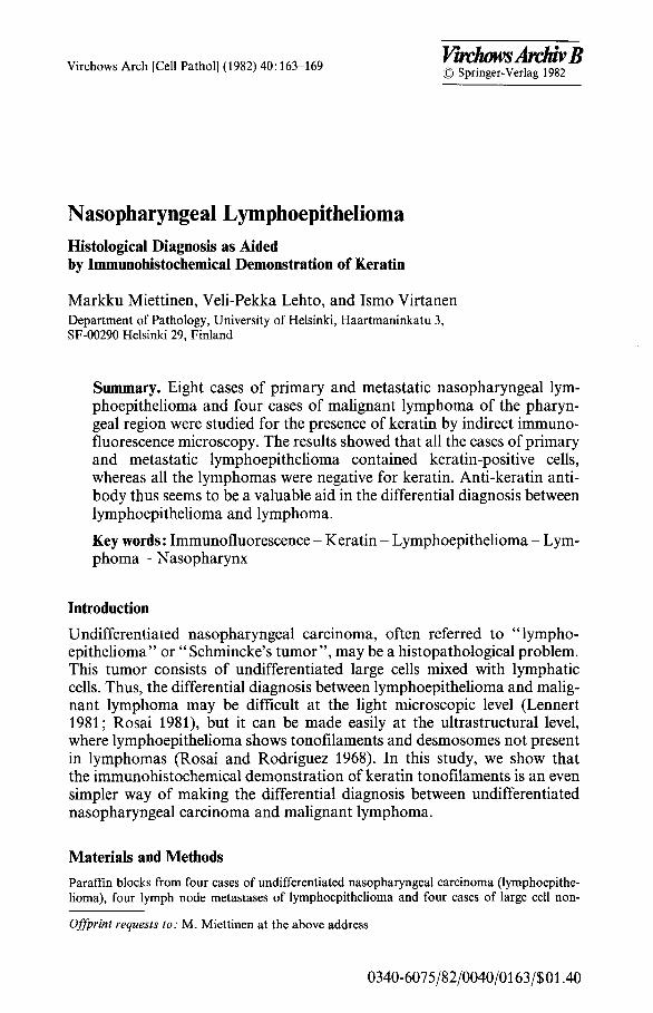
All o f the b i o p s y spec imens c o n t a i n e d u n i f o r m la rge r o u n d cells wi th ves icu- lar nuclei , p r o m i n e n t nucleol i a n d spa r se c y t o p l a s m . T h e la rge cells were m i x e d wi th smal l l y m p h o c y t e s a n d s o m e t i m e s f o r m e d s t r ands or syncy t ia l masses . Of ten , h o w e v e r , the epi thel ia l cells were d i spe r sed a n d fa i led to f o r m cell c lus ters (Fig. I a). I m m u n o f l u o r e s c e n c e s ta in ing wi th a n t i - k e r a t i n a n t i b o d y s h o w e d pos i t iv i ty in m a n y o f the la rge cells in all cases (Fig. 1 b). H o w e v e r , n o t all the la rge cells were pos i t ive fo r ke ra t in .
Fig. I a Nasopharyngeal lymphoepithelioma, where recognition of the large ceils as epithelial in type is difficult. Hematoxylin and eosin x 900. b Immunofluorescence staining with anti- keratin antibodies brightly decorates some cells. Magnification 900
Keratin in Lymphoepithelioma 165
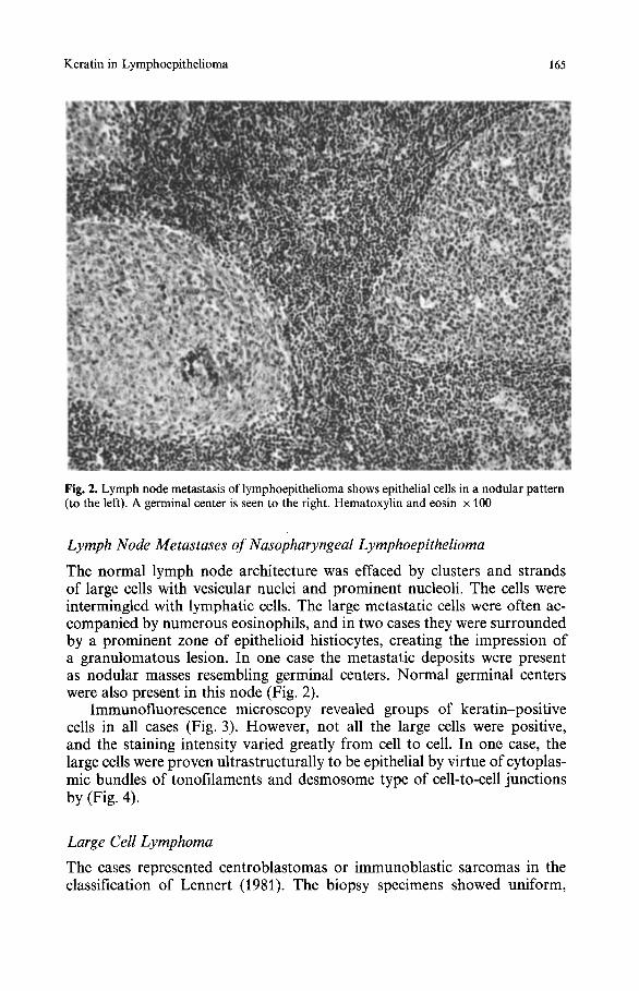
Fig. 2. Lymph node metastasis of lymphoepithelioma shows epithelial cells in a nodular pattern (to the left). A germinal center is seen to the right. Hematoxylin and eosin • 100
Lymph Node Metastases of Nasopharyngeal Lymphoepithelioma
The normal lymph node architecture was effaced by clusters and strands of large cells with vesicular nuclei and prominent nucleoli. The cells were intermingled with lymphatic cells. The large metastatic cells were often ac- companied by numerous eosinophils, and in two cases they were surrounded by a prominent zone of epithelioid histiocytes, creating the impression of a granulomatous lesion. In one case the metastatic deposits were present as nodular masses resembling germinal centers. Normal germinal centers were also present in this node (Fig. 2).
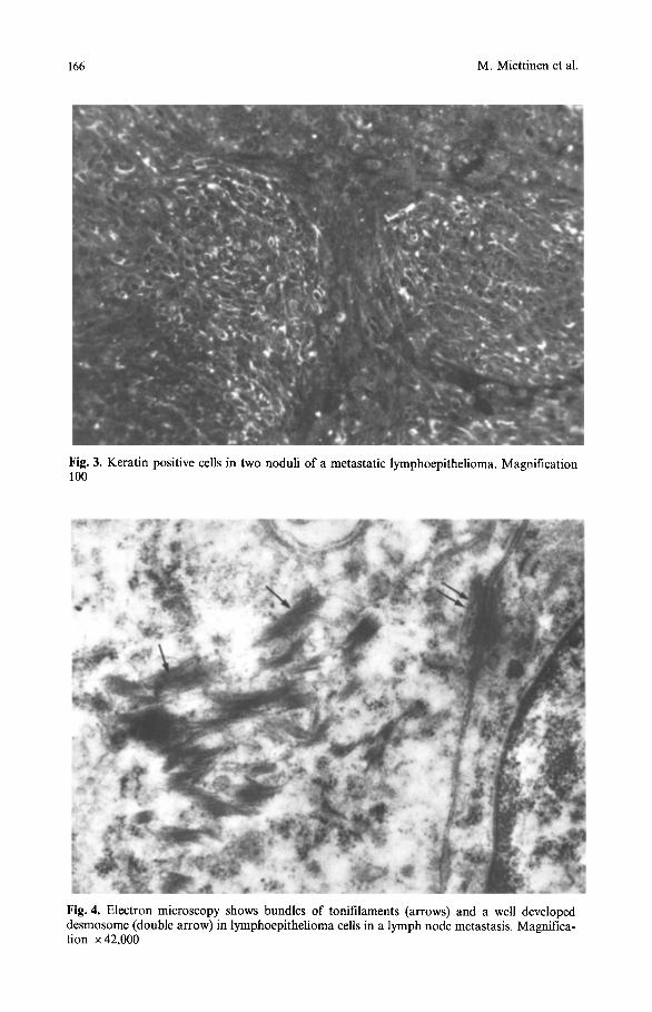
Immunofluorescence microscopy revealed groups of keratin-positive cells in all cases (Fig. 3). However, not all the large cells were positive, and the staining intensity varied greatly from cell to cell. In one case, the large cells were proven ultrastructurally to be epithelial by virtue of cytoplas- mic bundles of tonofilaments and desmosome type of cell-to-cell junctions by (Fig. 4).
Large Cell Lymphoma
The cases represented centroblastomas or immunoblastic sarcomas in the classification of Lennert (1981). The biopsy specimens showed uniform,
166 M. Miettinen et al.
Fig. 3. Keratin positive cells in two noduli of a metastatic lymphoepithelioma. Magnification 100
Fig. 4. Electron microscopy shows bundles of tonifilaments (arrows) and a well developed desmosome (double arrow) in lymphoepithelioma cells in a lymph node metastasis. Magnifica- tion x 42,000
Keratin in Lymphoepithetioma 167
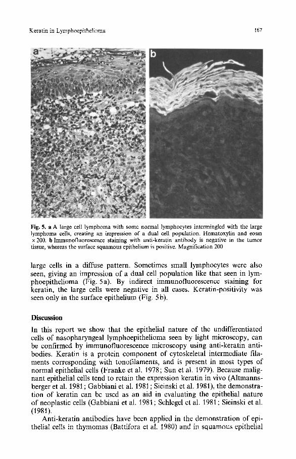
Fig. 5. a A large cell lymphoma with some normal lymphocytes intermingled with the large lymphoma cells, creating an impression of a dual cell population. Hematoxylin and eosin x 200. b Immunofluoreseenee staining with anti-keratin antibody is negative in the tumor tissue, whereas the surface squamous epithelium is positive. Magnification 200
large cells in a diffuse pattern. Sometimes small lymphocytes were also seen, giving an impression of a dual cell population like that seen in lym- phoepithelioma (Fig. 5a). By indirect immunofluorescence staining for keratin, the large cells were negative in all cases. Keratin-positivity was seen only in the surface epithelium (Fig. 5 b).
Discussion
In this report we show that the epithelial nature of the undifferentiated cells of nasopharyngeal lymphoepithelioma seen by light microscopy, can be confirmed by immunofluorescence microscopy using anti-keratin anti- bodies. Keratin is a protein component of cytoskeletal intermediate fila- ments corresponding with tonofitaments, and is present in most types of normal epithelial cells (Franke et al. 1978; Sun et al. 1979). Because malig- nant epithelial cells tend to retain the expression keratin in vivo (Altmanns- berger et al. 1981 ; Gabbiani et al. 1981 ; Sieinski et al. 1981), the demonstra- tion of keratin can be used as an aid in evaluating the epithelial nature of neoplastic cells (Gabbiani et al. 1981 ; Schlegel et al. 1981 ; Sieinski et al. (1981).
Anti-keratin antibodies have been applied in the demonstration of epi- thelial cells in thymomas (Battifora et al. 1980) and in squamous epithelial
168 M. Miettinen et al.
cells of craniopharyngeomas (Asa et al. 1981), as well as in the differential diagnosis between carcinomas and sarcomas (Gabbiani et al. 1981).
Lymph node metastases o f lymphoepithel ioma show a wide variation in their histological appearance. As noted in this study, metastatic lympho- epitheliomas may be accompanied by a prominent granulomatous reaction, which may even obscure the presence of neoplastic epithelial cells (Rennke and Lennert 1973). The differential diagnosis between lymphoepithel ioma and lymphoma can be made using anti-keratin antibodies, because epithelial cells are positive while lymphatic cells are negative (Gabbiani et al. 1981; Sieinski et al. 1981).
It has been suggested that the undifferentiated cells of lymphoepithelio- mas are derived f rom primitive squamous epithelial cells (Yeh 1962). Their squamous epithelial nature is supported by the ultrastructural demonstra- tion of prominent bundles of tonofi laments and typical well developed des- mosomes in the tumor cells (Rosai and Rodriguez 1968). However, the presence of keratin is not specific to squamous epithelial cells (Franke et al. 1978; Sun et al. 1979). Thus the demonstra t ion of keratin as such does not help in the evaluation of the type of a metastatic epithelial neoplasm. However, the evaluation o f subset polypeptides o f keratin filaments may offer the possibility o f elucidating the origin of a metastasis in the future (Franke et al. 1981).
Acknowledgements. The skilful technical assistance of Ms. Raili Taavela is kindly acknowl- edged. This study was supported by grants from the Finnish Medical Research Council the Ida Montin Foundation and the Association of Finnish Life Insurance Companies.
References
Altmansberger M, Osborn M, H61scher A, Schauer A, Weber K (1981) The distribution of keratin type intermediate filaments in human breast cancer .An immunohistological study. Virchows Archiv [Cell Pathol] 37 : 27%284
Asa SL, Kovacs K, Bilbao JM, Penz G (1981) Immunohistochemical localization of keratin in craniopharyngiomas and squamous cell nests of the human pituitary. Acta Neuropathol 54:257-260
Battifora H, Sun T-T, Bahu RM, Rao S (1980) The use of antikeratin antiserum as a diagnostic tool: thymoma versus lymphoma. Hum Pathol 11:635-641
Brozman M (1978) Immunohistochemical analysis of formaldehyde- and trypsin- or pepsin- treated material. Acta Histochem 63: 251-260
Franke WW, Weber K, Osborn M, Schmid E, Freudenstein C (1978) Antibody to prekeratin. Decoration of tonofilament-like arrays in various cells of epithelial character. Exp Cell Res 116: 42~445
Franke WW, Schiller DL, Moll R, Winter S, Schmid E, Engelbrecht I, Denk H, Krepler R, Platzer B (1981) Diversity of cytokeratins. Differentation specific expression of cytokera- tin polypeptides in epithelial cells and tissues. J Mol Biol 153:933-959
Gabbiani G, Kapanci Y, Barazzone P, Franke WW (1981) Immunohistochemical identification of intermediate-sized filaments in human neoplastic cells. Am J Pathol 104:206-216
Lennert K (1981) Histopathology of non-Hodgkin's lymphomas. Springer, Berlin, Heidelberg, New York
Rennke H, Lennert K (1973) Kfisig-tuberkuloide Reaktion bei Lymphknotenmetastasen lym- phoepithelialer Carcinome (Schminke-Tumoren). Virchows Arch [Pathol Anat) 358:241-247
Rosai J (1981) Ackerman's surgical pathology. C.V. Mosby, St. Louis, pp 165-166
Keratin in Lymphoepithelioma 169
Rosai J, Rodriguez HA (1968) Application of electron microscopy to the differential diagnosis of tumors. Arch Pathol Am J Clin Pathol 50:555-562
Schlegel R, Banks-Schlegel S, McLeod JA, Pinkus GS (1980) Immunoperoxidase localization of keratin in human neoplasms. A preliminary survey. Am J Pathol 101:41-50
Sieinski W, Dorsett B, Ioachim HL (1981) Identification of prekeratin by immunofluorescence staining in the differential diagnosis of tumors. Hum Pathol 12:452-458
Sun TT, Shih C, Green H (1979)Keratin cytoskeletons in epithelial cells of internal organs. Proc Natl Acad Sci USA 76: 2813-2817
Virtanen I, Lehto V-P, Lehtonen E, Vartio T, Stenman S, Kurki P, Wager P, Small JV, Dahl D, Badley RA (1981 a) Expression of intermediate filaments in cultured cells. J Cell Sci 50:45-63
Virtanen I, Koskull H, Lehto V-P, Vartio T, Aula P ~1981 b) Cultured human amniotic fluid cells characterized with antibodies against intermediate filaments in indirect immunofluores- cence microscopy. J Clin Invest 68:1348-1355
Yeh S (1962) A histological classification of carcinomas of the nasopharynx with a critical review as to the existence of lymphoepitheliomas. Cancer 15:895-919
Received April 16 / Accepted May 12, 1982
Related Documents