Sino-Nasal polyposis Presenter: Dr. Pradip Mallik Moderator: Prof. Th Sudhiranjan Department of Otorhinolaryngology Regional Institute of medical sciences, Imphal Dated: 5.3.2013

Welcome message from author
This document is posted to help you gain knowledge. Please leave a comment to let me know what you think about it! Share it to your friends and learn new things together.
Transcript
Sino-Nasal polyposis
Presenter: Dr. Pradip Mallik
Moderator: Prof. Th SudhiranjanDepartment of Otorhinolaryngology
Regional Institute of medical sciences, ImphalDated: 5.3.2013
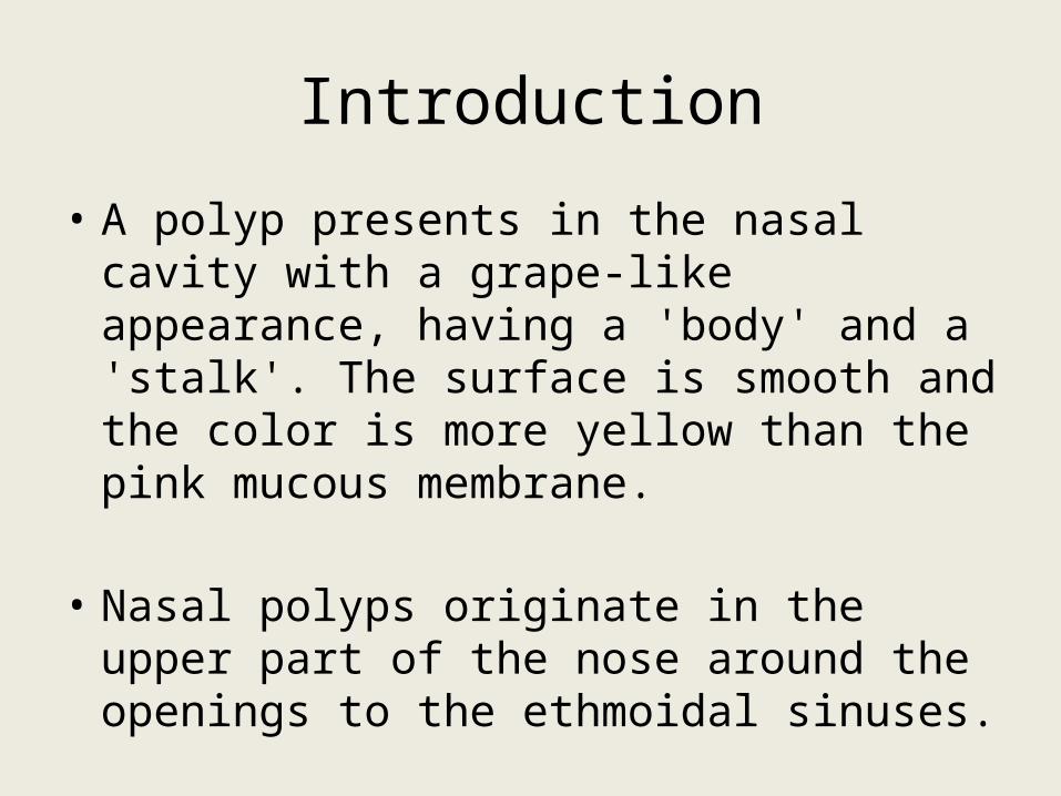
Introduction
• A polyp presents in the nasal cavity with a grape-like appearance, having a 'body' and a 'stalk'. The surface is smooth and the color is more yellow than the pink mucous membrane.
• Nasal polyps originate in the upper part of the nose around the openings to the ethmoidal sinuses.
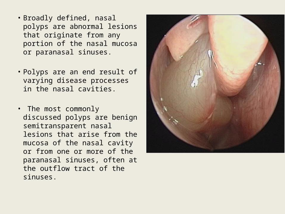
• Broadly defined, nasal polyps are abnormal lesions that originate from any portion of the nasal mucosa or paranasal sinuses.
• Polyps are an end result of varying disease processes in the nasal cavities.
• The most commonly discussed polyps are benign semitransparent nasal lesions that arise from the mucosa of the nasal cavity or from one or more of the paranasal sinuses, often at the outflow tract of the sinuses.
History of Nasal polyposis
• The earliest record of nasal polyps is found in Egyptian literature of approximately 2,000 years bc.
• Hippocrates (460–370 bce) referred to the “nasal growths” as “polypus” due to their resemblance to the sea-polyp, and this name has persisted to this day.
• Boerhaave, in 1744, was among the first to surmise that these growths resulted from elongation of the linings of the sinus membranes.
• In 1843, Frerichs and Billroth proposed that polyps were truly a hypertrophy of normal Sino nasal mucosa, as the epithelium covering the polyp was similar to the mucosa of the originating sinus.
• Advances in immunohistochemistry and immunobiology in the 1940s led to the first description of the predominance of eosinophil and lymphocyte populations in polyps.
• Messerklinger’s discoveries, functional endoscopic sinus surgery (FESS) was introduced in the late 1960s in Germany, and David Kennedy is credited for introducing FESS in the United States in 1985.
EPIDEMIOLOGY
• The prevalence of nasal polyps (NP) in the population has been grossly estimated as 1–4%.
• It increases with age, reaching a peak in those aged 50 years and older.
• Male: Female = 2:1
• An association between NP and allergic rhinitis (AR) is weak, with NP prevalence in patients with AR estimated between 1.5 and 1.7% and this incidence is similar to that of the general population.
• Nasal polyposis occurs with a high frequency in groups of patients having specific airway diseases

Disease Group Percentage
Allergic Rhinitis In Adult 0.1
In children 1.5
Non-allergic Rhinitis 5
Asthma in adults Allergic 5
Non-allergic 13
NSAID intolerance 36-72
NSAID intolerance+asthma 80
Allergic fungal rhinosinusitis
>80
Churg-strauss syndrome 50
Cystic fibrosis In children 10
In adults 40
Primary ciliary dyskinesia 40
• Genetic inheritance has been proposed as a possible etiology of NP. Studies have suggested that up to 14% of patients with NP have a family history of NP.
• In a Caucasian population, NP have been shown to have a strong eosinophilic component while NP in Asian countries show a neutrophilic pattern.
• Up to 80% of NSAID insensitive patients have NP.

ETIOLOGY and ASSOCIATED DISEASES
• The aspirin triad: A triad of nasal polyposis, aspirin and asthma was described (Samtard’s triad) and it represents the most aggressive forms of disease.
• It is a nonallergic entity and the intolerance is not confined to acetylsalicylic acid, as the patients react to other NSAIDs.
ALLERGY?
• It was believed that allergy is a significant cause of nasal polyposis as most of the polyps are characterized by tissue eosinophilia.
• Most studies have failed to show a higher occurrence of positive skin tests to inhaled allergens in patients with polyps than in the general population.
• According to Keith et al there is no deterioration of nasal symptoms or eosinophilia during the pollen season in polyp patients having a positive skin test to pollen. Thus, it appears that allergy is not a well documented cause or aggravating factor in nasal polyposis.

Cystic fibrosis:
• Cystic fibrosis (CF), also known as mucoviscidosis, is an autosomal recessive genetic disorder that affects most critically the lungs, and also the pancreas, liver, and intestine.
• It is characterized by abnormal transport of chloride and sodium across an epithelium, leading to thick, viscous secretions.
• Nasal endoscopy demonstated polyp in 45% of adults with cystic fibrosis.
• Mucosal abnormalities in the sinuses consistent with polypoid hyperplasia are demonstrated on computed tomography (CT) scans in almost all patients with CF.
• A sweat chloride test is recommended in any child with nasal polyps, to rule out cystic fibrosis.
Kartagener’s syndrome:
• Primary ciliary dyskinesia (PCD), also known as immotile ciliary syndrome.
• Rare, ciliopathic, autosomal recessive genetic disorder.
• It causes a defect in the action of the cilia lining the respiratory tract (lower and upper, sinuses, Eustachian tube, middle ear) and fallopian tube, and also of the flagella of sperm in males.
• Absent mucociliary clearance and recurrent bacterial infections result in nasal polyposis in about 40 percent of the patients. The recurrence rate is low after surgery.

Young syndrome
• It is a rare condition which consists of recurrent respiratory disease with
• Chronic rhinosinusitis, • Nasal polyps, • Bronchiectasis and • Azoospermia.

Pathogenesis & Pathophysiology
• The pathogenesis of nasal polyps explains how the polyps start and grow.
• The pathophysiology of nasal polyps explains the events and processes taking place in the outgrowth of nasal polyps.
• Several pathogenetic theories on the formation of nasal polyps have been published during the last 150 years that have been summarised previously These theories are based on oedema, an increase in tubulo-alveolar glands, the presence of the cysts of mucous glands and on mucous glands of NP.
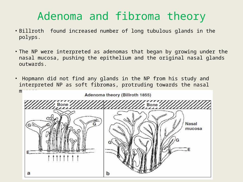
Adenoma and fibroma theory• Billroth found increased number of long tubulous glands in the polyps.
• The NP were interpreted as adenomas that began by growing under the nasal mucosa, pushing the epithelium and the original nasal glands outwards.
• Hopmann did not find any glands in the NP from his study and interpreted NP as soft fibromas, protruding towards the nasal mucosa.

Necrotizing Ethmoiditis Theory
• This theory supposes that ethmoiditis leads to periostitis and osteitis of the ethmoid bone and causes bone necrosis.
• Hayek argued strongly against this theory, based on the fact that he could not found bone necrosis in the ethmoid sinus.
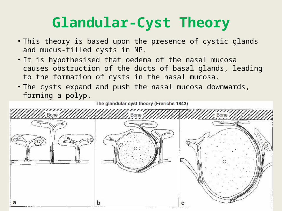
Glandular-Cyst Theory• This theory is based upon the presence of cystic glands and mucus-filled
cysts in NP.• It is hypothesised that oedema of the nasal mucosa causes obstruction of
the ducts of basal glands, leading to the formation of cysts in the nasal mucosa.
• The cysts expand and push the nasal mucosa downwards, forming a polyp.

Mucosal Exudate Theory
• Hayek believed that the formation of NP started via an exudate localised deep in the nasal mucosa, which pressed outwards caudally.
• According to this theory, both layers of the tubulo-alveolar sero-mucous nasal glands should be displaced outwards and be found in the distal part of the polyp.

Theory on Cystic Dilatationof the Excretory Duct of NasalGlands and Vessel Obstruction
• According to this theory in chronic inflammation of the nasal mucosa, excretory ducts of nasal tubulo-alveolar glands are obstructed, distended and dilated into cystic structures.
• The capillaries and veins (which are arranged around the excretory ducts and the gland mass) become stretched and obstructed, resulting in increased permeability, transudation and oedema.
• This theory has been used to explain polyp formation in cystic fibrosis.
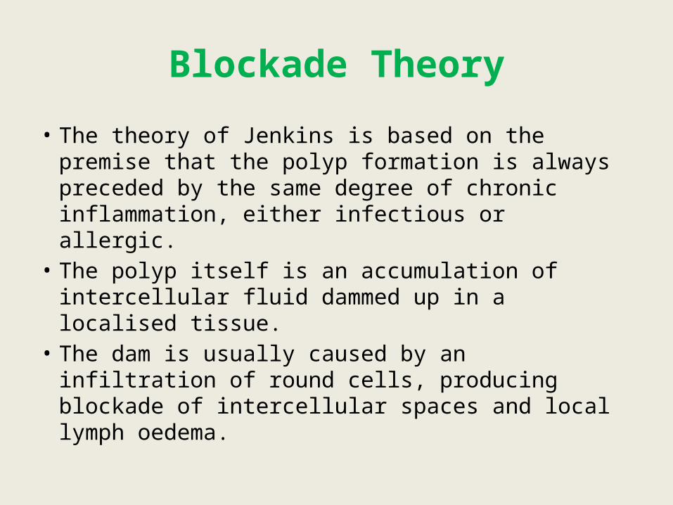
Blockade Theory
• The theory of Jenkins is based on the premise that the polyp formation is always preceded by the same degree of chronic inflammation, either infectious or allergic.
• The polyp itself is an accumulation of intercellular fluid dammed up in a localised tissue.
• The dam is usually caused by an infiltration of round cells, producing blockade of intercellular spaces and local lymph oedema.
Peri-Phlebitis and Peri-Lymphangitis Theory
• The theory of Eggston and Wolff is based upon the recurrent infections that lead to the blocking of intercellular fluid transport in the mucosa and oedema of the lamina propria.
• If the oedema involves major areas, the result is the prolapse of the mucosa and formation of polyps.
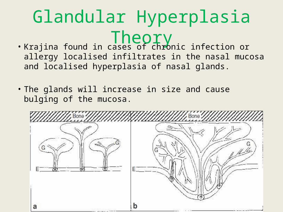
Glandular Hyperplasia Theory• Krajina found in cases of chronic infection or allergy
localised infiltrates in the nasal mucosa and localised hyperplasia of nasal glands.
• The glands will increase in size and cause bulging of the mucosa.
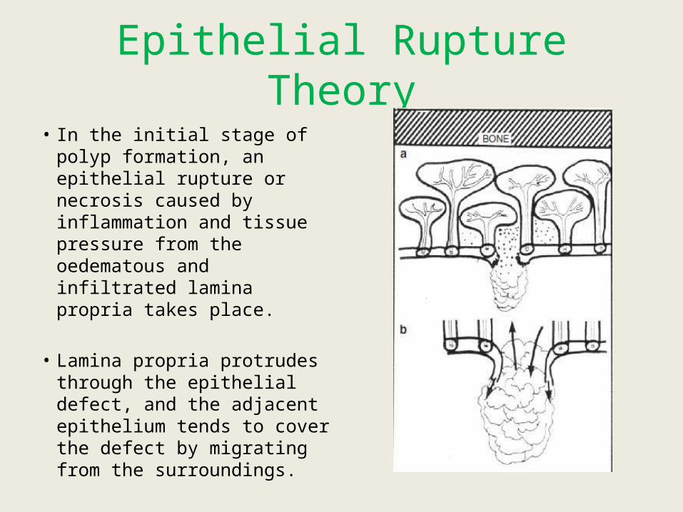
Epithelial Rupture Theory
• In the initial stage of polyp formation, an epithelial rupture or necrosis caused by inflammation and tissue pressure from the oedematous and infiltrated lamina propria takes place.
• Lamina propria protrudes through the epithelial defect, and the adjacent epithelium tends to cover the defect by migrating from the surroundings.
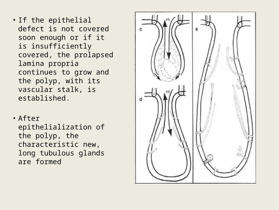
• If the epithelial defect is not covered soon enough or if it is insufficiently covered, the prolapsed lamina propria continues to grow and the polyp, with its vascular stalk, is established.
• After epithelialization of the polyp, the characteristic new, long tubulous glands are formed

Mucous gland in nasal polyp
• In most of the pathogenetic theories, the mucous glands have played a role.
• The glandular orifices are irregularly distributed, as there is no particular concentration of glands in the stalk or in the most distal end of the polyp.
• The density of glands in NP is considerably lower than in the nasal mucosa.
• The polyp glands are tubular, of different shapes and sizes and differ widely from those of the nasal glands.
• The most striking glands are the long tubular glands, which may be 1–8 mm of length.
• Some are very simple, narrow tubes other have prominences of small, round, alveolar bulges on their sides.

Long, simple tubularglands (a, f).
Long tubularglands with some branches(b–e).
Short, simple tubularglands (g).
Short, branchedtubular glands (h, i).
Tubular glands withflask-shaped dilatation(j, k).
Tubulo-alveolarglands, which are foundextremely rarely (l)
Cellular Infiltration
• Eosinophilic inflammation is an important feature in the pathogenesis of chronic Rhinosinusitis (CRS) with nasal polyps.
• The eosinophilic accumulation in the polyp stroma is basically caused by increased transendothelial migration and increased survival time in the tissue, where an increased concentration of interleukine 5 (IL-5) plays a major role.
• The increased amount of IL-5 is predominantly released from T-lymphocytes, independently of atopy, and the highest concentration has been found in polyps from patients with non-allergic asthma and acetylsalicylic acid (ASA) intolerance.
• These are the sub-groups of patients also known to exhibit the greatest accumulation of eosinophils
• In the ASA intolerant patients, a lowered prostaglandin E2 (PGE2) production has been observed.
• PGE2 has a significant anti-inflammatory activity, including inhibition of eosinophils.
• A possible intrinsic defect in PGE2 production might, therefore, be responsible for a further increase of eosinophilic accumulation in ASA intolerant patients.
Role of Staphylococcus aureus enterotoxins (SAE)
• Multiclonal IgE antibody formation to SAE can be seen in nasal polyp tissue, but rarely in CRS.
• It is positive in about 30-50% of the patients with NP and in about 60-80% of nasal polyp subjects with asthma

Nasal polyposis: aetiology and pathogenesis
Chemokines
TB
Cytokines Hyper IgE
Eosinophils ( apoptosis)
SuperantigensIL-5
ECP
Albumin
Eotaxin
Polyclonal IgE
Epithelial damage (barrier dysfunction)
chronic microbial trigger
S. Aureus enterotoxins: disease modifiers
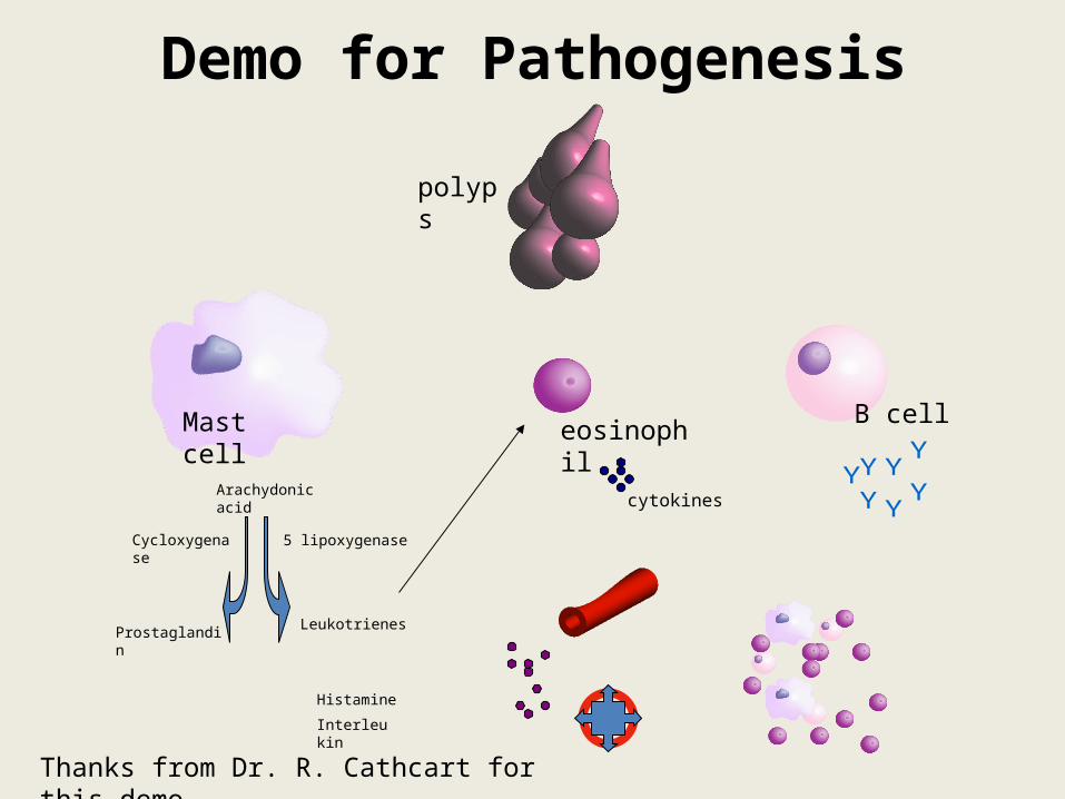
Demo for Pathogenesis
polyps
eosinophilB cellMast cell
Arachydonic acid
5 lipoxygenase
Leukotrienes
Interleukin
Prostaglandin
Histamine
Cycloxygenase
cytokines
Thanks from Dr. R. Cathcart for this demo
Histopathology
Macroscopic:• Macroscopically, most polyps have an edematous, smooth and shiny
appearance with a soft consistency compared with the surrounding nonpolypoid mucosa.
• The cut surface is usually pale, edematous with a translucent appearance.
• Polyps are generally mobile and often attached via a stalk to the underlying mucosa.
• Polyps commonly arise from the middle meatus and the sphenoethmoidal recess and are often bilateral. However, unilateral polyps are not uncommon.
• In long-standing polyps, the sinonasal bones may remodel and cause broadening of the nasal dorsum.
• A polyp arising from the maxillary sinus and into the nasal cavity is characteristic of antrochoanal polyp and is generally unilateral.
• A proportion of CRS with nasal polyps also has characteristic thick, dark, and tenacious mucus, termed eosinophilic mucus.
• This mucus is typically seen in allergic fungal sinusitis but is also present in patients with severe and recalcitrant polypoid CRS including cystic fibrosis and Sampter’s triad and in the lungs of allergic bronchopulmonary aspergillosis.
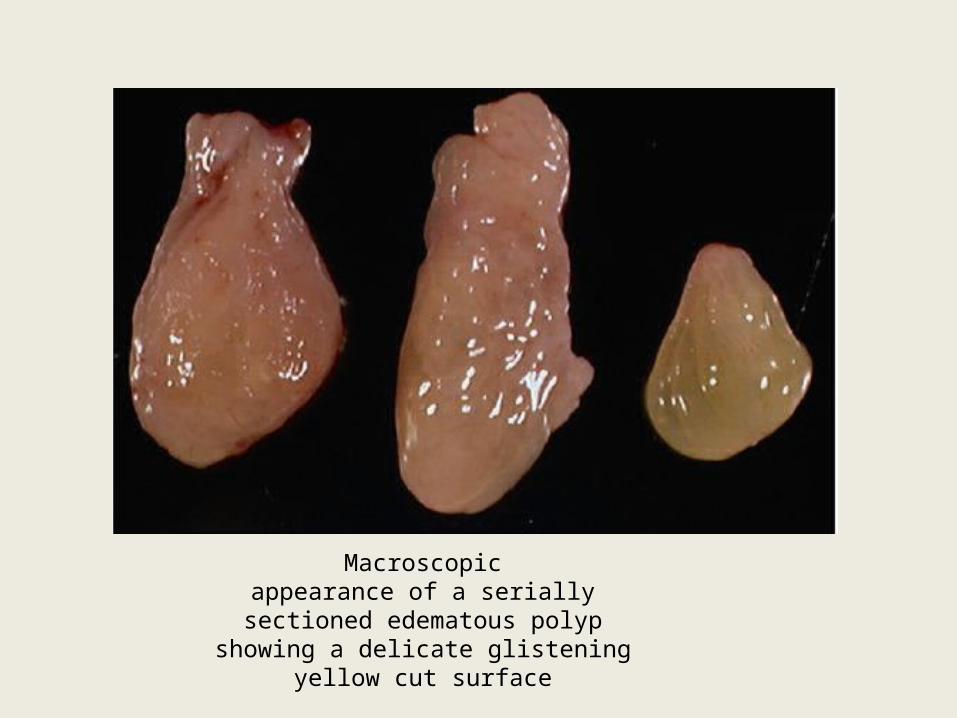
Macroscopicappearance of a serially
sectioned edematous polypshowing a delicate glistening
yellow cut surface

• Microscopic:
The major histological characteristics of nasal polyps and CRS mucosa compared with normal mucosa include
(1) Structural changes involving the epithelium, submucosa, and sometimes underlying bone; and
(2) The nature and degree of inflammatory cell infiltrate.
Polyps have historically been classified based on their histological structural appearance and the nature of predominant inflammatory cell population into 3 types.

(1) edematous, eosinophilic, or “ allergic” polyps:
• . Edematous and eosinophilic polyps are the most common type and are also known as “allergic” nasal polyps.
• These polyps are lined with respiratory epithelium with a range of mucosal alterations that include ulceration, granulation tissue, acute mucositis, epithelial and goblet cell hyperplasia, and squamous metaplasia.
• The basement membrane is often thickened, and there is abundant submucosal edema.
• Mucus retention cysts are common and varying amounts of mixed inflammatory cell infiltrates contain mostly eosinophils, plasma cells, and scattered lymphocytes.
• The edematous and eosinophilic polyps are seen in the whole spectrum of associated disorders including, EMCRS, allergic fungal sinusitis, Sampter’s triad, cystic fibrosis, and Churg–Strauss syndrome.

1.Edematous polyp
2. thickenedbasement membranes(arrows)
3.markedsubmucosal edema resultingin extensive clear spacebetween submucosalconnective tissue fibers.
4.scatteredthroughout are mixedinflammatory infiltratesincluding eosinophils and plasma cells
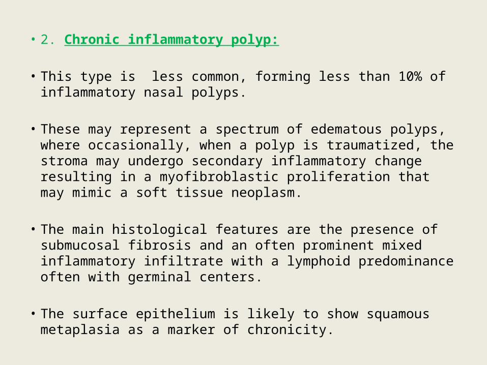
• 2. Chronic inflammatory polyp:
• This type is less common, forming less than 10% of inflammatory nasal polyps.
• These may represent a spectrum of edematous polyps, where occasionally, when a polyp is traumatized, the stroma may undergo secondary inflammatory change resulting in a myofibroblastic proliferation that may mimic a soft tissue neoplasm.
• The main histological features are the presence of submucosal fibrosis and an often prominent mixed inflammatory infiltrate with a lymphoid predominance often with germinal centers.
• The surface epithelium is likely to show squamous metaplasia as a marker of chronicity.
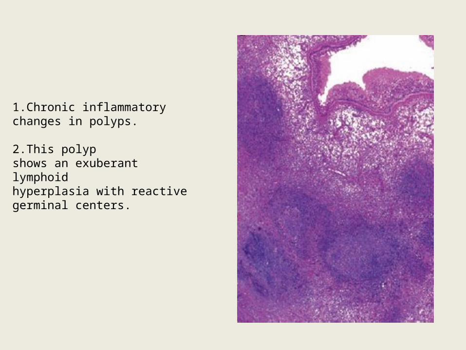
1.Chronic inflammatorychanges in polyps.
2.This polypshows an exuberant lymphoidhyperplasia with reactivegerminal centers.
3) Seromucinous, glandular polyps:• Polyps with hyperplasia of seromucinous
glands are less common.• Lesions in this category are relatively new and
somewhat controversial as to their relationship with true epithelial neoplasms, and include respiratory epithelial adenomatoid hamartoma and seromucinous hamartoma.
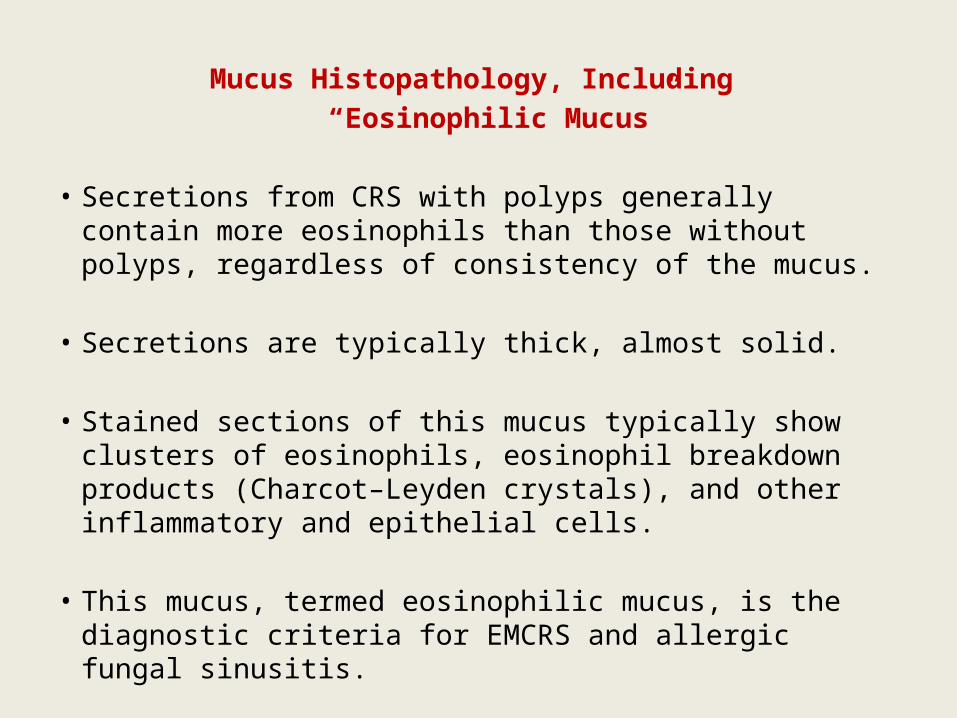
Mucus Histopathology, Including
“Eosinophilic Mucus”
• Secretions from CRS with polyps generally contain more eosinophils than those without polyps, regardless of consistency of the mucus.
• Secretions are typically thick, almost solid.
• Stained sections of this mucus typically show clusters of eosinophils, eosinophil breakdown products (Charcot–Leyden crystals), and other inflammatory and epithelial cells.
• This mucus, termed eosinophilic mucus, is the diagnostic criteria for EMCRS and allergic fungal sinusitis.

Antrochoanal polyp
• Antrochoanal polyps are the most common type of choanal polyp.
• Other sites of origin may be sphenoid, ethmoid, rarely septum, and inferior turbinate.
• Antrochoanal polyps represent 4–6% of all polyps, and in the pediatric population, up to 33%.
• They have an antral and choanal component. These typically arise from the posterior wall of maxillary antrum and often have a thin “neck” that passes through the maxillary sinus ostium (or accessory ostium).
• They are often unilateral, but may be bilateral on rare occasions.
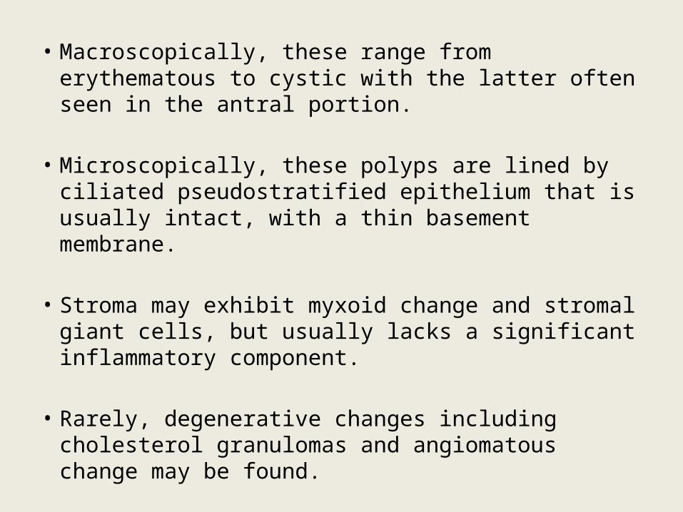
• Macroscopically, these range from erythematous to cystic with the latter often seen in the antral portion.
• Microscopically, these polyps are lined by ciliated pseudostratified epithelium that is usually intact, with a thin basement membrane.
• Stroma may exhibit myxoid change and stromal giant cells, but usually lacks a significant inflammatory component.
• Rarely, degenerative changes including cholesterol granulomas and angiomatous change may be found.
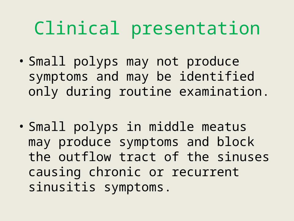
Clinical presentation
• Small polyps may not produce symptoms and may be identified only during routine examination.
• Small polyps in middle meatus may produce symptoms and block the outflow tract of the sinuses causing chronic or recurrent sinusitis symptoms.

Symptom-producing polyps can cause:
• 1) Nasal airway obstruction• 2) Post nasal drainage• 3) Dull headache• 4) Snoring• 5) Rhinorrhoea• 6) Hyposmia/anosmia• 7) Epistaxis if preset may give suspicion of
more serious nasal cavity lesions.
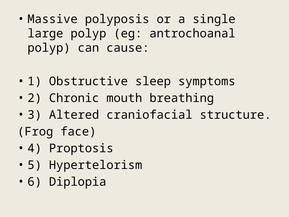
• Massive polyposis or a single large polyp (eg: antrochoanal polyp) can cause:
• 1) Obstructive sleep symptoms• 2) Chronic mouth breathing• 3) Altered craniofacial structure.
(Frog face)• 4) Proptosis• 5) Hypertelorism• 6) Diplopia

Diagnosis and scoring• Rhinoscopy: Large polyp can be identified by
anterior rhinoscopy.
• In contrast to hyperplastic turbinate, a polyp can be made to move by touching with a probe.
• Seen as multiple glistening, grayish white, smooth pedunculated mass in both side of nasal cavity.
• On probing insensitive to touch, do not bleed and probe can be passed all around the masses.

• Direct nasal endoscopy:
- Preferred method of examination.
- Can diagnose small polyp in middle meatus.
- It gives superior assessment of the extent of the disease and anatomical abnormalities
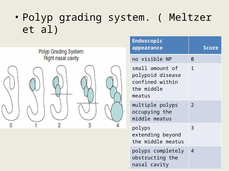
• Polyp grading system. ( Meltzer et al)
Endoscopic appearance Score
no visible NP 0
small amount of polypoid disease confined within the middlemeatus
1
multiple polyps occupying the middle meatus
2
polypsextending beyond the middle meatus
3
polyps completelyobstructing the nasal cavity
4
Imaging modalities• ›› CT scan is the primary modality used to evaluate patients with
NP, but MRI does have an occasional role.
• ›› Conventional radiographs are no longer within the standard of care for the evaluation of the paranasal sinuses.
• ›› Modern helical multislice CT scanners can produce reconstructed images of diagnostic quality in any plane of imaging.
• ›› Cystic fibrosis patients have preferential opacification of the paranasal sinuses, whereas non-CF patients with NP have preferential opacification of the nasal cavity.
• ›› On MRI, fungal infection may have very low signal on T2-weighted images, and may thus mimic aerated sinuses.

• Polyps appear radiographically as rounded nodules of soft tissue along the mucosal surfaces of the paranasal sinuses and nasal cavity.
• More radiodense than the surrounding mucosal thickening or secretions, which make them appear slightly brighter on CT.
• This pattern may be reversed if the secretions become inspissated.
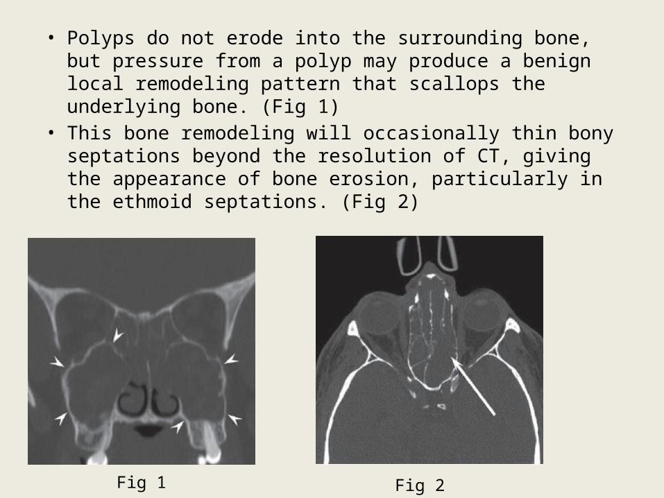
• Polyps do not erode into the surrounding bone, but pressure from a polyp may produce a benign local remodeling pattern that scallops the underlying bone. (Fig 1)
• This bone remodeling will occasionally thin bony septations beyond the resolution of CT, giving the appearance of bone erosion, particularly in the ethmoid septations. (Fig 2)
Fig 1 Fig 2

• In antronasal polyps sometimes, the polyp will be narrowed as it passes through the maxillary os, resulting in a dumbbell configuration on coronal CT. The medial wall of the maxillary sinus is usually bowed into the nasal cavity. (Fig 1)
• In antrochoanal polyp the most helpful radiologic feature is the mass itself extending into the nasopharynx. (Fig 2)
Fig 1 Fig 2
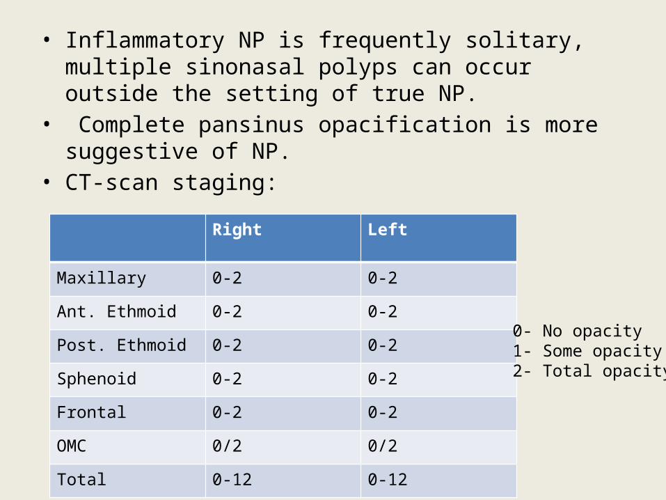
• Inflammatory NP is frequently solitary, multiple sinonasal polyps can occur outside the setting of true NP.
• Complete pansinus opacification is more suggestive of NP.• CT-scan staging:
Right Left
Maxillary 0-2 0-2
Ant. Ethmoid 0-2 0-2
Post. Ethmoid 0-2 0-2
Sphenoid 0-2 0-2
Frontal 0-2 0-2
OMC 0/2 0/2
Total 0-12 0-12
0- No opacity1- Some opacity2- Total opacity

• Middle meatus syndrome. (Fig 1)• A chronically obstructed sinus may form a mucocele. On CT,
the affected sinus enlarges and its walls become rounded outward. ( Fig 2)
Fig 1 Fig 2
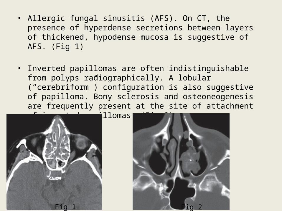
• Allergic fungal sinusitis (AFS). On CT, the presence of hyperdense secretions between layers of thickened, hypodense mucosa is suggestive of AFS. (Fig 1)
• Inverted papillomas are often indistinguishable from polyps radiographically. A lobular (“cerebriform”) configuration is also suggestive of papilloma. Bony sclerosis and osteoneogenesis are frequently present at the site of attachment of inverted papillomas. (Fig 2)
Fig 1 Fig 2

Laboratory Investigations
• Routine blood examination.
• Serological radioallergosorbent test (RAST) .
• Allergic skin testing for those who are having polyp associated with allergic rhinitis.
• Sweat chloride test/ genetic testing for custic fibrosis in any child with multiple benign nasal polyp.
• Nasal smear eosinophils may differentiate allergic from nonallergic sinus diseases.
• Biopsy from the polyp after evaluation by CT scan/MRI.

Histologic finding
NPs are characterised by • Pseudostratified ciliated columnar epithalium• Thickening of epithelial basement membrane• Stroma of NP is oedematous• Vascularization is poor and lacks innervation • Hyperplasia of seromucous gland when
comparing with inferior or middle turbinate.
• Eosinophils are most commonly found inflammatory cell in NP (found in 80-90% of polyps)
• Another inflammatory cell, neutroophil, occurs in 7% cases. This type of NP associates with CF, primary ciliary dyskinesia or Young syndrome.

Chemical mediators:
1) Cytokines: IL-1, Il-5 are found regularly
2) Growth factors:
• TNF: Alpha and beta found regularly• GM-CSF: Never – Intermittent• Platelet derived factor- present• Vascular permeable factors: Present• Vascular endothelial growth factor: Present• Insulin like growth factor 1: Present• Stem cell factor : Present

Immunoglobulins:
• IgG = Same level as MT or IT• IgA = Increased• IgM = Same• IgD = Same• IgE = Increased
Adhesion molecule:• Vascular adhesion molecule: Present• E & P selectin : Present

Treatment
• Treatment can either be medical and/or surgical.
• Oral and topical nasal steroid administration is the primary medical therapy.
• Antihistamines, decongestants provide little benefit.
• Immunotherapy is useful in treating associated allergic rhinitis.
• Antibiotic for bacterial super infections.
Intranasal corticosteroids
• Normally used intranasal steroids are Beclomethasone, Ciclesonide, Flunisolide, Fluticasone Propionate, Fluticasone furoate, Mometasone furoate, Triamcinolone Acetonide.
• They help in: • Reducing polyp size• Increase nasal patency• Reduction in rhinitis symptoms• Reduction in loss of sense of smell• Reduction in recurrence of polyp• Safety

Sideeffect of IntraNasal steroids
• - Excoriation and bleeding( can be reduced by cross hand technique)
• - Beclomethasone dipropionate nasal spray is associated with the onset of increased intraocular pressure.
• -Delay in growth in prepubescent children has led to an FDA warning on all INS
Systemic Corrticosteroids
• Can be given as tablets and depot-injections.• Oral prednisolone can be given at 25mg/daily
for 10-14 days.• As a depot-injection corresponds 100mg
prednisolone.• It may serve as medical polypectomy.
Risks of systemic steroid
• Insomnia,• personality change, • truncal obesity, • weight gain, • glaucoma, • cataracts, • osteoporosis (requires greater than 3 months usage), • peptic ulcer disease, and • increased incidence of infection

EPOS 2007

Surgical ManagementGeneral principles surgery for nasal polyposis:
• Selection of the patient who has exhausted medical management and remains symptomatic.
• The patient must understand the absolute necessity of often prolonged postoperative care.
• Selection of appropriate anesthetic method and preparation of the surgical field to maximize visualization and minimize bleeding.
• Accurate identification of surgical landmarks such as the lamina papyracea, anterior ethmoid artery, and skull base on preoperative CT imaging.

• Use of stereotactic image-guided surgery when necessary to confirm the skull base, cribriform plate, and lamina papyracea.
• Atraumatic removal of polyps and diseased bone while preserving as much mucosa as possible. Meticulous removal of osteitic bone and bony partitions.
• Ongoing postoperative medical management to prevent disease recurrence.

FESS
• Preoperative evaluation by radiology and endoscopic examination.
• Preoperatively, patients with evidence of active infections should be administered culture-directed antibiotics to help eradicate infection and to decrease mucosal inflammation.
• Administration of preoperative oral corticosteroid reduces the size of the polyp helps in proper visualization.
• Intravenous anesthesia with remifentanil and propofol, improved visual field with decreased blood loss has been demonstrated.
Steps• Polyps obstructing the nasal cavity or emanating from the middle meatus
are best addressed with the microdebrider.
• The maxillary sinus is best addressed with 0 and 30° endoscopes.
• Uncinate process is removed with sickle knife, backbiting forcep & microdebrider.
• The natural ostium of the maxillary sinus should be probed and widened and that any surgically created ostium is brought into continuity with the natural as in order to prevent recirculation of mucus.
• In the case of polyposis, large antrostomies are recommended. The sinus should be inspected with at minimum the 45° endoscope; a 70° endoscope is advised for analyzing the floor and anterior-most portions of the maxillary sinus to inspect for polyps and any infraorbital ethmoid cells that may obstruct the ostium. Inspissated secretions need to be evacuated, cultured, and sent to pathology for analysis of possible fungal disease.
• Anterior ethmoid cells should be meticulously dissected using through-cutting instruments and polypoid debris removed with the microdebrider.
• The first important landmark to identify is the medial orbital wall and the dissection is continued along the medial orbital wall rather than medially, because the skull base is significantly thicker and less likely to be injured laterally as opposed to medially where it slopes down into the middle turbinate.
• It is imperative that foci of ostetic bone along the lamina papyracea and middle turbinate be removed to help reduce inflammation and prevent polyp recurrence in these areas.
• Once the basal lamella is identified, the posterior ethmoid cells should be dissected in a similar fashion.
• Dissection of the ethmoid cavity typically occurs in an anterior to posterior fashion, identifying landmarks along the lamina papyracea and skull base until the anterior face of the sphenoid is encountered.
• The superior turbinate is next identified in order to enter the sphenoid sinus through the sphenoethmoidal recess and its natural ostium.
• Sinus is entered away from critical structures such as the carotid artery (dehiscent in up to 20% of cases) and the optic nerve (dehiscent in 5–10% of cases)
• This method also ensures that a sphenoethmoidal (Onodi) cell is not mistakenly treated as the sphenoid sinus.
• The sphenoid sinus should be opened widely from the skull base to lamina papyracea; care must be taken to avoid the septal branch of the sphenopalatine artery during this dissection.
• Polyps in sphenoid sinus is seen rarely
• Once the sphenoid sinus is opened, dissection along the skull base occurs in a retrograde fashion in order to clear lamella along the skull base until the frontal recess is encountered.
• The region of the anterior ethmoid artery is identified and avoided. It typically lies at the uppermost limit of the anterior wall of the ethmoidal bulla.
• If an injury to the anterior ethmoid artery occurs, bipolar electrocautery is used to coagulate the vessel.
• Frontal sinus is infrequently diseased with polyps; however, the frontal recess can be extensively diseased with polyps, resulting in obstruction of the frontal sinus itself.
• Much of the dissection involves angled instruments and endoscopes.
• In this region, preoperative review of the CT scans is critical, as a variety of drainage pathways may lead to the frontal sinus and a ethmoid air cells may invade the frontal sinus, complicating its anatomy and dissection, and polyps within the frontal recess can impair vision of these anomalies.
• Through-cutting instruments are used to dissect the anterior ethmoid lamella until the skull base is skeletonized and the anterior ethmoid artery identified, just posterior to the frontal recess.
• Partitions separating the frontal sinus from the supraorbital ethmoid cells are then taken down meticulously. The agger nasi cap is identified, resected, and frontal recess evaluated.

• After a complete dissection of the paranasal sinuses has been performed, the field should be inspected for loose fragments of bone, missed ethmoid partitions, and points of bleeding.
• Wherever possible, any exposed bone is removed.
• Merocel spacers are placed for 24 h and removed on postoperative day one when debridement occurs.
Postoperative management
• Surgery represents an adjunct to medical therapy in chronic rhinosinusitis with nasal polyposis.
• Postoperatively, failure is almost assured if the ongoing medical therapy to prevent recurrence is not administered.
• Routine postoperative debridement at first week of surgery of the sinonasal cavity is critical to assuring success.
• Culturedirected antibiotics in the postoperative setting should be utilized to limit iatrogenic infection, treat osteitis, and eradicate sources of inflammation.
• Oral corticosteroid acts as a potent anti-inflammatory in CRS with NP
• In diffuse nasal polyposis, the patient should be discharged with a tapering course of prednisone over a prolonged period, and overlapping administration of topical corticosteroids should begin before cessation of oral corticosteroids is considered.
• Patients unwilling to undergo oral corticosteroid therapy may benefit from topical administration of budesonide 0.5 mg/2 mL mixed in 240 mL normal saline for irrigation.
Other surgical techniques
• Nasalization procedure:
• The aim of the nasalisation procedure is to remove the ethmoidal mucosa as completely as possible without hazards, and to transform the ethmoidal labyrinth into a unique cavity opening into the nose.
• Aggressive Sinus Marsupialization Including the Endoscopic Modified Lothrop Procedure:
• The frontal recess clearance maximizes the natural ostium of the frontal sinus without enlarging it.
• The frontal drillout or EMLP creates the largest possible frontal ostium diameter for individual anatomical constraints.
• In patients who develop recurrent polyps after
surgery, the modified endoscopic Lothrop procedure, Draf III or frontal drillout produces
better outcomes, and in some cases a cure, than standard revision ESS.
Reasons for failure• Insufficient ethmoidectomy• Insufficient removal of septa• Insufficient drug concentration behind the septa• Insufficient cleaning of the polypoid mucosa behind the
septa• Pool for collection of secretions• Insufficient surgery of the frontal sinus• Polyps at the frontal recess and frontal ostium area• Polyps and mucoceles in the frontal sinus• Stenosis of frontal ostium• Insufficient surgery of the maxillary sinus

• Insufficient maxillary sinus ostium• Reclosure of the ostium• Decreased ventilation and drainage• Insufficient drainage due to thick secretions• Insufficient cleaning preop and postop• Insufficient opening of anterior wall of sphenoid sinus• Decreased ventilation• Decreased drainage• Insufficient drug concentration• Insufficient cleaning preop and postop• Free bony spicles• Granulation tissue and polyp recurrence
Reference.
1. Scott- Brown otorhinolaryngology
2. Cummings Otolaryngology
3. Nasal polyposis by T. Metin Önerci
Berrylin J. Ferguson
4.Pubmed indexed Journals.

• THANK YOU
THANK YOU
Related Documents