doi:10.1016/j.gca.2004.04.018 Nanoscale occurrence of Pb in an Archean zircon SATOSHI UTSUNOMIYA, 1 CHRIS S. PALENIK, 1 JOHN W. VALLEY, 2 AARON J. CAVOSIE, 2 SIMON A. WILDE, 3 and RODNEY C. EWING 1, * 1 Geological Sciences, University of Michigan, 425 East University Avenue, Ann Arbor, MI 48109-1063, USA 2 Department of Geology and Geophysics, University of Wisconsin, 1215 West Dayton Street, Madison, WI 53706, USA 3 Department of Applied Geology, Curtin Institute of Technology, GPO Box U1987, Perth, Australia (Received November 11, 2003; accepted in revised form April 26, 2004) Abstract—We report, for the first time, a direct, atomic-scale characterization of Pb in zircon (4.4 –3.1 Ga) from the early Archean Yilgarn craton in Australia using high-resolution HAADF-STEM. Two forms of Pb have been identified: Pb concentrated at 3 atom% as a nanoscale patch in zircon structure, and Pb concentrated within the amorphous domain created by fission fragment damage. The first result suggests that the Pb atoms directly substitute for Zr 4 in the zircon structure, and the latter observation demonstrates that Pb diffusion can occur through amorphous regions created by radiation damage, although volume diffusion is typically considered to be the dominant mechanism for Pb diffusion. Beyond the first percolation point, i.e., when the amorphous domains overlap and form a fully interconnected network of amorphous domains, there is a new pathway for the diffusion of Pb that is faster than volume diffusion through crystalline zircon. Copyright © 2004 Elsevier Ltd 1. INTRODUCTION Zircon (ZrSiO 4 ) is one of the most thoroughly studied of natural phases because of its predominant use in geochronology (Krogh, 1982; Bowring and Housh, 1995; Vervoort et al., 1996; Bowring et al., 1989; Amelin et al., 1999) and oxygen isotope studies (Valley, 2003). Because zircon is physically and chem- ically durable (Ewing, 1999), it has also been proposed as a waste form for the immobilization of plutonium from disman- tled nuclear weapons (Ewing et al., 1995; Ewing, 1999). In each of these applications, loss of trace elements, e.g., U, Pb and Pu, is of critical interest, and elemental loss may be much enhanced by radiation damage caused by the -decay of con- stituent actinides, 232 Th, 235 U, 238 U, and radionuclides in their decay series (Ewing, 2003). The fate of Pb is critical in U- Th-Pb isotopic dating, because Pb-loss due to radiation-en- hanced alteration of zircon is a common cause of discordant dates (Pidgeon et al., 1966; Craig, 1968). The diffusion of Pb, coupled with the diffusion of U and Th, has been extensively studied, and the Pb-diffusion rate is gen- erally considered to be faster than that of the tetravalent actin- ides U, Th and Pu (Bogomolov, 1991; Cherniak et al., 1997; Lee et al., 1997; Watson et al., 1997; Cherniak and Watson, 2000). The diffusivities of tetravalent cations, U, Th and Hf, in synthetic zircon were found to increase with decreasing ionic radius (Cherniak et al., 1997). Diffusion coefficients of U, Th and Pb have been systematically measured, the Pb diffusion coefficient is 4 orders of magnitude greater than that of U and Th at 1100°C (Lee et al., 1997), and the difference may be as high as 6 orders of magnitude (Cherniak and Watson, 2000). Recently, estimates of inward and outward diffusion rates of Pb in zircon have been reexamined at 1200 –1300°C, and the measured rates are 1–2 orders of magnitude slower than the previously determined values (Cherniak and Watson, 2000); nonetheless, Pb diffusion is still considerably faster than that of tetravalent U and Th. In addition to volume diffusion, amorphous domains created by radiation damage provide an additional pathway for Pb diffusion. The accumulation of radiation damage in zircon results from the -decay series for 238 U, 235 U, and 232 Th to 206 Pb (eight -decay events), 207 Pb (seven -decay events), and 208 Pb (six -decay events), respectively. The -decay event causes radiation damage to the atomic-scale periodicity by two separate, simultaneous process; (i) an alpha particle (helium nucleus) with an energy of 4.5 to 5.8 MeV dissipates most of its energy by electronic excitations along a trajectory of 10 –30 m, and (ii) an alpha recoil atom with an energy of 70 to 100 keV transfers most of its energy through elastic colli- sions creating a displacement cascade of approximately 1000 atoms. (Ewing et al., 1987, 2000, 2003). With accumulating radiation damage, the overlap of these amorphous domains creates interconnected pathways that may provide fast paths for diffusion. The formation of these pathways has been described and modeled using percolation theory, which defines two per- colation points (Salje et al., 1999; Ríos et al. 2000; Trachenko et al., 2003). At the first percolation point, the concentration of amorphous domains is large enough to form an interconnected pathway of overlapping amorphous domains. At this point there should be an increase in the diffusivity. At the second perco- lation point, the formerly interconnected crystalline matrix is reduced to a series of disconnected crystalline “islands” in an amorphous matrix. Based on crystal chemistry and experimental data, U and Th are known to substitute into the ZrO 7 coordination polyhedron of zircon as tetravalent cations (Speer, 1982). However, there is no consensus concerning the atomic location of Pb produced by the -decay of U and Th. Pb may occupy the Zr-site or be present in the amorphous domains created by -decay events. Indeed, a high concentration of Pb implanted into zircon was reported to form nanocrystals (Lian et al., 2003). Although a previous study (Cherniak and Watson, 2000) has pointed out that the valence of Pb in zircon must affect the Pb diffusion * Author to whom correspondence should be addressed, at Department of Geological Sciences, 425 East University Avenue, Ann Arbor, MI 48109-1063 ([email protected]). Pergamon Geochimica et Cosmochimica Acta, Vol. 68, No. 22, pp. 4679-4686, 2004 Copyright © 2004 Elsevier Ltd Printed in the USA. All rights reserved 0016-7037/04 $30.00 .00 4679

Welcome message from author
This document is posted to help you gain knowledge. Please leave a comment to let me know what you think about it! Share it to your friends and learn new things together.
Transcript
Pergamon
Geochimica et Cosmochimica Acta, Vol. 68, No. 22, pp. 4679-4686, 2004Copyright © 2004 Elsevier Ltd
Printed in the USA. All rights reserved
doi:10.1016/j.gca.2004.04.018
Nanoscale occurrence of Pb in an Archean zircon
SATOSHI UTSUNOMIYA,1 CHRIS S. PALENIK,1 JOHN W. VALLEY,2 AARON J. CAVOSIE,2 SIMON A. WILDE,3 and RODNEY C. EWING1,*
1Geological Sciences, University of Michigan, 425 East University Avenue, Ann Arbor, MI 48109-1063, USA2Department of Geology and Geophysics, University of Wisconsin, 1215 West Dayton Street, Madison, WI 53706, USA
3Department of Applied Geology, Curtin Institute of Technology, GPO Box U1987, Perth, Australia
(Received November 11, 2003; accepted in revised form April 26, 2004)
Abstract—We report, for the first time, a direct, atomic-scale characterization of Pb in zircon (4.4–3.1 Ga)from the early Archean Yilgarn craton in Australia using high-resolution HAADF-STEM. Two forms of Pbhave been identified: Pb concentrated at �3 atom% as a nanoscale patch in zircon structure, and Pbconcentrated within the amorphous domain created by fission fragment damage. The first result suggests thatthe Pb atoms directly substitute for Zr4� in the zircon structure, and the latter observation demonstrates thatPb diffusion can occur through amorphous regions created by radiation damage, although volume diffusionis typically considered to be the dominant mechanism for Pb diffusion. Beyond the first percolation point, i.e.,when the amorphous domains overlap and form a fully interconnected network of amorphous domains, thereis a new pathway for the diffusion of Pb that is faster than volume diffusion through crystalline
0016-7037/04 $30.00 � .00
zircon. Copyright © 2004 Elsevier Ltd
1. INTRODUCTION
Zircon (ZrSiO4) is one of the most thoroughly studied ofnatural phases because of its predominant use in geochronology(Krogh, 1982; Bowring and Housh, 1995; Vervoort et al., 1996;Bowring et al., 1989; Amelin et al., 1999) and oxygen isotopestudies (Valley, 2003). Because zircon is physically and chem-ically durable (Ewing, 1999), it has also been proposed as awaste form for the immobilization of plutonium from disman-tled nuclear weapons (Ewing et al., 1995; Ewing, 1999). Ineach of these applications, loss of trace elements, e.g., U, Pband Pu, is of critical interest, and elemental loss may be muchenhanced by radiation damage caused by the �-decay of con-stituent actinides, 232Th, 235U, 238U, and radionuclides in theirdecay series (Ewing, 2003). The fate of Pb is critical in U-Th-Pb isotopic dating, because Pb-loss due to radiation-en-hanced alteration of zircon is a common cause of discordantdates (Pidgeon et al., 1966; Craig, 1968).
The diffusion of Pb, coupled with the diffusion of U and Th,has been extensively studied, and the Pb-diffusion rate is gen-erally considered to be faster than that of the tetravalent actin-ides U, Th and Pu (Bogomolov, 1991; Cherniak et al., 1997;Lee et al., 1997; Watson et al., 1997; Cherniak and Watson,2000). The diffusivities of tetravalent cations, U, Th and Hf, insynthetic zircon were found to increase with decreasing ionicradius (Cherniak et al., 1997). Diffusion coefficients of U, Thand Pb have been systematically measured, the Pb diffusioncoefficient is �4 orders of magnitude greater than that of U andTh at 1100°C (Lee et al., 1997), and the difference may be ashigh as �6 orders of magnitude (Cherniak and Watson, 2000).Recently, estimates of inward and outward diffusion rates of Pbin zircon have been reexamined at 1200–1300°C, and themeasured rates are 1–2 orders of magnitude slower than thepreviously determined values (Cherniak and Watson, 2000);
* Author to whom correspondence should be addressed, at Department
of Geological Sciences, 425 East University Avenue, Ann Arbor, MI48109-1063 ([email protected]).4679
nonetheless, Pb diffusion is still considerably faster than that oftetravalent U and Th.
In addition to volume diffusion, amorphous domains createdby radiation damage provide an additional pathway for Pbdiffusion. The accumulation of radiation damage in zirconresults from the �-decay series for 238U, 235U, and 232Th to206Pb (eight �-decay events), 207Pb (seven �-decay events),and 208Pb (six �-decay events), respectively. The �-decayevent causes radiation damage to the atomic-scale periodicityby two separate, simultaneous process; (i) an alpha particle(helium nucleus) with an energy of �4.5 to 5.8 MeV dissipatesmost of its energy by electronic excitations along a trajectory of10–30 �m, and (ii) an alpha recoil atom with an energy of 70to 100 keV transfers most of its energy through elastic colli-sions creating a displacement cascade of approximately 1000atoms. (Ewing et al., 1987, 2000, 2003). With accumulatingradiation damage, the overlap of these amorphous domainscreates interconnected pathways that may provide fast paths fordiffusion. The formation of these pathways has been describedand modeled using percolation theory, which defines two per-colation points (Salje et al., 1999; Ríos et al. 2000; Trachenkoet al., 2003). At the first percolation point, the concentration ofamorphous domains is large enough to form an interconnectedpathway of overlapping amorphous domains. At this point thereshould be an increase in the diffusivity. At the second perco-lation point, the formerly interconnected crystalline matrix isreduced to a series of disconnected crystalline “islands” in anamorphous matrix.
Based on crystal chemistry and experimental data, U and Thare known to substitute into the ZrO7 coordination polyhedronof zircon as tetravalent cations (Speer, 1982). However, there isno consensus concerning the atomic location of Pb produced bythe �-decay of U and Th. Pb may occupy the Zr-site or bepresent in the amorphous domains created by �-decay events.Indeed, a high concentration of Pb implanted into zircon wasreported to form nanocrystals (Lian et al., 2003). Although aprevious study (Cherniak and Watson, 2000) has pointed out
that the valence of Pb in zircon must affect the Pb diffusion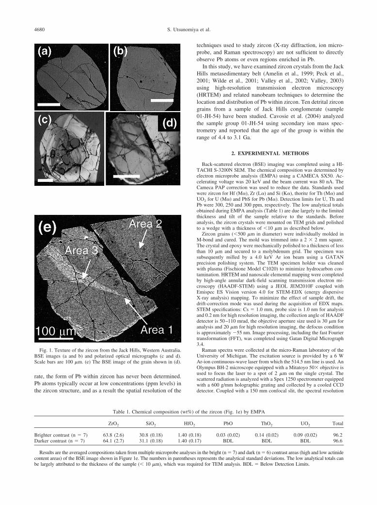
4680 S. Utsunomiya et al.
rate, the form of Pb within zircon has never been determined.Pb atoms typically occur at low concentrations (ppm levels) inthe zircon structure, and as a result the spatial resolution of the
Fig. 1. Texture of the zircon from the Jack Hills, Western Australia.BSE images (a and b) and polarized optical micrographs (c and d).Scale bars are 100 �m. (e) The BSE image of the grain shown in (d).
Table 1. Chemical composition (
ZrO2 SiO2
Brighter contrast (n � 7) 63.8 (2.6) 30.8 (0.18) 1.Darker contrast (n � 7) 64.1 (2.7) 31.1 (0.18) 1.
Results are the averaged compositions taken from multiple microprobe a
content areas) of the BSE image shown in Figure 1e. The numbers in parenthesesbe largely attributed to the thickness of the sample (� 10 �m), which was requitechniques used to study zircon (X-ray diffraction, ion micro-probe, and Raman spectroscopy) are not sufficient to directlyobserve Pb atoms or even regions enriched in Pb.
In this study, we have examined zircon crystals from the JackHills metasedimentary belt (Amelin et al., 1999; Peck et al.,2001; Wilde et al., 2001; Valley et al., 2002; Valley, 2003)using high-resolution transmission electron microscopy(HRTEM) and related nanobeam techniques to determine thelocation and distribution of Pb within zircon. Ten detrital zircongrains from a sample of Jack Hills conglomerate (sample01-JH-54) have been studied. Cavosie et al. (2004) analyzedthe sample group 01-JH-54 using secondary ion mass spec-trometry and reported that the age of the group is within therange of 4.4 to 3.1 Ga.
2. EXPERIMENTAL METHODS
Back-scattered electron (BSE) imaging was completed using a HI-TACHI S-3200N SEM. The chemical composition was determined byelectron microprobe analysis (EMPA) using a CAMECA SX50. Ac-celerating voltage was 20 keV and the beam current was 80 nA. TheCameca PAP correction was used to reduce the data. Standards usedwere zircon for Hf (M�), Zr (L�) and Si (K�), thorite for Th (M�) andUO2 for U (M�) and PbS for Pb (M�). Detection limits for U, Th andPb were 300, 250 and 300 ppm, respectively. The low analytical totalsobtained during EMPA analysis (Table 1) are due largely to the limitedthickness and tilt of the sample relative to the standards. Beforeanalysis, the zircon crystals were mounted on TEM grids and polishedto a wedge with a thickness of �10 �m as described below.
Zircon grains (�500 �m in diameter) were individually molded inM-bond and cured. The mold was trimmed into a 2 � 2 mm square.The crystal and epoxy were mechanically polished to a thickness of lessthan 10 �m and secured to a molybdenum grid. The specimen wassubsequently milled by a 4.0 keV Ar ion beam using a GATANprecision polishing system. The TEM specimen holder was cleanedwith plasma (Fischione Model C1020) to minimize hydrocarbon con-tamination. HRTEM and nanoscale elemental mapping were completedby high-angle annular dark-field scanning transmission electron mi-croscopy (HAADF-STEM) using a JEOL JEM2010F coupled withEmispec ES Vision version 4.0 for STEM-EDX (energy dispersiveX-ray analysis) mapping. To minimize the effect of sample drift, thedrift-correction mode was used during the acquisition of EDX maps.STEM specifications: Cs � 1.0 mm, probe size is 1.0 nm for analysisand 0.2 nm for high resolution imaging, the collection angle of HAADFdetector is 50–110 mrad, the objective aperture size used is 30 �m foranalysis and 20 �m for high resolution imaging, the defocus conditionis approximately �55 nm. Image processing, including the fast Fouriertransformation (FFT), was completed using Gatan Digital Micrograph3.4.
Raman spectra were collected at the micro-Raman laboratory of theUniversity of Michigan. The excitation source is provided by a 6 WAr-ion continuous-wave laser from which the 514.5 nm line is used. AnOlympus BH-2 microscope equipped with a Mitatoyo 50� objective isused to focus the laser to a spot of 2 �m on the single crystal. Thescattered radiation is analyzed with a Spex 1250 spectrometer equippedwith a 600 g/mm holographic grating and collected by a cooled CCDdetector. Coupled with a 150 mm confocal slit, the spectral resolution
f the zircon (Fig. 1e) by EMPA
PbO ThO2 UO2 Total
8) 0.03 (0.02) 0.14 (0.02) 0.09 (0.02) 96.27) BDL BDL BDL 96.6
in the bright (n � 7) and dark (n � 6) contrast areas (high and low actinide
wt%) o
HfO2
40 (0.140 (0.1
nalyses
represents the analytical standard deviations. The low analytical totals canred for TEM analysis. BDL � Below Detection Limits.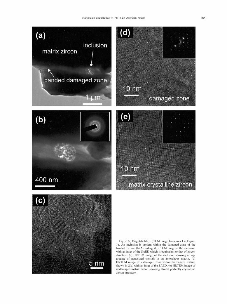
4681Nanoscale occurrence of Pb in an Archean zircon
Fig. 2. (a) Bright-field (BF)TEM image from area 1 in Figure1e. An inclusion is present within the damaged zone of thebanded texture. (b) An enlarged BFTEM image of the inclusionwith an inset of the SAED which is equivalent to that of zirconstructure. (c) HRTEM image of the inclusion showing an ag-gregate of nanosized crystals in an amorphous matrix. (d)HRTEM image of a damaged zone within the banded textureshown in 2(a) with an inset of the SAED. (e) HRTEM image ofundamaged matrix zircon showing almost perfectly crystalline
zircon structure.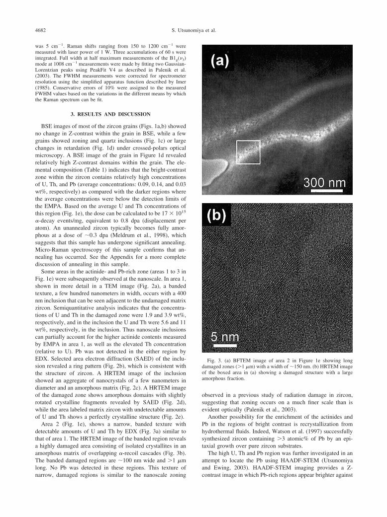
4682 S. Utsunomiya et al.
was 5 cm�1. Raman shifts ranging from 150 to 1200 cm�1 weremeasured with laser power of 1 W. Three accumulations of 60 s wereintegrated. Full width at half maximum measurements of the B1g(�3)mode at 1008 cm�1 measurements were made by fitting two Gaussian-Lorentzian peaks using PeakFit V4 as described in Palenik et al.(2003). The FWHM measurements were corrected for spectrometerresolution using the simplified apparatus function described by Imer(1985). Conservative errors of 10% were assigned to the measuredFWHM values based on the variations in the different means by whichthe Raman spectrum can be fit.
3. RESULTS AND DISCUSSION
BSE images of most of the zircon grains (Figs. 1a,b) showedno change in Z-contrast within the grain in BSE, while a fewgrains showed zoning and quartz inclusions (Fig. 1c) or largechanges in retardation (Fig. 1d) under crossed-polars opticalmicroscopy. A BSE image of the grain in Figure 1d revealedrelatively high Z-contrast domains within the grain. The ele-mental composition (Table 1) indicates that the bright-contrastzone within the zircon contains relatively high concentrationsof U, Th, and Pb (average concentrations: 0.09, 0.14, and 0.03wt%, respectively) as compared with the darker regions wherethe average concentrations were below the detection limits ofthe EMPA. Based on the average U and Th concentrations ofthis region (Fig. 1e), the dose can be calculated to be 17 � 1015
�-decay events/mg, equivalent to 0.8 dpa (displacement peratom). An unannealed zircon typically becomes fully amor-phous at a dose of �0.3 dpa (Meldrum et al., 1998), whichsuggests that this sample has undergone significant annealing.Micro-Raman spectroscopy of this sample confirms that an-nealing has occurred. See the Appendix for a more completediscussion of annealing in this sample.
Some areas in the actinide- and Pb-rich zone (areas 1 to 3 inFig. 1e) were subsequently observed at the nanoscale. In area 1,shown in more detail in a TEM image (Fig. 2a), a bandedtexture, a few hundred nanometers in width, occurs with a 400nm inclusion that can be seen adjacent to the undamaged matrixzircon. Semiquantitative analysis indicates that the concentra-tions of U and Th in the damaged zone were 1.9 and 3.9 wt%,respectively, and in the inclusion the U and Th were 5.6 and 11wt%, respectively, in the inclusion. Thus nanoscale inclusionscan partially account for the higher actinide contents measuredby EMPA in area 1, as well as the elevated Th concentration(relative to U). Pb was not detected in the either region byEDX. Selected area electron diffraction (SAED) of the inclu-sion revealed a ring pattern (Fig. 2b), which is consistent withthe structure of zircon. A HRTEM image of the inclusionshowed an aggregate of nanocrystals of a few nanometers indiameter and an amorphous matrix (Fig. 2c). A HRTEM imageof the damaged zone shows amorphous domains with slightlyrotated crystalline fragments revealed by SAED (Fig. 2d),while the area labeled matrix zircon with undetectable amountsof U and Th shows a perfectly crystalline structure (Fig. 2e).
Area 2 (Fig. 1e), shows a narrow, banded texture withdetectable amounts of U and Th by EDX (Fig. 3a) similar tothat of area 1. The HRTEM image of the banded region revealsa highly damaged area consisting of isolated crystallites in anamorphous matrix of overlapping �-recoil cascades (Fig. 3b).The banded damaged regions are �100 nm wide and �1 �mlong. No Pb was detected in these regions. This texture of
narrow, damaged regions is similar to the nanoscale zoningobserved in a previous study of radiation damage in zircon,suggesting that zoning occurs on a much finer scale than isevident optically (Palenik et al., 2003).
Another possibility for the enrichment of the actinides andPb in the regions of bright contrast is recrystallization fromhydrothermal fluids. Indeed, Watson et al. (1997) successfullysynthesized zircon containing �3 atomic% of Pb by an epi-taxial growth over pure zircon substrates.
The high U, Th and Pb region was further investigated in anattempt to locate the Pb using HAADF-STEM (Utsunomiyaand Ewing, 2003). HAADF-STEM imaging provides a Z-
Fig. 3. (a) BFTEM image of area 2 in Figure 1e showing longdamaged zones (�1 �m) with a width of �150 nm. (b) HRTEM imageof the boxed area in (a) showing a damaged structure with a largeamorphous fraction.
contrast image in which Pb-rich regions appear brighter against
e from
4683Nanoscale occurrence of Pb in an Archean zircon
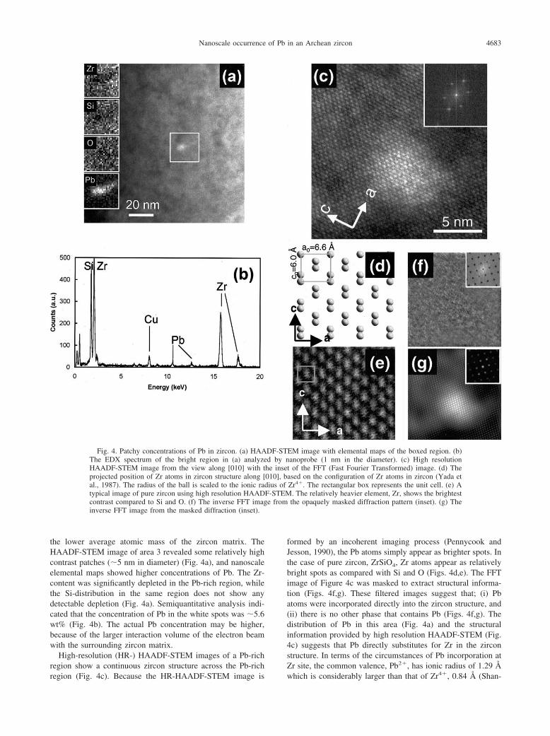
the lower average atomic mass of the zircon matrix. TheHAADF-STEM image of area 3 revealed some relatively highcontrast patches (�5 nm in diameter) (Fig. 4a), and nanoscaleelemental maps showed higher concentrations of Pb. The Zr-content was significantly depleted in the Pb-rich region, whilethe Si-distribution in the same region does not show anydetectable depletion (Fig. 4a). Semiquantitative analysis indi-cated that the concentration of Pb in the white spots was �5.6wt% (Fig. 4b). The actual Pb concentration may be higher,because of the larger interaction volume of the electron beamwith the surrounding zircon matrix.
High-resolution (HR-) HAADF-STEM images of a Pb-richregion show a continuous zircon structure across the Pb-rich
Fig. 4. Patchy concentrations of Pb in zircon. (a) HAAThe EDX spectrum of the bright region in (a) analyzeHAADF-STEM image from the view along [010] with tprojected position of Zr atoms in zircon structure along [al., 1987). The radius of the ball is scaled to the ionic ratypical image of pure zircon using high resolution HAADcontrast compared to Si and O. (f) The inverse FFT imaginverse FFT image from the masked diffraction (inset).
region (Fig. 4c). Because the HR-HAADF-STEM image is
formed by an incoherent imaging process (Pennycook andJesson, 1990), the Pb atoms simply appear as brighter spots. Inthe case of pure zircon, ZrSiO4, Zr atoms appear as relativelybright spots as compared with Si and O (Figs. 4d,e). The FFTimage of Figure 4c was masked to extract structural informa-tion (Figs. 4f,g). These filtered images suggest that; (i) Pbatoms were incorporated directly into the zircon structure, and(ii) there is no other phase that contains Pb (Figs. 4f,g). Thedistribution of Pb in this area (Fig. 4a) and the structuralinformation provided by high resolution HAADF-STEM (Fig.4c) suggests that Pb directly substitutes for Zr in the zirconstructure. In terms of the circumstances of Pb incorporation atZr site, the common valence, Pb2�, has ionic radius of 1.29 Å
EM image with elemental maps of the boxed region. (b)anoprobe (1 nm in the diameter). (c) High resolution
t of the FFT (Fast Fourier Transformed) image. (d) Theased on the configuration of Zr atoms in zircon (Yada etZr4�. The rectangular box represents the unit cell. (e) A. The relatively heavier element, Zr, shows the brightestthe opaquely masked diffraction pattern (inset). (g) The
DF-STd by nhe inse010], bdius ofF-STEM
which is considerably larger than that of Zr4�, 0.84 Å (Shan-
4684 S. Utsunomiya et al.
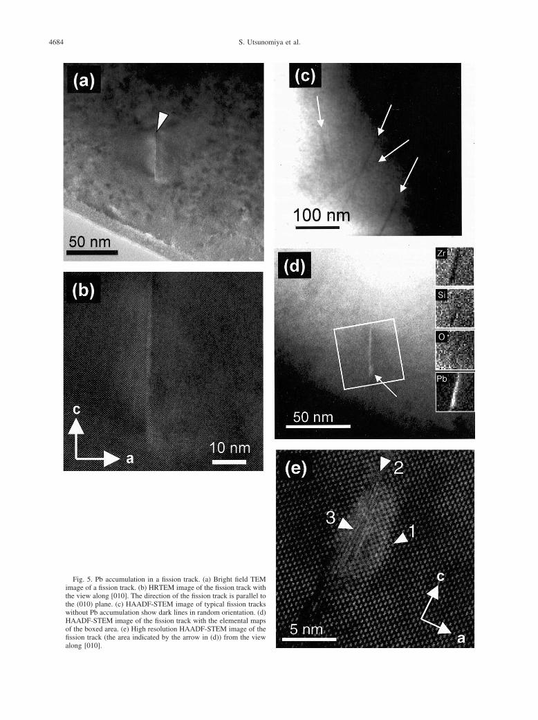
Fig. 5. Pb accumulation in a fission track. (a) Bright field TEMimage of a fission track. (b) HRTEM image of the fission track withthe view along [010]. The direction of the fission track is parallel tothe (010) plane. (c) HAADF-STEM image of typical fission trackswithout Pb accumulation show dark lines in random orientation. (d)HAADF-STEM image of the fission track with the elemental mapsof the boxed area. (e) High resolution HAADF-STEM image of thefission track (the area indicated by the arrow in (d)) from the view
along [010].
4685Nanoscale occurrence of Pb in an Archean zircon
non, 1976). A less common valence, Pb4�, has an ionic radius(Pb4� � 0.94 Å), compatible with possible substitution ofZr4�. Despite the size preference for Pb4�, the Pb-O system isdominated by PbO under typical geological conditions, with asmall stability field for native Pb. The stability field for PbO2
exists only under far more oxidizing conditions (Watson et al.,1997). However, it is possible that the compatibility of the sizeand charge can result in stabilization of the tetravalent oxida-tion state in the zircon structure. Because the concentration ofPb at this patch is only �3 atomic%, it is difficult to draw anyconclusions about the valence of Pb using only the structuralinformation obtained from the HRSTEM image.
In addition to the patchy occurrence of Pb, the Pb-rich areaalso revealed numerous fission tracks (�5 nm width) createdby high-energy (�100 MeV), heavy particles from spontaneousfission events (Figs. 5a,b). Although spontaneous fission of238U is infrequent (decay constant � 10�16/yr (Yada et al.,1987)), the old age and actinide content of these zircons suggestthat fission tracks are to be expected. Although a fission trackis typically up to 10 �m in length (Yada et al., 1981), the lengthof fission tracks observed here are �50 nm. The observablelength is limited mainly by the thickness of the imaged speci-men, which depends on the thickness through which a 200 keVelectron can penetrate and the random orientation of fissiontracks in three dimensions. The majority of the fission trackswere visible as streaks of dark contrast in the HAADF-STEMimages (Fig. 5c); however, one fission track showed brightcontrast (Fig. 5d) throughout the length of the entire track. Theelemental maps indicated that this fission track had a higherconcentration of Pb than the matrix. The orientation of thisfission track is parallel to the (011) plane (Fig. 5b), which isconsistent with channeling of fission fragment in the structure(Yada et al., 1987). The high resolution HAADF-STEM imageof the end of this fission track (Fig. 5e) shows: (I) The edge ofthe zircon lattice, indicated by an arrow 1, has a higher contrastthan the bulk zircon matrix, suggesting the incorporation of Pbatoms in the zircon lattice adjacent to the fission track. This isalso supported by the Pb elemental map in Figure 5d. (II) Thereis no continuous lattice within the track (arrow 2). The interfacebetween the zircon matrix and the fission track (arrow 3),shows a disordered array of bright contrast, suggesting that theaccumulated Pb is not in a crystalline phase or, if crystalline,without any preferred orientation relative to the surroundingzircon structure. In addition, the oxygen distribution in this areadoes not show any evidence of O-depletion in the fission track,which indicates that Pb is not present as elemental Pb.
The occurrence of Pb in the fission track is obviously notoriginal, since spontaneous fission occurred after zircon forma-tion. Thus, Pb probably accumulated in the track by diffusion,which, in the case of the images shown in Figures 5d and e,appears to be at the end of the fission track. A similar textureof U or Th accumulation in the fission track was not observed.In general, fission tracks are amorphous regions (Yada et al.,1981), as anticipated by irradiation experiments using highly-energetic heavy ions (Ewing et al., 2003). The Pb that hasaccumulated in the fission track observed here suggests that Pbcan diffuse through an amorphous domain, such as fissiontracks, as well as metamict domains resulting from the accu-mulation of �-decay event damage (e.g., the banded amorphous
regions in Figs. 2 and 3). In fact, previous experiments (Cher-niak et al., 1991; Cherniak, 1993) on the Pb diffusion rate inzircon have predicted that Pb diffusion through highly damagedpaths would show a higher diffusion rate. The above observa-tions are consistent with the percolation theory model of radi-ation damage accumulation in zircon, whereby, radiation dam-age accumulation results first in the formation of isolatedamorphous domains that then overlap as the damage accumu-lates, eventually creating a network of interconnected amor-phous domains that could serve as a fast diffusion pathway(Salje et al., 1999; Ríos et al., 2000; Trachenko et al., 2003).The first percolation point is defined as the dose at which thedamaged amorphous domains become fully interconnected.These results show the potential significance of Pb diffusionthrough amorphous domains that result from radiation-damage.
Acknowledgments—S.U. thanks J. F. Mansfield and C. J. Wauchope fortheir technical support in the Electron Microbeam Analysis Laboratoryat the University of Michigan and M. Kawasaki and S. Johnson ofJEOL, USA, for important advice on the use of the STEM. We verymuch appreciate the constructive review comments by Professor BruceWatson, Dr. David Cole, and two anonymous reviewers. We thankProfessor Lars Stixrude for access to the Raman microprobe (supportedby NSF EAR99-73050 to L. Stixrude). This work was supported by theDivision of Materials Science of the Office of Basic Energy Science ofthe U.S. Department of Energy (DE-FG02-97ER45656).
Associate editor: D. Cole
REFERENCES
Amelin Y., Lee D. C., Halliday A. N., and Pidgeon R. T. (1999) Natureof the Earth’s earliest crust from hafnium isotopes in single detritalzircons. Nature 399, 252–255.
Bogomolov Ye. S. (1991) Migration of lead in non-metamict zircon.Earth Planet. Sci. Lett. 107, 625–633.
Bowring S. A. and Housh T. (1995) The Earth’s early evolution.Science 269, 1535–1540.
Bowring S. A., King J. E., Housh T. B., Isachsen C. E., and PodosekF. A. (1989) Neodymium and lead isotope evidence for enrichedearly Archaean crust in North America. Nature 340, 222–225.
Cavosie A. J., Wilde S. A., Liu D., Valley J. W., and Weiblen P. W.(2004) Internal zoning and U-Th-Pb chemistry of Jack Hills detritalzircons: A mineral record of Early Archean (4404-1576 Ma) mag-matism. Precambrian Research. (in press).
Cherniak D. J. (1993) Lead diffusion in titanite and preliminary resultson the effects of radiation damage on Pb transport. Chem. Geol.110, 177–194.
Cherniak D. J., Lanford W. A., and Ryerson F. J. (1991) Lead diffusionin apatite and zircon using ion implantation and Rutherford Back-scattering techniques. Geochim. Cosmochim. Acta 55, 1663–1673.
Cherniak D. J., Hanchar J. M., and Watson E. B. (1997) Diffusion oftetravalent cations in zircon. Contrib. Mineral. Petrol. 127, 383–390.
Cherniak D. J. and Watson E. B. (2000) Pb diffusion in zircon. Chem.Geol. 172, 5–24.
Craig H. (1968) Zircon lead loss—A kinetic model. Science 159, 447.Ewing R. C., Lutz W., and Weber W. J. (1995) Zircon: A host-phase
for the disposal of weapons plutonium. J. Mater. Res. 10, 253–246.Ewing R. C. (1999) Nuclear waste forms for actinides. Proc. Nat. Acad.
Sci. USA 96, 3432–3439.Ewing R. C., Chakoumakos B. C., Lumpkin G. R., and Murakami T.
(1987) The metamict state. Mater. Res. Soc. Bull. 12, 58–66.Ewing R. C., Meldrum A., Wang L. M. and Wang S. X. (2000)
Radiation-induced amorphization. In Transformation Processes inMinerals (eds. S. A. T. Redfern and M. A. Carpenter), pp. 319–361.Rev. Mineral. Geochem. 39. Mineralogical Society of America.
Ewing R. C., Meldrum A., Wang L. M., Weber W. J., and CorralesL. R. (2003) Radiation damage in zircon. In Zircon (eds. J. M.
4686 S. Utsunomiya et al.
Hanchar and P. W. O. Hoskin), pp. 387–425. Rev. Mineral.Geochem. 53. Mineralogical Society of America.
Imer G. (1985) Zum Einfluss der Apparatefunktion auf die Bestim-mung von Streuquerschnitten und Lebensdauern aus optishchenPhononenspectren. Exp. Tech. Phys. 33, 501–506.
Krogh T. E. (1982) Improved accuracy of U-Pb zircon ages by thecreation of more concordant systems using an air abrasion tech-nique. Geochim. Cosmochim. Acta 46, 637–649.
Lee J. K. W., Williams I. S., and Ellis D. J. (1997) Pb, U and Thdiffusion in natural zircon. Nature 390, 159–162.
Lian J., Ríos S., Boatner L. A., Wang L. M., and Ewing R. C. (2003)Microstructural evolution and nanocrystal formation in Pb�-im-planted ZrSiO4 single crystals. J. Appl. Phys. 94, 5695–5703.
Meldrum A., Zinkle S. J., Boatner L. A., and Ewing R. C. (1998) Atransient liquid-like phase in the displacement cascades of zircon,hafnon and thorite. Nature 395, 56–58.
Nasdala L., Wenzel M., Vavra G., Irmer G., Wenzel T., and Kober B.(2001) Metamictisation of natural zircon: Accumulation versusthermal annealing of radioactivity-induced damage. Contrib. Min-eral. Petrol. 141, 125–144.
Nasdala L., Reiners P. W., Garver J. I., Kennedy A. K., Stern R. A.,Balan E., and Wirth R. (2004) Incomplete retention of radia-tion damage in zircon from Sri Lanka. Am. Mineral. 89, 219 –231.
Palenik C. S., Nasdala L., and Ewing R. C. (2003) Radiation damagein zircon. Am. Mineral. 88, 770–781.
Peck W. H., Valley J. W., Wilde S. A., and Graham C. M. (2001)Oxygen isotope ratios and rare earth elements in 3.3 to 4.4 Gazircons: Ion microprobe evidence for high �18O continental crustand oceans in the Early Archean. Geochim. Cosmochim. Acta 65,4215–4229.
Pennycook S. J. and Jesson D. E. (1990) High-resolution incoherentimaging of crystals. Phys. Rev. Lett. 64, 938–941.
Pidgeon R. T., O’Neil J. R., and Silver L. T. (1966) Uranium and leadisotopic stability in a metamict zircon under experimental hydro-thermal conditions. Science 154, 1538–1540.
Ríos S., Salje E. K. H., Zhang M., and Ewing R. C. (2000) Amor-phization in zircon: Evidence for direct impact damage. J. Phys.Condensed Matt. 12, 2401–2412.
Salje E. K. H., Chrosch J., and Ewing R. C. (1999) Is “metamictiza-tion” of zircon a phase transition? Am. Mineral. 84, 1107–1116.
Shannon R. D. (1976) Revised effective ionic radii and systematicstudies of interatomic distances in halides and chalcogenides. ActaCrystallogr. A 32, 751–767.
Speer J. A. (1982) Zircon. In Orthosilicates (ed. P. H. Ribbe), pp.67–112. Reviews in Mineralogy 5, Mineralogical Society of Amer-ica.
Trachenko K., Dove M. T., and Salje E. K. H. (2003) Large swellingand percolation in irradiated zircon. J. Phys. Condensed Matt. 15,L1–L7.
Utsunomiya S. and Ewing R. C. (2003) Application of high-angleannular dark field scanning transmission electron microscopy—energy dispersive X-ray spectrometry, and energy-filtered transmis-sion electron microscopy to the characterization of nanoparticles inthe environment. Environ. Sci. Technol. 37, 786–791.
Valley J. W. (2003) Oxygen isotopes in zircon. In Zircon (eds. J. M.Hanchar and P. W. O. Hoskin), pp. 343–385. Reviews in Mineral-ogy 53, Mineralogical Society of America.
Valley J. W., Peck W. H. King E. M., and Wilde S. A. (2002) Coolearly Earth. Geology 30, 351–354.
Vervoort J. D., Patchett P. J., Gehrels G. E., and Nutman A. P. (1996)Constraints on early Earth differentiation from hafnium and neody-mium isotopes. Nature 379, 624–627.
Watson E. B., Cherniak D. J., Hanchar J. M., Harrison T. M., and WarkD. A. (1997) The incorporation of Pb into zircon. Chem. Geol. 141,19–31.
Wilde S. A., Valley J. W. Peck W. H., and Graham C. M. (2001)Evidence from detrital zircons for the existence of continental crust
and oceans on the Earth 4.4 Gyr ago. Nature 409, 175–178.Yada K., Tanji T., and Sunagawa I. (1981) Application of latticeimagery to radiation damage investigation in natural zircon. Phys.Chem. Miner. 7, 47–52.
Yada K., Tanji T., and Sunagawa I. (1987) Radiation induced latticedefects in natural zircon (ZrSiO4) observed at atomic resolution.Phys Chem. Miner. 14, 197–204.
APPENDIX
Based on the U and Th concentration analyzed by EMPA, the doseaccumulated in the brighter contrast area (Fig. 1e) can be calculated tobe 17 � 1015 �-decay events/mg, equivalent to 0.8 dpa. Refer toPaleniket al. (2003) for the details of the dose calculations. Although theaccumulated dose calculated using EMPA data was 0.8 dpa and zirconshould become fully amorphous at �0.3 dpa at room temperature(Meldrum et al., 1998), nanoscale observation by HRTEM did notreveal any fully amorphous regions (Figs. 2 and 3).
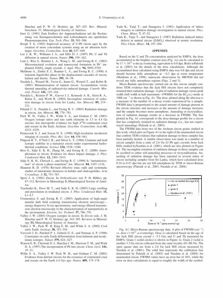
Micro-Raman spectroscopy carried out on this zircon sample con-firms TEM evidence that the Jack Hill zircons have not completelyretained their radiation damage. A plot of radiation damage versus peakwidth (full width at half maximum—FWHM) for the B1g(�3) mode at1008 cm�1 is shown in Fig. A1. The dose, plotted along the abscissa isa measure of the number of �-decay events experienced by a sample.FWHM data is proportional to the actual amount of damage present inthe zircon structure and increases as the amount of damage increasesand the sample becomes more amorphous. Annealing or recrystalliza-tion of radiation damage results in a decrease in FWHM. The lineplotted in Fig. A1 corresponds to the dose-damage profile for a zirconthat has completely retained its radiation damage (i.e., has not experi-enced annealing) (Palenik et al., 2003).
The FWHM data from two of the Archean zircon grains studied inthis work, which plot on Figure A1 to the right of the unannealed zirconline confirm TEM evidence that radiation damage in these samples hasbeen partially healed. A similar observation was made in a number ofzircon samples from a suite of 2.6 Ga (U-Pb age) crystals from JackHills studied byNasdala et al. (2001), which are also plotted on FigureA1. The incomplete retention of radiation damage in these samples canbe ascribed to either self-annealing processes or recrystallization. An-nealing of radiation damage has been reported in several suites ofzircon including samples from Sri Lanka, which have calculated dose0.16 to 0.47 dpa but are not full amorphous by TEM or micro-Ramanspectroscopy (Palenik et al., 2003; Nasdala et al., 2004).
Fig. A1. Micro-Raman spectroscopy data. A plot of FWHM (cm�1)vs. dose (�1015 �-events/mg). Dose is calculated based on the age ofthe Jack Hill zircon crystal (�3.3 Ga) and U and Th measured byEMPA. Grain 1 (solid circles) is shown in Figure 1e. Grain 2 (solid isanother 3.3 Ga zircon collected from the same locality (01-JH-54). Theopen square data are from a 2.6 Ga Jack Hill zircon measured byNasdala et al. (2001). The solid line represents the calibration linedetermined by Palenik et al. (2003) and Nasdala et al. (2004) forunannealed zircon. FWHM values have an error bar of 10%, while the
error on dose calculations is equal to roughly the width of the symbol.
Related Documents