RAVARIAN ET AL . VOL. 7 ’ NO. 10 ’ 8469–8483 ’ 2013 www.acsnano.org 8469 September 03, 2013 C 2013 American Chemical Society Nanoscale Chemical Interaction Enhances the Physical Properties of Bioglass Composites Roya Ravarian, † Xia Zhong, † Mike Barbeck, ‡ Shahram Ghanaati, ‡ Charles James Kirkpatrick, ‡ Ciara M. Murphy, §,^ Aaron Schindeler, §,^ Wojciech Chrzanowski, ) and Fariba Dehghani †, * † School of Chemical and Biomolecular Engineering, The University of Sydney, Sydney, NSW, Australia, ‡ Institute of Pathology, University Medical Center of the Johannes Gutenberg University, Mainz, Germany, § Orthopaedic Research & Biotechnology Unit, The Children's Hospital at Westmead, Sydney, Australia, ^ Discipline of Paediatrics and Child Health, Faculty of Medicine, The University of Sydney, Sydney, Australia, and ) Faculty of Pharmacy, The University of Sydney, Sydney, NSW, Australia B ioceramics such as bioglass are favor- able materials for bone grafting appli- cations due to the high biocompatibility and bonding capability to the host tissue in the body. 16 The surface of bioactive materi- als forms a biologically active hydroxyapatite layer, which provides the bonding interface with tissues. This apatite phase is chemically and structurally equivalent to the mineral phase in bone, providing interfacial bonding. 7 Among bioceramics, silica-based materials show great potential for bone tissue regen- eration applications. One of the major obsta- cles for the application of bioglass in bone repair is its poor mechanical properties, which make its manufacturing a challenging issue. Several attempts have been made to resolve the issues associated with the brittle and fragile structure of bioglass prior to bone grafting. 8 Introducing materials with a lower elastic modulus such as polymer matrices is a strategy to improve the mechanical proper- ties of bioglass and mimic the natural bone structure. 911 Poly(methyl methacrylate) (PMMA) is a synthetic polymer often used to augment the mechanical properties of bioglass. 8,1115 This polymer has broad biomedical applica- tions due to its self-hardening property and excellent mechanical properties. 16 It has been used for prosthetic fixation, as a bone substitute in orthopedics, and as a self- curing reagent in dental prosthetics. 17,18 PMMA-based bone cements were first in- troduced in 1960s. 19 Since then, progress has been made to improve their properties for fixation of implants and filling bone voids after removal of tumors or trauma. 13 For example, in orthopedic and dentistry operations, a mixture of methyl methacrylate * Address correspondence to [email protected]. Received for review April 30, 2013 and accepted September 3, 2013. Published online 10.1021/nn402157n ABSTRACT Bioglasses are favorable biomaterials for bone tissue engineering; however, their applications are limited due to their brittleness. In addition, the early failure in the interface is a common problem of composites of bioglass and a polymer with high mechanical strength. This effect is due to the phase separation, nonhomogeneous mixture, nonuniform mechanical strength, and different degradation properties of two compounds. To address these issues, in this study a nanoscale interaction between poly(methyl methacrylate) (PMMA) and bioactive glass was formed via silane coupling agent (3-trimethoxysilyl)propyl methacrylate (MPMA). A monolith was produced at optimum composition from this hybrid by the solgel method at 50 °C with a rapid gelation time (<50 min) that possessed superior physicochemical properties compared to pure bioglass and physical mixture. For instance, the Young's modulus of bioglass was decreased 40-fold and the dissolution rate of silica was retarded 1.5-fold by integration of PMMA. Prolonged dissolution of silica fosters bone integration due to the continuous dissolution of bioactive silica. The primary osteoblast cells were well anchored and cell migration was observed on the surface of the hybrid. The in vivo studies in mice demonstrated that the integrity of the hybrids was maintained in subcutaneous implantation. They induced mainly a mononuclear phagocytic tissue reaction with a low level of inflammation, while bioglass provoked a tissue reaction with TRAP-positive multinucleated giant cells. These results demonstrated that the presence of a nanoscale interaction between bioglass and PMMA affects the properties of bioglass and broadens its potential applications for bone replacement. KEYWORDS: hybrids . PMMA . bioglass . solgel . bone regenerative material . nanocomposites ARTICLE

Welcome message from author
This document is posted to help you gain knowledge. Please leave a comment to let me know what you think about it! Share it to your friends and learn new things together.
Transcript
RAVARIAN ET AL . VOL. 7 ’ NO. 10 ’ 8469–8483 ’ 2013
www.acsnano.org
8469
September 03, 2013
C 2013 American Chemical Society
Nanoscale Chemical InteractionEnhances the Physical Propertiesof Bioglass CompositesRoya Ravarian,† Xia Zhong,† Mike Barbeck,‡ Shahram Ghanaati,‡ Charles James Kirkpatrick,‡
Ciara M. Murphy,§,^ Aaron Schindeler,§,^ Wojciech Chrzanowski, ) and Fariba Dehghani†,*
†School of Chemical and Biomolecular Engineering, The University of Sydney, Sydney, NSW, Australia, ‡Institute of Pathology, University Medical Center of theJohannes Gutenberg University, Mainz, Germany, §Orthopaedic Research & Biotechnology Unit, The Children's Hospital at Westmead, Sydney, Australia, ^Discipline ofPaediatrics and Child Health, Faculty of Medicine, The University of Sydney, Sydney, Australia, and )Faculty of Pharmacy, The University of Sydney, Sydney, NSW, Australia
Bioceramics such as bioglass are favor-able materials for bone grafting appli-cationsdue to thehighbiocompatibility
and bonding capability to the host tissue inthe body.1�6 The surface of bioactive materi-
als forms a biologically active hydroxyapatite
layer, which provides the bonding interface
with tissues. This apatite phase is chemically
and structurally equivalent to the mineral
phase in bone, providing interfacial bonding.7
Among bioceramics, silica-based materials
show great potential for bone tissue regen-
eration applications. One of the major obsta-
cles for the application of bioglass in bone
repair is its poormechanical properties, which
make its manufacturing a challenging issue.
Several attempts have been made to resolve
the issues associated with the brittle and
fragile structure of bioglass prior to bone
grafting.8 Introducing materials with a lower
elastic modulus such as polymer matrices is astrategy to improve the mechanical proper-ties of bioglass and mimic the natural bonestructure.9�11
Poly(methyl methacrylate) (PMMA) is asynthetic polymer often used to augment
the mechanical properties of bioglass.8,11�15
This polymer has broad biomedical applica-tions due to its self-hardening property and
excellent mechanical properties.16 It has
been used for prosthetic fixation, as a bonesubstitute in orthopedics, and as a self-
curing reagent in dental prosthetics.17,18
PMMA-based bone cements were first in-troduced in 1960s.19 Since then, progress
has been made to improve their properties
for fixation of implants and filling bone
voids after removal of tumors or trauma.13
For example, in orthopedic and dentistry
operations, amixture ofmethylmethacrylate
* Address correspondence [email protected].
Received for review April 30, 2013and accepted September 3, 2013.
Published online10.1021/nn402157n
ABSTRACT Bioglasses are favorable biomaterials for bone tissue engineering; however, their
applications are limited due to their brittleness. In addition, the early failure in the interface is a
common problem of composites of bioglass and a polymer with high mechanical strength. This
effect is due to the phase separation, nonhomogeneous mixture, nonuniform mechanical strength,
and different degradation properties of two compounds. To address these issues, in this study a
nanoscale interaction between poly(methyl methacrylate) (PMMA) and bioactive glass was formed
via silane coupling agent (3-trimethoxysilyl)propyl methacrylate (MPMA). A monolith was produced
at optimum composition from this hybrid by the sol�gel method at 50 �C with a rapid gelation time(<50 min) that possessed superior physicochemical properties compared to pure bioglass and
physical mixture. For instance, the Young's modulus of bioglass was decreased 40-fold and the dissolution rate of silica was retarded 1.5-fold by integration
of PMMA. Prolonged dissolution of silica fosters bone integration due to the continuous dissolution of bioactive silica. The primary osteoblast cells were well
anchored and cell migration was observed on the surface of the hybrid. The in vivo studies in mice demonstrated that the integrity of the hybrids was
maintained in subcutaneous implantation. They induced mainly a mononuclear phagocytic tissue reaction with a low level of inflammation, while bioglass
provoked a tissue reaction with TRAP-positive multinucleated giant cells. These results demonstrated that the presence of a nanoscale interaction between
bioglass and PMMA affects the properties of bioglass and broadens its potential applications for bone replacement.
KEYWORDS: hybrids . PMMA . bioglass . sol�gel . bone regenerative material . nanocomposites
ARTIC
LE

RAVARIAN ET AL . VOL. 7 ’ NO. 10 ’ 8469–8483 ’ 2013
www.acsnano.org
8470
(MMA) monomer, initiators, and activators is placed inthe body, and the fixation occurs by in situ poly-merization.20 This approach, however, is not desirabledue to several issues including damage to the sur-rounding tissues as a result of temperature raise (70 �C)from an exothermic reaction; release of toxic com-pounds such as residual MMA monomer, initiators,and activators; and inertness and lack of bioactivity.These issues lead to the thickening of an interveningfibrous tissue layer; inhibition of cell function andgrowth/differentiation; increased inflammatory re-sponse and cell death/necrosis; and finally looseningof prosthesis/implants.11,13,21 Several approaches havebeen attempted to resolve the problems of PMMA-based implants. For example, the amount of MMAmonomer was reduced with the addition of PMMApolymer to the mixture. This reduced the heat genera-tion during the exothermic reaction, minimizing thedamage to surrounding tissues.22�24 However, thismethod still does not eradicate the issue of addingMMA monomer for in situ polymerization and lowbioactivity.Bioglass powder was added as a filler to PMMA-
based biomaterials to enhance their biocompatibilityand bioactivity.13,14,23,25 The amount and particle sizeof bioglass powder have a significant effect on thebioactivity of PMMA�bioglass physically mixed com-posites. Decreasing the particle size of bioglass in-creases the surface area, resulting in higher exposureof bioactive compounds to the surrounding tissue,hence enhancing the bioactivity of the implant.12,23,26
However, physicalmixtures of PMMAandbioglassmaybenonuniform due to the lack of adhesion between thesetwo materials and the absence of molecular interaction.Phase separation is a challenging issue in manufactur-
ing ceramic�polymer composites (physical mixtures)due to the different inherent physicochemical character-istics of these two compounds. This effect leads to theformation of cavities in their interfaces and suddenchanges in the structural properties of the material.27,28
Discrepancies in the physical properties, such as degra-dation rate and mechanical strength, increase the risk ofimplant failure.12,29 Additionally a UV- or photosensitiveinitiator may be added to the composite for in situ
polymerization of MMA monomer to promote homoge-neous mixing of these two compounds.30 This strategyaccelerates the gelation time of these composites, whichis favorable for injectable bone implants. However, thesereagents may be toxic to surrounding tissues.Organic�inorganic hybrids have been introduced as
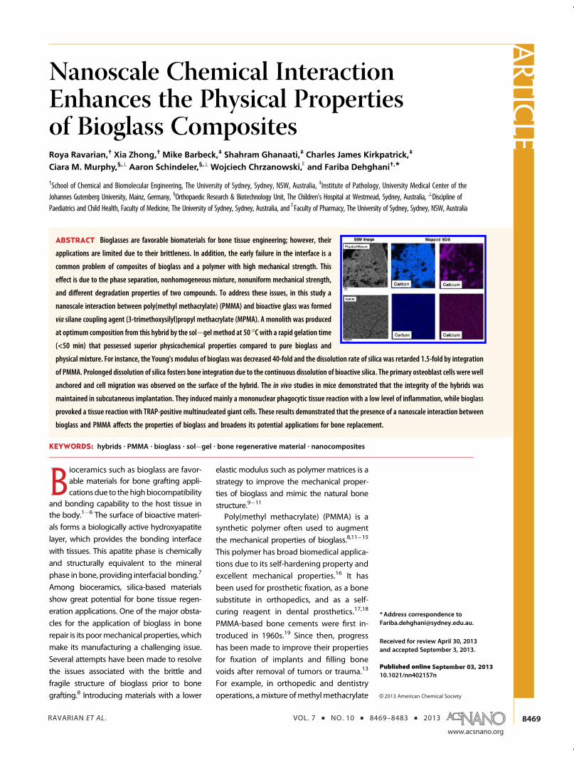
an alternative approach to address the issue of phaseseparation in physical mixing.31 The interaction betweenPMMA�bioglass ranges from weak van der Waals andhydrogen bondings (class I hybrid) to strong covalentbonds (class II hybrid). Organosilane coupling agentssuch as 3-(trimethoxysilyl)propyl methacrylate (MPMA),shown in Figure 1, are molecules with both organic and
inorganic moieties that provide the active sites for cova-lent bonding between organic and inorganicmolecules.32
These hybrids are commonly produced by the sol�gelmethod atmoderate temperatures, which is the favorablemethod for the incorporation of an organic compoundinto the structure of an inorganic substance withoutthermal degradation.33 Furthermore, the sol�gel methodis efficient in creating a homogeneous distribution be-tween two phases as a result of dissolving both com-pounds in a common solvent. The presence of MPMAorganosilane coupling agent results in the formation ofSi�O�Si covalent bonds to the silica network and forma-tion of a homogeneous mixture of PMMA�bioglass. Theschematic molecular structures of PMMA, MPMA, andPMMA�bioglass are shown in Figure 1.PMMA�silica hybrids have been used in optics,34
mechanics,34�36 and electronics.36,37 Few studiessynthesized and characterized the physical and che-mical properties of PMMA�silica hybrids and intro-duced this material as a candidate for bone cements38
and dental39,40 applications. The bioactivity of PMMA�silica hybrids was confirmed by the formation of anapatite layer on the surface.16,29 In comparison withpure PMMA polymer, the cell response was also pro-moted in samples containing silica.41,42 However, thesestudies have not investigated the impact of covalentbonding at the molecular level on bioactivity, physico-chemical, and biological properties of hybrids, whichwas one of the objectives for our study.MPMA has been used as a coupling agent to en-
hance chemical bonding between bioglass and PMMAto fabricate hybrids with superior properties. It wasperceived thatMPMAcomposition has a negligible effecton the molecular interaction between PMMA�bioglassand formation of the hybrid.43 However, we have pre-viously demonstrated that the composition ofMPMAhasa significant impact on chemical conjugation betweenPMMA and silica and formation of homogeneoushybrids.44 The hybrids produced from a 0.1 molar ratioof MPMA:MMA were transparent, underlining the ab-sence of phase separation; however, at lowermolar ratios(i.e., 0.004, 0.02), samples were opaque due to phaseseparation between PMMA and bioglass. This observa-tion was confirmed by the results of one- and two-dimensional solid-state NMR, indicating strong intermo-lecular interaction between bioglass and PMMA at amolecular level when using 0.1 molar ratio of MPMA:MMA, while at lower ratios this interaction was low.44
Additionally, this nanoscale interaction shifted the de-gradation temperature of this hybrid fabricated at 0.1molar ratio of MPMA:MMA to 400 �C, which was at least10% higher than PMMA and hybrids prepared fromMPMA:MMA < 0.1 (mol ratio).43,44 The PMMA-co-MPMAproduced at highermolar ratios ofMPMA:MMA (e.g. >0.1)was very viscous and involved complicated purificationsteps. Therefore, a 0.1 MPMA:MMA molar ratio wasdeemed as the optimum composition for the fabrication
ARTIC
LE
RAVARIAN ET AL . VOL. 7 ’ NO. 10 ’ 8469–8483 ’ 2013
www.acsnano.org
8471
of bioglass�PMMA hybrids. It is important to note that itwas viable to fabricate hybrids with various ratios ofbioglass:PMMAwhen using a 0.1MPMA:MMAmolar ratio.The morphology, mechanical properties, and degra-
dation profiles are the crucial factors for bone implantapplications. Therefore, in this study, we aim to inves-tigate the effect of intermolecular interactions on theproperties of PMMA�bioglass. A 0.1 MPMA:PMMAmolar ratio was used to fabricate hybrids, and theirproperties were compared with a physical mixture andneat bioglass. Analytical methods such as scanningelectron microscopy (SEM), energy dispersive spectro-metry (EDS), atomic force microscopy (AFM), and scan-ning transmission electron microscopy (STEM) wereused to investigate the surface properties of thesecomposites. Furthermore, the effect of covalent bond-ing and molecular interaction of hybrids on the me-chanical strength and degradation profile wasassessed. Finally, in vitro and in vivo assays wereconducted to compare the bioactivity, cell adhesionproperties, and cytotoxicity of hybrids with bioglassand a physical mixture.
RESULTS AND DISCUSSION
In this study the physical properties of the hybrid(H1) were compared with the physical mixture ofPMMA�bioglass (H0). Both pure bioglass and PMMAhomopolymer were considered as control samples, asshown in Table 1. It was observed that only H1 formeda one-phase, transparent, three-dimensional (3D), mono-lith structure, which is due to the formation of covalentbondingbetweenMPMAand silanol groups of bioglass.44
However, in the absence of MPMA, the sol�gel methodwas not efficient for the creation of a uniform mixture ofPMMA and bioglass, and two separate phases of a thinfilm of polymer and a gel structure of brittle bioglasswereformed. Previous studies have shown that it is viableto create a 3D structure of PMMA�bioglass of differentcomposition by in situ polymerization ofMMAmonomersin the presence of bioglass as a filler. However, these
products are still not as uniform as hybrids and exhibit anonhomogenous degradation profile and nonuniformdistribution of mechanical force due to the differentload-bearing characteristics of polymer and bioglassparticles.45
The hybrid and bioglass were formed within 5 and120 h by the sol�gel method at room temperature,respectively. However, it was found that by increasingthe temperature from 25 �C to 50 �C the gelation timesof H1 and bioglass were decreased dramatically to 3 hand 45 min, respectively. These data demonstrate thesignificant impact of temperature on the formation of asilica network.46,47 The faster gelation of H1 comparedto bioglass was due to the presence of strong Si�Cbonds.46,48 Rapid gelation is favorable for the fabrica-tion of in situ products in clinical applications.
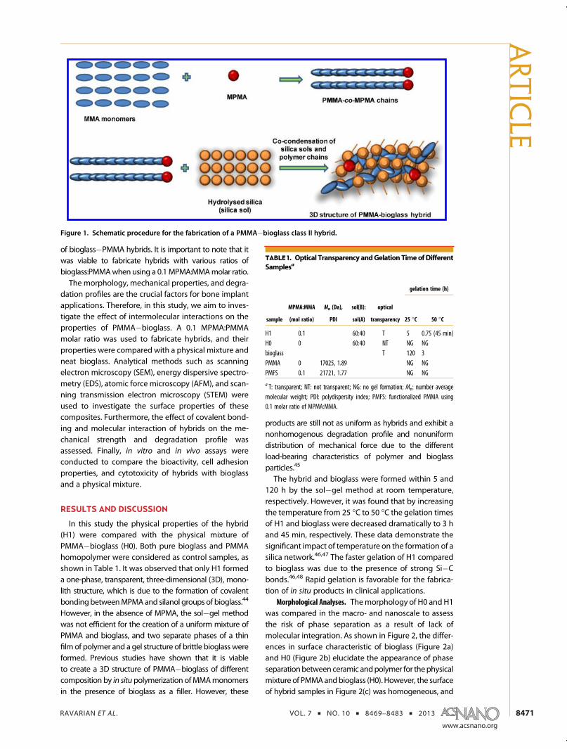
Morphological Analyses. Themorphology of H0 andH1was compared in the macro- and nanoscale to assessthe risk of phase separation as a result of lack ofmolecular integration. As shown in Figure 2, the differ-ences in surface characteristic of bioglass (Figure 2a)and H0 (Figure 2b) elucidate the appearance of phaseseparationbetweenceramic andpolymer for thephysicalmixture of PMMAandbioglass (H0). However, the surfaceof hybrid samples in Figure 2(c) was homogeneous, and
Figure 1. Schematic procedure for the fabrication of a PMMA�bioglass class II hybrid.
TABLE 1. Optical Transparency andGelationTimeofDifferent
Samplesa
gelation time (h)
sample
MPMA:MMA
(mol ratio)
Mn (Da),
PDI
sol(B):
sol(A)
optical
transparency 25 �C 50 �C
H1 0.1 60:40 T 5 0.75 (45 min)H0 0 60:40 NT NG NGbioglass T 120 3PMMA 0 17025, 1.89 NG NGPMFS 0.1 21721, 1.77 NG NG
a T: transparent; NT: not transparent; NG: no gel formation; Mn: number averagemolecular weight; PDI: polydispersity index; PMFS: functionalized PMMA using0.1 molar ratio of MPMA:MMA.
ARTIC
LE
RAVARIAN ET AL . VOL. 7 ’ NO. 10 ’ 8469–8483 ’ 2013
www.acsnano.org
8472
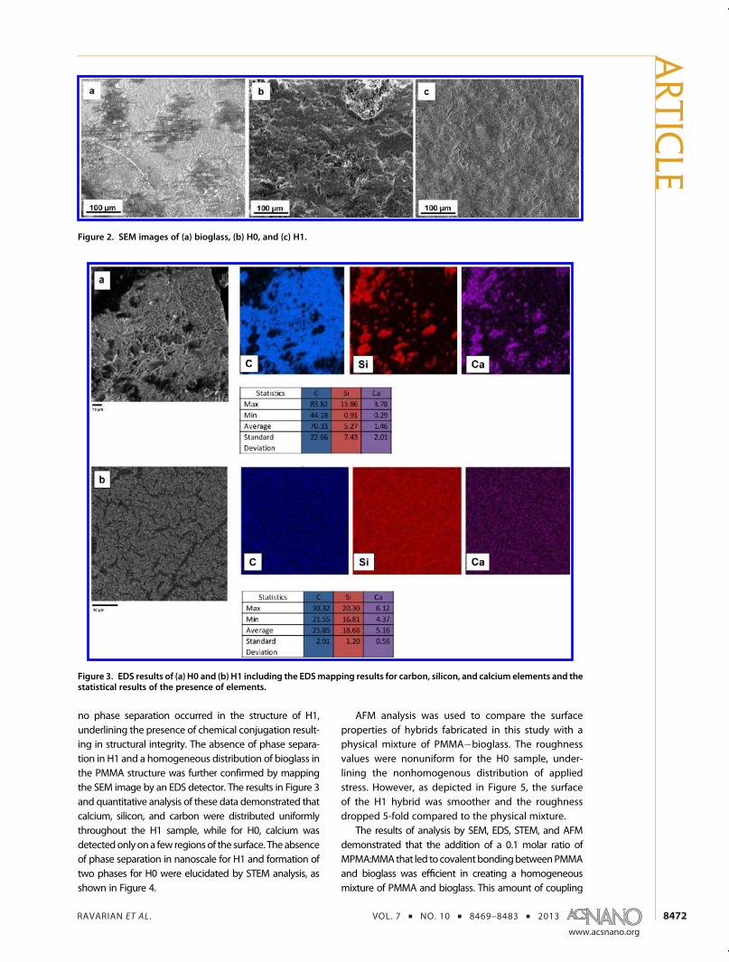
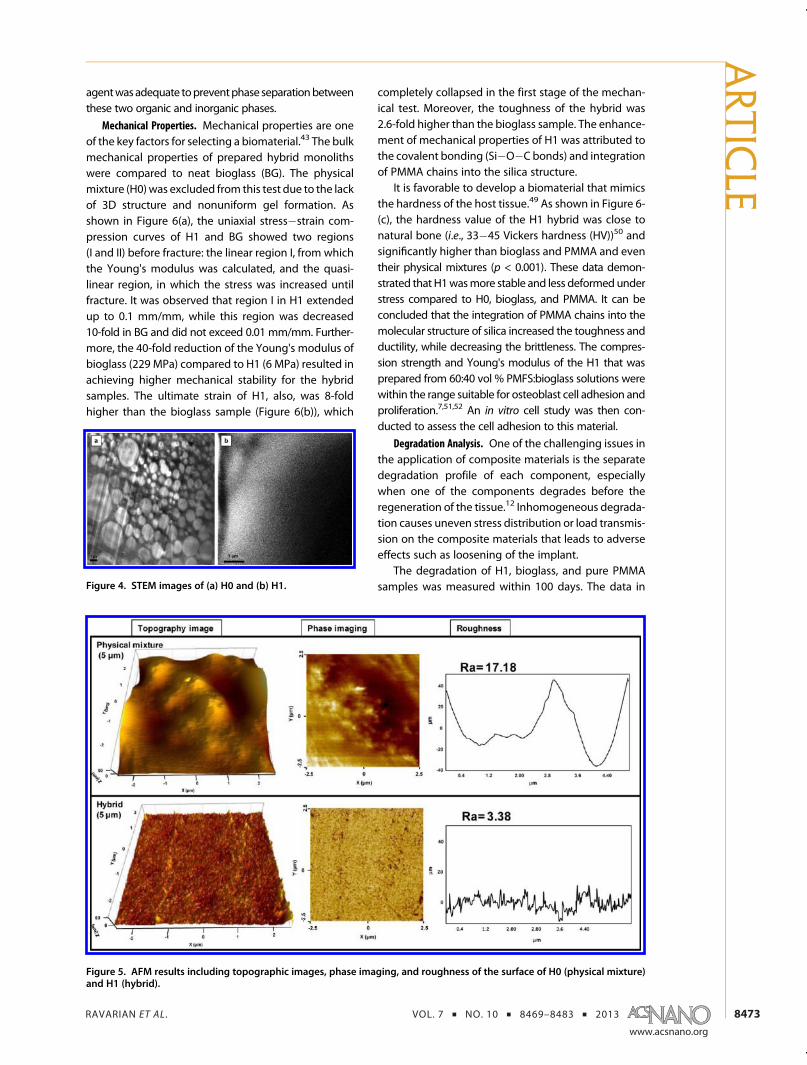
no phase separation occurred in the structure of H1,underlining the presence of chemical conjugation result-ing in structural integrity. The absence of phase separa-tion in H1 and a homogeneous distribution of bioglass inthe PMMA structure was further confirmed by mappingthe SEM image by an EDS detector. The results in Figure 3and quantitative analysis of these data demonstrated thatcalcium, silicon, and carbon were distributed uniformlythroughout the H1 sample, while for H0, calcium wasdetectedonlyona few regionsof the surface. Theabsenceof phase separation in nanoscale for H1 and formation oftwo phases for H0 were elucidated by STEM analysis, asshown in Figure 4.
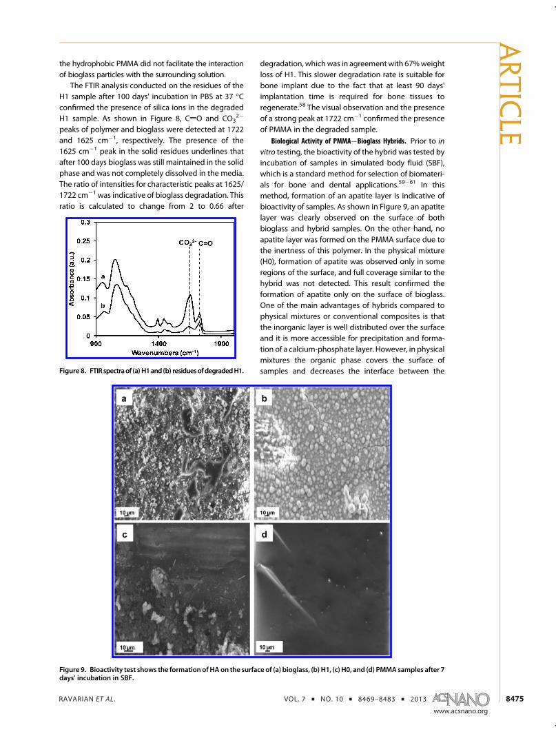
AFM analysis was used to compare the surfaceproperties of hybrids fabricated in this study with aphysical mixture of PMMA�bioglass. The roughnessvalues were nonuniform for the H0 sample, under-lining the nonhomogenous distribution of appliedstress. However, as depicted in Figure 5, the surfaceof the H1 hybrid was smoother and the roughnessdropped 5-fold compared to the physical mixture.
The results of analysis by SEM, EDS, STEM, and AFMdemonstrated that the addition of a 0.1 molar ratio ofMPMA:MMAthat led tocovalentbondingbetweenPMMAand bioglass was efficient in creating a homogeneousmixture of PMMA and bioglass. This amount of coupling
Figure 2. SEM images of (a) bioglass, (b) H0, and (c) H1.
Figure 3. EDS results of (a) H0 and (b) H1 including the EDSmapping results for carbon, silicon, and calcium elements and thestatistical results of the presence of elements.
ARTIC
LE
RAVARIAN ET AL . VOL. 7 ’ NO. 10 ’ 8469–8483 ’ 2013
www.acsnano.org
8473
agentwasadequate topreventphase separationbetweenthese two organic and inorganic phases.
Mechanical Properties. Mechanical properties are oneof the key factors for selecting a biomaterial.43 The bulkmechanical properties of prepared hybrid monolithswere compared to neat bioglass (BG). The physicalmixture (H0)was excluded from this test due to the lackof 3D structure and nonuniform gel formation. Asshown in Figure 6(a), the uniaxial stress�strain com-pression curves of H1 and BG showed two regions(I and II) before fracture: the linear region I, from whichthe Young's modulus was calculated, and the quasi-linear region, in which the stress was increased untilfracture. It was observed that region I in H1 extendedup to 0.1 mm/mm, while this region was decreased10-fold in BG and did not exceed 0.01 mm/mm. Further-more, the 40-fold reduction of the Young's modulus ofbioglass (229 MPa) compared to H1 (6 MPa) resulted inachieving higher mechanical stability for the hybridsamples. The ultimate strain of H1, also, was 8-foldhigher than the bioglass sample (Figure 6(b)), which
completely collapsed in the first stage of the mechan-ical test. Moreover, the toughness of the hybrid was2.6-fold higher than the bioglass sample. The enhance-ment of mechanical properties of H1 was attributed tothe covalent bonding (Si�O�C bonds) and integrationof PMMA chains into the silica structure.
It is favorable to develop a biomaterial that mimicsthe hardness of the host tissue.49 As shown in Figure 6-(c), the hardness value of the H1 hybrid was close tonatural bone (i.e., 33�45 Vickers hardness (HV))50 andsignificantly higher than bioglass and PMMA and eventheir physical mixtures (p < 0.001). These data demon-strated that H1wasmore stable and less deformed understress compared to H0, bioglass, and PMMA. It can beconcluded that the integration of PMMA chains into themolecular structure of silica increased the toughness andductility, while decreasing the brittleness. The compres-sion strength and Young's modulus of the H1 that wasprepared from 60:40 vol % PMFS:bioglass solutions werewithin the range suitable for osteoblast cell adhesion andproliferation.7,51,52 An in vitro cell study was then con-ducted to assess the cell adhesion to this material.
Degradation Analysis. One of the challenging issues inthe application of composite materials is the separatedegradation profile of each component, especiallywhen one of the components degrades before theregeneration of the tissue.12 Inhomogeneous degrada-tion causes uneven stress distribution or load transmis-sion on the composite materials that leads to adverseeffects such as loosening of the implant.
The degradation of H1, bioglass, and pure PMMAsamples was measured within 100 days. The data inFigure 4. STEM images of (a) H0 and (b) H1.
Figure 5. AFM results including topographic images, phase imaging, and roughness of the surface of H0 (physical mixture)and H1 (hybrid).
ARTIC
LE

RAVARIAN ET AL . VOL. 7 ’ NO. 10 ’ 8469–8483 ’ 2013
www.acsnano.org
8474
Figure 7 show that PMMA has no weight loss duringthis period; however, bioglass was rapidly degradedand completely dissolved after 83 days. The mechan-ism of degradation of bioglass is well established in theliterature.45,53�55 During the degradation of bioglass,SiO2 is gradually dissolved and results in producingsilicic acid, which is excreted from the body.45,53�55
However, the degradation rate of SiO2 matrices is afunction of parameters such as the composition ofprecursors, water:TEOS molar ratio, pH, and networkconnectivity of the silica matrix that is shown by Qn
species.56 Qn shows the number of Si�O�Si bondsaround each silicon. For example, the high value of Q3
and low value of Q4 are indicative of a less compactnetwork and hence a faster degradation rate. Previousstudies also showed that the covalent bonding be-tween a bioglass and a polymer had a negligible effecton the mechanism of degradation that is based onreleasing the silica ion.45,57
The composition of bioglass in this study was TEOS:water:HCl:CC = 1:8:0.01:0.2 with 41% Q3 and 53% Q4
species, which were calculated from 29Si NMR analysis.44
The data in Figure 7 demonstrated that these character-istics led to the degradation rate of 0.92% per day and0.65% per day for bioglass and H1 samples, respectively.The zero-order kinetics of bioglass degradation was inagreementwithother studies.53 The additionof PMMA tobioglass by covalent bonding (H1) resulted in impedingthe degradation rate of bioglass by 1.5-fold and main-taining nearly 30% of its original weight after 100 days of
incubation. It was demonstrated that the mechanism ofthe new bone formation at the implant interface and thehost tissue continues until there is ion exchangebetweenthe implant and body fluids.58 Therefore, retarding thedegradation of the silica matrix in H1 provides a longerperiod for ion exchange, hence resulting in strongerinteraction with the host tissue and formation of thickerapatite for bone formation. It is assumed that duringgradual degradation of bioactive glass innate hydroxya-patite and extracellularmatrix are regenerated in situ thatmimic the required mechanical strength for bone regen-eration. In addition, no weight loss was observed in thedegradation profile of H0, which was due to the fact that
Figure 6. Mechanical properties of bioglass and H1 samples: (a) stress�strain diagram, (b) ultimate strain, (c) microhardnessvalues (*** represents p < 0.001).
Figure 7. Degradation profiles of hybrid compared withpure PMMAandbioglass (the datawere extrapolated for BGuntil day 83).
ARTIC
LE
RAVARIAN ET AL . VOL. 7 ’ NO. 10 ’ 8469–8483 ’ 2013
www.acsnano.org
8475
the hydrophobic PMMA did not facilitate the interactionof bioglass particles with the surrounding solution.
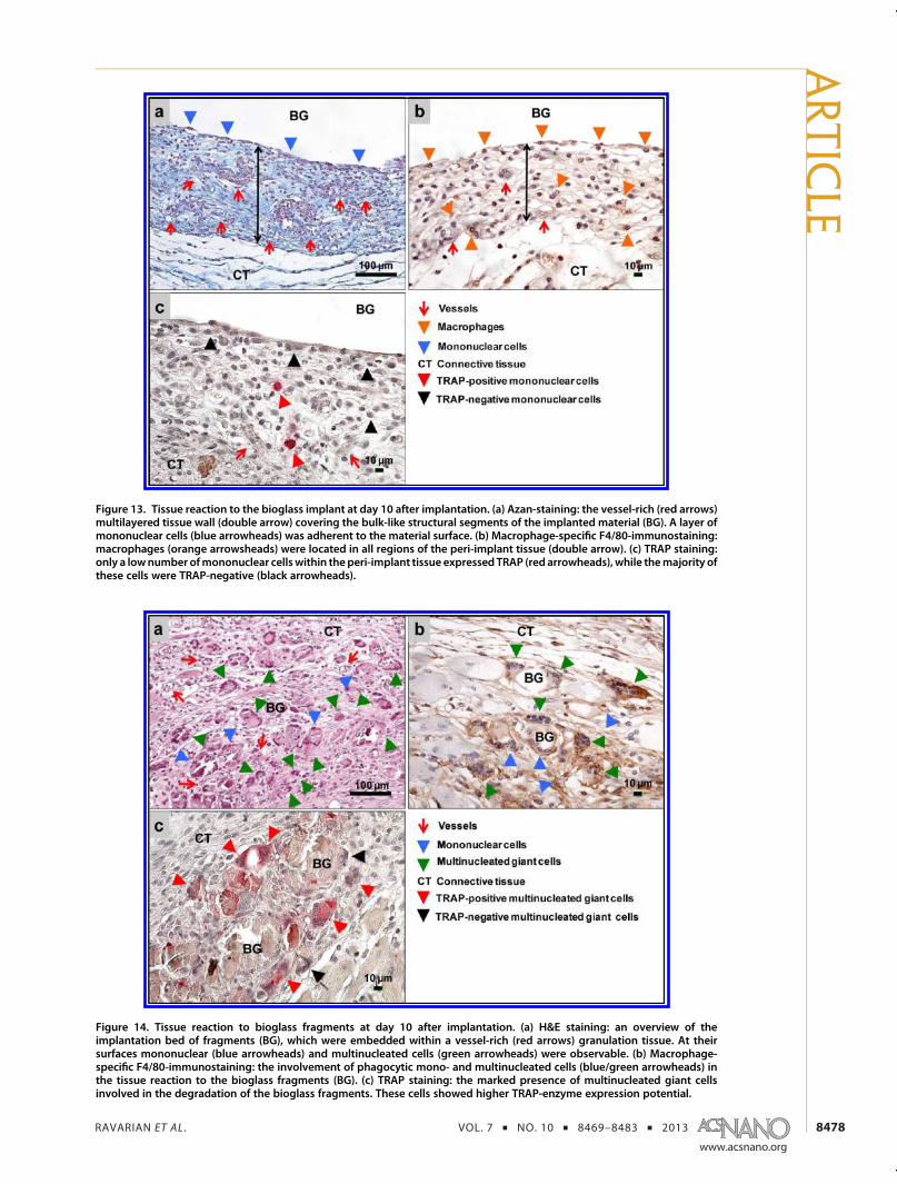
The FTIR analysis conducted on the residues of theH1 sample after 100 days' incubation in PBS at 37 �Cconfirmed the presence of silica ions in the degradedH1 sample. As shown in Figure 8, CdO and CO3
2�
peaks of polymer and bioglass were detected at 1722and 1625 cm�1, respectively. The presence of the1625 cm�1 peak in the solid residues underlines thatafter 100 days bioglass was still maintained in the solidphase and was not completely dissolved in the media.The ratio of intensities for characteristic peaks at 1625/1722 cm�1 was indicative of bioglass degradation. Thisratio is calculated to change from 2 to 0.66 after
degradation, whichwas in agreementwith 67%weightloss of H1. This slower degradation rate is suitable forbone implant due to the fact that at least 90 days'implantation time is required for bone tissues toregenerate.58 The visual observation and the presenceof a strong peak at 1722 cm�1 confirmed the presenceof PMMA in the degraded sample.
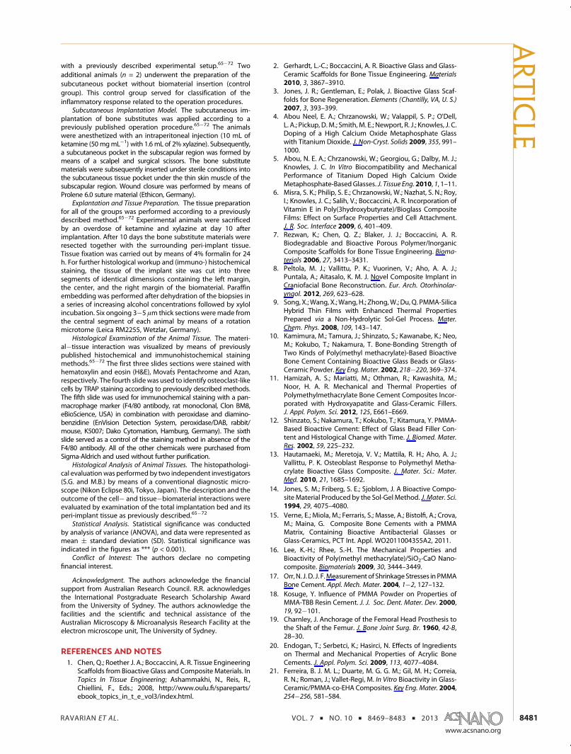
Biological Activity of PMMA�Bioglass Hybrids. Prior to in
vitro testing, the bioactivity of the hybrid was tested byincubation of samples in simulated body fluid (SBF),which is a standard method for selection of biomateri-als for bone and dental applications.59�61 In thismethod, formation of an apatite layer is indicative ofbioactivity of samples. As shown in Figure 9, an apatitelayer was clearly observed on the surface of bothbioglass and hybrid samples. On the other hand, noapatite layer was formed on the PMMA surface due tothe inertness of this polymer. In the physical mixture(H0), formation of apatite was observed only in someregions of the surface, and full coverage similar to thehybrid was not detected. This result confirmed theformation of apatite only on the surface of bioglass.One of the main advantages of hybrids compared tophysical mixtures or conventional composites is thatthe inorganic layer is well distributed over the surfaceand it is more accessible for precipitation and forma-tion of a calcium-phosphate layer. However, in physicalmixtures the organic phase covers the surface ofsamples and decreases the interface between theFigure 8. FTIRspectraof (a) H1and(b) residuesofdegradedH1.
Figure 9. Bioactivity test shows the formation of HAon the surface of (a) bioglass, (b) H1, (c) H0, and (d) PMMAsamples after 7days' incubation in SBF.
ARTIC
LE

RAVARIAN ET AL . VOL. 7 ’ NO. 10 ’ 8469–8483 ’ 2013
www.acsnano.org
8476
inorganic phase and calcium-phosphate ions.32 Further-more, the formation of apatite on the surface of H1implies that the presence of inert PMMA polymer witha composition of 60:40 PMFS:bioglass had a negligibleimpact on bioactivity.
In Vitro Attachment, Proliferation, and Osteoblastic Differ-entiation. Physical mixture (H0) and hybrid (H1) ofPMMA-bioglass were analyzed in vitro for their cellattachment and osteoblast growth properties. Thesematerials were compared with their constituentmaterials, pure PMMA and bioglass. The bioglass wasmarkedly brittle and prone to thermal and mechanicalshock-induced fracturing.
Cells were found to adhere to all surfaces andsupported attachment after 4 days in culture, as visua-lized by scanning electronmicroscopy (Figure 10). Cellsshowed a more flattened morphology, indicative ofincreased attachment on bioglass compared to PMMA.Notably thismorphologywas bettermaintained on thehybrid H1 compared to H0, suggesting the improvedhomogeneity of the PMFS-bioglass gave more consis-tent attachment.
Cell growth was measured by viability assay oncultured primary osteoblasts at days 1 and 7 aftercell seeding. No significant difference was seen be-tween the samples after 24 h; however the H1 hybridshowed a remarkable increase after 7 days, which wassuperior to all other samples including the physical
mixture in terms of cell viability. The cell viabilitynumber of H1 was close to the control (TCPS) after7 days of culture (p > 0.05). A trend toward increasedviability was seen in tissue culture plastic controls,although this is a surface optimized for cell attachmentand growth.
Finally, the alkaline phosphatase (ALP) activity ofsamples was assessed. Human preosteoblasts weregrown on H0, H1, PMMA, and bioglass materials andtreated with osteogenic differentiation media to in-duce expression of mature osteoblast markers. Robuststaining was seen on cells grown on tissue cultureplastic as a positive control (Figure 12). Notably,blue ALPþ cells were seen on the H1-treated hybridsamples but not on the H0 samples or on the PMMAor bioglass alone. These data suggest that thehybrid material may have superior properties as abone replacement material for early osteoblastdifferentiation.
In Vivo Study. Histological Results: Bioglass Group.
The brittleness of bioglass resulted in the formationof bulk-like structures within the implantation bed andfragments of various sizes ranging up to small particlesafter 10 days. As shown in Figure 13 and Figure 14, theimplantation of this material provoked two differentcellular reactions. Around the bulk-like structures with-in the implantation bed granulocytes and mononu-clear cells, mainly macrophages and lymphocytes, were
Figure 10. SEM images of (a) pure PMMA, (b) H0, (c) H1, and (d) bioglass. White arrows show the individual narrowed cells onthe surface of samples.
ARTIC
LE

RAVARIAN ET AL . VOL. 7 ’ NO. 10 ’ 8469–8483 ’ 2013
www.acsnano.org
8477
embedded within a well-vascularized granulation tissue(Figure 13(a)). The immunohistochemical detection ofthe macrophage-specific F4/80 antigen showed thatthese cells were mainly detectable as a monolayer at thematerial�tissue interface and loosely distributed withinthe material�adherent granulation tissue (Figure 13(b)).Furthermore, only a small percentage of cells within theperi-implant tissuewas shown to express tartrate-resistantacid phosphatase (TRAP) (Figure 13(c)).
The tissue reaction to bioglass fragments was domi-nated by multinucleated giant cells in addition to theabove-mentionedmononuclear cells (Figure 14(a,b)). Theimmunochemical detection of the F4/80 antigen addi-tionally revealed the dominance of macrophages amongall material-adherent mononuclear cells (Figure 14(b)).Furthermore, TRAP staining revealed that within the im-plantation bed of fragments both TRAP-positive mono-nuclear and multinucleated cells dominated when com-pared to thosewithout TRAP expression (control samples)
(Figure 14(c)). We postulate that the occurrence of themultinucleated giant cells within the bioglass might bemainly size-related and less associated with the potentialmaterial impurity.
Histological Results: H1 Group. Better mechanicalproperties and less brittleness of H1 samples in com-parison to the bioglass resulted in almost no materialfragments within the peri-implant tissue during im-plantation and explantation (Figure 15). Thus, only onecellular reaction pattern was detected. The histologicalanalysis at day 10 after implantation revealed that theH1 implant was embedded within a cell- and vessel-rich connective tissue (Figure 15). This tissue waslocalized as a relatively thick wall along the material�tissue interface and contained macrophages, granulo-cytes, and lymphocytes aswell as fibroblasts. The tissuereaction was comparable to that toward the bioglassbulk-like structures. It should be noted that no cell andtissue penetration into the material core was observeddue to the lack of porosity. Directly at the surface of theH1 implant predominantly macrophages were located(Figure 15). Furthermore, a fewer number of multi-nucleated cells was detectable within the tissue adja-cent to the material (Figure 15(b)). TRAP detectionrevealed that only a small number of cells at the surfaceof the H1 implant showed expression of this enzyme(Figure 15(c)).
In this study an in vivo pilot study was performed inorder to assess the tissue reaction to bioglass and H1samples. The histological evaluation has revealed thatduring the implantation phase of 10 days bioglassunderwent a fragmentationmost likely due to physicalforces during animal movement rather than tissuepenetration into the material. Consequently, this
Figure 11. Viability assay of H1 hybrid sample compared tothe PMMA and bioglass (*** represents p < 0.001).
Figure 12. ALP staining on materials showing ALPþ cells (blue) after 4 days of osteogenic differentiation. Positive stainingwas seen on H1 hybrid and tissue culture plastic, but not on PMMA, bioglass, or H0 material.
ARTIC
LE
RAVARIAN ET AL . VOL. 7 ’ NO. 10 ’ 8469–8483 ’ 2013
www.acsnano.org
8478
Figure 14. Tissue reaction to bioglass fragments at day 10 after implantation. (a) H&E staining: an overview of theimplantation bed of fragments (BG), which were embedded within a vessel-rich (red arrows) granulation tissue. At theirsurfaces mononuclear (blue arrowheads) and multinucleated cells (green arrowheads) were observable. (b) Macrophage-specific F4/80-immunostaining: the involvement of phagocytic mono- and multinucleated cells (blue/green arrowheads) inthe tissue reaction to the bioglass fragments (BG). (c) TRAP staining: the marked presence of multinucleated giant cellsinvolved in the degradation of the bioglass fragments. These cells showed higher TRAP-enzyme expression potential.
Figure 13. Tissue reaction to the bioglass implant at day 10 after implantation. (a) Azan-staining: the vessel-rich (red arrows)multilayered tissue wall (double arrow) covering the bulk-like structural segments of the implanted material (BG). A layer ofmononuclear cells (blue arrowheads) was adherent to the material surface. (b) Macrophage-specific F4/80-immunostaining:macrophages (orange arrowsheads) were located in all regions of the peri-implant tissue (double arrow). (c) TRAP staining:only a lownumber ofmononuclear cellswithin theperi-implant tissue expressed TRAP (red arrowheads), while themajority ofthese cells were TRAP-negative (black arrowheads).
ARTIC
LE

RAVARIAN ET AL . VOL. 7 ’ NO. 10 ’ 8469–8483 ’ 2013
www.acsnano.org
8479
material induced two different cellular reactions. Thebulk-like structures induced a mainly mononuclearinflammatory response, while the fragments inducedmultinucleated giant cells. The occurrence of the latterwas independent of fragment size. Overall, the im-plantation bed of this group was relatively extensive,which again can be attributed to the presence of themultinucleated giant cells, which are known to producevascular endothelial growth factor (VEGF) among othersubstances responsible for material degradation. H1 un-dergoes negligible fragmentation during the observationperiod, which can be attributed to its higher mechanicalstability. This material induced mainly mononuclear cellsand a relatively low amount of multinucleated giant cells.Interestingly, the latter did not show a high TRAP expres-sion, and it was observed that the implantation bed of H1was well vascularized.
CONCLUSIONS
PMMA�bioglass hybrids were produced by thesol�gel method in the presence of chemical bonding
that integrated organic and inorganic components.This molecular level interaction addressed the issueof phase separation that is commonly observed in thepreparation of physical mixtures of bioglass with apolymer. In addition, the mechanical properties ofthe hybrid acquired at an optimum composition ofMPMA:MMA were significantly improved comparedwith bioglass; the Young's modulus of the hybrid wasdecreased 40-fold and its hardness was 16-fold higherthan pure bioglass. The chemical bonding of PMMAwith bioglass resulted in prolonging the degradation ofbioglass, which may be favorable for bone regenera-tion and in situ drug release applications. Additionally,the results of in vitro and in vivo studies demonstratedthat the molecular level interaction had no adverseeffect on biocompatibility of bioglass and in factsignificantly enhanced its integrity and reduced thelevel of inflammation. Therefore, the fabricated hybridin this study is a viable alternative for bioglass andPMMA�bioglass physical mixtures as bone regenera-tive materials.
MATERIALS AND METHODS
Materials. Precursors required for the synthesis of PMMA-co-MPMA copolymer including MPMA, R,R0-azoisobutyronitrile(AIBN), and N,N0-dimethylformamide (DMF) were purchased fromSigma and used as received. Methyl methacrylate purchased from
Sigma was used after distillation under reduced pressure. Hydro-chloric acid (HCl; Merck), tetraethyl orthosilicate (TEOS; Sigma),calcium chloride dihydrate (CaCl2. 3 2H2O (CC); Ajax Finechem PtyLtd.), tetrahydrofuran (THF; Merck), and deionized water were usedfor fabrication of the inorganic solution and the hybrid.
Figure 15. Tissue reaction to the H1 implant at day 10. (a) Macrophage-specific F4/80-immunostaining: an overview of thedistribution ofmononuclear cells within the peri-implant tissue (double arrow) within the subcutaneous (CT) layer of the CD1mouse. Most of the cells at the surface as well as the periphery were identified as macrophages (orange arrowheads). (b) H&Estaining: a granulation tissue was located at the surface of the implant. (c) TRAP staining: only a small amount of cells at thesurface of the material as well as within the material-adherent tissue (CT) showed signs of TRAP activity (red arrowheads).Most of the cells were TRAP-negative (black arrowheads).
ARTIC
LE

RAVARIAN ET AL . VOL. 7 ’ NO. 10 ’ 8469–8483 ’ 2013
www.acsnano.org
8480
Preparation of Bioglass, PMMA-co-MPMA, and Pure PMMA Solutions.TEOS was mixed with deionized water and HCl and stirred for30 min followed by addition of calcium chloride dihydrate. Acommon calcium source for the preparation of sol�gel-derivedbioglasses is calcium nitrate tetrahydrate; however, in thisstudy, calcium chloride was used to minimize the risk of toxicityresulting from nitrate byproduct.62 The precursors were mixedwith a molar ratio of TEOS:water:HCl:CC = 1:8:0.01:0.2, and thesolution is referred to as sol(A) for convenience.
A free radical polymerization technique was used for thesynthesis of PMMA-co-MPMA with MPMA:MMA = 0.1 mol ratiocoded as PMFS and PMMA (without MPMA) using AIBN as aninitiator. Precursors were mixed in a Schlenk flask (MMA:AIBN =200 (mol ratio), DMF (20 mL)) and degassed by three freeze�pump�thaw cycles. Polymerization was conducted at 70 �C fora period of 12 h. The polymer was purified by precipitation indiethyl ether followed by filtration and drying under vacuum.PMFS or PMMA was dissolved in THF with a concentration of10 wt %, and the solutions were labeled as sol(B) and sol(C),respectively.
Hybrid Formation. Sol(A) and sol(B) were mixed in the volu-metric ratio of sol(B):sol(A) = 60:40, thenmechanically stirred for1 h to obtain a homogeneous and well-dispersed solution. Thiscomposition was selected due to the fact that bioglass compo-sition was shown to have no significant impact on the networkcharacteristics and molecular integration of hybrids.
It was then kept sealed until a gel formed, then was dried atambient temperature and subsequently at 37 �C for a period of7 days at each temperature. The product was then dried in avacuum oven at 40 �C for a period of 2 days. This temperatureprofile was developed for drying the samples to removeresidues of solvents and maintain the monolith structure.
Physical Mixture Formation. Sol(A) and sol(C) were mixed(sol(C):sol(A) = 60:40 vol %) and mechanically stirred for 1 h.Thesameprocedurewas followed todry these samplesashybrids.Athin film of polymer was obtained with a gel structure of bioglass,which was subsequently ground andwell-mixed as a powder. Afterthis, the powderwas dissolved in THF (10wt%) and cast on a Tefloncontainer, which was then vacuum-dried at 40 �C for 2 days.
Characterizations. Scanning Electron Microscopy�Energy Dis-persive Spectrometry (SEM-EDS). The surface microstructuresand crystal phase formed on the specimens were analyzed byfield emission scanning electron microscopy (FE-SEM; ZeissULTRA Plus). This instrument was equippedwith a Bruker XFlash4010 EDS detector with high-speed acquisition and hypermap-ping capability. Samples were mounted on aluminum stubsusing conductive carbon paint, then gold coated by using anEmitech K7550X instrument prior to SEM analysis.
Scanning Transmission Electron Microscopy (STEM). STEManalysis was conducted to investigate the interaction betweenthe phases in nanoscale. Samples were ground to powders,embedded in epoxy resins, andmicrotomedwith Leica Ultracutultramicrotomes (UC7) for 100 nm layers. The layers wereharvested and seated on carbon grids prior to STEM analysis(Zeiss ULTRA Plus).
Atomic Force Microscopy (AFM). The surface topography ofthe prepared hybrid samples was characterized by an atomicforce microscope (Asylum Research, MFP-3D-BIO) in ac modeusing a silicon nitride tip (AC160). In order to identify theuniformity of distribution and separation of bioglass withinthe polymer structure, phase images were recorded. A thin filmwas produced to examine the surface properties.
Mechanical Testing. Hybrid samples were prepared in theform of monoliths, and uniaxial compression tests were per-formed in an unconfined state with a 1000 N load cell by Instron(model 5543). Dimensions of the samples were 6.61( 0.05 mmdiameter and 1.2( 0.1mmheight. The samples were subjectedto a loading, and a Young's modulus was obtained as thetangent slope of the stress�strain curve between 0% and10% strain level. The area under the compressive stress�straincurves was calculated for measuring the toughness of thesamples. Three samples were examined for each group forstatistical analysis.
Microhardness measurements were performed using Vick-ers indentations at loads of 50 g and an indentation time of 5 s at
25 �C using a microhardness tester (Buehler, Lake Bluff, IL;1105D). Six tests were conducted for each sample for statisticalanalysis.
Degradation Assessment. The degradation rate of sampleswas tested by measuring the change in sample weight overtime under simulated physiological conditions. Three sampleswere kept in PBS at 37 �C, and at different time intervals theywere removed from the degradation medium, rinsed threetimes with deionized water, and dried prior to weighing. Themeasurement was continued for a period of 100 days.
Attenuated Total Reflection Fourier Transform Infrared (ATR-FTIR) Spectroscopy. The molecular structure of the hybrid andthe degradation products of samples after leaving in PBS for 100days were analyzed by ATR-FTIR spectroscopy (FTIR; Nicolet6700, Thermo Fisher Scientific Inc.). The samples were scannedat a speed of 32 scan/min.
Bioactivity. The samples were characterized for their bioac-tivity by incubation in simulated body fluid63 at 37 �C for periodsof 1, 3, 7, and 14 days. Samples were washed twice withdeionized water to remove any residue of minerals absorbedon the surface. The dried monoliths were examined to deter-mine the formation of an apatite layer on the surface.
Cell Attachment, Morphology, and Proliferation. Human pre-osteoblasts (HOB) were used to assess the cell interaction withthe samples. Complete osteoblast growth media (Invitrogen)was used to culture the HOB cells, which were incubated at37 �C in thepresenceof 5%CO2 and95%humidity. Themediawasrefreshed every 3 days until the cells approached confluence.
The samples (12 mm diameter� 1 mm height) were placedinto a well plate and kept in 70% ethanol for 1 h for sterilization,followed by rinsing with PBS three times. Samples were thenexposed to UV light for 30 min and were washed with freshmedium at 37 �C overnight. The substrates were placed intowell plates, seeded with cells at a density of 2 � 105 cells/mL,and kept in culture for 7 days. Cell morphologywas examined atday 4 of culture by SEM (FE-SEM; Zeiss ULTRA plus) analysis afterbeing fixed with glutaraldehyde according to a previouslypublishedmethod.64 Viability wasmeasured at 1 day and 7 dayspostseeding. Cellular viability was assessed using the CellTiter96 Aqueous One Solution cell proliferation assay kit (Promega)according to the manufacturer's instructions. Briefly, scaffoldswere incubated with the viability solution for 30 min at 37 �Cand read using a spectrophotometer at 595 nm. All sampleswere analyzed triple for statistical analysis. Tissue culture poly-styrene (TCPS) was used as a control.
In Vitro Osteogenic Differentiation and Alkaline PhosphataseActivity. To initiate osteogenic differentiation, samples weretransferred to media supplemented with ascorbic acid (50 μg/mL), β-glycerophosphate (10mM), and BMP-2 (200 ng/mL) after48 h of culture with cells. The time of transferring the samples tothis media was considered as day 0 for the alkaline phosphatase(ALP) test.
ALP is an osteogenic marker that is expressed by differen-tiating osteoblasts. ALP staining was carried out on pure PMMAand bioglass, their physical mixture, and hybrid at day 4.Samples were fixed with gluteraldehyde, washed with PBS,and incubated in TRIS buffer (1 M, 9.4 pH) for 5 min. Thescaffolds were then stained in Naphthol AS-BI phosphate(Sigma) as a substrate and Fast Blue (Sigma) as the stain.Scaffolds were washed with H2O to remove excess stainingbefore imaging. Cells alone on tissue culture plastic were usedas a control. Images were captured using a Leica MZ6 micro-scope with a QImaging Micropublisher 5.0 camera.
In Vivo Study. Experimental Design of the in Vivo Pilot Study.The in vivo experiments were carried out after approval from theCommittee on theUse of Live Animals in Teaching and Researchof the State of Rhineland-Palatinate, Germany. A total of 10female 4�6-week-old CD1-mice (Charles River Laboratories,Germany) were reared under standard experimental conditionsat the in vivo Laboratory Animal Unit at the Institute ofPathology of the Johannes Gutenberg University of Mainz,Germany. The animals were randomly divided into three ex-perimental groups. Animals of the first two groups (n = 4 and 8animals in total) underwent a subcutaneous implantation withthe bioglass and prepared hybrid in this study in accordance
ARTIC
LE
RAVARIAN ET AL . VOL. 7 ’ NO. 10 ’ 8469–8483 ’ 2013
www.acsnano.org
8481
with a previously described experimental setup.65�72 Twoadditional animals (n = 2) underwent the preparation of thesubcutaneous pocket without biomaterial insertion (controlgroup). This control group served for classification of theinflammatory response related to the operation procedures.
Subcutaneous Implantation Model. The subcutaneous im-plantation of bone substitutes was applied according to apreviously published operation procedure.65�72 The animalswere anesthetized with an intraperitoneal injection (10 mL ofketamine (50mgmL�1) with 1.6mL of 2% xylazine). Subsequently,a subcutaneous pocket in the subscapular region was formed bymeans of a scalpel and surgical scissors. The bone substitutematerials were subsequently inserted under sterile conditions intothe subcutaneous tissue pocket under the thin skin muscle of thesubscapular region. Wound closure was performed by means ofProlene 6.0 suture material (Ethicon, Germany).
Explantation and Tissue Preparation. The tissue preparationfor all of the groups was performed according to a previouslydescribed method.65�72 Experimental animals were sacrificedby an overdose of ketamine and xylazine at day 10 afterimplantation. After 10 days the bone substitute materials wereresected together with the surrounding peri-implant tissue.Tissue fixation was carried out by means of 4% formalin for 24h. For further histological workup and (immuno-) histochemicalstaining, the tissue of the implant site was cut into threesegments of identical dimensions containing the left margin,the center, and the right margin of the biomaterial. Paraffinembedding was performed after dehydration of the biopsies ina series of increasing alcohol concentrations followed by xylolincubation. Six ongoing 3�5 μm thick sections weremade fromthe central segment of each animal by means of a rotationmicrotome (Leica RM2255, Wetzlar, Germany).
Histological Examination of the Animal Tissue. The materi-al�tissue interaction was visualized by means of previouslypublished histochemical and immunohistochemical stainingmethods.65�72 The first three slides sections were stained withhematoxylin and eosin (H&E), Mova�ts Pentachrome and Azan,respectively. The fourth slidewas used to identify osteoclast-likecells by TRAP staining according to previously described methods.The fifth slide was used for immunochemical staining with a pan-macrophage marker (F4/80 antibody, rat monoclonal, Clon BM8,eBioScience, USA) in combination with peroxidase and diamino-benzidine (EnVision Detection System, peroxidase/DAB, rabbit/mouse, K5007; Dako Cytomation, Hamburg, Germany). The sixthslide served as a control of the staining method in absence of theF4/80 antibody. All of the other chemicals were purchased fromSigma-Aldrich and used without further purification.
Histological Analysis of Animal Tissues. The histopathologi-cal evaluationwas performed by two independent investigators(S.G. and M.B.) by means of a conventional diagnostic micro-scope (Nikon Eclipse 80i, Tokyo, Japan). The description and theoutcome of the cell� and tissue�biomaterial interactions wereevaluated by examination of the total implantation bed and itsperi-implant tissue as previously described.65�72
Statistical Analysis. Statistical significance was conductedby analysis of variance (ANOVA), and data were represented asmean ( standard deviation (SD). Statistical significance wasindicated in the figures as *** (p < 0.001).
Conflict of Interest: The authors declare no competingfinancial interest.
Acknowledgment. The authors acknowledge the financialsupport from Australian Research Council. R.R. acknowledgesthe International Postgraduate Research Scholarship Awardfrom the University of Sydney. The authors acknowledge thefacilities and the scientific and technical assistance of theAustralian Microscopy & Microanalysis Research Facility at theelectron microscope unit, The University of Sydney.
REFERENCES AND NOTES1. Chen, Q.; Roether J. A.; Boccaccini, A. R. Tissue Engineering
Scaffolds from Bioactive Glass and Composite Materials. InTopics In Tissue Engineering; Ashammakhi, N., Reis, R.,Chiellini, F., Eds.; 2008, http://www.oulu.fi/spareparts/ebook_topics_in_t_e_vol3/index.html.
2. Gerhardt, L.-C.; Boccaccini, A. R. Bioactive Glass and Glass-Ceramic Scaffolds for Bone Tissue Engineering. Materials2010, 3, 3867–3910.
3. Jones, J. R.; Gentleman, E.; Polak, J. Bioactive Glass Scaf-folds for Bone Regeneration. Elements (Chantilly, VA, U. S.)2007, 3, 393–399.
4. Abou Neel, E. A.; Chrzanowski, W.; Valappil, S. P.; O'Dell,L. A.; Pickup, D. M.; Smith, M. E.; Newport, R. J.; Knowles, J. C.Doping of a High Calcium Oxide Metaphosphate Glasswith Titanium Dioxide. J. Non-Cryst. Solids 2009, 355, 991–1000.
5. Abou, N. E. A.; Chrzanowski, W.; Georgiou, G.; Dalby, M. J.;Knowles, J. C. In Vitro Biocompatibility and MechanicalPerformance of Titanium Doped High Calcium OxideMetaphosphate-BasedGlasses. J. Tissue Eng. 2010, 1, 1–11.
6. Misra, S. K.; Philip, S. E.; Chrzanowski, W.; Nazhat, S. N.; Roy,I.; Knowles, J. C.; Salih, V.; Boccaccini, A. R. Incorporation ofVitamin E in Poly(3hydroxybutyrate)/Bioglass CompositeFilms: Effect on Surface Properties and Cell Attachment.J. R. Soc. Interface 2009, 6, 401–409.
7. Rezwan, K.; Chen, Q. Z.; Blaker, J. J.; Boccaccini, A. R.Biodegradable and Bioactive Porous Polymer/InorganicComposite Scaffolds for Bone Tissue Engineering. Bioma-terials 2006, 27, 3413–3431.
8. Peltola, M. J.; Vallittu, P. K.; Vuorinen, V.; Aho, A. A. J.;Puntala, A.; Aitasalo, K. M. J. Novel Composite Implant inCraniofacial Bone Reconstruction. Eur. Arch. Otorhinolar-yngol. 2012, 269, 623–628.
9. Song, X.; Wang, X.;Wang, H.; Zhong,W.; Du, Q. PMMA-SilicaHybrid Thin Films with Enhanced Thermal PropertiesPrepared via a Non-Hydrolytic Sol-Gel Process. Mater.Chem. Phys. 2008, 109, 143–147.
10. Kamimura, M.; Tamura, J.; Shinzato, S.; Kawanabe, K.; Neo,M.; Kokubo, T.; Nakamura, T. Bone-Bonding Strength ofTwo Kinds of Poly(methyl methacrylate)-Based BioactiveBone Cement Containing Bioactive Glass Beads or Glass-Ceramic Powder. Key Eng. Mater. 2002, 218�220, 369–374.
11. Hamizah, A. S.; Mariatti, M.; Othman, R.; Kawashita, M.;Noor, H. A. R. Mechanical and Thermal Properties ofPolymethylmethacrylate Bone Cement Composites Incor-porated with Hydroxyapatite and Glass-Ceramic Fillers.J. Appl. Polym. Sci. 2012, 125, E661–E669.
12. Shinzato, S.; Nakamura, T.; Kokubo, T.; Kitamura, Y. PMMA-Based Bioactive Cement: Effect of Glass Bead Filler Con-tent and Histological Change with Time. J. Biomed. Mater.Res. 2002, 59, 225–232.
13. Hautamaeki, M.; Meretoja, V. V.; Mattila, R. H.; Aho, A. J.;Vallittu, P. K. Osteoblast Response to Polymethyl Metha-crylate Bioactive Glass Composite. J. Mater. Sci.: Mater.Med. 2010, 21, 1685–1692.
14. Jones, S. M.; Friberg, S. E.; Sjoblom, J. A Bioactive Compo-site Material Produced by the Sol-Gel Method. J. Mater. Sci.1994, 29, 4075–4080.
15. Verne, E.; Miola, M.; Ferraris, S.; Masse, A.; Bistolfi, A.; Crova,M.; Maina, G. Composite Bone Cements with a PMMAMatrix, Containing Bioactive Antibacterial Glasses orGlass-Ceramics, PCT Int. Appl. WO2011004355A2, 2011.
16. Lee, K.-H.; Rhee, S.-H. The Mechanical Properties andBioactivity of Poly(methyl methacrylate)/SiO2-CaO Nano-composite. Biomaterials 2009, 30, 3444–3449.
17. Orr, N. J. D. J. F.Measurement of ShrinkageStresses in PMMABone Cement. Appl. Mech. Mater. 2004, 1�2, 127–132.
18. Kosuge, Y. Influence of PMMA Powder on Properties ofMMA-TBB Resin Cement. J. J. Soc. Dent. Mater. Dev. 2000,19, 92�101.
19. Charnley, J. Anchorage of the Femoral Head Prosthesis tothe Shaft of the Femur. J. Bone Joint Surg. Br. 1960, 42-B,28–30.
20. Endogan, T.; Serbetci, K.; Hasirci, N. Effects of Ingredientson Thermal and Mechanical Properties of Acrylic BoneCements. J. Appl. Polym. Sci. 2009, 113, 4077–4084.
21. Ferreira, B. J. M. L.; Duarte, M. G. G. M.; Gil, M. H.; Correia,R. N.; Roman, J.; Vallet-Regi, M. In Vitro Bioactivity in Glass-Ceramic/PMMA-co-EHA Composites. Key Eng. Mater. 2004,254�256, 581–584.
ARTIC
LE
RAVARIAN ET AL . VOL. 7 ’ NO. 10 ’ 8469–8483 ’ 2013
www.acsnano.org
8482
22. Vallo, C. I. Residual Monomer Content in Bone CementsBased on Poly(methyl methacrylate). Polym. Int. 2000, 49,831–838.
23. Mousa, W. F.; Kobayashi, M.; Shinzato, S.; Kamimura, M.;Neo, M.; Yoshihara, S.; Nakamura, T. Biological andMechanical Properties of PMMA-Based Bioactive BoneCements. Biomaterials 2000, 21, 2137–2146.
24. Hasenwinkel, J. M.; Lautenschlager, E. P.; Wixson, R. L.;Gilbert, J. L. A Novel High-Viscosity, Two-Solution AcrylicBone Cement: Effect of Chemical Composition on Proper-ties. J. Biomed. Mater. Res. 1999, 47, 36–45.
25. Zulfikar, M. A.; Wahab, M. A.; Hilal, N. Preparation andCharacterization of Novel Porous PMMA-SiO2 HybridMembranes. Desalination 2006, 192, 262–270.
26. Chrzanowski, W.; Abou Neel, E. A.; Lee, K. Y.; Bismarck, A.;Young,A.M.; Hart, A.D.;Dalby,M. J.; Knowles, J. C. TailoringCellBehavioronPolymersby the Incorporationof TitaniumDopedPhosphate Glass Filler. Adv. Eng. Mater. 2010, 12, B298–B308.
27. Guild, F. J.; Kinloch, A. J.; Taylor, A. C. Particle Cavitation inRubber Toughened Epoxies: The Role of Particle Size.J. Mater. Sci. 2010, 45, 3882–3894.
28. Hsieh, T. H.; Kinloch, A. J.; Masania, K.; Sohn, L. J.; Taylor, A. C.;Sprenger, S. The Toughness of Epoxy Polymers and FibreComposites Modified with Rubber Microparticles and SilicaNanoparticles. J. Mater. Sci. 2010, 45, 1193–1210.
29. Rhee, S.-H.; Choi, J.-Y. Preparation of a Bioactive Poly-(methyl methacrylate)/Silica Nanocomposite. J. Am. Cer-am. Soc. 2002, 85, 1318–1320.
30. Burdick, J. A.; Peterson, A. J.; Anseth, K. S. Conversion andTemperature Profiles during the Photoinitiated Polymer-ization of Thick Orthopedic Biomaterials. Biomaterials2001, 22, 1779–1786.
31. Mammeri, F.; Le, B. E.; Rozes, L.; Sanchez, C. MechanicalProperties of Hybrid Organic-Inorganic Materials. J. Mater.Chem 2005, 15, 3787–3811.
32. Poologasundarampillai, G.; Ionescu, C.; Tsigkou, O.;Murugesan, M.; Hill, R. G.; Stevens, M. M.; Hanna, J. V.;Smith, M. E.; Jones, J. R. Synthesis of Bioactive Class II Poly-(γ-Glutamic Acid)/Silica Hybrids for Bone Regeneration.J. Mater. Chem. 2010, 20, 8952–8961.
33. Mellon, V.; Rinaldi, D.; Bourgeat-Lami, E.; D'Agosto, F. BlockCopolymers of γ-Methacryloxypropyltrimethoxysilaneand Methyl Methacrylate by RAFT Polymerization. A NewClass of Polymeric Precursors for the Sol-Gel Process.Macromolecules 2005, 38, 1591–1598.
34. Alvarado-Rivera, J.; Munoz-Saldana, J.; Ramirez-Bon, R.Nanoindentation Testing of SiO2-PMMA Hybrid Films onAcrylic Substrates with Variable Coupling Agent Content.J. Sol-Gel Sci. Technol. 2010, 54, 312–318.
35. Avila-Herrera, C. A.; Gomez-Guzman, O.; Almaral-Sanchez,J. L.; Yanez-Limon, J. M.; Munoz-Saldana, J.; Ramirez-Bon, R.Mechanical and Thermal Properties of SiO2-PMMA Mono-liths. J. Non-Cryst. Solids 2006, 352, 3561–3566.
36. Chang, T. C.; Wang, Y. T.; Hong, Y. S.; Chiu, Y. S. Organic-Inorganic Hybrid Materials. V. Dynamics and Degradationof Poly(methyl methacrylate) Silica Hybrids. J. Polym. Sci.,Part A: Polym. Chem. 2000, 38, 1972–1980.
37. Morales-Acosta, M. D.; Quevedo-Lopez, M. A.; Gnade, B. E.;Ramirez-Bon, R. PMMA-SiO2 Organic-Inorganic HybridFilms: Determination of Dielectric Characteristics. J. Sol-Gel Sci. Technol. 2011, 58, 218–224.
38. Yang, J.-M.; Lu, C.-S.; Hsuu, Y.-G.; Shih, C.-H.Mechanical Proper-ties of Acrylic Bone Cement Containing PMMA-SiO2 HybridSol-Gel Material. J. Biomed. Mater. Res. 1997, 38, 143–154.
39. Liu, Q.; Ding, J.; Chambers, D. E.; Debnath, S.; Wunder, S. L.;Baran, G. R. Filler-Coupling Agent-Matrix Interactions inSilica/Polymethylmethacrylate Composites. J. Biomed.Ma-ter. Res. 2001, 57, 384–393.
40. Wei, Y.; Jin, D.; Wei, G.; Yang, D.; Xu, J. Novel Organic-Inorganic Chemical Hybrid Fillers for Dental CompositeMaterials. J. Appl. Polym. Sci. 1998, 70, 1689–1699.
41. Rhee, S.-H.; Hwang, M.-H.; Choi, J.-Y. Effect of Silica Contentin PMMA/Silica Hybrids Containing Calcium Salt on Cal-cium Phosphate Formation and Cell Responses. Key Eng.Mater. 2003, 240�242, 183–186.
42. Rhee, S.-H.; Hwang, M.-H.; Si, H.-J.; Choi, J.-Y. BiologicalActivities of Osteoblasts on Poly(methyl methacrylate)/Silica Hybrid Containing Calcium Salt. Biomaterials 2003,24, 901–906.
43. Wei, Y.; Yang, D.; Bakthavatchalam, R. Thermal Stability andHardness of New Polyacrylate-Silica Hybrid Sol-Gel Mate-rials. Mater. Lett. 1992, 13, 261–266.
44. Ravarian, R.; Wei, H.; Rawal, A.; Hook, J.; Chrzanowski, W.;Dehghani, F. Molecular Interactions in Coupled PMMA-Bioglass Hybrid Networks. J. Mater. Chem. B 2013, 1, 1835–1845.
45. Mahony, O.; Tsigkou, O.; Ionescu, C.; Minelli, C.; Ling, L.;Hanly, R.; Smith, M. E.; Stevens, M. M.; Jones, J. R. Silica-Gelatin Hybrids with Tailorable Degradation and Mechan-ical Properties for Tissue Regeneration. Adv. Funct. Mater.2010, 20, 3835–3845.
46. Martin, R. A.; Yue, S.; Hanna, J. V.; Lee, P. D.; Newport, R. J.;Smith, M. E.; Jones, J. R. Characterizing the HierarchicalStructures of Bioactive Sol-Gel Silicate Glass and HybridScaffolds for Bone Regeneration. Philos. Trans. R. Soc., A2012, 370, 1422–1443.
47. Colby, M. W.; Osaka, A.; Mackenzie, J. D. Effects of Tem-perature on Formation of Silica Gel. J. Non-Cryst. Solids1986, 82, 37–41.
48. Zou, H.; Wu, S.; Shen, J. Polymer/Silica Nanocomposites:Preparation, Characterization, Properties, and Applica-tions. Chem. Rev. (Washington, DC, U. S.) 2008, 108,3893–3957.
49. Rho, J. Y.; Roy, M.; Pharr, G. M. Comments on 'ElasticModulus and Hardness of Cortical and Trabecular BoneLamellae Measured by Nanoindentation in the HumanFemur'. J Biomech. 2000, 33, 1335–1337.
50. Ohman, C.; Baleani, M.; Perilli, E.; Dall'Ara, E.; Tassani, S.;Baruffaldi, F.; Viceconti, M. Mechanical Testing of Cancel-lous Bone from the Femoral Head: Experimental ErrorsDue to Off-Axis Measurements. J. Biomech. 2007, 40,2426–2433.
51. Xu, C.; Su, P.; Chen, X.; Meng, Y.; Yu,W.; Xiang, A. P.;Wang, Y.Biocompatibility and Osteogenesis of Biomimetic Bio-glass-Collagen-Phosphatidylserine Composite Scaffoldsfor Bone Tissue Engineering. Biomaterials 2010, 32,1051–1058.
52. Puertolas, J. A.; Vadillo, J. L.; Sanchez-Salcedo, S.; Nieto, A.;Gomez-Barrena, E.; Vallet-Regi, M. Compression Behaviourof Biphasic Calcium Phosphate and Biphasic CalciumPhosphate-Agarose Scaffolds for Bone Regeneration. ActaBiomater. 2011, 7, 841–847.
53. Viitala, R.; Jokinen,M.; Rosenholm, J. B. Mechanistic Studieson Release of Large and Small Molecules from Biodegrad-able SiO2. Int. J. Pharm. 2007, 336, 382–390.
54. Nieto, A.; Areva, S.; Wilson, T.; Viitala, R.; Vallet-Regi, M. CellViability in Wet Silica Gel. Acta Biomater. 2009, 5, 3478–3487.
55. Ahola, M. S.; Sailynoja, E. S.; Raitavuo, M. H.; Vaahtio, M. M.;Salonen, J. I.; Yli-Urpo, A. U. O. In Vitro Release of Heparinfrom Silica Xerogels. Biomaterials 2001, 22, 2163–2170.
56. Viitala, R.; Jokinen,M.; Maunu, S. L.; Jalonen, H.; Rosenholm,J. B. Chemical Characterization of Bio-Resorbable Sol-GelDerived SiO2Matrices Prepared at Protein-Compatible pH.J. Non-Cryst. Solids 2005, 351, 3225–3234.
57. Poologasundarampillai, G.; Yu, B.; Tsigkou, O.; Valliant, E.;Yue, S.; Lee, P. D.; Hamilton, R.W.; Stevens, M.M.; Kasuga, T.;Jones, J. R. Bioactive Silica-Poly(γ-glutamic acid) Hybridsfor Bone Regeneration: Effect of Covalent Coupling onDissolution and Mechanical Properties and Fabrication ofPorous Scaffolds. Soft Matter 2012, 8, 4822–4832.
58. De Aza, P. N.; Luklinska, Z. B.; Santos, C.; Guitian, F.; De, A. S.Mechanism of Bone-Like Formation on a Bioactive Implantin Vivo. Biomaterials 2003, 24, 1437–1445.
59. Kokubo, T.; Hanakawa, M.; Kawashita, M.; Minoda, M.;Beppu, T.; Miyamoto, T.; Nakamura, T. Apatite FormationonNon-Woven Fabric of Carboxymethylated Chitin in SBF.Biomaterials 2004, 25, 4485–4488.
60. Kokubo, T.; Takadama, H. HowUseful Is SBF in Predicting inVivo Bone Bioactivity? Biomaterials 2006, 27, 2907–2915.
ARTIC
LE

RAVARIAN ET AL . VOL. 7 ’ NO. 10 ’ 8469–8483 ’ 2013
www.acsnano.org
8483
61. Bohner, M.; Lemaitre, J. Can Bioactivity Be Tested in Vitrowith SBF Solution? Biomaterials 2009, 30, 2175–2179.
62. Valliant, E. M.; Jones, J. R. Softening Bioactive Glass forBone Regeneration: Sol-Gel Hybrid Materials. Soft Matter2011, 7, 5083–5095.
63. Chrzanowski, W.; Yeow, W. J.; Rohanizadeh, R.; Dehghani,F. Bone Bonding Ability - How to Measure It? RSC Adv.2012, 2, 9214–9223.
64. Zhong, X.; Dehghani, F. Fabrication of Biomimetic Poly-(propylene carbonate) Scaffolds by Using Carbon Dioxideas a Solvent, Monomer and Foaming Agent. Green Chem.2012, 14, 2523–2533.
65. Ghanaati, S.; Barbeck, M.; Detsch, R.; Deisinger, U.; Hilbig,U.; Rausch, V.; Sader, R.; Unger, R. E.; Ziegler, G.; Kirkpatrick,C. J. The Chemical Composition of Synthetic Bone Sub-stitutes Influences Tissue Reactions in Vivo: Histologicaland Histomorphometrical Analysis of the Cellular Inflam-matory Response to Hydroxyapatite, Beta-TricalciumPhosphate and Biphasic Calcium Phosphate Ceramics.Biomed. Mater. (Bristol, U. K.) 2012, 7, 015005/1–015005/14.
66. Ghanaati, S. M.; Thimm, B. W.; Unger, R. E.; Orth, C.; Kohler,T.; Barbeck, M.; Mueller, R.; Kirkpatrick, C. J. Collagen-Embedded Hdroxylapatite-Beta-Tricalcium Phosphate-Silicon Dioxide Bone Substitute Granules Assist RapidVascularization and Promote Cell Growth. Biomed. Mater.(Bristol, U. K.) 2010, 5, 025004/1–025004/11.
67. Ghanaati, S.; Schlee, M.; Webber, M. J.; Willershausen, I.;Barbeck, M.; Balic, E.; Goerlach, C.; Stupp, S. I.; Sader, R. A.;Kirkpatrick, C. J. Evaluation of the Tissue Reaction to a NewBilayered Collagen Matrix in Vivo and Its Translation to theClinic. Biomed. Mater. (Bristol, U. K.) 2011, 6, 015010/1–015010/12.
68. Ghanaati, S.; Orth, C.; Unger, R. E.; Barbeck, M.; Webber,M. J.; Motta, A.; Migliaresi, C.; Kirkpatrick, C. J. Fine-TuningScaffolds for Tissue Regeneration: Effects of Formic AcidProcessing on Tissue Reaction to Silk Fibroin. J. Tissue Eng.Regener. Med. 2010, 4, 464–472.
69. Ghanaati, S.; Orth, C.; Barbeck, M.; Willershausen, I.; Thimm,B. W.; Booms, P.; Stuebinger, S.; Landes, C.; Sader, R. A.;Kirkpatrick, C. J. Histological and HistomorphometricalAnalysis of a Silica Matrix Embedded NanocrystallineHydroxyapatite Bone Substitute Using the SubcutaneousImplantation Model in Wistar Rats. Biomed. Mater. (Bristol,U. K.) 2010, 5, 035005/1–035005/11.
70. Ghanaati, S.; Barbeck, M.; Orth, C.; Willershausen, I.; Thimm,B. W.; Hoffmann, C.; Rasic, A.; Sader, R. A.; Unger, R. E.;Peters, F.; Kirkpatrick, C. J. Influence of β-Tricalcium Phos-phate Granule Size and Morphology on Tissue Reaction inVivo. Acta Biomater. 2010, 6, 4476–4487.
71. Ghanaati, S.; Barbeck, M.; Hilbig, U.; Hoffmann, C.; Unger,R. E.; Sader, R. A.; Peters, F.; Kirkpatrick, C. J. An InjectableBone Substitute Composed of Beta-Tricalcium PhosphateGranules, Methylcellulose and Hyaluronic Acid InhibitsConnective Tissue Influx Into Its Implantation Bed in Vivo.Acta Biomater. 2011, 7, 4018–4028.
72. Ghanaati, S. Non-Cross-Linked Porcine-Based Collagen I-IIIMembranes Do Not Require High Vascularization Rates forTheir Integration within the Implantation Bed: A ParadigmShift. Acta Biomater. 2012, 8, 3061–3072.
ARTIC
LE
Related Documents