Nanophotonic Ionization for Ultratrace and Single-Cell Analysis by Mass Spectrometry Bennett N. Walker, Jessica A. Stolee, and Akos Vertes* W. M. Keck Institute for Proteomics Technology and Applications, Department of Chemistry, The George Washington University, Washington, District of Columbia 20052, United States * S Supporting Information ABSTRACT: Recent mechanistic studies have indicated that at subwavelength post diameters and selected aspect ratios nanopost arrays (NAPA) exhibit ion yield resonances (Walker, B. N.; Stolee, J. A.; Pickel, D. L.; Retterer, S. T.; Vertes, A. J. Phys. Chem. C 2010, 114, 4835−4840). In this contribution we explore the analytical utility of these optimized structures as matrix-free platforms for laser desorption ionization mass spectrometry (LDI-MS). Using NAPA, we show that high ionization efficiencies enable the detection of ultratrace amounts of analytes (e.g., ∼800 zmol of verapamil) with a dynamic range spanning up to 4 orders of magnitude. Due to the clean nanofabrication process and the lack of matrix material, minimal background interferences are present in the low-mass range. We demonstrate that LDI from NAPA ionizes a broad class of small molecules including pharmaceuticals, natural products, metabolites, and explosives. Quantitation of resveratrol in red wine samples shows that the analysis of targeted analytes in complex mixtures is feasible with minimal sample preparation using NAPA-based LDI. We also describe how multiple metabolite species can be directly detected in single yeast cells deposited on the NAPA chip. Twenty-four metabolites, or 4% of the yeast metabolome, were identified in the single-cell spectra. M atrix-assisted laser desorption ionization (MALDI), introduced in the late 1980s, has become a mainstream ionization source for the mass spectral analysis of peptides and proteins. 1,2 Although MALDI is an efficient soft ionization method for large molecules, spectral interferences due to the applied matrix limit its use for small molecules, e.g., pharmaceuticals and metabolites. To minimize these interfer- ences and improve sensitivity in laser desorption ionization (LDI) mass spectrometry (MS), nanoporous materials 3,4 and nanostructures, such as nanowires, 5 nanorods, 6 and nano- particles, 2,7 have been explored. Although silicon nanowires have high aspect ratios due to their vapor−liquid−solid growth synthesis technique, they tend to be thinner and longer than the posts in nanopost arrays (NAPA). As a result they are more prone to melting and ablation at elevated laser fluences. This can result in the detection of silicon clusters in the mass spectra. 8 Laser irradiation of plasmonic structures enables ion formation from small organic adsorbates based on surface plasmon resonances. 9 As a recent comprehensive review indicates, 10 the search for a platform that exhibits high sensitivity, broad molecular coverage, and long-term stability continues. Recently proposed systems include meso- and microstructured silicon, such as nanocavities, 11 microtips, 12 and microcolumns. 13 The most broadly recognized method, desorption ionization on silicon (DIOS), 3 utilizes nanoporous silicon to trap the analyte molecules prior to laser irradiation and in some cases can achieve limits of detection down to ∼800 ymol. 14 Nanoporous silicon offers high-performance character- istics with simpler fabrication than NAPA. Precise control over the morphology of the porous structure, however, is less straightforward, and certain photonic properties, e.g., the polarization dependence of the ion yield, have not been reported. The high sensitivity of nanostructured substrates for LDI enables the analysis of single cells. Recently, LDI-MS has been used to analyze single cells from algae 15 and to chemically image single plant 16 and mammalian cells. 4,17 These platforms can also be used to study intrapopulation heterogeneity and to better understand cell phenotypes in multicellular organisms. Typical single-cell studies have been performed on relatively large cells, with volumes exceeding 500 fL. To analyze smaller, e.g., microbial cells, ultrasensitive techniques are required. Emerging understanding of the physical processes contribu- ting to the LDI of adsorbates from nanostructures points to energy and plume confinement, near-field effects, and the surface properties of these substrates. For example, LDI from size-selected gold nanoparticles (AuNPs) in the ≤10 nm range showed the effect of quantum confinement on the ionization efficiency of biomolecules and peptides. 18 For silicon nano- cavity arrays, a direct relationship was observed between the ion yields and the surface cavity sizes. 19 Ion production is not only dependent on the structural properties of the substrate but also on its surface chemistry. 20,21 For example, changes to the Received: May 8, 2012 Accepted: August 8, 2012 Published: August 8, 2012 Article pubs.acs.org/ac © 2012 American Chemical Society 7756 dx.doi.org/10.1021/ac301238k | Anal. Chem. 2012, 84, 7756−7762

Welcome message from author
This document is posted to help you gain knowledge. Please leave a comment to let me know what you think about it! Share it to your friends and learn new things together.
Transcript

Nanophotonic Ionization for Ultratrace and Single-Cell Analysis byMass SpectrometryBennett N. Walker, Jessica A. Stolee, and Akos Vertes*
W. M. Keck Institute for Proteomics Technology and Applications, Department of Chemistry, The George Washington University,Washington, District of Columbia 20052, United States
*S Supporting Information
ABSTRACT: Recent mechanistic studies have indicated thatat subwavelength post diameters and selected aspect ratiosnanopost arrays (NAPA) exhibit ion yield resonances (Walker,B. N.; Stolee, J. A.; Pickel, D. L.; Retterer, S. T.; Vertes, A. J.Phys. Chem. C 2010, 114, 4835−4840). In this contribution weexplore the analytical utility of these optimized structures asmatrix-free platforms for laser desorption ionization massspectrometry (LDI-MS). Using NAPA, we show that highionization efficiencies enable the detection of ultratraceamounts of analytes (e.g., ∼800 zmol of verapamil) with a dynamic range spanning up to 4 orders of magnitude. Due to theclean nanofabrication process and the lack of matrix material, minimal background interferences are present in the low-massrange. We demonstrate that LDI from NAPA ionizes a broad class of small molecules including pharmaceuticals, naturalproducts, metabolites, and explosives. Quantitation of resveratrol in red wine samples shows that the analysis of targeted analytesin complex mixtures is feasible with minimal sample preparation using NAPA-based LDI. We also describe how multiplemetabolite species can be directly detected in single yeast cells deposited on the NAPA chip. Twenty-four metabolites, or 4% ofthe yeast metabolome, were identified in the single-cell spectra.
Matrix-assisted laser desorption ionization (MALDI),introduced in the late 1980s, has become a mainstream
ionization source for the mass spectral analysis of peptides andproteins.1,2 Although MALDI is an efficient soft ionizationmethod for large molecules, spectral interferences due to theapplied matrix limit its use for small molecules, e.g.,pharmaceuticals and metabolites. To minimize these interfer-ences and improve sensitivity in laser desorption ionization(LDI) mass spectrometry (MS), nanoporous materials3,4 andnanostructures, such as nanowires,5 nanorods,6 and nano-particles,2,7 have been explored. Although silicon nanowireshave high aspect ratios due to their vapor−liquid−solid growthsynthesis technique, they tend to be thinner and longer thanthe posts in nanopost arrays (NAPA). As a result they are moreprone to melting and ablation at elevated laser fluences. Thiscan result in the detection of silicon clusters in the massspectra.8 Laser irradiation of plasmonic structures enables ionformation from small organic adsorbates based on surfaceplasmon resonances.9 As a recent comprehensive reviewindicates,10 the search for a platform that exhibits highsensitivity, broad molecular coverage, and long-term stabilitycontinues. Recently proposed systems include meso- andmicrostructured silicon, such as nanocavities,11 microtips,12
and microcolumns.13 The most broadly recognized method,desorption ionization on silicon (DIOS),3 utilizes nanoporoussilicon to trap the analyte molecules prior to laser irradiationand in some cases can achieve limits of detection down to ∼800ymol.14 Nanoporous silicon offers high-performance character-istics with simpler fabrication than NAPA. Precise control over
the morphology of the porous structure, however, is lessstraightforward, and certain photonic properties, e.g., thepolarization dependence of the ion yield, have not beenreported.The high sensitivity of nanostructured substrates for LDI
enables the analysis of single cells. Recently, LDI-MS has beenused to analyze single cells from algae15 and to chemicallyimage single plant16 and mammalian cells.4,17 These platformscan also be used to study intrapopulation heterogeneity and tobetter understand cell phenotypes in multicellular organisms.Typical single-cell studies have been performed on relativelylarge cells, with volumes exceeding 500 fL. To analyze smaller,e.g., microbial cells, ultrasensitive techniques are required.Emerging understanding of the physical processes contribu-
ting to the LDI of adsorbates from nanostructures points toenergy and plume confinement, near-field effects, and thesurface properties of these substrates. For example, LDI fromsize-selected gold nanoparticles (AuNPs) in the ≤10 nm rangeshowed the effect of quantum confinement on the ionizationefficiency of biomolecules and peptides.18 For silicon nano-cavity arrays, a direct relationship was observed between the ionyields and the surface cavity sizes.19 Ion production is not onlydependent on the structural properties of the substrate but alsoon its surface chemistry.20,21 For example, changes to the
Received: May 8, 2012Accepted: August 8, 2012Published: August 8, 2012
Article
pubs.acs.org/ac
© 2012 American Chemical Society 7756 dx.doi.org/10.1021/ac301238k | Anal. Chem. 2012, 84, 7756−7762

chemistry of silicon surfaces through silylation14 or the additionof surfactants22 have been found to improve ionizationefficiency or promote fragmentation. In nanostructure−initiatormass spectrometry (NIMS)4 selecting the appropriate poresize, overall porosity, surface polarity, and infusing the poreswith initiator compounds resulted in enhanced LDI perform-ance.Quasiperiodic laser-induced silicon microcolumn arrays
(LISMA)13 can be produced by the exposure of polishedsilicon surfaces to picosecond or femtosecond laser pulses inaqueous environments. In addition to low limits of detection,they exhibit photonic ion production, such as polarization andincidence angle-dependent ion yields.23,24 Changing theprocessing environment to other liquids or gases has a limitedeffect on the surface morphology (e.g., column height,diameter, and periodicity) and on the arrangement of themicrocolumns.25−28 Thus, a systematic optimization of theLISMA morphology for LDI is limited by the geometriesaccessible through the laser surface structuring process.Nanofabrication of silicon NAPA enables a precise control
over the three crucial dimensions, height (H), diameter (D),and periodicity (P), in a wide range. Ion production on NAPAexhibits the photonic properties similar to LISMA. In addition,for NAPA with subwavelength diameter posts ion yieldresonances were observed at certain aspect ratios.29
In this paper we explore the analytical capabilities of NAPAfor a broad class of molecules varying in molecular weight,volatility, and polarity. Spectra obtained from the ionization ofadsorbate molecules by NAPA exhibit minimal interferencesfrom background ions resulting in low chemical noise. We alsoshow that NAPA can be used for the analysis of targetcompounds in complex mixtures, including untreated singleyeast cells.
■ EXPERIMENTAL SECTIONNanopost Array Fabrication. The fabrication and
characterization of NAPA was conducted at Oak RidgeNational Laboratory, Center for Nanophase Material Sciences,and was explained in depth previously.29 Briefly, low-resistivityp-type silicon wafers are spin-coated with resist and patterns ofcircles with D = 150 nm diameters and P = 337 nm periodicitiesare rendered on the wafer by e-beam lithography to producepatterns over 500 μm × 500 μm areas per NAPA chip. A 10 nmchromium layer is deposited onto the wafer, and nanopostswith H = 1200 nm height are produced by highly anisotropicreactive ion etching. The selected D, P, and H combinationcorresponded to the values found for a strong ion yieldresonance.29 The dimensions and uniformity of the resultingNAPA were inspected by a scanning electron microscope(SEM) (FEI, Nova Nanolab 600 DualBeam) system to verifythe success of the fabrication process. An SEM image of atypical NAPA used in this study is shown in the inset of Figure1b.Sample Preparation. Substance P acetate salt hydrate
(Arg-Pro-Lys-Pro-Gln-Gln-Phe-Phe-Gly-Leu-Met-NH2), angio-tensin I (Asp-Arg-Val-Tyr-Ile-His-Pro-Phe-His-Leu), bradyki-nin (Arg-Pro-Pro-Gly-Phe-Ser-Pro-Phe-Arg), leucine enkepha-lin (Tyr-Gly-Gly-Phe-Leu), verapamil, resveratrol, tenoxicam,3-nitroaniline, loratadine, melatonin, propranolol, pseudoephe-drine, ketoprofen, 1,3-dinitrobenzene, HMX (octogen), RDX(cyclonite), D-(+)-glucose solution (10%), yeast extract,peptone, yeast (Saccharomyces cerevisiae, type II), and HPLCgrade methanol and water were purchased from Sigma-Aldrich
(St. Louis, MO). Serial dilutions of verapamil and resveratrolwere made to determine their limits of detection and dynamicrange using LDI from NAPA. Red wines were purchased from alocal grocery store and, to avoid overloading the NAPA chip,diluted 1000 times in HPLC grade water before beingdeposited onto NAPA. Yeast were cultured by combining 10mL of water, 2.5 mL of glucose solution, 0.25 g of peptone,0.125 g of yeast extract, and 0.26 g of S. cerevisiae (type II) in a50 mL beaker. The beaker was covered with Parafilm andincubated at 30 °C and 150 rpm for 48 h in a MaxQ 4000shaker (Thermo Scientific, Waltham, MA).
Mass Spectrometry. Aliquots of sample solutions (0.5 μL)were deposited on the NAPA chips and allowed to air-dry. Thechips were then attached to a standard MALDI plate usingdouble-sided carbon tape. LDI-MS experiments were con-ducted using a curved field reflectron time-of-flight (TOF)mass spectrometer (Axima CFR, Shimadzu-Kratos, Manchester,U.K.) equipped with a 337 nm wavelength nitrogen laser.Averaged mass spectra were acquired from 100 laser shots usinga 2.5 kV extraction voltage with a 100 ns delay and a 20 kVaccelerating voltage. Laser pulse energies were selected justabove the ionization threshold in all experiments.
Yeast Cell Deposition. Single yeast cells were depositedonto the NAPA surface using a nanospray emitter (SilicaTip,New Objective, Woburn, MA) with a 10 μm tip diametermounted on a translation stage. The cell suspension wassupplied to the emitter by a syringe pump (SP100I, WPI,Aston, U.K.). The number of cells deposited onto the NAPAchip was verified by optical microscopy. Only chips with asingle yeast cell were used in the single-cell experiments.Contact mode atomic force microscopy (AFM; MPP-11100,
Figure 1. Comparison of positive ion LDI mass spectra of substance Pfrom NAPA with (a) off-resonance dimensions of H = 800 nm postheight and D = 400 nm post diameter, i.e., an aspect ratio of H/D = 2,and (b) dimensions of H = 1000 nm post height and D = 100 nm postdiameter at the resonance aspect ratio of H/D = 10 indicateapproximately 10 times higher ion yields for the latter. The amount ofsubstance P deposited on the NAPA structures in this experiment is 1pmol. Insets show the SEM image of the corresponding NAPAstructures.
Analytical Chemistry Article
dx.doi.org/10.1021/ac301238k | Anal. Chem. 2012, 84, 7756−77627757

Veeco, Camarillo, CA) with a high aspect ratio probe tip wasused to capture detailed images of the yeast cell on the NAPAsurface.
■ RESULTS AND DISCUSSION
Resonance versus Off-Resonance Performance. Ini-tially we compared the analytical performance of structureswith various aspect ratios. Figure 1 depicts the SEM images oftwo representative structures (see the insets) and thecorresponding mass spectra. The mass spectra show very lownoise levels and four substance P-related peaks ([M + H −14]+, [M + H]+, [M + Na]+, and [M + K]+). When the spectrain Figure 1a (H = 800 nm, D = 400 nm, and H/D = 2) andFigure 1b (H = 1000 nm, D = 100 nm, and H/D = 10) arecompared, it is clear that the structure with the resonant aspectratio of H/D = 10, in Figure 1b, produces superior ionintensities; in this case the signal is increased 10-fold. Furtherincreasing the aspect ratio, however, leads to a dramatic drop inthe ion yields. This resonant behavior is similar to theheightened response of conventional antennas at particularlengths. Varying the periodicity, however, resulted in limited orno changes in the ion yields. An important advantage of usingNAPA with resonant geometry is the significantly reducedionization threshold. The reduced fluence needed for thesestructures is less likely to induce fragmentation.In the analytical applications below, we use NAPA with D =
150 nm and an optimum aspect ratio of H/D = 8. These NAPAprovide slightly higher ion yields than the D = 200 nm posts atan optimum aspect ratio of H/D = 6 determined earlier.29 SEMimages after LDI experiments with the 150 nm post diameterindicate that melting does not occur. Thus, the D = 150 nm H/D = 8 system represents a sufficiently robust NAPA with highsensitivity.Analysis of Small Molecules. The large structural and
chemical diversity of small organic and biomolecules makes thedevelopment of an ion source with broad coverage difficult.Compounds with different volatilities and polarities in themolar mass range between 50 and 1500 Da were chosen for MSanalysis by LDI from NAPA in positive and negative modes.Similar to LISMA, NAPA exhibits a decline in sensitivity as theanalyte molecular mass approaches 2000 Da. Spectra obtainedfrom ∼1 pmol or less of verapamil, resveratrol, tenoxicam, 3-nitroaniline, loratadine, melatonin, propranolol, pseudoephe-drine, ketoprofen, 1,3-dinitrobenzene, HMX, RDX, substanceP, angiotensin I, bradykinin, and leucine enkephalin exhibitedstrong quasimolecular ion peaks and high signal-to-noise ratiosin all cases. Apart from the occasional alkali ion signal, the low-mass range was devoid of chemical noise. Some examples ofthese spectra are shown in Figure 2.Melatonin, a naturally occurring hormone, has a relatively
low melting point of 117 °C and a low vapor pressure ofunknown value. According to the NIST/EPA/NIH massspectral library (ver. 2.0f), electron impact mass spectra ofmelatonin exhibit extensive fragmentation with abundantfragments at m/z 160 and 173. The NAPA-based positive ionmass spectrum of melatonin (100 fmol deposited, see Figure2a) shows a dominant peak corresponding to the protonatedmolecule, potassium ion signal in the low-mass range, and avery weak potassiated melatonin peak. Although it is notutilized here, on NAPA at high laser fluences structure-specificfragment ions begin to emerge from high- and low-energyfragmentation channels.30
The room-temperature vapor pressure and the melting pointof the beta blocker propranolol are 1 × 10⁻⁸ Torr and 163 °C,respectively. The dominant peaks of the electron impactspectrum are m/z 72 and 30, with a molecular ion peak ofinsignificant abundance. The ionization of propranolol (seeFigure 2b for the spectrum of a 100 fmol sample) and the otherstudied pharmaceuticals on NAPA requires lower laser fluencesthan peptides. This results in very clean spectra with negligibleion fragmentation. Due to their presence in solvents at tracelevels, sodium and potassium ions are often observed as well aslow-intensity quasimolecular sodium and potassium adductsalongside the abundant protonated molecule.High explosives typically show elevated melting points (205
and 278 °C for RDX and HMX, respectively) and very low
Figure 2. High signal-to-noise ratio mass spectra were acquired inpositive ion mode for (a) 100 fmol of the natural hormone melatoninand (b) 100 fmol of the beta blocker propranolol, as well as for (c) 1pmol of the high explosive HMX in negative ion mode.
Analytical Chemistry Article
dx.doi.org/10.1021/ac301238k | Anal. Chem. 2012, 84, 7756−77627758

vapor pressures at room temperature (3 × 10⁻¹² and 3 × 10⁻¹⁵
Torr for RDX and HMX, respectively). The electron impactmass spectra of these energetic materials do not have molecularion peaks, and the two most abundant peaks at m/z 46 and 30do not carry structure-specific information. In contrast, thedominant peak in the negative ion mode NAPA spectrum is themolecular ion resulting from a proton loss (see the spectrumfor 1 pmol of HMX in Figure 2c). Two additional minor peaksare observed due to the successive loss of two NO2 groups.Results on the 16 compounds from these three groups of
small organics show that NAPA can be used for the detection ofdiverse molecules in both positive and negative mode withminimal degradation or spectral interferences.Figures of Merit and Stability. The figures of merit were
determined using verapamil, a pharmaceutical and a commontest compound in mass spectrometry. A limit of detection of800 zmol indicated the potential of NAPA-based LDI-MS forultratrace analysis. The inset in Figure 3 shows acceptable
signal-to-noise ratio in the molecular ion region of the massspectrum for the limiting case. To ascertain quantitationcapabilities, the dynamic range was determined by depositingvarious amounts of verapamil onto the NAPA chip andmeasuring the corresponding peak areas of the quasimolecularions (protonated, sodiated, and potassiated) at constant laserfluence (see Figure 3). Two linear regions were observed fordeposited amounts between 8 × 10⁻¹⁹ and 8 × 10⁻¹⁵ mol, aswell as 2 × 10⁻¹³ and 2 × 10⁻⁹ mol. Linear regression for thesetwo segments produces slopes of 0.23 ± 0.02 and 0.20 ± 0.01,respectively, with correlation coefficients R2 > 0.96 (see thegray dashed lines in Figure 3). These attributes indicate thatNAPA is especially well-suited for the analysis of severelyvolume-limited samples, e.g., the study of metabolites in singlecells.A possible explanation of the two linear ranges with different
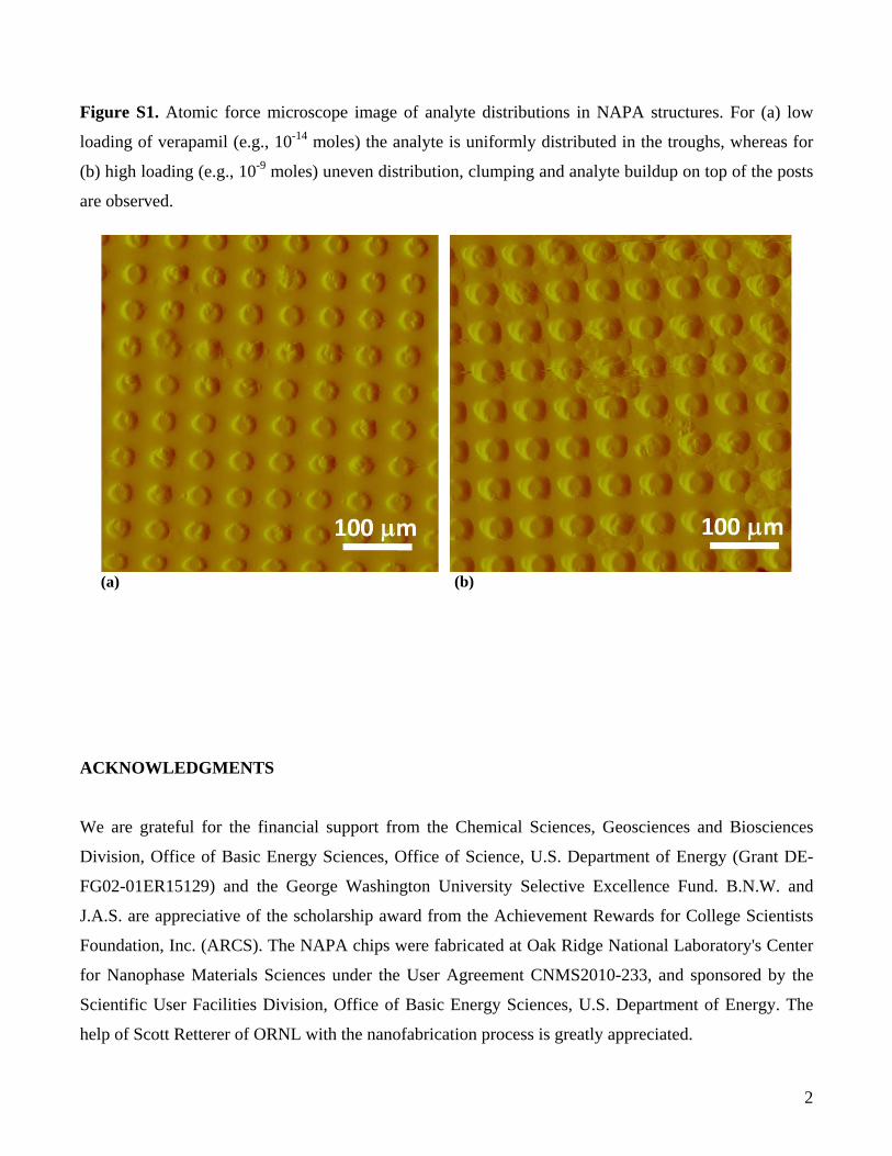
slopes can be based on the differences in the loading of theNAPA structure with the sample. As shown by AFM in FigureS1 of the Supporting Information, for low loading of verapamil(10−14 mol) the analyte is uniformly distributed in the troughs,
whereas for high loading (10−9 mol) uneven distribution,clumping, and analyte buildup on top of the posts are observed.A decrease in signal intensity is observed for the highest
amount of deposited verapamil (see the data point for 2 × 10⁻⁸
mol in Figure 3). This signal drop off is believed to be due tooverloading the NAPA structure with the sample. AFM images(see Figure S1b of the Supporting Information) indicate thatwhen large amounts of sample are deposited onto the NAPAstructure it begins to fill in the troughs and obscure theinteraction between the laser radiation and the silicon posts.This reduced interaction can result in lower levels of energydissipation in the posts and diminished near-field enhancementof the electric field around the posts. Thus, it is important toremember that the overall sample capacity of these NAPA chipsis approximately 2 nmol.To assess the long-term stability of the NAPA structures,
chips were stored in low-humidity environment for 7 months.Comparison of mass spectra before and after the extendedstorage (not shown) indicated no significant decrease inionization efficiency. A slight increase in the spectral noise level,however, was noticed. A likely source of the increased noise iscontamination during storage. These results suggest that thenanopost performance degradation during extended storage isnegligible.Although NAPA structures are intended to be disposed of
after a single use, we have found that, with proper cleaning, i.e.,rinsing with methanol and water followed by sonication, theycan be reused for additional analyses. This is especially true atanalyte concentrations above the ultratrace levels. Inspection ofthe NAPA structures by SEM imaging indicates the lack ofstructural damage on the nanoposts after laser irradiation atfluence levels close to the ion generation threshold. This helpswith the reusability of NAPA and the shot-to-shot stability ofthe signal during the LDI-MS experiments.
Analysis of Mixtures. As most analytical problems involvecomplex mixtures rather than pure samples, it is important toexplore potential interferences and ion suppression effects. Forexample, peptide mixtures are commonly analyzed inproteomics applications. A mass spectrum from an equimolarpeptide mixture containing 1 pmol amounts of leucineenkephalin, bradykinin, angiotensin I, and substance P, each,deposited onto a NAPA chip is shown in Figure 4a. All of thecomponents are present as intact quasimolecular ions, althoughnot with equal intensities. The response from angiotensin I issignificantly weaker than from the other peptides, especially ifwe take into account the intensities of the two fragment ions, x1from leucine enkephalin and a3 from substance P. The isotopeion peaks in the inset are well-resolved, and their intensitiesclosely resemble the calculated isotope distribution of substanceP.Quantitation of targeted compounds in complex mixtures is
another task commonly performed in, e.g., food or environ-mental analysis. For example, recent investigations have found acorrelation between a reduction in cardiovascular andcerebrovascular risks and a moderate consumption of redwine.31,32 These studies claimed that resveratrol, a stilbenoidwith a monoisotopic mass of 228.079 Da, and proanthocyani-din in the wine helped to maintain cardiac health and couldincrease life expectancy. Thus, it became important to rapidlyprofile resveratrol and other compounds in commercial wines33
and to follow the transformations of resveratrol in humanmetabolism.34 Representative concentrations of trans-resvera-trol in red wine range between 0.4 and 63 μM,31,35 and its
Figure 3. Molecular ion intensities for verapamil as a function of thesample amount deposited on the NAPA chip. On the basis of the massspectra a detection limit of ∼800 zmol is established. See thecorresponding spectrum zoomed around the molecular ion peak in theinset. The dashed lines indicate the regressions for the two linearregions in the response curve.
Analytical Chemistry Article
dx.doi.org/10.1021/ac301238k | Anal. Chem. 2012, 84, 7756−77627759

quantitation typically requires a separation step followed bynegative ion electrospray MS.36 Due to the presence ofhundreds of compounds in wine samples, the identification ofresveratrol through MS relies on accurate mass measurements,the determination of isotope distribution, and ultimately,fragmentation studies.To prevent overloading the NAPA chip by the abundant
components of the wine, a 1000-times dilution in water wasperformed before analysis. To obtain the LDI mass spectrum ofred wine, 0.5 μL of the diluted sample was deposited on aNAPA. A typical negative ion spectrum, shown in Figure 4b,contains ∼100 peaks. Fifteen of them, including resveratrol, areputatively identified based on accurate masses within a ± 6mDa margin, addition of resveratrol standard to the sample,isotope distribution patterns (see the bottom inset in Figure4b), and information from previous studies.32,33,37,38 Toestablish the figures of merit for resveratrol analysis onNAPA, standard samples were analyzed in a wide range ofconcentrations. The top inset in Figure 4b shows the peak
intensity as a function of the deposited moles of resveratrol inthe 2 × 10⁻¹⁷ to 2 × 10⁻¹⁰ mol range. On the basis of this data,a detection limit of 20 amol was established with a 3 orders ofmagnitude range of quantitation between 2 × 10⁻¹⁶ and 2 ×10⁻¹³ mol. Linear regression in this range yielded a correlationcoefficient of R2 = 0.95. From this curve, a combinedconcentration of cis- and trans-resveratrol of ∼68 μM wasfound for the studied red wine samples, which is within themargin of error for the high values found in the literature.31
Single-Cell Analysis. Detection of metabolites from singlemicrobial cells requires highly sensitive multispecies techni-ques.39 The estimated amounts of primary metabolites within abudding yeast (S. cerevisiae) cell, with a volume of only 30 fL,are typically between 10 amol and 30 fmol.40 On the basis of itsfigures of merit, LDI-MS from NAPA is expected to besufficiently sensitive for producing mass spectra in this range.As the yeast metabolic network is relatively small and has beenthoroughly analyzed,40 the identification of metabolites isrelatively simple.Single yeast cells were directly deposited onto NAPA chips
and analyzed by LDI-MS in positive and negative ion mode.Figure 5a shows an AFM image of a yeast cell on a NAPAstructure before laser exposure. During LDI, laser excitation ofthe posts ruptures the cell, exposing and ionizing theintracellular components. Figure 5b, shows a representative
Figure 4. (a) Positive ion LDI mass spectrum of a four-peptidemixture containing leucine enkephalin, bradykinin, angiotensin I, andsubstance P from NAPA shows the molecular ion peaks and twoknown fragments. The inset shows the isotope distribution pattern ofthe protonated substance P molecule. (b) Negative ion LDI massspectrum of red wine from NAPA. Ten of the 15 putatively assignedcompounds, including resveratrol, are labeled. The zoomed spectrumin the range of the isotope distribution pattern for resveratrol is in thebottom inset. The top inset depicts the quantitative LDI responsefrom NAPA for a wide range of deposited resveratrol amounts. Thevalues determined for red wine (vertical dashed line) are well withinthe range of quantitation (fitted dashed line in gray).
Figure 5. (a) Atomic force microscope image of a single yeast celldeposited on a NAPA before LDI-MS analysis. (b) LDI mass spectrumof a single yeast cell from NAPA in the mass range of metabolites.Four of the ∼24 putatively assigned metabolites are labeled.
Analytical Chemistry Article
dx.doi.org/10.1021/ac301238k | Anal. Chem. 2012, 84, 7756−77627760

mass spectrum from a single-cell experiment with some of theassigned metabolites. Initially multicell spectra were acquiredfor identification of the peaks with higher fidelity. On the basisof the spectra for multiple cells, accurate mass measurements,isotope distribution patterns, literature data, and databasesearches41 were used to assign the peaks. In total, 24biochemicals, corresponding to ∼4% of the known metab-olome, were detected (see Table 1). On the basis of theseassignments, the coverage of major biochemical pathways was29% with at least one metabolite in a pathway identified. In oneexample, 5 out of 12 metabolites involved in the threonine andmethionine biosynthesis superpathway were assigned. Detaileddirect analysis of single cells with LDI from NAPA can improveour understanding of how individual cells grow, develop, andrespond to environmental stress, as well as reveal themetabolites involved in chemical communication between cells.
■ CONCLUSIONS
Mechanistic studies on NAPA structures, a representative ofnanophotonic ion sources, indicated the existence of selectgeometries that produce a resonance-like enhancement in ionyields. In this contribution, LDI on NAPA with a resonantstructure is shown to enable the ultratrace level detection ofsmall molecules in the mass range between 50 and 1500 Da.Limits of detection for verapamil and resveratrol are establishedat 800 zmol and 20 amol, respectively. A comparison of LISMAand NAPA indicates improved figures of merit for the latter.For example, typical limit of detection for LISMA was 10 fmolfor the P14R synthetic peptide. Quantitation with the most
commonly used LDI method, MALDI, is only possible in anarrow range of concentrations. With NAPA we show adynamic range of 3−4 orders of magnitude for resveratrol andverapamil. On the basis of investigating spectral responses from16 compounds, NAPA promises to be a robust LDI platformfor the analysis of a broad range of small molecules includingpharmaceuticals, natural products, explosives, and metabolites.Initial stability experiments indicate unchanged LDI perform-ance for at least 7 months of storage in a low-humidityenvironment. The most significant disadvantage of the NAPAplatform is its involved fabrication. It requires a nanofabricationfacility in a clean room. This process, however, confers avaluable advantage, i.e., the extremely low chemical backgrounddue to the clean fabrication environment.Turning to real-world samples, we show examples of
detecting and quantitating targeted compounds in complexmixtures. Resveratrol is readily detected in red wine without theneed for a separation method. After depositing a drop ofdiluted red wine on a NAPA chip, we obtain an negative ionLDI spectrum exhibiting ∼100 peaks and determine thecombined concentration of cis- and trans-resveratrol. In anotherexample, a severely volume-limited sample is analyzed onNAPA. A single yeast cell with a typical volume of ∼30 fL issufficient to produce a complex LDI spectrum. In these spectrawe are able to tentatively assign 24 metabolites, whichcorrespond to a 4% coverage of the yeast metabolome.Metabolite profiling of single cells opens the door to studyingcellular heterogeneity, a crucial step in understanding cell-to-cell variations in physiology and environmental response. A
Table 1. Metabolites Detected in a Single Yeast Cell by LDI from NAPA
no. assigned metabolite ion calcd m/z measd m/z Δ m/z
1 amino propanol [M − H]− 74.061 74.048 0.0132 isobutyrate/acetoin [M − H]− 87.040 87.033 0.0073 cadaverine [M + H]+ 103.136 103.132 0.0044 proline [M + H]+ 116.053 116.071 −0.0185 fumaric acid [M + H]+ 117.019 117.010 0.0096 aspartate semialdehyde/amino oxobutanoate [M + H]+ 118.050 117.990 0.0607 threonine/homoserine [M + K]+ 158.060 158.021 0.0398 threonine/homoserine [M + H]+ 120.066 120.035 0.0319 glutamic semialdehyde/aminolevulinate/aminooxopentanoate [M + H]+ 132.066 132.030 0.03610 methionine [M − H]− 148.040 148.070 −0.03011 xanthine/xylitol [M − H]− 151.026 151.050 −0.024
151.06012 indole/valine/aminopentanoate [M + K]+ 156.021 156.050 −0.029
156.04313 dihydroxyacetone phosphatidic acid/glyceraldehyde phosphate/phospholactate/sulfolactate [M + H]+ 171.005 170.996 0.009
171.005171.005170.997
14 glycerol phosphatidic acid/dehydroshikimate [M + H]+ 173.022 173.021 0.022173.045
15 histidinol [M + K]+ 180.037 180.052 −0.01516 iditol [M + H]+ 183.086 183.097 −0.01117 phosphohomoserine [M + H]+ 200.030 200.060 −0.03018 lipoamide [M + H]+ 206.067 206.093 −0.02619 pantothenate [M − H]− 218.103 218.122 −0.01920 cytidine [M + H]+ 244.093 244.040 0.05321 inosine [M − H]− 267.070 267.090 −0.02022 naringenin [M − H]− 271.061 271.023 0.03823 sedoheptulose phosphatidic acid [M + H]+ 290.160 290.101 0.05924 phosphoribosylformylglycineamidine [M + H]+ 314.075 314.070 0.005
Analytical Chemistry Article
dx.doi.org/10.1021/ac301238k | Anal. Chem. 2012, 84, 7756−77627761

crucial step in entering that field is to develop high-throughputcapabilities for the NAPA-based analytical platforms. Experi-ments to analyze single mammalian cells on NAPA areunderway and will be reported in a separate contribution.
■ ASSOCIATED CONTENT*S Supporting InformationAdditional information as noted in text. This material isavailable free of charge via the Internet at http://pubs.acs.org.
■ AUTHOR INFORMATIONCorresponding Author*E-mail: [email protected]. Phone: +1 202-994-2717. Fax: +1202-994-5873.
NotesThe authors declare no competing financial interest.
■ ACKNOWLEDGMENTSWe are grateful for the financial support from the ChemicalSciences, Geosciences and Biosciences Division, Office of BasicEnergy Sciences, Office of Science, U.S. Department of Energy(Grant DE-FG02-01ER15129) and the George WashingtonUniversity Selective Excellence Fund. B.N.W. and J.A.S. areappreciative of the scholarship award from the AchievementRewards for College Scientists Foundation, Inc. (ARCS). TheNAPA chips were fabricated at Oak Ridge NationalLaboratory’s Center for Nanophase Materials Sciences underthe user agreement CNMS2010-233 and sponsored by theScientific User Facilities Division, Office of Basic EnergySciences, U.S. Department of Energy. The help of ScottRetterer of ORNL with the nanofabrication process is greatlyappreciated.
■ REFERENCES(1) Karas, M.; Hillenkamp, F. Anal. Chem. 1988, 60, 2299−2301.(2) Tanaka, K.; Waki, H.; Ido, Y.; Akita, S.; Yoshida, Y.; Yoshida, T.Rapid Commun. Mass Spectrom. 1988, 2, 151−153.(3) Wei, J.; Buriak, J. M.; Siuzdak, G. Nature 1999, 399, 243−246.(4) Northen, T. R.; Yanes, O.; Northen, M. T.; Marrinucci, D.;Uritboonthai, W.; Apon, J.; Golledge, S. L.; Nordstrom, A.; Siuzdak, G.Nature 2007, 449, 1033−1036.(5) Go, E. P.; Apon, J. V.; Luo, G.; Saghatelian, A.; Daniels, R. H.;Sahi, V.; Dubrow, R.; Cravatt, B. F.; Vertes, A.; Siuzdak, G. Anal. Chem.2005, 77, 1641−1646.(6) Castellana, E. T.; Gamez, R. C.; Gomez, M. E.; Russell, D. H.Langmuir 2010, 26, 6066−6070.(7) Wen, X. J.; Dagan, S.; Wysocki, V. H. Anal. Chem. 2007, 79, 434−444.(8) Luo, G. H.; Chen, Y.; Daniels, H.; Dubrow, R.; Vertes, A. J. Phys.Chem. B 2006, 110, 13381−13386.(9) Owega, S.; Lai, E. P. C.; Bawagan, A. D. O. Anal. Chem. 1998, 70,2360−2365.(10) Stolee, J. A.; Walker, B. N.; Zorba, V.; Russo, R. E.; Vertes, A.Phys. Chem. Chem. Phys. 2012, 14, 8453−8471.(11) Xiao, Y. S.; Retterer, S. T.; Thomas, D. K.; Tao, J. Y.; He, L. J.Phys. Chem. C 2009, 113, 3076−3083.(12) Gorecka-Drzazga, A.; Dziuban, J.; Drzazga, W.; Kraj, A.;Silberring, J. J. Vac. Sci. Technol., B 2005, 23, 819−823.(13) Chen, Y.; Vertes, A. Anal. Chem. 2006, 78, 5835−5844.(14) Trauger, S. A.; Go, E. P.; Shen, Z. X.; Apon, J. V.; Compton, B.J.; Bouvier, E. S. P.; Finn, M. G.; Siuzdak, G. Anal. Chem. 2004, 76,4484−4489.(15) Amantonico, A.; Urban, P. L.; Fagerer, S. R.; Balabin, R. M.;Zenobi, R. Anal. Chem. 2010, 82, 7394−7400.
(16) Holscher, D.; Shroff, R.; Knop, K.; Gottschaldt, M.; Crecelius,A.; Schneider, B.; Heckel, D. G.; Schubert, U. S.; Svatos, A. Plant J.2009, 60, 907−918.(17) Rubakhin, S. S.; Romanova, E. V.; Nemes, P.; Sweedler, J. V.Nat. Methods 2011, 8, S20−S29.(18) McLean, J. A.; Stumpo, K. A.; Russell, D. H. J. Am. Chem. Soc.2005, 127, 5304−5305.(19) Finkel, N. H.; Prevo, B. G.; Velev, O. D.; He, L. Anal. Chem.2005, 77, 1088−1095.(20) Kruse, R. A.; Li, X. L.; Bohn, P. W.; Sweedler, J. V. Anal. Chem.2001, 73, 3639−3645.(21) Thomas, J. J.; Shen, Z. X.; Crowell, J. E.; Finn, M. G.; Siuzdak,G. Proc. Natl. Acad. Sci. U.S.A. 2001, 98, 4932−4937.(22) Nordstrom, A.; Apon, J. V.; Uritboonthal, W.; Go, E. P.;Siuzdak, G. Anal. Chem. 2006, 78, 272−278.(23) Walker, B. N.; Razunguzwa, T.; Powell, M.; Knochenmuss, R.;Vertes, A. Angew. Chem., Int. Ed. 2009, 48, 1669−1672.(24) Stolee, J. A.; Walker, B. N.; Chen, Y.; Vertes, A. AIP Conf. Proc.2010, 1278, 98−110.(25) Her, T. H.; Finlay, R. J.; Wu, C.; Mazur, E. Appl. Phys. A: Mater.Sci. Process. 2000, 70, 383−385.(26) Wu, C.; Crouch, C. H.; Zhao, L.; Carey, J. E.; Younkin, R.;Levinson, J. A.; Mazur, E.; Farrell, R. M.; Gothoskar, P.; Karger, A.Appl. Phys. Lett. 2001, 78, 1850−1852.(27) Pedraza, A. J.; Fowlkes, J. D.; Guan, Y. F. Appl. Phys. A: Mater.Sci. Process. 2003, 77, 277−284.(28) Crouch, C. H.; Carey, J. E.; Warrender, J. M.; Aziz, M. J.; Mazur,E.; Genin, F. Y. Appl. Phys. Lett. 2004, 84, 1850−1852.(29) Walker, B. N.; Stolee, J. A.; Pickel, D. L.; Retterer, S. T.; Vertes,A. J. Phys. Chem. C 2010, 114, 4835−4840.(30) Stolee, J. A.; Vertes, A. Phys. Chem. Chem. Phys. 2011, 13, 9140−9146.(31) Baur, J. A.; Sinclair, D. A. Nat. Rev. Drug Discovery 2006, 5, 493−506.(32) Bertelli, A. A. A.; Das, D. K. J. Cardiovasc. Pharmacol. 2009, 54,468−476.(33) Carpentieri, A.; Marino, G.; Amoresano, A. Anal. Bioanal. Chem.2007, 389, 969−982.(34) Yu, C. W.; Shin, Y. G.; Chow, A.; Li, Y. M.; Kosmeder, J. W.;Lee, Y. S.; Hirschelman, W. H.; Pezzuto, J. M.; Mehta, R. G.; vanBreemen, R. B. Pharm. Res. 2002, 19, 1907−1914.(35) Goldberg, D. M.; Ng, E.; Karumanchiri, A.; Diamandis, E. P.;Soleas, G. J. Am. J. Enol. Vitic. 1996, 47, 415−420.(36) Careri, M.; Corradini, C.; Elviri, L.; Nicoletti, I.; Zagnoni, I. J.Agric. Food Chem. 2004, 52, 6868−6874.(37) Mazzuca, P.; Ferranti, P.; Picariello, G.; Chianese, L.; Addeo, F.J. Mass Spectrom. 2005, 40, 83−90.(38) Alcalde-Eon, C.; Escribano-Bailon, M. T.; Santos-Buelga, C.;Rivas-Gonzalo, J. C. Anal. Chim. Acta 2006, 563, 238−254.(39) Amantonico, A.; Oh, J. Y.; Sobek, J.; Heinemann, M.; Zenobi, R.Angew. Chem., Int. Ed. 2008, 47, 5382−5385.(40) Forster, J.; Famili, I.; Fu, P.; Palsson, B. O.; Nielsen, J. GenomeRes. 2003, 13, 244−253.(41) Saccharomyces Genome Database. http://www.yeastgenome.org(accessed July 29, 2012).
Analytical Chemistry Article
dx.doi.org/10.1021/ac301238k | Anal. Chem. 2012, 84, 7756−77627762

1
Supporting Information for
Nanophotonic Ionization for Ultratrace and Single-Cell
Analysis by Mass Spectrometry
Bennett N. Walker, Jessica A. Stolee, and Akos Vertes*
W. M. Keck Institute for Proteomics Technology and Applications, Department of Chemistry,
The George Washington University, Washington, District of Columbia 20052, United States
* To whom correspondence should be addressed. E-mail: [email protected]. Phone: +1 (202) 994-2717.
Fax: +1 (202) 994-5873.
2
Figure S1. Atomic force microscope image of analyte distributions in NAPA structures. For (a) low
loading of verapamil (e.g., 10-14 moles) the analyte is uniformly distributed in the troughs, whereas for
(b) high loading (e.g., 10-9 moles) uneven distribution, clumping and analyte buildup on top of the posts
are observed.
(a) (b)
ACKNOWLEDGMENTS
We are grateful for the financial support from the Chemical Sciences, Geosciences and Biosciences
Division, Office of Basic Energy Sciences, Office of Science, U.S. Department of Energy (Grant DE-
FG02-01ER15129) and the George Washington University Selective Excellence Fund. B.N.W. and
J.A.S. are appreciative of the scholarship award from the Achievement Rewards for College Scientists
Foundation, Inc. (ARCS). The NAPA chips were fabricated at Oak Ridge National Laboratory's Center
for Nanophase Materials Sciences under the User Agreement CNMS2010-233, and sponsored by the
Scientific User Facilities Division, Office of Basic Energy Sciences, U.S. Department of Energy. The
help of Scott Retterer of ORNL with the nanofabrication process is greatly appreciated.
Related Documents