Nanoparticle engineered TRAIL-overexpressing adipose-derived stem cells target and eradicate glioblastoma via intracranial delivery Xinyi Jiang a , Sergio Fitch b , Christine Wang b , Christy Wilson c , Jianfeng Li a , Gerald A. Grant c , and Fan Yang a,b,1 a Department of Orthopaedic Surgery, Stanford University, Stanford, CA 94305; b Department of Bioengineering, Stanford University, Stanford, CA 94305; and c Department of Neurosurgery, Stanford University, Stanford, CA 94305 Edited by Robert Langer, Massachusetts Institute of Technology, Cambridge, MA, and approved October 17, 2016 (received for review September 29, 2016) Glioblastoma multiforme (GBM) is one of the most intractable of human cancers, principally because of the highly infiltrative nature of these neoplasms. Tracking and eradicating infiltrating GBM cells and tumor microsatellites is of utmost importance for the treat- ment of this devastating disease, yet effective strategies remain elusive. Here we report polymeric nanoparticle-engineered human adipose-derived stem cells (hADSCs) overexpressing tumor necro- sis factor-related apoptosis-inducing ligand (TRAIL) as drug-delivery vehicles for targeting and eradicating GBM cells in vivo. Our results showed that polymeric nanoparticle-mediated transfection led to robust up-regulation of TRAIL in hADSCs, and that TRAIL-expressing hADSCs induced tumor-specific apoptosis. When transplanted in a mouse intracranial xenograft model of patient-derived glioblastoma cells, hADSCs exhibited long-range directional migration and in- filtration toward GBM tumor. Importantly, TRAIL-overexpressing hADSCs inhibited GBM growth, extended survival, and reduced the occurrence of microsatellites. Repetitive injection of TRAIL-over- expressing hADSCs significantly prolonged animal survival com- pared with single injection of these cells. Taken together, our data suggest that nanoparticle-engineered TRAIL-expressing hADSCs exhibit the therapeutically relevant behavior of “seek-and-destroy” tumortropic migration and could be a promising therapeutic ap- proach to improve the treatment outcomes of patients with malig- nant brain tumors. nanoparticle | adipose-derived stem cells | TRAIL | glioblastoma | tumor microsatellites G lioblastoma multiforme (GBM) is the most common and aggressive subtype of malignant brain tumor. Current state- of-the-art treatment consisting of surgical resection in combi- nation with radiation and chemotherapy for GBM fails to address its highly infiltrative nature, often leaving behind micro- scopic tumor satellites, which results in tumor recurrence. Thera- peutic gene delivery by direct injection of viral vectors into the primary brain tumor or postoperative tumor resection cavity has also largely failed to reach outgrowing tumor micrometastatic nests of glioma cells at sites distant from the main tumor mass as well as infiltrating glioma cells in the adjacent brain. As such, it is imperative to develop novel treatment strategies that focus specifically on targeting and eliminating the disseminated neo- plastic burden within the brain. Human stem cells have shown promise as a drug delivery ve- hicle for targeting infiltrating brain cancer cells that cannot be removed by surgery. Recent research shows that transplanted human neural stem cells (NSCs; hNSCs) possess remarkable tumortropic migratory capacity in vitro and in vivo; hNSCs have been used for the delivery of cytotoxic and immunomodulatory therapies (1–5) and were recently approved for clinical trials. However, the clinical translational potential of hNSCs may be hindered by ethical concerns associated with their isolation, expansion, and associated immune response (6, 7). Bone marrow- derived human mesenchymal stem cells (hMSCs) exhibit selec- tive tropism similar to hNSCs, migrating significant distances to target gliomas (8). Unlike hNSCs and hMSCs, human adipose tissue-derived stem cells (hADSCs) represent an abundant and easily accessible autologous source of stem cells, with fewer ethical concerns associated with their use. Additionally, unmod- ified hADSCs remain free of oncogenic transformation for at least 8 mo when injected into immunocompromised mice, dem- onstrating more oncogenic resistance than human bone marrow- derived stem cells (9). Therefore, hADSCs derived from fat tissue could represent a better alternative for stem cell-based cancer gene therapy. By using stem cells as drug delivery vehicles, different bi- ological drugs have been delivered, including chemotherapeutic agents, prodrugs (10–13), and genetic signals (14). One limita- tion of delivering chemotherapeutic agents is that they generally could not differentiate cancerous cells from normal cells. To avoid undesirable cytotoxicity to normal cells, here we have specifically chosen the full length of tumor necrosis factor- related apoptosis-inducing ligand (TRAIL), a type II membrane- bound protein that can rapidly induce apoptosis in a variety of cancers while leaving normal cell types intact. TRAIL induces apoptosis by binding to its death receptors DR4 and DR5, forming a homotrimeric complex (TRAIL-R1/D4 and TRAIL- R2/D5), causing caspase-8 activation in the death-inducing signaling complex; this complex can directly cleave caspase-3 to activate extrinsic pathways or cleave the Bcl-2–inhibitory Significance Current treatment for glioblastoma multiforme (GBM) fails to address its highly infiltrative nature; treatment often leaves behind microscopic neoplastic satellites, resulting in eventual tumor recurrence. Here we report polymeric nanoparticle- engineered human adipose-derived stem cells (hADSCs) over- expressing the cancer-specific TNF-related apoptosis-inducing ligand for targeting and eradicating glioblastoma cells. Engi- neered hADSCs exhibited long-range directional migration to- ward tumor in patient-derived GBM orthotropic xenografts and showed significant inhibition of tumor growth and extension of animal survival. Repetitive injection further prolonged animal survival compared with single injection. Together, our data sug- gest that nanoparticle-engineered hADSCs exhibit the thera- peutically relevant behavior of “seek-and-destroy” tumortropic migration, and may offer a promising therapy for substantial enhancement of GBM treatment. Author contributions: X.J., C. Wang, G.A.G., and F.Y. designed research; X.J. and S.F. performed research; C. Wilson contributed new reagents/analytic tools; X.J., S.F., C. Wang, C. Wilson, J.L., G.A.G., and F.Y. analyzed data; and X.J., S.F., G.A.G., and F.Y. wrote the paper. The authors declare no conflict of interest. This article is a PNAS Direct Submission. 1 To whom correspondence should be addressed. Email: [email protected]. This article contains supporting information online at www.pnas.org/lookup/suppl/doi:10. 1073/pnas.1615396113/-/DCSupplemental. www.pnas.org/cgi/doi/10.1073/pnas.1615396113 PNAS | November 29, 2016 | vol. 113 | no. 48 | 13857–13862 MEDICAL SCIENCES

Welcome message from author
This document is posted to help you gain knowledge. Please leave a comment to let me know what you think about it! Share it to your friends and learn new things together.
Transcript
Nanoparticle engineered TRAIL-overexpressingadipose-derived stem cells target and eradicateglioblastoma via intracranial deliveryXinyi Jianga, Sergio Fitchb, Christine Wangb, Christy Wilsonc, Jianfeng Lia, Gerald A. Grantc, and Fan Yanga,b,1
aDepartment of Orthopaedic Surgery, Stanford University, Stanford, CA 94305; bDepartment of Bioengineering, Stanford University, Stanford, CA 94305;and cDepartment of Neurosurgery, Stanford University, Stanford, CA 94305
Edited by Robert Langer, Massachusetts Institute of Technology, Cambridge, MA, and approved October 17, 2016 (received for review September 29, 2016)
Glioblastoma multiforme (GBM) is one of the most intractable ofhuman cancers, principally because of the highly infiltrative natureof these neoplasms. Tracking and eradicating infiltrating GBM cellsand tumor microsatellites is of utmost importance for the treat-ment of this devastating disease, yet effective strategies remainelusive. Here we report polymeric nanoparticle-engineered humanadipose-derived stem cells (hADSCs) overexpressing tumor necro-sis factor-related apoptosis-inducing ligand (TRAIL) as drug-deliveryvehicles for targeting and eradicating GBM cells in vivo. Our resultsshowed that polymeric nanoparticle-mediated transfection led torobust up-regulation of TRAIL in hADSCs, and that TRAIL-expressinghADSCs induced tumor-specific apoptosis. When transplanted in amouse intracranial xenograft model of patient-derived glioblastomacells, hADSCs exhibited long-range directional migration and in-filtration toward GBM tumor. Importantly, TRAIL-overexpressinghADSCs inhibited GBM growth, extended survival, and reduced theoccurrence of microsatellites. Repetitive injection of TRAIL-over-expressing hADSCs significantly prolonged animal survival com-pared with single injection of these cells. Taken together, our datasuggest that nanoparticle-engineered TRAIL-expressing hADSCsexhibit the therapeutically relevant behavior of “seek-and-destroy”tumortropic migration and could be a promising therapeutic ap-proach to improve the treatment outcomes of patients with malig-nant brain tumors.
nanoparticle | adipose-derived stem cells | TRAIL | glioblastoma |tumor microsatellites
Glioblastoma multiforme (GBM) is the most common andaggressive subtype of malignant brain tumor. Current state-
of-the-art treatment consisting of surgical resection in combi-nation with radiation and chemotherapy for GBM fails toaddress its highly infiltrative nature, often leaving behind micro-scopic tumor satellites, which results in tumor recurrence. Thera-peutic gene delivery by direct injection of viral vectors into theprimary brain tumor or postoperative tumor resection cavity hasalso largely failed to reach outgrowing tumor micrometastaticnests of glioma cells at sites distant from the main tumor mass aswell as infiltrating glioma cells in the adjacent brain. As such, it isimperative to develop novel treatment strategies that focusspecifically on targeting and eliminating the disseminated neo-plastic burden within the brain.Human stem cells have shown promise as a drug delivery ve-
hicle for targeting infiltrating brain cancer cells that cannot beremoved by surgery. Recent research shows that transplantedhuman neural stem cells (NSCs; hNSCs) possess remarkabletumortropic migratory capacity in vitro and in vivo; hNSCs havebeen used for the delivery of cytotoxic and immunomodulatorytherapies (1–5) and were recently approved for clinical trials.However, the clinical translational potential of hNSCs may behindered by ethical concerns associated with their isolation,expansion, and associated immune response (6, 7). Bone marrow-derived human mesenchymal stem cells (hMSCs) exhibit selec-tive tropism similar to hNSCs, migrating significant distances to
target gliomas (8). Unlike hNSCs and hMSCs, human adiposetissue-derived stem cells (hADSCs) represent an abundant andeasily accessible autologous source of stem cells, with fewerethical concerns associated with their use. Additionally, unmod-ified hADSCs remain free of oncogenic transformation for atleast 8 mo when injected into immunocompromised mice, dem-onstrating more oncogenic resistance than human bone marrow-derived stem cells (9). Therefore, hADSCs derived from fat tissuecould represent a better alternative for stem cell-based cancergene therapy.By using stem cells as drug delivery vehicles, different bi-
ological drugs have been delivered, including chemotherapeuticagents, prodrugs (10–13), and genetic signals (14). One limita-tion of delivering chemotherapeutic agents is that they generallycould not differentiate cancerous cells from normal cells. Toavoid undesirable cytotoxicity to normal cells, here we havespecifically chosen the full length of tumor necrosis factor-related apoptosis-inducing ligand (TRAIL), a type II membrane-bound protein that can rapidly induce apoptosis in a variety ofcancers while leaving normal cell types intact. TRAIL inducesapoptosis by binding to its death receptors DR4 and DR5,forming a homotrimeric complex (TRAIL-R1/D4 and TRAIL-R2/D5), causing caspase-8 activation in the death-inducingsignaling complex; this complex can directly cleave caspase-3to activate extrinsic pathways or cleave the Bcl-2–inhibitory
Significance
Current treatment for glioblastoma multiforme (GBM) fails toaddress its highly infiltrative nature; treatment often leavesbehind microscopic neoplastic satellites, resulting in eventualtumor recurrence. Here we report polymeric nanoparticle-engineered human adipose-derived stem cells (hADSCs) over-expressing the cancer-specific TNF-related apoptosis-inducingligand for targeting and eradicating glioblastoma cells. Engi-neered hADSCs exhibited long-range directional migration to-ward tumor in patient-derived GBM orthotropic xenografts andshowed significant inhibition of tumor growth and extension ofanimal survival. Repetitive injection further prolonged animalsurvival compared with single injection. Together, our data sug-gest that nanoparticle-engineered hADSCs exhibit the thera-peutically relevant behavior of “seek-and-destroy” tumortropicmigration, and may offer a promising therapy for substantialenhancement of GBM treatment.
Author contributions: X.J., C. Wang, G.A.G., and F.Y. designed research; X.J. and S.F.performed research; C. Wilson contributed new reagents/analytic tools; X.J., S.F., C. Wang,C. Wilson, J.L., G.A.G., and F.Y. analyzed data; and X.J., S.F., G.A.G., and F.Y. wrotethe paper.
The authors declare no conflict of interest.
This article is a PNAS Direct Submission.1To whom correspondence should be addressed. Email: [email protected].
This article contains supporting information online at www.pnas.org/lookup/suppl/doi:10.1073/pnas.1615396113/-/DCSupplemental.
www.pnas.org/cgi/doi/10.1073/pnas.1615396113 PNAS | November 29, 2016 | vol. 113 | no. 48 | 13857–13862
MED
ICALSC
IENCE
S

BH3-domain–containing protein to activate the intrinsic pathway(15, 16). A recombinant soluble version of the transmembrane deathligand TRAIL has shown compelling preclinical results as apotential cancer therapeutic agent, but only rare clinical re-sponses have been observed in clinical trials, possibly because ofinsufficient tumor exposure (17, 18) and/or weak engagement of theextrinsic pathway (19, 20). On the contrary, in the case of viral-mediated TRAIL gene therapy, limitations arise from triggeringimmune responses and unintended genomic integration events (21,22), which remain a key bottleneck for broad clinical application.Here, we sought to develop biodegradable nanoparticle-engi-
neered stem cells to efficiently express the suicide protein TRAILfor targeting and eradicating microsatellites and infiltrating gliomacells in patient-derived adult glioblastoma orthotropic xenograftmodels. Compared with methods that use nonviral gene therapyalone, this combined polymer/stem cell approach takes advantageof the stem cell’s ability to track microsatellites and infiltratingglioma cells. TRAIL secreted on the surface of hADSCs binds thedeath receptors DR4 and DR5 on tumor cells and triggers tumorcell death. hADSCs were modified with TRAIL DNA via aminogroup-ended poly(β-amino ester)s (PBAEs), hydrolytically bio-degradable polymers that can condense DNA to form nano-particles. We showed that polymeric nanoparticle-engineeredhADSCs led to robust TRAIL up-regulation in hADSCs com-pared with cells transfected with plasmid control DNA (pCDNA).Patient-derived glioma xenograft cells cocultured with TRAIL-expressing hADSCs exhibited significant cell death 48 h afterexposure. In vivo, GBM xenograft mice treated with nanoparticle-engineered TRAIL-expressing hADSCs significantly inhibited tu-mor growth in mice and substantially prolonged animal survivalcompared with hADSCs modified with pCDNA-laden nanopar-ticles, naïve hADSCs, or PBS solution. Repetitive injection ofTRAIL-overexpressing hADSCs significantly prolonged animalsurvival compared with single injection of those cells. Together,our results suggest that nanoparticle-engineered hADSCs couldserve as cellular vehicles for targeting and eradicating GBM cellsin a smart “seek-and-destroy” fashion, thereby improving thetreatment outcomes of this devastating disease.
ResultsBiodegradable Nanoparticle-Mediated hADSCs Transfection in Vitro.Primary hADSCs are relatively resistant to transfection withnonviral methods. Using a leading commercially available trans-fection reagent (Lipofectamine 2000), the highest transfectionefficiency we achieved was ∼7.37 ± 0.96%, which is far fromsufficient to achieve therapeutic efficacy for clinical application.Based on our recent work on biodegradable nanoparticle-me-diated transfection in stem cells (23, 24), we further systemati-cally optimized the nanoparticle-fabrication formulation and thetransfection protocol with our custom-developed amino groupend-modified PBAEs. As shown via agarose gel electrophoresis,the DNA plasmid was completely condensed in polymer nano-particles when the weight ratio of polymer and plasmid codedwith recombinant enhanced GFP (pEGFP) in nanoparticles wasgreater than 10:1 (Fig. S1A). For a deep investigation of the bio-chemophysical properties of PBAE nanoparticles, we systemicallymeasured the particle size and zeta potential of nanoparticles withdifferent formulations. Along with the increase of polymer ratio,the size of the nanoparticles significantly decreased as shown ina dynamic light scattering assay, and, because of the binding ofproteins in the serum, the particles showed increased size in fullsupplemented Dulbecco’s modified Eagle medium containing10% (vol/vol) FBS compared with that in deionized water (Fig.S1B). Nanoparticles suspended in deionized water have a posi-tive charge, whereas the surface charge of nanoparticles becomesslightly negative when suspended in cell-culture medium (Fig.S1C). Using GFP as a reporter gene, we further titrated thenanoparticle-mediated transfection formulation by using hADSCs.
By using a leading nanoparticle formulation with a weight ratio ofpolymer to pEGFP of 20:1, we achieved ∼5.48-fold higher trans-fection efficiency in hADSCs vs. a control group transfected usingLipofectamine 2000 (Fig. S2). Dynamic light scattering analysisshowed the leading PBAE nanoparticles in deionized water have aZ-average diameter of ∼183.6 nm (Fig. 1A) and a zeta potentialof ∼40.06 mV (Fig. 1B), and transmission electron microscope(TEM) revealed that the nanoparticles exhibited a spherical shape(Fig. 1C and Fig. S3).
Biodegradable Nanoparticle-Mediated Transfection Induced MembraneTRAIL Expression by hADSCs in Vitro. Next, we transfected hADSCswith the plasmid vector encoding the native full-length TRAIL(pTRAIL) using the leading PBAE nanoparticles. Forty-eight hoursafter transfection, hADSCs were harvested for assays of TRAILexpression. Flow cytometry indicated that 68.41 ± 2.65% of nano-particle-transfected hADSCs expressed TRAIL (Fig. 1D). Geneexpression analysis via quantitative real-time PCR (qRT-PCR)revealed a 546.07-fold increase in TRAIL expression after trans-fection with nanoparticles (Fig. 1E). Immunoblotting of lysatesprepared from cells harvested 48 h after nanoparticle trans-fection indicated that hADSCs produce TRAIL protein (Fig. 1F).Immunostaining showed a strong fluorescence signal fromTRAIL antibody in phTAIL-laden nanoparticle-transfectedhADSCs, whereas pCDNA-laden nanoparticle-transfected hADSCs
Fig. 1. Polymeric nanoparticles containing TRAIL plasmid DNA led to robustup-regulation of TRAIL in hADSCs. (A–C) Physicochemical characterization ofpEGFP-laden PBAE nanoparticles with a PBAE:pEGFP weight ratio of 20:1.Size distribution (A) and zeta potential (B) of nanoparticles determinedby dynamic light scattering. (C) TEM imaging confirmed that PBAE poly-mer condensed DNA-forming nanoparticles. (D) Flow cytometry analysis ofTRAIL+ cell percentage (**P < 0.01) and (E) qRT-PCR reveals the fold increaseof TRAIL mRNA expression in hADSCs vs. GAPDH mRNA expression 48 h aftertransfection with optimized pTRAIL-laden PBAE nanoparticles (i.e., NPs/pTRAIL; **P < 0.01). (F) Western blot assay of TRAIL protein expression inhADSCs transfected with NPs/pTRAIL or pCDNA-laden PBAE nanoparticles(i.e., NPs/pCDNA). (G) Immunostaining confirmed TRAIL expression in hADSCs;hADSCs transfected with NPs/pCDNA showed minimal TRAIL signals. Red in-dicates TRAIL, green indicates F-actin, blue indicates cell nucleus. (Scale bars:10 μm.) (H) The viability of cells was analyzed via the MTS assay 48 h aftertransfection with NPs/pTRAIL. hADSCs transfected with NPs/pTRAIL exhibitedno change in viability comparedwith naïve controls and those transfected withNPs/pCDNA. Untransfected, unmanipulated parallel cultures of hADSCs rep-resented 100% viability.
13858 | www.pnas.org/cgi/doi/10.1073/pnas.1615396113 Jiang et al.
displayed no fluorescence (Fig. 1G). It has been reported that in-fected cells can secrete TRAIL as a soluble protein into the culturemedium, which can be monitored via ELISA (25). However, we didnot detect soluble TRAIL in culture medium from nanoparticle-transfected hADSCs. These results demonstrate that all TRAILexpressed in hADSCs was membrane-bound. To determine thecytotoxicity of nanoparticle-induced TRAIL expression in hADSCs,we performed a cellTiter 96 AQueous one solution cell prolife-ration assay (MTS); there was no significant change in viability innanoparticle-treated hADSCs vs. naïve hADSCs (Fig. 1H).
TRAIL Induces Apoptosis in Glioma Cells but Not in hADSCs andNormal Astrocytes. To test the specificity of TRAIL cytotoxicityagainst glioma cells and hADSCs, we performed TUNEL immu-nostaining on cells transfected with phTRAIL nanoparticles. Wedetected a significant decrease in the viability of GBM cells,whereas hADSCs did not exhibit decreased viability 48 h aftertransfection (Fig. S4).Next, we cocultured plasmid human TRAIL-laden nanoparticles
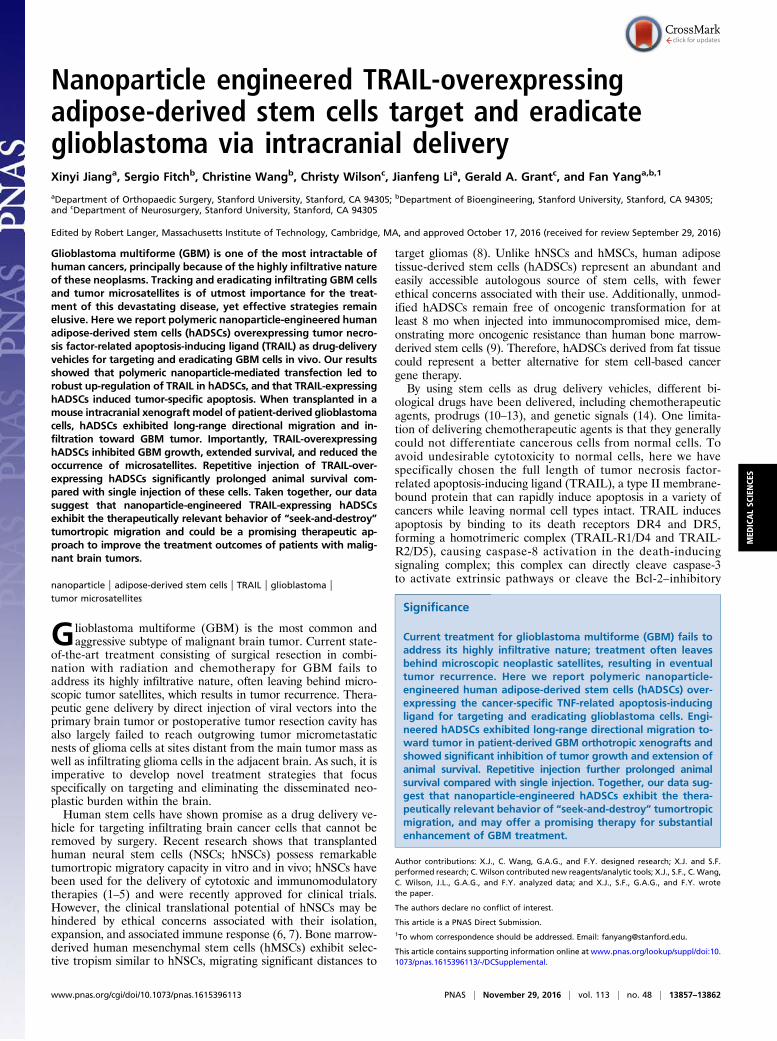
(NPs/pTRAIL)-engineered hADSCs with patient-derived malig-nant glioma xenograft cells (D-270 MG) for 48 h. This cocultureresulted in significant apoptosis and death in glioma cells (Fig. 2 Aand B), whereas glioma cells cocultured with hADSCs transfectedwith plasmid control DNA-laden nanoparticles (NPs/pCDNA) didnot demonstrate significant apoptotic activity (Fig. 2 A and B). Thecaspases are a family of closely related enzymes crucial to TRAIL-induced apoptosis. zVADfmk is a cell-permeable pan-caspaseinhibitor. Application of this compound to 10:1 coculturedglioma cells and hADSCs caused an obvious reduction in deathand apoptosis, confirming the importance of caspase pathwaysin the mechanism of TRAIL-induced glioma cell death (Fig. 2A and B).To confirm that the glioma cell population was responsible for
the dead and apoptotic populations, the GBM cells were labeledwith the fluorescent dye Dil before the annexin-V apoptosis as-say. Our data showed that the apoptotic and dead cells camespecifically from the glioma cell population, and that 35.7 ± 4.3%of the glioma cell population was dead or apoptotic, comparedwith only 4.2 ± 0.6% of the hADSCs population (P < 0.001; Fig. 3A and C). Normal astrocytes were used as a negative controlto verify the cytotoxic specificity of TRAIL-expressing hADSCsagainst normal brain cells; coculture with TRAIL-expressing
hADSCs did not increase normal astrocyte death and apoptosis(Fig. 3 B and C).To further demonstrate caspase activation, we examined cas-
pase-8 and caspase-3 activation and cleavage of the caspasesubstrate proteolytic poly(ADP-ribose) polymerase (PARP) incell lysis. Coincidental with the annexin V staining data from flowcytometry assay, caspase-8 and caspase-3 activation and PARPcleavage were detected in GBM cells cocultured with NPs/pTRAIL-transfected hADSCs but not in cocultures of NPs/pCDNA-engi-neered hADSCs with GBM cells, NPs/pTRAIL-hADSCs alone, orGBM cells alone (Fig. 3D). These results indicate a rapid activationof caspase-8 and the caspase cascade in TRAIL-induced gliomacell death when GBM cells were cocultured with NPs/pTRAIL-modified hADSCs.
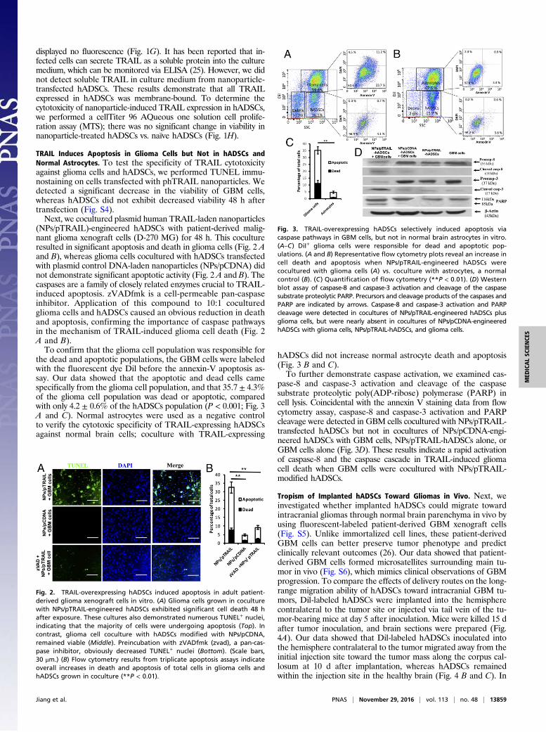
Tropism of Implanted hADSCs Toward Gliomas in Vivo. Next, weinvestigated whether implanted hADSCs could migrate towardintracranial gliomas through normal brain parenchyma in vivo byusing fluorescent-labeled patient-derived GBM xenograft cells(Fig. S5). Unlike immortalized cell lines, these patient-derivedGBM cells can better preserve tumor phenotype and predictclinically relevant outcomes (26). Our data showed that patient-derived GBM cells formed microsatellites surrounding main tu-mor in vivo (Fig. S6), which mimics clinical observations of GBMprogression. To compare the effects of delivery routes on the long-range migration ability of hADSCs toward intracranial GBM tu-mors, Dil-labeled hADSCs were implanted into the hemispherecontralateral to the tumor site or injected via tail vein of the tu-mor-bearing mice at day 5 after inoculation. Mice were killed 15 dafter tumor inoculation, and brain sections were prepared (Fig.4A). Our data showed that Dil-labeled hADSCs inoculated intothe hemisphere contralateral to the tumor migrated away from theinitial injection site toward the tumor mass along the corpus cal-losum at 10 d after implantation, whereas hADSCs remainedwithin the injection site in the healthy brain (Fig. 4 B and C). In
Fig. 2. TRAIL-overexpressing hADSCs induced apoptosis in adult patient-derived glioma xenograft cells in vitro. (A) Glioma cells grown in coculturewith NPs/pTRAIL-engineered hADSCs exhibited significant cell death 48 hafter exposure. These cultures also demonstrated numerous TUNEL+ nuclei,indicating that the majority of cells were undergoing apoptosis (Top). Incontrast, glioma cell coculture with hADSCs modified with NPs/pCDNA,remained viable (Middle). Preincubation with zVADfmk (zvad), a pan-cas-pase inhibitor, obviously decreased TUNEL+ nuclei (Bottom). (Scale bars,30 μm.) (B) Flow cytometry results from triplicate apoptosis assays indicateoverall increases in death and apoptosis of total cells in glioma cells andhADSCs grown in coculture (**P < 0.01).
Fig. 3. TRAIL-overexpressing hADSCs selectively induced apoptosis viacaspase pathways in GBM cells, but not in normal brain astrocytes in vitro.(A–C) DiI+ glioma cells were responsible for dead and apoptotic pop-ulations. (A and B) Representative flow cytometry plots reveal an increase incell death and apoptosis when NPs/pTRAIL-engineered hADSCs werecocultured with glioma cells (A) vs. coculture with astrocytes, a normalcontrol (B). (C ) Quantification of flow cytometry (**P < 0.01). (D) Westernblot assay of caspase-8 and caspase-3 activation and cleavage of the caspasesubstrate proteolytic PARP. Precursors and cleavage products of the caspases andPARP are indicated by arrows. Caspase-8 and caspase-3 activation and PARPcleavage were detected in cocultures of NPs/pTRAIL-engineered hADSCs plusglioma cells, but were nearly absent in cocultures of NPs/pCDNA-engineeredhADSCs with glioma cells, NPs/pTRAIL-hADSCs, and glioma cells.
Jiang et al. PNAS | November 29, 2016 | vol. 113 | no. 48 | 13859
MED
ICALSC
IENCE
S
the tumor brain, Dil-labeled hADSCs mostly occurred at the tu-mor margin (Fig. 4D). Interestingly, hADSCs colocalized with theinfiltrating GBM cells at a distance from the main tumor mass(Fig. 4E). Our in vivo antitumor efficacy study also revealedobvious migration of NPs/pTRAIL-engineered hADSCs within10 d, with appreciable TRAIL secretion after intratumoral (i.t.)administration (Fig. 6A); migrating hADSCs remained at theborder of the tumor, where it interfaced with normal tissue atday 10. Thus, hADSCs demonstrated a robust migratory capacityand glioma tropism in vivo and exhibited an ability to “seek”infiltrating glioma cells, which is significant for GBM treatment.In contrast, when hADSCs were injected through a tail vein,no hADSCs were detected in the brain (Fig. S7), suggestinglocal delivery within the brain is required for effective tumortargeting.
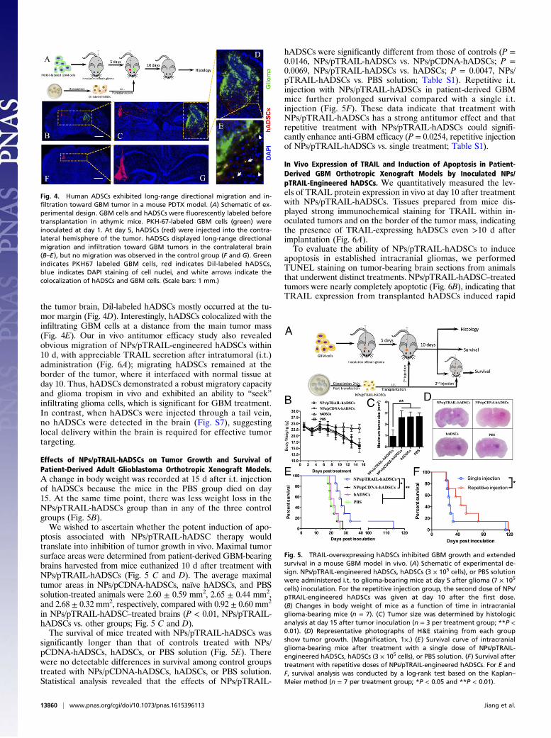
Effects of NPs/pTRAIL-hADSCs on Tumor Growth and Survival ofPatient-Derived Adult Glioblastoma Orthotropic Xenograft Models.A change in body weight was recorded at 15 d after i.t. injectionof hADSCs because the mice in the PBS group died on day15. At the same time point, there was less weight loss in theNPs/pTRAIL-hADSCs group than in any of the three controlgroups (Fig. 5B).We wished to ascertain whether the potent induction of apo-
ptosis associated with NPs/pTRAIL-hADSC therapy wouldtranslate into inhibition of tumor growth in vivo. Maximal tumorsurface areas were determined from patient-derived GBM-bearingbrains harvested from mice euthanized 10 d after treatment withNPs/pTRAIL-hADSCs (Fig. 5 C and D). The average maximaltumor areas in NPs/pCDNA-hADSCs, naïve hADSCs, and PBSsolution-treated animals were 2.60 ± 0.59 mm2, 2.65 ± 0.44 mm2,and 2.68 ± 0.32 mm2, respectively, compared with 0.92 ± 0.60 mm2
in NPs/pTRAIL-hADSC–treated brains (P < 0.01, NPs/pTRAIL-hADSCs vs. other groups; Fig. 5 C and D).The survival of mice treated with NPs/pTRAIL-hADSCs was
significantly longer than that of controls treated with NPs/pCDNA-hADSCs, hADSCs, or PBS solution (Fig. 5E). Therewere no detectable differences in survival among control groupstreated with NPs/pCDNA-hADSCs, hADSCs, or PBS solution.Statistical analysis revealed that the effects of NPs/pTRAIL-
hADSCs were significantly different from those of controls (P =0.0146, NPs/pTRAIL-hADSCs vs. NPs/pCDNA-hADSCs; P =0.0069, NPs/pTRAIL-hADSCs vs. hADSCs; P = 0.0047, NPs/pTRAIL-hADSCs vs. PBS solution; Table S1). Repetitive i.t.injection with NPs/pTRAIL-hADSCs in patient-derived GBMmice further prolonged survival compared with a single i.t.injection (Fig. 5F). These data indicate that treatment withNPs/pTRAIL-hADSCs has a strong antitumor effect and thatrepetitive treatment with NPs/pTRAIL-hADSCs could signifi-cantly enhance anti-GBM efficacy (P = 0.0254, repetitive injectionof NPs/pTRAIL-hADSCs vs. single treatment; Table S1).
In Vivo Expression of TRAIL and Induction of Apoptosis in Patient-Derived GBM Orthotropic Xenograft Models by Inoculated NPs/pTRAIL-Engineered hADSCs. We quantitatively measured the lev-els of TRAIL protein expression in vivo at day 10 after treatmentwith NPs/pTRAIL-hADSCs. Tissues prepared from mice dis-played strong immunochemical staining for TRAIL within in-oculated tumors and on the border of the tumor mass, indicatingthe presence of TRAIL-expressing hADSCs even >10 d afterimplantation (Fig. 6A).To evaluate the ability of NPs/pTRAIL-hADSCs to induce
apoptosis in established intracranial gliomas, we performedTUNEL staining on tumor-bearing brain sections from animalsthat underwent distinct treatments. NPs/pTRAIL-hADSC–treatedtumors were nearly completely apoptotic (Fig. 6B), indicating thatTRAIL expression from transplanted hADSCs induced rapid
Fig. 4. Human ADSCs exhibited long-range directional migration and in-filtration toward GBM tumor in a mouse PDTX model. (A) Schematic of ex-perimental design. GBM cells and hADSCs were fluorescently labeled beforetransplantation in athymic mice. PKH-67-labeled GBM cells (green) wereinoculated at day 1. At day 5, hADSCs (red) were injected into the contra-lateral hemisphere of the tumor. hADSCs displayed long-range directionalmigration and infiltration toward GBM tumors in the contralateral brain(B–E), but no migration was observed in the control group (F and G). Greenindicates PKH67 labeled GBM cells, red indicates Dil-labeled hADSCs,blue indicates DAPI staining of cell nuclei, and white arrows indicate thecolocalization of hADSCs and GBM cells. (Scale bars: 1 mm.)
Fig. 5. TRAIL-overexpressing hADSCs inhibited GBM growth and extendedsurvival in a mouse GBM model in vivo. (A) Schematic of experimental de-sign. NPs/pTRAIL-engineered hADSCs, hADSCs (3 × 10⁵ cells), or PBS solutionwere administered i.t. to glioma-bearing mice at day 5 after glioma (7 × 10⁵
cells) inoculation. For the repetitive injection group, the second dose of NPs/pTRAIL-engineered hADSCs was given at day 10 after the first dose.(B) Changes in body weight of mice as a function of time in intracranialglioma-bearing mice (n = 7). (C) Tumor size was determined by histologicanalysis at day 15 after tumor inoculation (n = 3 per treatment group; **P <0.01). (D) Representative photographs of H&E staining from each groupshow tumor growth. (Magnification, 1×.) (E) Survival curve of intracranialglioma-bearing mice after treatment with a single dose of NPs/pTRAIL-engineered hADSCs, hADSCs (3 × 10⁵ cells), or PBS solution. (F) Survival aftertreatment with repetitive doses of NPs/pTRAIL-engineered hADSCs. For E andF, survival analysis was conducted by a log-rank test based on the Kaplan–Meier method (n = 7 per treatment group; *P < 0.05 and **P < 0.01).
13860 | www.pnas.org/cgi/doi/10.1073/pnas.1615396113 Jiang et al.
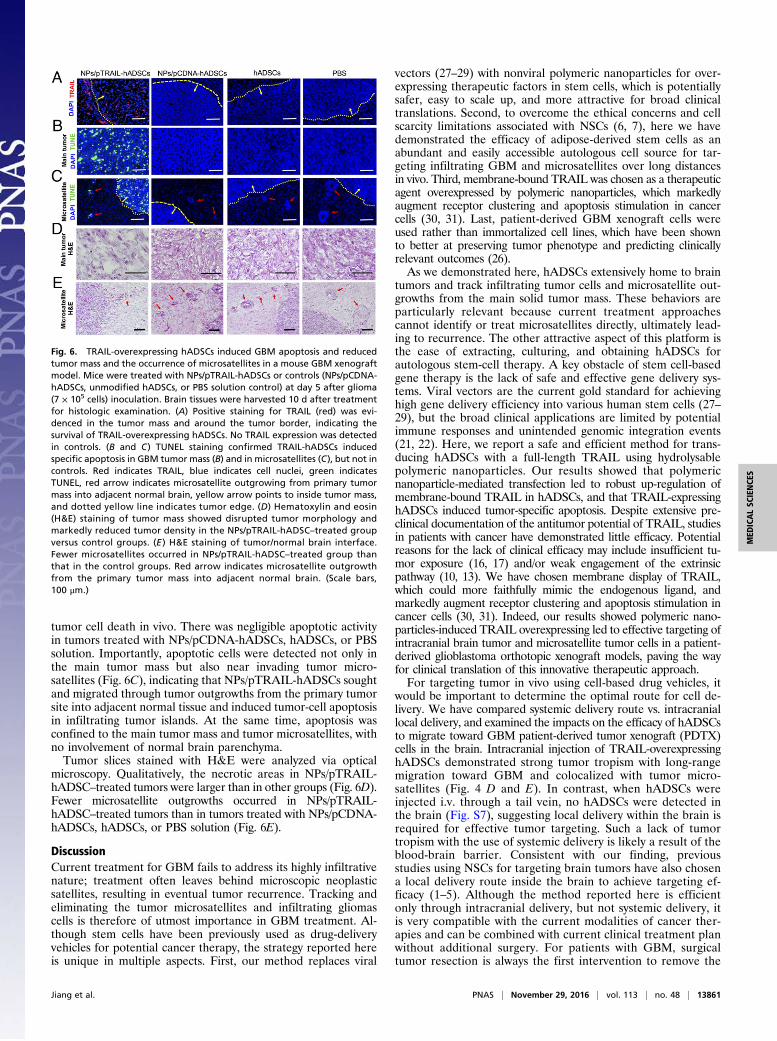
tumor cell death in vivo. There was negligible apoptotic activityin tumors treated with NPs/pCDNA-hADSCs, hADSCs, or PBSsolution. Importantly, apoptotic cells were detected not only inthe main tumor mass but also near invading tumor micro-satellites (Fig. 6C), indicating that NPs/pTRAIL-hADSCs soughtand migrated through tumor outgrowths from the primary tumorsite into adjacent normal tissue and induced tumor-cell apoptosisin infiltrating tumor islands. At the same time, apoptosis wasconfined to the main tumor mass and tumor microsatellites, withno involvement of normal brain parenchyma.Tumor slices stained with H&E were analyzed via optical
microscopy. Qualitatively, the necrotic areas in NPs/pTRAIL-hADSC–treated tumors were larger than in other groups (Fig. 6D).Fewer microsatellite outgrowths occurred in NPs/pTRAIL-hADSC–treated tumors than in tumors treated with NPs/pCDNA-hADSCs, hADSCs, or PBS solution (Fig. 6E).
DiscussionCurrent treatment for GBM fails to address its highly infiltrativenature; treatment often leaves behind microscopic neoplasticsatellites, resulting in eventual tumor recurrence. Tracking andeliminating the tumor microsatellites and infiltrating gliomascells is therefore of utmost importance in GBM treatment. Al-though stem cells have been previously used as drug-deliveryvehicles for potential cancer therapy, the strategy reported hereis unique in multiple aspects. First, our method replaces viral
vectors (27–29) with nonviral polymeric nanoparticles for over-expressing therapeutic factors in stem cells, which is potentiallysafer, easy to scale up, and more attractive for broad clinicaltranslations. Second, to overcome the ethical concerns and cellscarcity limitations associated with NSCs (6, 7), here we havedemonstrated the efficacy of adipose-derived stem cells as anabundant and easily accessible autologous cell source for tar-geting infiltrating GBM and microsatellites over long distancesin vivo. Third, membrane-bound TRAIL was chosen as a therapeuticagent overexpressed by polymeric nanoparticles, which markedlyaugment receptor clustering and apoptosis stimulation in cancercells (30, 31). Last, patient-derived GBM xenograft cells wereused rather than immortalized cell lines, which have been shownto better at preserving tumor phenotype and predicting clinicallyrelevant outcomes (26).As we demonstrated here, hADSCs extensively home to brain
tumors and track infiltrating tumor cells and microsatellite out-growths from the main solid tumor mass. These behaviors areparticularly relevant because current treatment approachescannot identify or treat microsatellites directly, ultimately lead-ing to recurrence. The other attractive aspect of this platform isthe ease of extracting, culturing, and obtaining hADSCs forautologous stem-cell therapy. A key obstacle of stem cell-basedgene therapy is the lack of safe and effective gene delivery sys-tems. Viral vectors are the current gold standard for achievinghigh gene delivery efficiency into various human stem cells (27–29), but the broad clinical applications are limited by potentialimmune responses and unintended genomic integration events(21, 22). Here, we report a safe and efficient method for trans-ducing hADSCs with a full-length TRAIL using hydrolysablepolymeric nanoparticles. Our results showed that polymericnanoparticle-mediated transfection led to robust up-regulation ofmembrane-bound TRAIL in hADSCs, and that TRAIL-expressinghADSCs induced tumor-specific apoptosis. Despite extensive pre-clinical documentation of the antitumor potential of TRAIL, studiesin patients with cancer have demonstrated little efficacy. Potentialreasons for the lack of clinical efficacy may include insufficient tu-mor exposure (16, 17) and/or weak engagement of the extrinsicpathway (10, 13). We have chosen membrane display of TRAIL,which could more faithfully mimic the endogenous ligand, andmarkedly augment receptor clustering and apoptosis stimulation incancer cells (30, 31). Indeed, our results showed polymeric nano-particles-induced TRAIL overexpressing led to effective targeting ofintracranial brain tumor and microsatellite tumor cells in a patient-derived glioblastoma orthotopic xenograft models, paving the wayfor clinical translation of this innovative therapeutic approach.For targeting tumor in vivo using cell-based drug vehicles, it
would be important to determine the optimal route for cell de-livery. We have compared systemic delivery route vs. intracraniallocal delivery, and examined the impacts on the efficacy of hADSCsto migrate toward GBM patient-derived tumor xenograft (PDTX)cells in the brain. Intracranial injection of TRAIL-overexpressinghADSCs demonstrated strong tumor tropism with long-rangemigration toward GBM and colocalized with tumor micro-satellites (Fig. 4 D and E). In contrast, when hADSCs wereinjected i.v. through a tail vein, no hADSCs were detected inthe brain (Fig. S7), suggesting local delivery within the brain isrequired for effective tumor targeting. Such a lack of tumortropism with the use of systemic delivery is likely a result of theblood-brain barrier. Consistent with our finding, previousstudies using NSCs for targeting brain tumors have also chosena local delivery route inside the brain to achieve targeting ef-ficacy (1–5). Although the method reported here is efficientonly through intracranial delivery, but not systemic delivery, itis very compatible with the current modalities of cancer ther-apies and can be combined with current clinical treatment planwithout additional surgery. For patients with GBM, surgicaltumor resection is always the first intervention to remove the
Fig. 6. TRAIL-overexpressing hADSCs induced GBM apoptosis and reducedtumor mass and the occurrence of microsatellites in a mouse GBM xenograftmodel. Mice were treated with NPs/pTRAIL-hADSCs or controls (NPs/pCDNA-hADSCs, unmodified hADSCs, or PBS solution control) at day 5 after glioma(7 × 10⁵ cells) inoculation. Brain tissues were harvested 10 d after treatmentfor histologic examination. (A) Positive staining for TRAIL (red) was evi-denced in the tumor mass and around the tumor border, indicating thesurvival of TRAIL-overexpressing hADSCs. No TRAIL expression was detectedin controls. (B and C) TUNEL staining confirmed TRAIL-hADSCs inducedspecific apoptosis in GBM tumor mass (B) and in microsatellites (C), but not incontrols. Red indicates TRAIL, blue indicates cell nuclei, green indicatesTUNEL, red arrow indicates microsatellite outgrowing from primary tumormass into adjacent normal brain, yellow arrow points to inside tumor mass,and dotted yellow line indicates tumor edge. (D) Hematoxylin and eosin(H&E) staining of tumor mass showed disrupted tumor morphology andmarkedly reduced tumor density in the NPs/pTRAIL-hADSC–treated groupversus control groups. (E ) H&E staining of tumor/normal brain interface.Fewer microsatellites occurred in NPs/pTRAIL-hADSC–treated group thanthat in the control groups. Red arrow indicates microsatellite outgrowthfrom the primary tumor mass into adjacent normal brain. (Scale bars,100 μm.)
Jiang et al. PNAS | November 29, 2016 | vol. 113 | no. 48 | 13861
MED
ICALSC
IENCE
S

bulk solid tumor, which exposes a cavity for the injection oftherapeutic cells/agents. Nonviral engineered hADSCs can beinjected during the same surgery of bulk tumor removal totarget and eradicate the infiltrating tumor microsatellites thatcannot be surgically removed. The promise of such an approachhas been demonstrated recently by injecting viral engineeredNSCs and bone marrow-derived MSCs into brain cavity afterbulk tumor resection (1, 2). Together, our results suggest thatlocal intracranial delivery of nonviral modified hADSCs ismore effective than systemic delivery for targeting infiltratingmicrosatellite cells to prevent brain tumor recurrence. i.t. in-jection of TRAIL-overexpressing hADSCs not only significantlyinhibited main GBM growth and extended survival, but also reducedthe occurrence of microsatellites. Furthermore, repetitive injectionof TRAIL-overexpressing hADSCs significantly prolonged animalsurvival compared with single injection of these cells. As such,our findings add credence to the elimination of disseminatedmicrosatellites or residual neoplastic foci in GBM patients in aseek-and-destroy manner by engrafting nanoparticle-inducedTRAIL-expressing autologous stem cells.
Materials and MethodsAll reagents and additional procedures used in this study, including primary cellisolation and culture and cell characterization, polymeric nanoparticle synthesisand characterization, optimization of transfection formulation, immunocyto-chemical and immunohistochemical assays, qRT-PCR, Western blotting, cellviability analysis, in vitro coculture and apoptosis analysis, flow cytometry, andstatistical analyses are described in SI Materials and Methods. All animal pro-cedures were performed with approval from the appropriate animal care anduse committee (Stanford University Administrative Panel on Laboratory Ani-mal Care and Use Committee). All procedures adhered to the US NationalInstitutes of Health Guide for the Care and Use of Laboratory Animals.
ACKNOWLEDGMENTS. This work was supported by an Alliance forCancer Gene Therapy Young Investigator Award Grant (to F.Y.), a StanfordSchool of Medicine Dean’s postdoctoral fellowship (X.J.), a Child Health Re-search Institute Grant and postdoctoral fellowship (X.J.), National Institutes ofHealth Grants R01DE024772 (to F.Y.) and K08 NS075144 (to G.A.G), Na-tional Science Foundation CAREER Award CBET-1351289 (to F.Y.), CaliforniaInstitute for Regenerative Medicine Tools and Technologies Award RT3-07804(to F.Y.), a Stanford Chem-H Institute Biomaterials Seed Grant (to F.Y.), theStanford Bio-X Interdisciplinary Initiative Program (F.Y.), a Stanford Child HealthResearch Institute Faculty Scholar Award (to F.Y.), and the Arline and Pete HarmanEndowed Faculty Scholar Stanford Child Health Research Institute (G.A.G.).
1. Kauer TM, Figueiredo J-L, Hingtgen S, Shah K (2011) Encapsulated therapeutic stemcells implanted in the tumor resection cavity induce cell death in gliomas. NatNeurosci 15(2):197–204.
2. Redjal N, Zhu Y, Shah K (2015) Combination of systemic chemotherapy with local stemcell delivered S-TRAIL in resected brain tumors. Stem Cells 33(1):101–110.
3. Bagci-Onder T, Du W, Figueiredo J-L, Martinez-Quintanilla J, Shah K (2015) Targetingbreast to brain metastatic tumours with death receptor ligand expressing therapeuticstem cells. Brain 138(pt 6):1710–1721.
4. Aboody KS, et al. (2000) Neural stem cells display extensive tropism for pathology inadult brain: Evidence from intracranial gliomas. Proc Natl Acad Sci USA 97(23):12846–12851.
5. Aboody KS, et al. (2013) Neural stem cell–mediated enzyme/prodrug therapy forglioma: Preclinical studies. Sci Transl Med 5(184):184ra159.
6. Arsenijevic Y, et al. (2001) Isolation of multipotent neural precursors residing in thecortex of the adult human brain. Exp Neurol 170(1):48–62.
7. Yu JJ, et al. (2006) Immunomodulatory neural stem cells for brain tumour therapy.Expert Opin Biol Ther 6(12):1255–1262.
8. Sasportas LS, et al. (2009) Assessment of therapeutic efficacy and fate of engineeredhuman mesenchymal stem cells for cancer therapy. Proc Natl Acad Sci USA 106(12):4822–4827.
9. Vilalta M, et al. (2008) Biodistribution, long-term survival, and safety of human adi-pose tissue-derived mesenchymal stem cells transplanted in nude mice by high sen-sitivity non-invasive bioluminescence imaging. Stem Cells Dev 17(5):993–1003.
10. Pacioni S, et al. (2015) Mesenchymal stromal cells loaded with paclitaxel induce cy-totoxic damage in glioblastoma brain xenografts. Stem Cell Res Ther 6(1):194.
11. Bonomi A, et al. (2015) Human amniotic mesenchymal stromal cells (hAMSCs) aspotential vehicles for drug delivery in cancer therapy: An in vitro study. Stem Cell ResTher 6(1):155.
12. Pessina A, et al. (2011) Mesenchymal stromal cells primed with paclitaxel provide anew approach for cancer therapy. PLoS One 6(12):e28321.
13. Levy O, et al. (2016) A prodrug-doped cellular Trojan Horse for the potential treat-ment of prostate cancer. Biomaterials 91:140–150.
14. Levy O, et al. (2013) mRNA-engineered mesenchymal stem cells for targeted deliveryof interleukin-10 to sites of inflammation. Blood 122(14):e23–e32.
15. Wang S, El-Deiry WS (2003) TRAIL and apoptosis induction by TNF-family death re-ceptors. Oncogene 22(53):8628–8633.
16. Wang S (2008) The promise of cancer therapeutics targeting the TNF-related apo-ptosis-inducing ligand and TRAIL receptor pathway. Oncogene 27(48):6207–6215.
17. Herbst RS, et al. (2010) Phase I dose-escalation study of recombinant human Apo2L/TRAIL, a dual proapoptotic receptor agonist, in patients with advanced cancer. J ClinOncol 28(17):2839–2846.
18. Kelley SK, et al. (2001) Preclinical studies to predict the disposition of Apo2L/tumornecrosis factor-related apoptosis-inducing ligand in humans: Characterization ofin vivo efficacy, pharmacokinetics, and safety. J Pharmacol Exp Ther 299(1):31–38.
19. Soria JC, et al. (2011) Randomized phase II study of dulanermin in combination withpaclitaxel, carboplatin, and bevacizumab in advanced non–small-cell lung cancer.J Clin Oncol 29(33):4442–51.
20. Ashkenazi A, Holland P, Eckhardt SG (2008) Ligand-based targeting of apoptosis incancer: The potential of recombinant human apoptosis ligand 2/Tumor necrosis fac-tor-related apoptosis-inducing ligand (rhApo2L/TRAIL). J Clin Oncol 26(21):3621–3630.
21. Brown BD, et al. (2007) In vivo administration of lentiviral vectors triggers a type Iinterferon response that restricts hepatocyte gene transfer and promotes vectorclearance. Blood 109(7):2797–2805.
22. Nayak S, Herzog RW (2010) Progress and prospects: Immune responses to viral vec-tors. Gene Ther 17(3):295–304.
23. Yang F, et al. (2009) Gene delivery to human adult and embryonic cell-derived stemcells using biodegradable nanoparticulate polymeric vectors. Gene Ther 16(4):533–546.
24. Yang F, et al. (2010) Genetic engineering of human stem cells for enhanced angio-genesis using biodegradable polymeric nanoparticles. Proc Natl Acad Sci USA 107(8):3317–3322.
25. Kim SM, et al. (2008) Gene therapy using TRAIL-secreting human umbilical cordblood-derived mesenchymal stem cells against intracranial glioma. Cancer Res 68(23):9614–9623.
26. Hidalgo M, et al. (2014) Patient-derived xenograft models: An emerging platform fortranslational cancer research. Cancer Discov 4(9):998–1013.
27. Ehtesham M, et al. (2002) Induction of glioblastoma apoptosis using neural stem cell-mediated delivery of tumor necrosis factor-related apoptosis-inducing ligand. CancerRes 62(24):7170–7174.
28. Grisendi G, et al. (2010) Adipose-derived mesenchymal stem cells as stable source oftumor necrosis factor-related apoptosis-inducing ligand delivery for cancer therapy.Cancer Res 70(9):3718–3729.
29. Loebinger MR, Eddaoudi A, Davies D, Janes SM (2009) Mesenchymal stem cell deliveryof TRAIL can eliminate metastatic cancer. Cancer Res 69(10):4134–4142.
30. Mitchell MJ, Wayne E, Rana K, Schaffer CB, King MR (2014) TRAIL-coated leukocytesthat kill cancer cells in the circulation. Proc Natl Acad Sci USA 111(3):930–935.
31. Nair PM, et al. (2015) Enhancing the antitumor efficacy of a cell-surface death ligandby covalent membrane display. Proc Natl Acad Sci USA 112(18):5679–5684.
32. Keir ST, Friedman HS, Reardon DA, Bigner DD, Gray LA (2013) Mibefradil, a noveltherapy for glioblastoma multiforme: Cell cycle synchronization and interlacedtherapy in a murine model. J Neurooncol 111(2):97–102.
13862 | www.pnas.org/cgi/doi/10.1073/pnas.1615396113 Jiang et al.
Related Documents