This journal is © the Owner Societies 2015 Phys. Chem. Chem. Phys., 2015, 17, 28977--28984 | 28977 Cite this: Phys. Chem. Chem. Phys., 2015, 17, 28977 Nanometer-scale hydrogen ‘portals’ for the control of magnesium hydride formation† Chia-Jung Chung,‡ Chinmay Nivargi‡ and Bruce Clemens* Magnesium and Mg-based material systems are attractive candidates for hydrogen storage but limited by unsuitable thermodynamic and kinetic properties. In particular, the kinetics are too slow at room temperature and atmospheric pressure. To study the hydride formation kinetics in a controlled way, we have designed a unique ‘nanoportal’ structure of Pd nanoparticles deposited on epitaxial Mg thin films, through which the hydride will nucleate only under Pd nanoparticles. We propose a growth mechanism for the hydrogenation reaction in the nanoportal structure, which is supported by scanning electron microscopy (SEM) images of hydrogenated samples exhibiting consistent results. Interestingly, the grain boundaries of Mg films play an important role in hydride nucleation and growth processes. Kinetic modeling based on the Johnson–Mehl–Avrami–Kolmogorov (JMAK) formalism seems to agree with the two-dimensional nucleation and growth mechanism hypothesized and the overall reaction rate is limited by hydrogen flux through the interface between the Pd nanoparticle and the underlying Mg film. The fact that in our structure Mg can be transformed completely into MgH 2 with only a small percentage of Pd nanoparticles offers possibilities for future on-board storage applications. 1 Introduction Magnesium hydride is an attractive material for hydrogen storage due to its high reversible hydrogen mass capacity of 7.6 wt%. However, a high desorption temperature (B265–320 1C for 1 bar H 2 pressure depending on ref. 1 and 2) and slow room-temperature hydrogen sorption kinetics pose difficulties for on-board applications. 3 Thermodynamic destabilization of the system can be achieved through alloying with other elements, 4–6 exploiting the excess interfacial energy in multi- layers 7 or intermixing energy with a catalyst, 8 leading to an increase in the equilibrium hydrogenation pressure at room temperature for practical use. The Mg hydrogenation reaction is also sluggish on its own and is further restricted due to the presence of a native oxide on the Mg surface. High temperatures of at least 400 1C are needed to crack the MgO layer and dissociate the hydrogen, and even then the absorption/desorption of hydrogen takes place over a period of hours. 9,10 The hydrogenation is often catalyzed at lower temperatures by Pd deposited on its surface, in contact with the pristine Mg, enabling dissociation of the H 2 molecules into H atoms which then diffuse through and react with Mg. In fact, in our experiments, we did not see a significant rate of hydrogen sorption at room temperature without the presence of the Pd catalyst. The nucleation occurs at Pd–Mg (or the catalyst/additive-Mg interface) followed by growth due to the diffusion of H atoms to the growth front. After nucleation, the kinetics are limited by the slow diffusion of H atoms to the reaction front, since the diffusion of H atoms through the already-grown magnesium hydride is eight to ten orders of magnitude slower than diffusion through a metal. 11,12 The formation of a ‘blocking’ layer of MgH 2 has been described previously in the literature and hence the diffusion length scale for complete reaction needs to be kept below 30–50 mm. 1,13–15 The hydrogenation kinetics can be substantially improved by reducing the diffusion length scale using nanosized materials. 10,16–18 The kinetics are also enhanced upon addition of the catalyst or alloying additives. Transition metals or their oxides are the most investigated. A good overview of the research on Mg based materials for hydrogen storage was provided in previous reviews. 19,20 However, the connection between the nanostructure and its effect on the kinetic properties of these materials is still unclear. Different studies use a variety of materials, catalysts and geometries, making identification of the underlying mechanisms very difficult. In order to critically examine the effects of the nanostructure on the kinetics of nucleation and growth, we have designed a Pd nanoparticle ‘nanoportal’ structure, illustrated in Fig. 1(a). This unique structure enables a physical and temporal decoupling of nucleation and growth processes, by allowing the nucleation to Department of Materials Science and Engineering, Stanford University, Stanford, CA - 94305, USA. E-mail: [email protected] † Electronic supplementary information (ESI) available. See DOI: 10.1039/ c5cp04515k ‡ These authors contributed equally to this work. Received 30th July 2015, Accepted 29th September 2015 DOI: 10.1039/c5cp04515k www.rsc.org/pccp PCCP PAPER Open Access Article. Published on 29 September 2015. Downloaded on 10/28/2021 5:03:33 PM. This article is licensed under a Creative Commons Attribution-NonCommercial 3.0 Unported Licence. View Article Online View Journal | View Issue

Welcome message from author
This document is posted to help you gain knowledge. Please leave a comment to let me know what you think about it! Share it to your friends and learn new things together.
Transcript

This journal is© the Owner Societies 2015 Phys. Chem. Chem. Phys., 2015, 17, 28977--28984 | 28977
Cite this:Phys.Chem.Chem.Phys.,
2015, 17, 28977
Nanometer-scale hydrogen ‘portals’ for thecontrol of magnesium hydride formation†
Chia-Jung Chung,‡ Chinmay Nivargi‡ and Bruce Clemens*
Magnesium and Mg-based material systems are attractive candidates for hydrogen storage but limited
by unsuitable thermodynamic and kinetic properties. In particular, the kinetics are too slow at room
temperature and atmospheric pressure. To study the hydride formation kinetics in a controlled way, we
have designed a unique ‘nanoportal’ structure of Pd nanoparticles deposited on epitaxial Mg thin films,
through which the hydride will nucleate only under Pd nanoparticles. We propose a growth mechanism
for the hydrogenation reaction in the nanoportal structure, which is supported by scanning electron
microscopy (SEM) images of hydrogenated samples exhibiting consistent results. Interestingly, the grain
boundaries of Mg films play an important role in hydride nucleation and growth processes. Kinetic
modeling based on the Johnson–Mehl–Avrami–Kolmogorov (JMAK) formalism seems to agree with the
two-dimensional nucleation and growth mechanism hypothesized and the overall reaction rate is limited
by hydrogen flux through the interface between the Pd nanoparticle and the underlying Mg film. The
fact that in our structure Mg can be transformed completely into MgH2 with only a small percentage of
Pd nanoparticles offers possibilities for future on-board storage applications.
1 Introduction
Magnesium hydride is an attractive material for hydrogenstorage due to its high reversible hydrogen mass capacity of7.6 wt%. However, a high desorption temperature (B265–320 1Cfor 1 bar H2 pressure depending on ref. 1 and 2) and slowroom-temperature hydrogen sorption kinetics pose difficultiesfor on-board applications.3 Thermodynamic destabilizationof the system can be achieved through alloying with otherelements,4–6 exploiting the excess interfacial energy in multi-layers7 or intermixing energy with a catalyst,8 leading to anincrease in the equilibrium hydrogenation pressure at roomtemperature for practical use.
The Mg hydrogenation reaction is also sluggish on its ownand is further restricted due to the presence of a native oxide onthe Mg surface. High temperatures of at least 400 1C are neededto crack the MgO layer and dissociate the hydrogen, and eventhen the absorption/desorption of hydrogen takes place over aperiod of hours.9,10 The hydrogenation is often catalyzed atlower temperatures by Pd deposited on its surface, in contactwith the pristine Mg, enabling dissociation of the H2 moleculesinto H atoms which then diffuse through and react with Mg.
In fact, in our experiments, we did not see a significant rate ofhydrogen sorption at room temperature without the presenceof the Pd catalyst. The nucleation occurs at Pd–Mg (or thecatalyst/additive-Mg interface) followed by growth due to thediffusion of H atoms to the growth front. After nucleation,the kinetics are limited by the slow diffusion of H atoms to thereaction front, since the diffusion of H atoms through thealready-grown magnesium hydride is eight to ten orders ofmagnitude slower than diffusion through a metal.11,12 Theformation of a ‘blocking’ layer of MgH2 has been describedpreviously in the literature and hence the diffusion length scalefor complete reaction needs to be kept below 30–50 mm.1,13–15
The hydrogenation kinetics can be substantially improved byreducing the diffusion length scale using nanosized materials.10,16–18
The kinetics are also enhanced upon addition of the catalyst oralloying additives. Transition metals or their oxides are the mostinvestigated. A good overview of the research on Mg basedmaterials for hydrogen storage was provided in previousreviews.19,20 However, the connection between the nanostructureand its effect on the kinetic properties of these materials is stillunclear. Different studies use a variety of materials, catalysts andgeometries, making identification of the underlying mechanismsvery difficult.
In order to critically examine the effects of the nanostructureon the kinetics of nucleation and growth, we have designed aPd nanoparticle ‘nanoportal’ structure, illustrated in Fig. 1(a).This unique structure enables a physical and temporal decouplingof nucleation and growth processes, by allowing the nucleation to
Department of Materials Science and Engineering, Stanford University, Stanford,
CA - 94305, USA. E-mail: [email protected]
† Electronic supplementary information (ESI) available. See DOI: 10.1039/c5cp04515k‡ These authors contributed equally to this work.
Received 30th July 2015,Accepted 29th September 2015
DOI: 10.1039/c5cp04515k
www.rsc.org/pccp
PCCP
PAPER
Ope
n A
cces
s A
rtic
le. P
ublis
hed
on 2
9 Se
ptem
ber
2015
. Dow
nloa
ded
on 1
0/28
/202
1 5:
03:3
3 PM
. T
his
artic
le is
lice
nsed
und
er a
Cre
ativ
e C
omm
ons
Attr
ibut
ion-
Non
Com
mer
cial
3.0
Unp
orte
d L
icen
ce.
View Article OnlineView Journal | View Issue
28978 | Phys. Chem. Chem. Phys., 2015, 17, 28977--28984 This journal is© the Owner Societies 2015
occur only under the Pd nanoparticles, followed by growth due tohydrogen flux through the Pd catalyst nanoparticle ‘portals’. As aresult, it can be employed to study hydrogenation reactions undera wide variety of carefully controllable experimental conditions.This has potential to provide great insight into the underlyingreaction mechanisms.
The nanoportal structure also allows the examination of thereaction using Scanning Electron Microscopy (SEM) and anoptical transmission technique based on hydrogenography,21
since the catalyst nanoparticles do not obscure the observationof the underlying phase transformation. We propose a reactionmechanism for this structure and then correlate a kinetic modelthat we have developed based on the Johnson–Mehl–Avrami–Kolmogorov (JMAK) formalism22 with the experimental observa-tions. The results suggest that the overall reaction rate depends onthe flux of hydrogen atoms through the interface between the Pdcatalyst nanoparticle and the underlying Mg film.
We found that, even for the thin Mg films studied here,Pd nanoparticles representing a mass fraction of only a fewpercent can be effective catalysts, increasing reaction rates byorders of magnitude over bare Mg. The investigation of reactionmechanisms with this novel nanostructure can pave the wayto rational designs of hydrogen storage systems with betterstorage properties for future on-board applications.
2 Experimental2.1 Sputtering of Pd nanoparticles and sample preparation
Samples were prepared in a UHV sputtering chamber consistingof two sputtering zones; one was for conventional thin film growthand the second was a high-pressure condensation zone forproducing nanoparticles.23 Epitaxial Mg films of 22 nm thicknessin the (001) orientation were grown on (001) Al2O3 at roomtemperature (for more details see ref. 24) using DC magnetronsputtering in a chamber zone with a base pressure of 5� 10�9 torrand an Ar deposition pressure of 1.3 millitorr. In order toinvestigate the effect of the grain structure on hydrogenationkinetics, non-epitaxial Mg films of the same thickness were grownon oxidized (100) Si wafers using the same growth conditions.
Pd nanoparticles were generated by a nanoparticle source fromMantis Deposition Ltd, consisting of a sputter source operating ina condensation zone held at high (hundreds of millitorr) Arpressure, and a quadrupole mass filter. By controlling thecondensation zone length and pressure, and by using the massfilter, particle sizes in the range of 1–20 nm can be produced.The generated Pd nanoclusters were then impinged upon theas-deposited Mg film. A detailed description of the sputteringconditions is provided in the ESI.†
2.2 X-Ray diffraction
Symmetric y � 2y scans and rocking curve scans were performedto examine the overall structure and preference of out-of-planetexture, while f scans after aligning on known peaks away fromthe sample normal were used to determine the presence ofin-plane orientation induced by epitaxial alignment with thesubstrate. More information is provided in the ESI.†
2.3 Optical transmission measurement
The samples were charged with hydrogen (99.999%) in anoptical measurement chamber. A high-power LED with a wave-length of 625 nm was used as the light source in the measure-ments. The LED output was modulated at 316 Hz to allow forlock-in detection in order to increase the signal-to-noise ratio.Since the 635 nm light was strongly absorbed by Mg but notMgH2, monitoring the optical transmission allowed real-time,direct measurement of the fraction of the Mg converted tohydride.21 The schematic of the setup and the pictures of thesample before/after hydrogenation can be found in the ESI.†
3 Results and discussion3.1 Construction of the nanoportal structure
The nanoportal structure consists of Pd nanoparticle catalystsforming portals for the dissociated H atoms to diffuse throughand then nucleate and grow the hydride in the underlying Mgfilm. To form this structure we first deposit a Mg film usingconventional UHV sputter deposition onto either sapphireor oxidized Si substrates (the Mg growth process is described
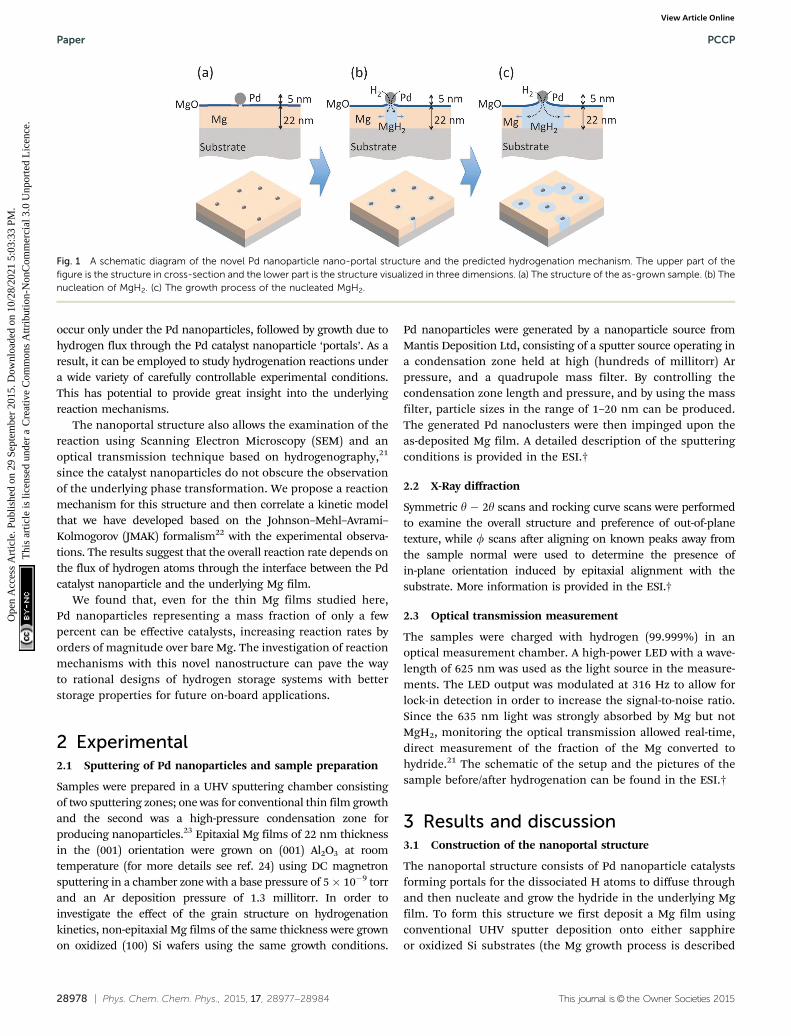
Fig. 1 A schematic diagram of the novel Pd nanoparticle nano-portal structure and the predicted hydrogenation mechanism. The upper part of thefigure is the structure in cross-section and the lower part is the structure visualized in three dimensions. (a) The structure of the as-grown sample. (b) Thenucleation of MgH2. (c) The growth process of the nucleated MgH2.
Paper PCCP
Ope
n A
cces
s A
rtic
le. P
ublis
hed
on 2
9 Se
ptem
ber
2015
. Dow
nloa
ded
on 1
0/28
/202
1 5:
03:3
3 PM
. T
his
artic
le is
lice
nsed
und
er a
Cre
ativ
e C
omm
ons
Attr
ibut
ion-
Non
Com
mer
cial
3.0
Unp
orte
d L
icen
ce.
View Article Online

This journal is© the Owner Societies 2015 Phys. Chem. Chem. Phys., 2015, 17, 28977--28984 | 28979
in more detail in ref. 24). This is followed by the deposition ofPd nanoparticles, formed by inert gas condensation23 of asputtered Pd atom flux, combined with in situ mass filteringto produce uniformly distributed, size-selected Pd nanoparticles.The Pd nanoparticle deposition immediately follows the Mg filmgrowth in the same deposition environment without breakingvacuum, leading to an intimate contact (and presumably somereaction8) between the Pd nanoparticles and the Mg film. Theresulting structure is then exposed to air, whereupon a B2 nmthick native MgO layer forms on the remaining Mg film that is notcovered by the Pd nanoparticles. This oxide layer effectively blockshydrogen from reacting with the underlying Mg. The final structureis shown schematically in Fig. 1(a).
Structural characterization of Mg films. The as-grown Mgthin films were analyzed using X-ray diffraction to determinethe relative crystallographic orientations of Mg films andsapphire substrates. For both samples grown on sapphireand oxidized silicon substrates, the Mg films exhibited a single(001) out-of-plane orientation, as shown in the X-ray high-angley � 2y diffraction pattern of Fig. 2. The rocking curve of
Mg(002) had a FWHM of B11 on (001) Al2O3 and B61 onoxidized Si, which indicates that the Mg(002) planes were moreclosely aligned with the substrate for the samples deposited onsapphire. For the samples grown on sapphire substrates, the fscan of Al2O3(202) and Mg(101) in Fig. 2(c) shows six Mg peakswith a FWHM of B61, indicating that Mg had an epitaxial,in-plane alignment, with a 301 rotation relative to the sapphiresubstrate. Although the Mg films on the sapphire substrateswere not single crystal, they had a high degree of out-of-planeand in-plane alignment, and hence were free of high-anglegrain boundaries. We refer to these Mg films as epitaxial sincethey have a crystallographic orientation influenced by thesingle-crystal substrate. Mg films grown on oxidized Si sub-strates had out-of-plane alignment but no epitaxial, in-planealignment, and thus had high-angle grain boundaries due tothe grains’ random orientation with respect to rotation aboutthe surface normal.
Nanoportal reaction proposed mechanism. The geometryand the mechanism of the nanoportal-enabled hydrogenationreaction are shown schematically in Fig. 1. Hydrogen gas isdisassociated by the Pd catalyst, and hydrogen is locally intro-duced to the Mg film through the intimate contact at theinterface between the Mg and the Pd nanoparticle, thereby alsolocalizing the nucleation sites for hydride growth. Importantly,we observe no hydrogenation reaction without the Pd nano-particles, and we observe negligible hydrogenation reactionrates when the Pd nanoparticles are deposited after the Mgfilm was exposed to air, forming the MgO native oxide. So it isclear that both the presence of the nanoparticles and theintimate contact between the Pd nanoparticles and the under-lying Mg film are necessary to allow the hydrogenation reactionat an appreciable rate.
Localizing the nucleation sites results in an ability to studyreaction kinetics with unprecedented control. By controllingthe density of nanoportals we can vary the nucleation densityand examine new growth behaviors. For example, when thedistance between nucleation sites is large compared to the filmthickness (true for all the samples discussed here) the hydridegrowth will occur as two-dimensional lateral growth for most ofthe reaction processes, since vertical growth will rapidly belimited by the film thickness (Fig. 1(b) and (c)). By followingthis growth process we can examine the limiting mechanismsas well as explore nucleation behavior.
3.2 Visualization of the reaction progress
In order to visualize the nucleation and growth processes ofMgH2, we used field emission scanning electron microscopy(FEI Magellan XHR SEM) to examine the morphology and thesize of the hydride at different stages of the hydrogenationreaction. To minimize beam damage to the hydride, we imagedthe samples at a relatively low voltage of 2 kV with a probecurrent of 13 pA. Owing to the electronic property differencebetween Mg and MgH2, the hydride phase, which had a lowersecondary electron yield, appeared darker in the SEM than theuntransformed metallic Mg. Therefore, the SEM allowed directobservation of the shape and the size of the hydride regions.
Fig. 2 X-ray diffraction y � 2y scans of as-deposited 22 nm Mg with Pdnanoparticles. (a) Samples that were grown on a sapphire substrate.(b) Samples that were grown on a silicon substrate. The unlabeled peaksare all from substrates. (c) X-ray diffraction phi scans of an epitaxial Mg film.The sample was grown on a (0001) Al2O3 substrate with Pd nanoparticlesdeposited on the surface.
PCCP Paper
Ope
n A
cces
s A
rtic
le. P
ublis
hed
on 2
9 Se
ptem
ber
2015
. Dow
nloa
ded
on 1
0/28
/202
1 5:
03:3
3 PM
. T
his
artic
le is
lice
nsed
und
er a
Cre
ativ
e C
omm
ons
Attr
ibut
ion-
Non
Com
mer
cial
3.0
Unp
orte
d L
icen
ce.
View Article Online

28980 | Phys. Chem. Chem. Phys., 2015, 17, 28977--28984 This journal is© the Owner Societies 2015
Fig. 3 shows SEM images of the hydrogenated Mg filmsgrown on sapphire and oxidized Si substrates, respectively,with an areal Pd nanoparticle coverage of 1.4%. Clearly thestructural difference in the parent Mg phase, as shown by X-raydiffraction, altered both the extent of the hydrogenation reac-tion and the morphology of the hydride. For the epitaxial Mgfilms, the hydride regions were nearly circular, while showingsome overlap of neighboring regions. These phenomena indi-cate that the hydride reaction rate was roughly independent ofthe in-plane crystallographic direction within a grain. However,the hydride regions on the non-epitaxial Mg films were moreirregular in shape and much smaller. It appears that theshape of the hydride region in the non-epitaxial Mg films wasinfluenced by the underlying Mg grain structure, coupled with avariation in the difficulty with which the growing hydride wasable to cross the different grain boundaries between adjacentgrains. The shape of the hydride regions appears to mimic thegrain structure of the parent Mg phase, indicating that the hydridegrowth is impaired during transit across grain boundaries. Furtherexamination can explore further the connection between thisgrowth impediment and grain boundary orientation, but it is clearthat the small angle grain boundaries present in the sample grownon sapphire offer much less interference to hydride growth.
We note that for both epitaxial and non-epitaxial samples,the spacing between the hydride regions is much larger thanthe spacing between the Pd nanoparticles, and that there are Pdnanoparticles over unreacted Mg regions of the film, or morethan one Pd nanoparticle on each nucleated hydride patch.This implies that only a fraction of the Pd nanoparticles areeffective in nucleating a hydride region. We suspect that thefailure of many nanoparticles to result in hydride formation is
due to the lack of direct contact between the Pd nanoparticleand the Mg film, perhaps due to oxidation in the imperfectvacuum of the deposition environment, or oxidation of theinterface subsequent to nanoparticle deposition and exposureto air.
It is also notable that the hydride regions in a given film ata given stage of reaction are approximately the same size,which indicates that each hydride region begins to grow atapproximately the same time. This result indicates that nuclea-tion takes place early in the reaction process, and that once theeffective nucleation sites are used up, no further nucleationoccurs. Finally, we note that the spacing between hydrideregions is much larger than the film thickness, so that muchof the transformation occurs via two-dimensional growth in theplane of the film after the hydride spans the thickness of the film,which is consistent with our predicted growth mechanism.
To extract the time-dependence of the hydride growth behaviorwe used SEM to observe the size of the hydride patches in sampleswith the same nominal nanoportal areal density (B1.7%) butexposed to hydrogen for varying reaction times. The averageradius (extracted by image analysis using ImageJ25) as a functionof reaction time is shown in Fig. 4. We see that the growth ratedr/dt decreases greatly with time. Fitting to a growth law r B tm
leads to unsatisfactory fits and to a value of m B 0.3, which isincompatible with growth limited by either diffusion throughthe growing hydride or by flux through the nanoparticle–filminterface,15 both of which would produce a r B t1/2 behavior fortwo-dimensional growth (see ESI†). At longer times, the observedgrowth rate decreases more rapidly than the r B t0.3 of the fittedfunction, leading to the conclusion that the effectiveness of thenanoportals might be decreasing with time. We speculate thatthis decrease may be due to further oxidation of the nanoparticle–film interface, resulting in a decrease in the effective nanoportal
Fig. 3 SEM micrographs of hydride growth in Mg films. (a) and (c) SEM ofhydride growth in an epitaxial Mg film grown on a sapphire substrate.(b) and (d) SEM of hydride growth in a non-epitaxial Mg film grown on asilicon substrate. Both samples were hydrogenated under 700 torr H2 atroom temperature for 8.5 minutes. Hydride regions are dark due to thelower electron yield. Pd nanoparticles are small bright spots in the pictures.B59% of the area is reacted for the epitaxial film, compared to B52% forthe non-epitaxial film.
Fig. 4 Model fits to explain the time dependence of the measuredaverage radii of the hydride patches grown in epitaxial Mg samples. Theagreement is seen to be much better for the models incorporating theoxidation under the portals.
Paper PCCP
Ope
n A
cces
s A
rtic
le. P
ublis
hed
on 2
9 Se
ptem
ber
2015
. Dow
nloa
ded
on 1
0/28
/202
1 5:
03:3
3 PM
. T
his
artic
le is
lice
nsed
und
er a
Cre
ativ
e C
omm
ons
Attr
ibut
ion-
Non
Com
mer
cial
3.0
Unp
orte
d L
icen
ce.
View Article Online

This journal is© the Owner Societies 2015 Phys. Chem. Chem. Phys., 2015, 17, 28977--28984 | 28981
area. This possibility is discussed more fully below. Note thatthese observations of nanoparticle diameter are taken in the timeand nucleation density regime before coalescence occurs, so thedecreased growth rate is not due to the impingement of growinghydride regions.
To explore the effect of oxidation of a nanoportal on thehydride growth rate, we considered both the interfacial anddiffusive flux limited encroachment of the oxide under thenanoportal. This is illustrated schematically in Fig. 5. Thisleads to a simple expression for the nanoportal area and hencethe flux of the H atoms through the nanoportals as a functionof time, which in turn can be used to obtain an expression ofthe dependence of the radius of an individual hydride patch asa function of time. Balancing the nanoportal flux and thehydride growth in a time increment dt for mass conservation,we find:
jHAnpdt = cH(2prh)dr
where jH is the hydrogen flux per unit active nanoportal areaAnp, cH is the concentration of hydrogen in the hydride, h isthe film thickness and r is the radius of the hydride region.If Anp = const., we find
r ¼ffiffiffiffiffiffiffiffiffiffiffiffiffiffijHAnpt
pcHh
r
so r B t1/2 as mentioned above. Allowing the area of thenanoportal to decrease with time with either interface ordiffusion limited oxide encroachment as shown in Fig. 5 givesrelatively simple analytical expressions with different timedependencies (see ESI†) and a much better fit to the hydrideradius behavior as shown in Fig. 4. The dependence is verysimilar to a power law at small times, before slowing downas the portal shrinks, and eventually pinches off. As can beseen, for the interface limited oxide encroachment, assumingthe average initial radius of the nanoportal to be 5 nm, thenanoportal pinches off at B516 s.
3.3 Optical transmission measurement of reaction progress
In order to further examine hydride growth behavior using thenanoportal mechanism, an optical transmission technique wasused to monitor the hydrogenation kinetics of Mg films withvarying Pd nanoparticle surface coverage. This monitoring ispossible due to the difference in optical transmission of MgH2,which is essentially transparent for light at l = 625 nm ascompared to the Mg metal which is reflective. The reaction
fraction was calculated by taking the light transmission of thesample at a given reaction time and then normalized by the totalchange of transmission when the sample was fully reacted, when itbecame fully transparent. The reacted fraction as a function oftime is shown in Fig. 6. Samples with a higher coverage of Pdnanoparticles had faster hydrogenation kinetics, as seen in Fig. 6,due to the increased Pd nanoparticle density and hence a higherdensity of nucleation sites. Thus a higher coverage of Pd nano-particles results in a higher density of effective nanoportals, eventhough, as mentioned above, only a fraction of Pd nanoparticlesare effective as nucleation sites. We also observe that samples thatare exposed to air for a longer period of time do not react asquickly as those that have shorter air exposure. This is consistentwith the growth behavior discussed above, which we explain by adecrease in the effective area of a nanoportal over time, even in thehydrogenation environment. These observations show that thenature of the interface between the nanoparticle and the under-lying film is critical for an effective nanoportal function. Futurework will explore ways to improve nanoparticle–film interaction.
3.4 Kinetic modeling of the reaction progress
In order to further understand the limiting mechanisms of thegrowth behavior, we constructed a kinetic model based on thewell-known JMAK formalism to understand the limiting stepsfor the reaction. Here we follow Christian’s treatment26 to applythis approach to our nanoportal mechanism. Consideringfirst the nucleation behavior, we observe that since nucleationoccurs under active nanoportals, we initially have a fixednumber (per area) of possible sites where nucleation can occur.If the sites nucleate with average time constant tN, then we findfor the nucleation rate per area:
I ¼ �dNdt¼ N0
tNe�t=tN
Fig. 5 The portal pinches off under the Pd nanoparticle. On the right is anexaggerated top view showing a decreased area of the portal and hence alowered H flux.
Fig. 6 Effect of Pd nanoparticle coverage on the hydrogenation reactionrate. Hydrogenation reaction curves of samples with different Pd nano-particle coverages (B1.7 to B6.3%) grown on epitaxial Mg films.
PCCP Paper
Ope
n A
cces
s A
rtic
le. P
ublis
hed
on 2
9 Se
ptem
ber
2015
. Dow
nloa
ded
on 1
0/28
/202
1 5:
03:3
3 PM
. T
his
artic
le is
lice
nsed
und
er a
Cre
ativ
e C
omm
ons
Attr
ibut
ion-
Non
Com
mer
cial
3.0
Unp
orte
d L
icen
ce.
View Article Online

28982 | Phys. Chem. Chem. Phys., 2015, 17, 28977--28984 This journal is© the Owner Societies 2015
where N0 is the initial number density of available nucleationsites and N is its (decreasing) value as a function of time.
Next we consider growth of the nuclei as a function of time.A nucleus that begins growth at time t and grows as a two-dimensional disk will have area at time t given by:
AtðtÞ ¼ pðttvðtÞdt
� �2
For either diffusion limited or nanoportal flux limited (withconstant nanoportal area) we find:
At(t) = c[(t � t)1/2]2 = c(t � t)
A more general expression might be considered r B (t � t)m
and At(t) B (t � t)2m, however the physical process of pinchingoff of the nanoportal area will give more complex behavior.
If we add up all the area of growing hydride regions,ignoring impingement and counting phantom nuclei that arepresent in already transformed regions we find the extendedarea:
AeðtÞ ¼ A0
ðt0
IðtÞAtðtÞdt
where A0 is the total sample area. The actual hydride area A(t) isthen found in the normal fashion by assuming that during atime increment dt only a fraction (1 � A(t)/A0) of the extendedarea increase occurs in the non-transformed region of thesample.
A(t) = A0(1 � e�Ae(t)/A0)
Comparing this model with the observed transformedfraction f = A(t)/A0 behavior we can investigate the nucleationbehavior and growth limiting mechanisms. To explore nuclea-tion behavior we first fit the short-time (t r 700 s) experimentaldata using a power law growth behavior (r B tm). We findrelatively good agreement with the optical behavior as shown inFig. 7, with an exponent of 0.2 r m r 0.5 which is roughlyconsistent with the fits we obtained for the radius behaviordiscussed above. We find that we are unable to extract mean-ingful nucleation times tN for samples with low nanoparticledensity, and that the nucleation time we extract for the samplewith high density is short (B54 s), but comparable to thereaction time of around 100 seconds. This indicates that thenucleation is complete in the low nanoparticle density samplesbefore significant reaction takes place, while for the highnanoparticle density samples there is nucleation and growthoccurring during the reaction. The quality of the fit decreases ifwe include longer reaction times, indicating that the power lawgrowth behavior is not good for a long time. This is consistentwith the hydride radius behavior discussed above and with theidea that the nanoportals become less effective with time, perhapsdue to pinching off by MgO growth under the nanoportal.
To explore this we incorporated the hydride growth behaviorassociated with the decreasing nanoportal area due to the oxideencroachment discussed above (see ESI,† for the model). Theoxide encroachment was modeled as both interfacial as well as
diffusive flux limited growth, leading to a decreasing area of thenanoportal, and hence a slowdown of the growth of the hydridepatch from the expected r B t1/2 behavior. The resulting fits forthe interfacial flux limited model are shown in Fig. 8. As canbe seen, the model is able to describe the behavior fairlyadequately over the entire reaction time. The diffusive fluxlimited model shows somewhat similar behavior. This modelwas considered only for the low coverage samples for whichthe oxide encroachment before reaction completion was signi-ficant. The fast reacting sample essentially reacted fully beforeencroachment became appreciable.
Fig. 7 Model fits to observed short-time behavior using power-lawgrowth behavior. The solid lines are fits to the data and the best fit tothe parameter m is shown.
Fig. 8 Model fits to observed behavior incorporating portal pinch-off andan additional leak flux through MgO, for the low coverage samples. Thesolid lines are fits to the data.
Paper PCCP
Ope
n A
cces
s A
rtic
le. P
ublis
hed
on 2
9 Se
ptem
ber
2015
. Dow
nloa
ded
on 1
0/28
/202
1 5:
03:3
3 PM
. T
his
artic
le is
lice
nsed
und
er a
Cre
ativ
e C
omm
ons
Attr
ibut
ion-
Non
Com
mer
cial
3.0
Unp
orte
d L
icen
ce.
View Article Online

This journal is© the Owner Societies 2015 Phys. Chem. Chem. Phys., 2015, 17, 28977--28984 | 28983
Using this approach, it was again found that the nucleationtime is short compared to the reaction times, and hencenucleation is effectively instantaneous. This results in hydridepatches of about the same size, as observed in the SEM.The time of nanoportal pinch-off varies across samples withdifferent nanoportal densities. It is also presumably affected bythe amount of time it was exposed to air before hydrogenationand the specific variation of nanoparticle sizes across samples,which is hard to completely account for in the modeling.However, for a similar nanoparticle density, it was found thatthe time for the nanoportals to completely pinch-off wascomparable for the fits to the transmission measurements(B350�1340 s depending on interfacial or diffusive flux limita-tion) and the radius measurements (B516�950 s). These timesare very sensitive to the initial radius of the nanoportals, which,of course, have a size distribution that is not included in themodeling.
Although the portals eventually pinch off, the reaction is stillseen to proceed slowly, indicating that the MgO layer on top ofMg and under the nanoparticle might be slightly permeable toH atoms dissociated by Pd, through cracks or defects. This islikelier after appreciable nucleation and growth, as opposed toat the beginning of the reaction where there is no significantnucleation yet. This is because of the growing hydride having a32% larger volume,8 compared to Mg, that might contribute toa decreased integrity of the oxide layer on top. A constant fluxterm was incorporated into the fits to account for this behavior,leading to much better agreement at long and short times, asshown. A better engineering of the Mg–Pd nanoparticle inter-face could help avoid this slowdown and pinch-off. A potentialway would be to accelerate the nanoparticles towards thesample by means of a voltage bias during deposition so as tohave a more intimate contact with the underlying Mg film – toprevent oxidation or cause more Mg–Pd intermixing. Investiga-tions to verify this hypothesis are ongoing in our lab.
4 Conclusions
In summary, we have developed a novel Pd nanoportal structurethat enables us to study the hydride nucleation and growthmechanism by physically controlling the nucleation sites of thehydride. X-ray diffraction analysis and SEM observations show theeffect of the Mg crystallographic structure on the hydride growth –high-angle grain boundaries seemed to impede hydride growth.The SEM micrographs exhibited consistent hydride growth beha-vior as predicted. We developed a simple kinetic model based onthe JMAK formalism, which was able to provide significant insightinto the nucleation and growth behaviors. It seemed to suggestthat the reaction is limited by flux of the H atoms across theinterface between the Pd nanoportals and the MgH2 nuclei. Thenanoportals get constricted and eventually might pinch off overtime due to oxidation underneath the portals during sampletransfer or the hydrogenation reaction. A modification to themodel incorporating effect of this oxidation gave better agreementwith the determined radius of the hydride patches as a function of
time as well as the optical transmission through the sample thatindicates the reacted fraction.
The amount of Pd used in this study is much less than theconventional Pd/Mg thin film structures. With only a smallpercentage of Pd nanoparticles, the Mg film can be fullytransformed into hydride. The separation between the nano-particles is less than 30–50 mm of a blocking layer, thusenabling full conversion. Therefore, the storage capacity issignificantly increased with this nanoportal structure comparedto Pd thin film catalyst structures. This makes it promising forfuture on-board storage applications. We hope to do further workto apply this novel structure to understand and design otherhydrogen storage systems.
Acknowledgements
The authors would like to thank Stephen T. Kelly for useful discus-sions. This work was supported as part of the Center on Nano-structuring for Efficient Energy Conversion (CNEEC) at StanfordUniversity, an Energy Frontier Research Center funded by the U.S.Department of Energy, Office of Basic Energy Sciences under AwardNumber DE-SC0001060. Part of this work was performed at theStanford Nano Shared Facilities (SNSF) at Stanford University.
References
1 B. Vigeholm, J. Kjøller, B. Larsen and A. Pedersen, J. Less-Common Met., 1983, 89, 135–144.
2 B. Tanguy, J. Soubeyroux, M. Pezat, J. Portier andP. Hagenmuller, Mater. Res. Bull., 1976, 11, 1441–1448.
3 L. Schlapbach and A. Zuttel, Nature, 2001, 414, 353–358.4 J. J. Vajo, F. Mertens, C. C. Ahn, R. C. Bowman and B. Fultz,
J. Phys. Chem. B, 2004, 108, 13977–13983.5 X. Tan, L. Wang, C. M. B. Holt, B. Zahiri, M. H. Eikerling and
D. Mitlin, Phys. Chem. Chem. Phys., 2012, 14, 10904–10909.6 C. Zhou, Z. Z. Fang, J. Lu, X. Luo, C. Ren, P. Fan, Y. Ren and
X. Zhang, J. Phys. Chem. C, 2014, 118, 11526–11535.7 P. Kalisvaart, B. Shalchi-Amirkhiz, R. Zahiri, B. Zahiri,
X. Tan, M. Danaie, G. Botton and D. Mitlin, Phys. Chem.Chem. Phys., 2013, 15, 16432–16436.
8 C. J. Chung, S.-C. Lee, J. Groves, E. Brower, R. Sinclair andB. M. Clemens, Phys. Rev. Lett., 2012, 108, 106102.
9 A. Zaluska, L. Zaluski and J. Strom Olsen, J. Alloys Compd.,1999, 288, 217–225.
10 W. Oelerich, T. Klassen and R. Bormann, J. Alloys Compd.,2001, 322, L5–L9.
11 J. Cermak and L. Kral, Acta Mater., 2008, 56, 2677–2686.12 J. Renner and H. Grabke, Z. Metalkd., 1978, 69, 639–642.13 C. M. Stander, Z. Phys. Chem., 1977, 104, 229–238.14 P. Kalisvaart, E. Luber, H. Fritzsche and D. Mitlin, Chem.
Commun., 2011, 47, 4294–4296.15 S. T. Kelly and B. M. Clemens, J. Appl. Phys., 2010, 108,
013521.16 W. Oelerich, T. Klassen and R. Bormann, J. Alloys Compd.,
2001, 315, 237–242.
PCCP Paper
Ope
n A
cces
s A
rtic
le. P
ublis
hed
on 2
9 Se
ptem
ber
2015
. Dow
nloa
ded
on 1
0/28
/202
1 5:
03:3
3 PM
. T
his
artic
le is
lice
nsed
und
er a
Cre
ativ
e C
omm
ons
Attr
ibut
ion-
Non
Com
mer
cial
3.0
Unp
orte
d L
icen
ce.
View Article Online

28984 | Phys. Chem. Chem. Phys., 2015, 17, 28977--28984 This journal is© the Owner Societies 2015
17 M. Fichtner, J. Engel, O. Fuhr, O. Kircher and O. Rubner,J. Mater. Sci. Eng. B, 2004, 108, 42–47.
18 A. Borgschulte, R. J. Westerwaal, J. H. Rector, B. Dam andR. Griessen, Appl. Phys. Lett., 2004, 85, 4884.
19 K.-F. Aguey-Zinsou and J.-R. Ares-Fernandez, Energy Environ.Sci., 2010, 3, 526.
20 C. Webb, J. Phys. Chem. Solids, 2015, 84, 96–106.21 R. Gremaud, C. Broedersz, D. Borsa, A. Borgschulte, P. Mauron,
H. Schreuders, J. Rector, B. Dam and R. Griessen, Adv. Mater.,2007, 19, 2813–2817.
22 M. Avrami, J. Chem. Phys., 1939, 7, 1103.23 M. Gracia-Pinilla, E. Martınez, G. S. Vidaurri and E. Perez-
Tijerina, Nanoscale Res. Lett., 2010, 5, 180–188.24 R. Kelekar, H. Giffard, S. T. Kelly and B. M. Clemens, J. Appl.
Phys., 2007, 101, 114311.25 C. A. Schneider, W. S. Rasband and K. W. Eliceiri, Nat.
Methods, 2012, 9, 671–675.26 J. Christian, The Theory Of Transformations In Metals
And Alloys, Oxford University Press, Oxford, 3rd edn,1975.
Paper PCCP
Ope
n A
cces
s A
rtic
le. P
ublis
hed
on 2
9 Se
ptem
ber
2015
. Dow
nloa
ded
on 1
0/28
/202
1 5:
03:3
3 PM
. T
his
artic
le is
lice
nsed
und
er a
Cre
ativ
e C
omm
ons
Attr
ibut
ion-
Non
Com
mer
cial
3.0
Unp
orte
d L
icen
ce.
View Article Online
Related Documents