Nanomedicine — nanoparticles, molecular biosensors and targeted gene/drug delivery for combined single-cell diagnostics and therapeutics Tan W. Prow1, Jose H. Salazar1, William A. Rose1, Jacob N. Smith', Lisa Reece1, Andrea A. Fontenot1, Nan A. Wang', R. Stephen Lloyd2, James F. Leary1 Molecular Cytometry Unit; 'Department of Internal Medicine, University of Texas Medical Branch, Galveston, TX 77555; 2Center for Research on Occupational and Environmental Toxicology, Oregon Health Sciences Center, Portland, OR 97239 ABSTRACT Next generation nanomedicine technologies are being developed to provide for continuous and linked molecular diagnostics and therapeutics. Research is being performed to develop "sentinel nanoparticles" which will seek out diseased (e.g. cancerous) cells, enter those living cells, and either perform repairs or induce those cells to die through apoptosis. These nanoparticles are envisioned as multifunctional "smart drug delivery systems". The nanosystems are being developed as multilayered nanoparticles (nanocrystals, nanocapsules) containing cell targeting molecules, intracellular re-targeting molecules, molecular biosensor molecules, and drugs/enzymes/gene therapy. These "nanomedicine systems" are being constructed to be autonomous, much like present-day vaccines, but will have sophisticated targeting, sensing, and feedback control systems — much more sophisticated than conventional antibody-based therapies. The fundamental concept ofnanomedicine is to not to just kill all aberrant cells by surgery, radiation therapy, or chemotherapy. Rather it is to fix cells, when appropriate, one cell-at-a- time, to preserve and re-build organ systems. When cells should not be fixed, such as in cases where an improperly repaired cell might give rise to cancer cells, the nanomedical therapy would be to induce apoptosis in those cells to eliminate them without the damaging bystander effects of the inflammatory immune response system reacting to necrotic cells or those which have died from trauma or injury. The ultimate aim ofnanomedicine is to combine diagnostics and therapeutics into "real-time medicine", using where possible in-vivo cytometry techniques for diagnostics and therapeutics. A number ofindividual components ofthese multi-component nanoparticles are already working in in- vitro and ex-vivo cell and tissue systems. Work has begun on construction of integrated nanomedical systems. Keywords: nanomedicine; nanoparticles, molecular biosensors, drug delivery, gene therapy; flow cytometry; confocal microscopy; molecular diagnostics 1. INTRODUCTION 1.1 What is nanomedicine? Nanomedicine is a fundamentally different paradigm for medicine. It is "nano" not only because it uses nanometer scaled tools, but also because it employs a cell-by-cell regenerative and repair philosophy working at the single cell level rather than at the organ level. Medicine has progressed from primitive surgery to detailed microsurgery on small regions of organs. Invited Paper Advanced Biomedical and Clinical Diagnostic Systems II, edited by Gerald E. Cohn, Warren S. Grundfest, David A. Benaron, Tuan Vo-Dinh, Proceedings of SPIE Vol. 5318 (SPIE, Bellingham, WA, 2004) · 1605-7422/04/$15 · doi: 10.1117/12.547922 1

Welcome message from author
This document is posted to help you gain knowledge. Please leave a comment to let me know what you think about it! Share it to your friends and learn new things together.
Transcript

Nanomedicine — nanoparticles, molecular biosensors and targetedgene/drug delivery for combined single-cell diagnostics and
therapeutics
Tan W. Prow1, Jose H. Salazar1, William A. Rose1, Jacob N. Smith', Lisa Reece1, Andrea A.Fontenot1, Nan A. Wang', R. Stephen Lloyd2, James F. Leary1
Molecular Cytometry Unit; 'Department of Internal Medicine, University of Texas Medical Branch,Galveston, TX 77555; 2Center for Research on Occupational and Environmental Toxicology,
Oregon Health Sciences Center, Portland, OR 97239
ABSTRACTNext generation nanomedicine technologies are being developed to provide for continuous and
linked molecular diagnostics and therapeutics. Research is being performed to develop "sentinelnanoparticles" which will seek out diseased (e.g. cancerous) cells, enter those living cells, and eitherperform repairs or induce those cells to die through apoptosis. These nanoparticles are envisioned asmultifunctional "smart drug delivery systems".
The nanosystems are being developed as multilayered nanoparticles (nanocrystals, nanocapsules)containing cell targeting molecules, intracellular re-targeting molecules, molecular biosensormolecules, and drugs/enzymes/gene therapy. These "nanomedicine systems" are being constructedto be autonomous, much like present-day vaccines, but will have sophisticated targeting, sensing,and feedback control systems — much more sophisticated than conventional antibody-basedtherapies. The fundamental concept ofnanomedicine is to not to just kill all aberrant cells bysurgery, radiation therapy, or chemotherapy. Rather it is to fix cells, when appropriate, one cell-at-a-time, to preserve and re-build organ systems. When cells should not be fixed, such as in cases wherean improperly repaired cell might give rise to cancer cells, the nanomedical therapy would be toinduce apoptosis in those cells to eliminate them without the damaging bystander effects of theinflammatory immune response system reacting to necrotic cells or those which have died fromtrauma or injury.
The ultimate aim ofnanomedicine is to combine diagnostics and therapeutics into "real-timemedicine", using where possible in-vivo cytometry techniques for diagnostics and therapeutics. Anumber ofindividual components ofthese multi-component nanoparticles are already working in in-vitro and ex-vivo cell and tissue systems. Work has begun on construction of integrated nanomedicalsystems.
Keywords: nanomedicine; nanoparticles, molecular biosensors, drug delivery, gene therapy; flowcytometry; confocal microscopy; molecular diagnostics
1. INTRODUCTION
1.1 What is nanomedicine? Nanomedicine is a fundamentally different paradigm for medicine. Itis "nano" not only because it uses nanometer scaled tools, but also because it employs a cell-by-cellregenerative and repair philosophy working at the single cell level rather than at the organ level.Medicine has progressed from primitive surgery to detailed microsurgery on small regions of organs.
Invited Paper
Advanced Biomedical and Clinical Diagnostic Systems II, edited by Gerald E. Cohn,Warren S. Grundfest, David A. Benaron, Tuan Vo-Dinh, Proceedings of SPIE Vol. 5318(SPIE, Bellingham, WA, 2004) · 1605-7422/04/$15 · doi: 10.1117/12.547922
1
The next step is nanomedicine which performs "nanosurgery" inside living single cells to eitherrepair those cells or to induce un-repairable cells to die by natural programmed cell death, avoidingthe effects of a inflammatory response by the body's immune system which in many cases can bemore severe than the original disease itself.
1.2 Some goals for nanomedicine: A goal ofmodem medicine is to provide earlier diagnostics sothat diseases can be treated when they are most treatable. Dramatic results have been achieved in anumber of diseases. However, diagnostics and therapeutics have not yet been combined in acontinuous system to not only diagnose, but also to treat, at the earliest possible stage — perhapsbefore actual symptoms appear. The three conventional treatments for cancer are (1) surgicalremoval ofthe tumor, (2) radiation therapy, and (3) chemotherapy. Nanomedicine attempts to makesmart decisions to either remove specific cells by induced apoptosis or repair them one cell-at-a-time. Single cell treatments will be based on molecular biosensor information that controlssubsequent drug delivery to that single cell. Such a system would also need to be semi-autonomous,with pre-determined decisions points for when a diagnosed condition warrants treatment. Clinicaldecisions are usually much delayed by the negative potential side effects of that treatment,particularly ifit turns out to be a misdiagnosis. Ifboth the accuracy oftreatment and theminimization of unfavorable side effects and bystander effects could be brought to acceptable levels,the consequences of faster autonomous treatments in continuous response to in-vivo moleculardiagnostics with slightly higher probabilities ofmisdiagnosis could be tolerated. In this paper wediscuss work still in the early stages of such a nanomedical system.
Conventional medicine is not readily available to much ofhumanity because it is labor-intensive.Medical labor is sophisticated and expensive. Nanomedicine will be much more preventive,combining very early diagnostics with initial therapeutics. In some ways it could resemble a smart,multifunctional vaccine. It might also be less expensive because it will minimize use of expensehuman experts for early diagnostics, and potentially could be mass produced and distributed.1.3 Potential pitfalls of nanomedicine: We believe that it is important to discuss some of thepotential pitfalls, as well as the promise, in nanomedical systems for future drug-gene delivery. Webelieve that the technology, while still in its infancy, has great promise to achieve these objectives.But a realistic examination ofthe tradeoffs must also be assessed. For example, some nanosystemsvery good for in-vitro work may have attributes that may preclude or limit their use in-vivo. Amongthese are the potentially long-term cytotoxic effects of some nanomaterials. The cytotoxicity of somenanomaterials in atom-by-atom or molecule-by-molecule nano self-assembly may prove to be verydifferent from those same materials in elemental form. Also, cytotoxicity is a far more complexeffect than simple cell killing, or even induction of apoptosis (programmed cell death). Ifthe geneexpression pattern is disturbed, those cells may not function, proliferate, or differentiate properly.There is currently very little "cytotoxicity" data for nanomaterials in biological systems.
Nonetheless, it should be kept in mind that one ofthe main barriers remaining in modernmedicine is the effective and controlled delivery of drugs or genes to the targeted cells, with minimaldisturbance ofnon-targeted cells. The development ofhighly efficient and well-controlled deliveryof drugs or genes to cells would represent a significant advance to modern medicine. That factshould be kept in mind in these still early days of nanomedical systems development. While thereare risks in the new systems, there are many problems of the existing old systems, too readilyaccepted, because there have been no available alternatives. The proper development ofnanomedicine will require the balancing of risks with opportunities.
2 Proc. of SPIE Vol. 5318
1.4 Need for a nanoparticle gene delivery system: Existing gene delivery systems have a varietyoflimitations (De Smedt, Demeester, and Hennink 2000). Liposome based gene transfer methodshave relatively low transfection rates, are difficult to produce in a specific size range, can beunstable in the blood stream, and are difficult to target to specific tissues (Dc Smedt, Demeester, andHennink 2000). Injection ofnaked DNA, RNA, and modified RNA directly into the blood streamleads to clearance ofthe injected nucleic acids with minimal beneficial outcome (Sandberg et al.2000). As such, there is currently a need for a gene delivery system which has minimal side effectsbut high affectivity and efficiency. One such system could be that of the self-assemblednanoparticles coated with targeting biomolecules (Lvov and Caruso 2001).1.5 Application of nanomedicine to radiation damage in astronauts: In addition to describingthe general concepts ofnanomedicine, we show a particular application being developed for NASAas part of their nanomedicine for astronauts program. New paradigms of any technology, includingnanomedicine, are often first developed for extreme environments andlor extreme circumstances.Longer voyage space exploration qualifies on both fronts. For example, on a manned roundtripvoyage to Mars and exploration on the surface of that planet, astronauts will encounter levels ofradiation that are impossible to shield. There will also be no hospitals with either diagnostic ortherapeutic treatments. The signal delays in Earth-Mars communications represents a majorchallenge to telemedicine, and largely precludes procedures requiring real-time Earth control. Anysystems brought along must be small, low weight, intelligent, and autonomous. While there may belarge radiation damage effects, the most likely scenario is a series of fractionated radiation doses,each of which is not necessarily a pivotal event, but whose accumulation can lead to furtherdownstream events such as organ injury or cancer. The strategy ofnanomedicine would be to try torepair the radiation damage on a continuous basis using DNA repair enzymes in nanoparticlesystems targeted to cells likely to have experienced radiation as a counter-measure to more seriousradiation injury at the organ level.
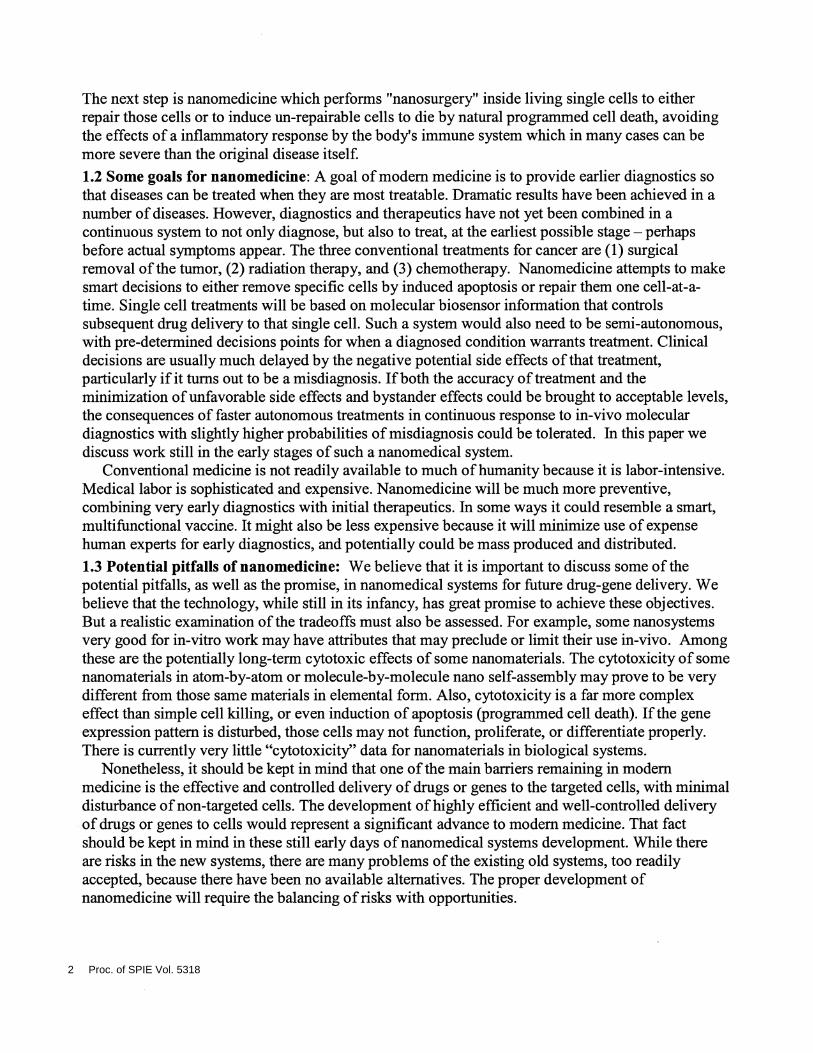
2. MATERIALS AND METHODSIn the early developmental stages of this project we have used a variety of experimental model
systems which are very reproducible and testable, as described below. A basic strategy for ananomedical system applied to the problem of repairing radiation damage in astronauts on acontinuous basis is shown in the concept diagram of Figure 1.
Targeted0
Is the DNAdamage
Yes present?
Figure 1 : A nanomedicineconcept applied to theproblem of continuous repairofradiation damage inastronauts. A terrestrialapplication would betargeted repair of radiation-damaged normal tissueinjured during radiationtreatment of cancer patients.
Daiie is /Ii . . (\ Di i1Kt
repairable\4::::lJPcc1s1on
ierairaNe
Express Inducetherapeutic DNA programmed cell
repair genes death (apoptosis)
Proc. of SPIE Vol. 5318 3

2.1 Nanoparticles: There are two general categories ofnanoparticles (with many variations)currently being used for applications in in-vitro and in-vivo nanomedicine: (1) nanoparticle coreswith single or multilayered coatings, (2) hollow nanoparticle capsules without cores.
Nanoparticle cores materials vary greatly, the most common being made from gold, silica, orsemiconductor materials. Some are made from magnetic materials which can be useful for recoveryofgene products within cells (Prow et al., 2004a). Many are now commercially available. Most ofthese nanoparticles must be coated for two general purposes. First, some of them are not water-soluble. To exist and to function in an in-vivo aqueous environment, some of these hydrophobicmaterials must be marked with a layer ofhydrophilic molecules. Second, some ofthese materials arecytotoxic to cells and tissues. Typically these are covered with lipophilic or other organic moleculesto provide a barrier between the cell and the core nanoparticle materials.
Hollow nanocapsules without cores come in a variety of sizes and materials. The simplest onesconsist of single or multiple layered liposomes, which are designed to fuse with the lipophilicmolecules of a cell membrane and then to spill the contents of the liposome into the interior of thecell. More complex, layer-by-layer assembly nanocapsules are being made by some research groups.These nanocapsules are self-assembling by alternating charged layers ofpolymers and similarmaterials. In other work we have begun to develop nanosystems based on this concept (Prow et al.,2004b). These nanocapsules are potentially biodegradable and may be less cytotoxic to biologicalsystems, although more detailed studies need to be conducted.
2.2 Nanoparticle targeting: Nanoparticle targeting can be accomplished in a variety ofways. Butthe two most common, as shown in this paper, are use of antibodies (e.g. anti-CD95 antibody) boundto the nanoparticle outer surface, or coating ofthe outer surface ofthe nanoparticles with moleculesthat are the ligands for cellular receptors (e.g. mannose to target nanoparticles to liver cells whichhave mannose receptors). While antibody targeting is very common for in-vitro applications, theiruse in-vivo can be problematical since some ofthese targeting antibodies can illicit an immuneresponse from the human or animal. The mannose represents less of a problem in this regard becausethe body already recognizes mannose and does not tend to mount an immune response against it.In the particular application ofnanomedicine for astronauts we are using up-regulation and transportofthe CD95 molecule to the radiation (or oxidative stress) damaged cell. Amounts ofcell surfaceCD95 vary in roughly a dose dependent maimer with radiation exposure (Sheard, 2001). So CD95serves as the initial surrogate biomarker for radiation damage. We also have modeled radiationdosed cells with two cell lines, one ofwhich expresses no CD95 (human MOLT-4 monocyte cellline) and another cell line (BJAB) which expresses high quantities of cell surface CD95 . Onceinside, the nanoparticle system performs a secondary check for oxidative stress which is highlycorrelated to radiation exposure using a biosensor sensitive to the presence ofreactive oxygenspecies molecules. Since exact radiation exposure is difficult to control, we have used, in initialstudies, a chemical which produces the same oxidative stress as radiation but in an easily dosedmanner.
The other significant difference between in-vitro and in-vivo targeting is the great difference inspecificity required. Cells are usually not rare in-vitro, while targeted cells are almost always rare in-vivo. Rare cell targeting presents considerable challenges in terms of specificity. Considering thenumber ofpossible interactions in-vivo, the specificity of the overall targeting system must, in mostcases, be better than a million to one. No antibodies alone have this degree of specificity. To solvethis problem, Boolean combinations of antibodies must be chosen (for review, see Leary, 1994). Thegood news is that these levels of specificity can be reached with antibody combinations of two
4 Proc. of SPIE Vol. 5318
positive biomarkers and one negative biomarker. The correct repertoire of these three biomarkers (orcocktails ofbiomarkers as are frequently used for detection ofrare stem cells)2.3 Cell entry facilitation: Most people do not realize that targeting ofnanoparticles to the cells ofinterest for nanomedicine represents only the first part of a long and complex journey. Ananoparticle is roughly one billionth the volume of a cell. So the interior of the cell representsanother new universe to the nanoparticle. The nanoparticle and/or its contents must get successfullyfrom the outside to the inside of a cell. How it enters the cell can control its subsequent fate. Andsimply dumping the drug or gene contents of the nanoparticle into the interior of the cell, whileperhaps superior to dumping the drug or gene into the body for general circulation in thebloodstream, still does not guarantee that the drug or gene gets to the intracellular site where it canhave therapeutic action. A further step of intracellular targeting is really required to be effective inthis process. We have used three entry facilitation methods: (1) arginine-repeat peptides, (2)LipofectamineTM coatings to promote fusion ofnanoparticles with the cell membrane, and (3)artificial tat-specific sequences, the entry and nuclear targeting molecule used by HIV-1.
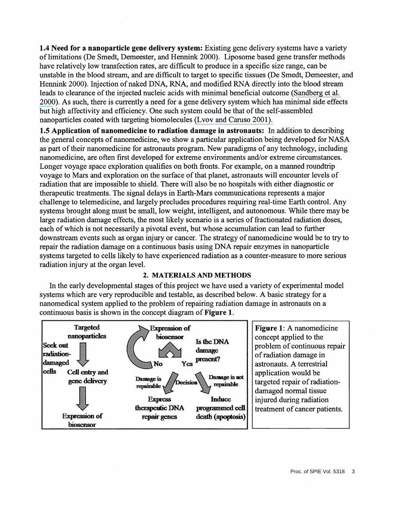
2.4 Intracellular targeting: We have used a variety of specific amino acid localization sequences todeliver and anchor delivery ofmolecules to three intracellular regions ofthe cell: (1) theendoplasmic reticulum (ER), (2) the mitochondria, and (3) the nucleus. An important technology tovisualize and study the proper localization ofnanoparticles to their intracellular targets is confocalmicroscopy. Since to allow for improved detection ofnanoparticles we used a Zeiss 510 METAconfocal microscope with multispectral imaging capabilities which correct for color overlaps on apixel-by-pixel basis within each optical section. This spectral unmixing algorithm (Bearman et al.,2002), referred to as "emission fingerprinting" provides for improved color deconvolution comparedto use ofconventional optical bandpass filters as shown in Figure 2.
"Emission Fingerprinting"Convenhional Contlied
Flow Diagram of a Spectral Unmixing Algorithm -çto tman eye
. 'I op&aI fliersDetein*ie ch&actedstlc Spectra lbr Each Fluosescent Dye (Cr Inçout / , \ gieenfrom a dase) dye speca. . . . . ;pfrdJ pixels only
I ' D&convolvedd
Measwetest sanWleand retrieve cbaictedslic specbbm for conçison : psxels only
to mdividu dye specira. f
II Deconçose measuied specum OfteStSanAe lito de conçonentspecha. . ., ,
DetenTEleweiqIds of each dye to quanlitate tatensity of each dye piesent.2 lmae conlatoEig only Image conlatemg only
those pixels of this _____ those pixels of thus md
Unnds the speckel ovedsp by seesaIiig outthe usages acconfmg to the gm specinimby thef specinirn byihe -remeniig image of test sançle stakied wiltu each dye. appmpnate suty appropnate ensuty
Mapled from: Beannan at .. U.S. Patent 6.403.328(2002)
Figure 2: (A) The basic concept of emission fingerprinting is shown in schematic form. Byknowing directly or indirectly, all of the spectral contributions of each dye or probe plus theautofluorescence spectrum of a cell, the colors can be "unmixed" on a pixel-by-pixel basis oneach plane of a multi-plane confocal image. (B) The algorithm essentially fits the overall colorcurve using regions of each dye or component spectrum that are less contaminated with theoverlap of other colors. The resulting emission fingerprinting technique can be superior to theuse of conventional optical filters which still leave considerable optical overlap.
Proc. of SPIE Vol. 5318 5

2.5 Molecularbiosensing of the intracellular environment: Thus far we have explored both viralbiosensors and reactive oxygen species (ROS) biosensors. This paper will describe use of ROSbiosensors (Zhu and Fahi, 2000) coupled to an eGFP reporter gene which fluoresces green whenactivated. Many biosensors share certain characteristics as shown in Figure 3.
2.6 Controlled drug-gene delivery: One ofthe ways we have tried to provide controlled drug/genedelivery to living single cells, is to attach the gene therapeutic to the biosensor. That way thetherapeutic gene is only produced as long as the biosensor sees its target molecule. In this case weare developing a transient gene therapy for DNA repair ofradiation-damaged DNA based on thebiosensing ofreactive oxygen species which has high correlation to radiation exposure.
An alternative treatment to repair is to accelerate the cell's natural "programmed cell death" orapoptosis. This is particularly important in the case oftrying to repair radiation damaged cells inastronauts. Ifthe cell is not repairable, we would prefer to have it die in a way that does not triggerinflammatory responses ofthe immune system which can, in many diseases and injuries, be ofgreater danger to the person than the disease or injury itself. In this application it is important to notallow cancerous cells to arise from mutations produced by radiation damage.
3. DATA/RESULTS3.1 Comparison of molecular immunochemical and nanoparticle targeting: While the twoprocesses are indeed quite different, fluorescent nanoparticle labeling can yield similar results tothose of conventional fluorescent antibody labeling techniques as shown in Figure 4. In this case weused very large nanoparticles (approximately 500 nm diameter) in order to visualize the nanoparticlelabeling pattern. Improved concordance of nanoparticle labeling with molecular labeling is obtainedwhen nanoparticles are 100 nm or less diameters (data not shown, Prow thesis, 2004).
Taie1Eg to EiitiaI Cleavage domali sensitive Reporter molecule (e.g.region of mierest to target molecule GFP or lucilerese)
For exançle. This biosensor can t designed to sense a target m The cel.Then target to The nucleus lü alow for 1ranscrtion of a gene lbr Therapy
TMD CD iii:.
Figure 3: Manymolecular biosensorsused for diagnosticshave targeting regions,a cleavage domain thatis sensitive to enzymesor proteases, and afluorescent reportermolecule.
tTA
Figure 4: Nanoparticles withtargeting molecules can label cells ina manner that gives similar results toconventional labeling with antibodies.However, as the nanoparticles getlarger, they tend to bind in a"quantized" manner that leads toincreasing discordance of results withincreasing nanoparticle size.
6 Proc. of SPIE Vol. 5318
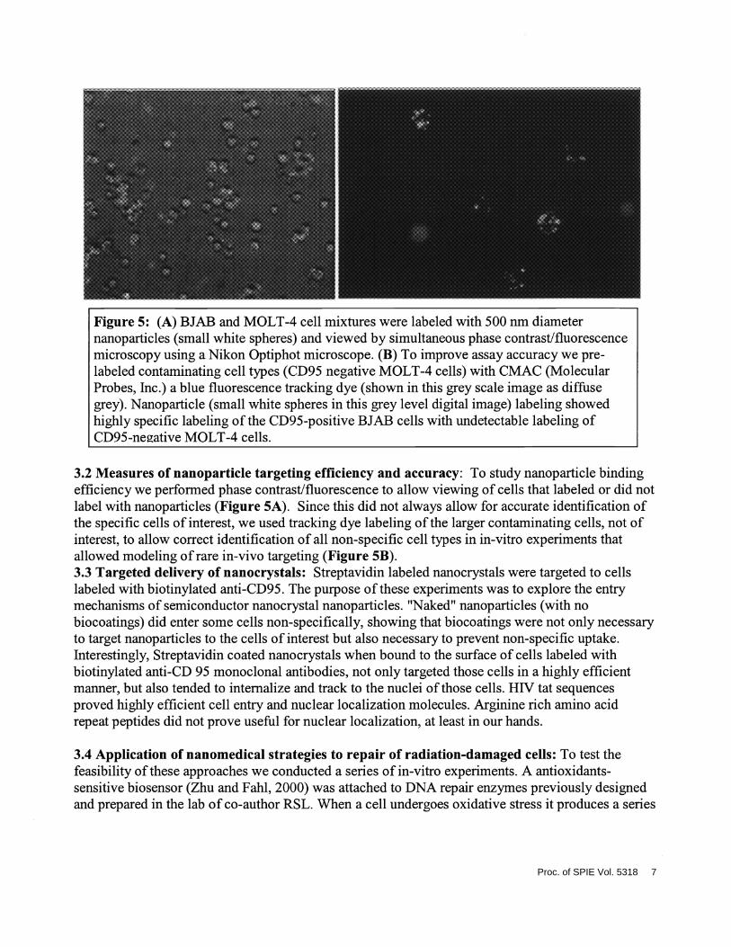
Figure 5: (A) BJAB and MOLT-4 cell mixtures were labeled with 500 urn diameternanoparticles (small white spheres) and viewed by simultaneous phase contrast/fluorescencemicroscopy using a Nikon Optiphot microscope. (B) To improve assay accuracy we pre-labeled contaminating cell types (CD95 negative MOLT-4 cells) with CMAC (MolecularProbes, Inc.) a blue fluorescence tracking dye (shown in this grey scale image as diffusegrey). Nanoparticle (small white spheres in this grey level digital image) labeling showedhighly specific labeling ofthe CD95-positive BJAB cells with undetectable labeling ofCD95-neative MOLT-4 cells.
3.2 Measures of nanoparticle targeting efficiency and accuracy: To study nanoparticle bindingefficiency we performed phase contrast/fluorescence to allow viewing of cells that labeled or did notlabel with nanoparticles (Figure 5A). Since this did not always allow for accurate identification ofthe specific cells of interest, we used tracking dye labeling of the larger contaminating cells, not ofinterest, to allow correct identification of all non-specific cell types in in-vitro experiments thatallowed modeling ofrare in-vivo targeting (Figure 5B).3.3 Targeted delivery of nanocrystals: Streptavidin labeled nanocrystals were targeted to cellslabeled with biotinylated anti-CD95. The purpose ofthese experiments was to explore the entrymechanisms of semiconductor nanocrystal nanoparticles. "Naked" nanoparticles (with nobiocoatings) did enter some cells non-specifically, showing that biocoatings were not only necessaryto target nanoparticles to the cells of interest but also necessary to prevent non-specific uptake.Interestingly, Streptavidin coated nanocrystals when bound to the surface of cells labeled withbiotinylated anti-CD 95 monoclonal antibodies, not only targeted those cells in a highly efficientmanner, but also tended to internalize and track to the nuclei ofthose cells. HW tat sequencesproved highly efficient cell entry and nuclear localization molecules. Arginine rich amino acidrepeat peptides did not prove useful for nuclear localization, at least in our hands.
3.4 Application of nanomedical strategies to repair of radiation-damaged cells: To test thefeasibility of these approaches we conducted a series of in-vitro experiments. A antioxidants-sensitive biosensor (Zhu and Fahl, 2000) was attached to DNA repair enzymes previously designedand prepared in the lab of co-author RSL. When a cell undergoes oxidative stress it produces a series
Proc. of SPIE Vol. 5318 7
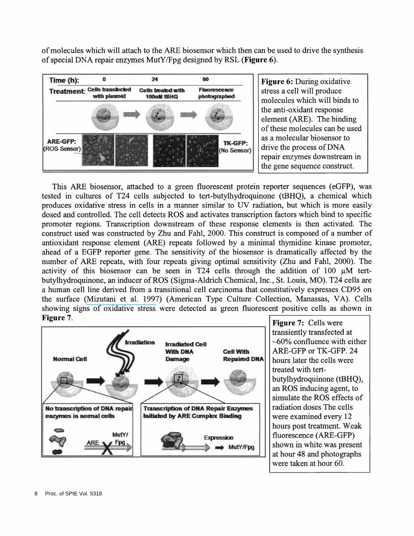
of molecules which will attach to the ARE biosensor which then can be used to drive the synthesisof special DNA repair enzymes MutYIFpg designed by RSL (Figure 6).
(RcX:: SCflS(
This ARE biosensor, attached to a green fluorescent protein reporter sequences (eGFP), wastested in cultures of T24 cells subjected to tert-butyihydroquinone (tBHQ), a chemical whichproduces oxidative stress in cells in a manner similar to UV radiation, but which is more easilydosed and controlled. The cell detects ROS and activates transcription factors which bind to specificpromoter regions. Transcription downstream of these response elements is then activated. Theconstruct used was constructed by Zhu and Fahl, 2000. This construct is composed of a number ofantioxidant response element (ARE) repeats followed by a minimal thymidine kinase promoter,ahead of a EGFP reporter gene. The sensitivity of the biosensor is dramatically affected by thenumber of ARE repeats, with four repeats giving optimal sensitivity (Zhu and Fahl, 2000). Theactivity of this biosensor can be seen in T24 cells through the addition of 1 00 jiM tert-butyihydroquinone, an inducer ofROS (Sigma-Aldrich Chemical, Inc., St. Louis, MO). T24 cells area human cell line derived from a transitional cell carcinoma that constitutively expresses CD95 onthe surface (Mizutani et al. 1997) (American Type Culture Collection, Manassas, VA). Cellsshowing signs of oxidative stress were detected as green fluorescent positive cells as shown inFigure 7
lime(h): 0
Treatmentw
Foocecephotrmiphed
Figure 6: During oxidativestress a cell will producemolecules which will binds tothe anti-oxidant responseelement (ARE). The bindingofthese molecules can be usedas a molecular biosensor todrive the process of DNArepair enzymes downstream inthe gene sequence construct.
Figure 7: Cells weretransiently transfected at6O% confluence with eitherARE-GFP or TK-GFP. 24hours later the cells weretreated with tert-butylhydroquinone (tBHQ),an ROS inducing agent, tosimulate the ROS effects ofradiation doses The cellswere examined every 12hours post treatment. Weakfluorescence (ARE-GFP)shown in white was presentat hour 48 and photographswere taken at hour 60.
8 Proc. of SPIE Vol. 5318

To test whether DNA repair enzymes could be introduced into cells damaged by actual UV radiationto accelerate normal DNA repair mechanisms by activating a second repair pathway, not normallyexpressed in human cells because one enzyme, a glycosylase, is absent in normal human cells(Figure 8). ______________________________________________
rs Strategy for assisting DNA repair in human cells> k humans. there is ONLY ONE mechanism to repa UV-Educed damage to DNA
I Immune system suppressed &-24 hrs.I DNA damage mmoval takes 24-48 hrs.
> However. sTçI& organisms have TWO and somelimes THREErepal systems> Oeofihese repel systems is parbaly pesest m humans. BUTwe are MISSINGthe FiRSTSTEP ________In' Humansare
'S' rrisstgttisrepwrx vr:e4svI ASE enzyme wtich can
I I betranslected untohuinancels
AP LYASE-i--- APEMXJIJQkAME
! I I I
PaIJ I'QIAimi •URRUS ITM—1Lull!! POIjtortXRCC1 LffI
Ii!Jm/xtLcclUsing nanoparticle/bEosensor technology we can supply this rrássing
first step to eithance DNA repair to human cell&
Figure 8: (A) Assisted DNA repair concept for DNA damage to both nuclear and mitochondrialDNA. (B) A specific strategy for assisting DNA repair ofUV damage in human cells.
This normally absent repair mechanism was activated by transfection of a glycosylase containinggene sequence which also contained an eGFP reporter molecule. UV damage can occur in bothnuclear and mitochondrial DNA. Since many molecules introduced into a cell frequently track non-specifically to the nucleus, we tested the ability of intracellular localization sequences to guide theserepair molecules to the mitochondria. In the absence of localization sequences, the repair moleculesdid not appear to track to specific regions of the cell, as shown by the diffuse staining of Figure 9.However when mitochondrial localization sequences were attached to the repair enzymes, theytracked to the mitochondria as shown by confocal microscopy (Figures 9B and 9C). Either transient
or stable gene therapy was demonstrated.In the present state of gene therapy concerns about patient
safety, our efforts are concentrated on the production of potentiallyuseful transient gene therapies using nanoparticle systems. To test
the actual effectiveness ofthese DNA repair enzymes inside living
uv "Assisted DNA RepairNanpahcks ernce
. aa cozyew A
Figure 9: (A) T4 transfected DNA repair enzyme with nolocalization anchoring sequence (note diffuse fluorescence)with transient expression (Wt-T4-PDG-GFP in CHO-XPG.Transient expression. 1 OOx objective)(B) T4 transfected DNA repair enzyme with mitochondriallocalization anchoring sequence, with transient expression(MLS35-T4-PDG-GFP in CHO XPG. lOOx objective)(C) T4 transfected DNA repair enzyme with mitochondriallocalization anchoring sequence, with stable expression(MLS18-T4-PDG-GFP in hXPA. lOOx objective)
Proc. of SPIE Vol. 5318 9
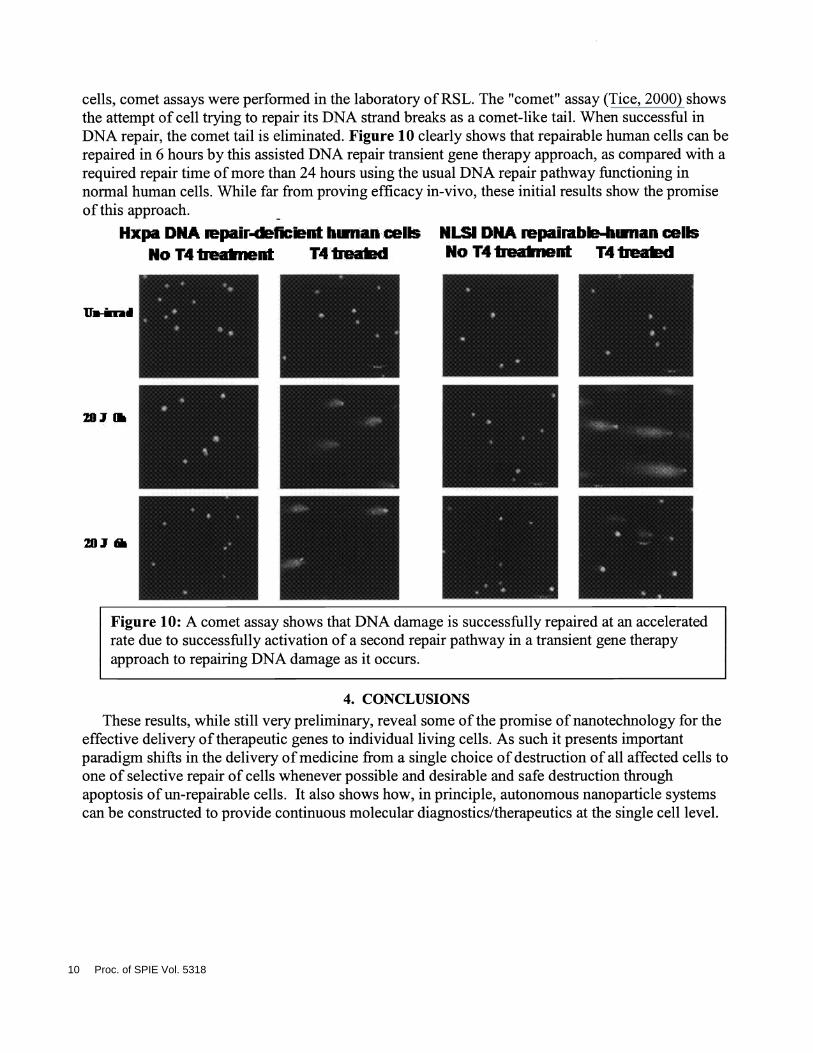
cells, comet assays were performed in the laboratory of RSL. The "comet" assay (Tice, 2000) showsthe attempt of cell trying to repair its DNA strand breaks as a comet-like tail. When successful inDNA repair, the comet tail is eliminated. Figure 10 clearly shows that repairable human cells can berepaired in 6 hours by this assisted DNA repair transient gene therapy approach, as compared with arequired repair time of more than 24 hours using the usual DNA repair pathway functioning innormal human cells. While far from proving efficacy in-vivo, these initial results show the promiseof this approach. -
Hxpa DNA iepair-dehcmnthiinancellsNo T4 Ireaknent T4 Iiead
Figure 10: A comet assay shows that DNA damage is successfully repaired at an acceleratedrate due to successfully activation of a second repair pathway in a transient gene therapyapproach to repairing DNA damage as it occurs.
4. CONCLUSIONSThese results, while still very preliminary, reveal some of the promise of nanotechnology for the
effective delivery oftherapeutic genes to individual living cells. As such it presents importantparadigm shifts in the delivery of medicine from a single choice of destruction of all affected cells toone of selective repair of cells whenever possible and desirable and safe destruction throughapoptosis of un-repairable cells. It also shows how, in principle, autonomous nanoparticle systemscan be constructed to provide continuous molecular diagnostics/therapeutics at the single cell level.
NLSI DNA repairab-hiinan cellsNo T4 Iieaknent T4 Iiead
207 III
201
10 Proc. of SPIE Vol. 5318
5. ACKNOWLEDGEMENTS
The authors gratefully acknowledge the contributions ofnanocrystals (by Dr. Nicholas Kotov,University ofMichigan). This work is described in more detail in other co-authored manuscriptscurrently pending publication.
This research was funded by the Biomolecular, Physics and Chemistry Program under NASA-Ames grant NAS2-02059.
6. REFERENCES
Bearman, G.H., Frasier, S.E., Lanford, R.D. System and method for monitoring cellular activity.U.S. Patent 6,403,328. 2002.Dc Smedt, S. C., J. Demeester, and W. B. Hennink. Cationic polymer based gene delivery systems.Pharm Res 17, no. 2: 1 13-126, 2000.
Leary, J. F. Strategies for rare cell detection and isolation. Methods Cell Biol 42 Pt B: 33 1-358,1994.Lvov, Y. and F. Caruso. Biocolloids with ordered urease multilayer shells as enzymatic reactors.Anal Chem 73, no. 17: 4212-4217, 2001.Mamedov, A. A., A. Belov, M. Giersig, N. N. Mamedova, and N. A. Kotov. Nanorainbows: Gradedsemiconductor films from quantum dots. J Am Chem Soc 123, no. 3 1 : 7738-7739, 2001.
Mizutani, Y., Y. Okada, 0. Yoshida, M. Fukumoto, and B. Bonavida. Doxorubicin sensitizes humanbladder carcinoma cells to fas-mediated cytotoxicity. Cancer 79, no. 6: 1 1 80-1 1 89, 1997.
Prow T.W., Rijnbrand, R., Wang, N., Salazar, J., Leary, J. F. Transient Gene Expression from DNATethered Magnetic Nanoparticles. Manuscript submitted for publication 2004a.
Prow T.W., Kotov, N.A., Lvov, Y.M., Rijnbrand, R, Leary, J.F. Nanoparticles, MolecularBiosensors, and Multispectral Confocal Microscopy. Journal of Molecular Histology (accepted)2004b.
Prow, T.W. Nanomedicine: Targeted nanoparticles for the delivery ofbiosensors and therapeuticgenes. Ph.D. thesis, University of Texas Medical Branch, Galveston, TX. 2004Sandberg, J. A., C. D. Sproul, K. S. Blanchard, L. Bellon, D. Sweedler, J. A. Powell, F. A. Caputo,D. J. Kornbrust, V. P. Parker, T. J. Parry, and L. M. Blatt. 2000. Acute toxicology andpharmacokinetic assessment of a ribozyme (angiozyme) targeting vascular endothelial growth factorreceptor mRNA in the cynomolgus monkey. Antisense Nucleic Acid Drug Dev 10, no. 3: 153-62.
Sheard, M.A. Ionizing radiation as a response-enhancing agent for CD95-mediated apoptosis. Tnt. J.Cancer 96: 213-220, 2001.
Tice RR, Agurell E, Anderson D, Burlinson B, Hartmann A, Kobayashi H, Miyamae Y, Rojas E,Ryu JC, Sasaki YF. Single cell gel/comet assay: guidelines for in vitro and in vivo genetictoxicology testing. Environ Mol Mutagen 35(3):206-221, 2000.Zhu, M. and W. E. Fahl Development of a green fluorescent protein microplate assay for thescreening of chemopreventive agents. Anal Biochem 287, no. 2: 2 10-217, 2000.
Proc. of SPIE Vol. 5318 11
Related Documents


