Int. J. Mol. Sci. 2011, 12, 3303-3321; doi:10.3390/ijms12053303 International Journal of Molecular Sciences ISSN 1422-0067 www.mdpi.com/journal/ijms Review Nanomedicine: Application Areas and Development Prospects Houria Boulaiz 1, *, Pablo J. Alvarez 1 , Alberto Ramirez 1 , Juan A. Marchal 1 , Jose Prados 1 , Fernando Rodrí guez-Serrano 1 , Macarena Perán 2 , Consolación Melguizo 1 and Antonia Aranega 1, * 1 Department of Human Anatomy and Embryology, Institute of Biopathology and Regenerative Medicine (IBIMER), School of Medicine, University of Granada, Granada 18071, Spain; E-Mails: [email protected] (P.J.A.); [email protected] (A.R.); [email protected] (J.A.M.); [email protected] (J.P.); [email protected] (F.R.-S.); [email protected] (C.M.) 2 Department of Health Sciences, University of Jaén, Jáen 23071, Spain; E-Mail: [email protected] * Authors to whom correspondence should be addressed; E-Mails: [email protected] (H.B.); [email protected] (A.A.); Tel.:+34-958-243534; Fax: +34-958-246296. Received: 31 March 2011; in revised form: 6 May 2011 / Accepted: 16 May 2011 / Published: 19 May 2011 Abstract: Nanotechnology, along with related concepts such as nanomaterials, nanostructures and nanoparticles, has become a priority area for scientific research and technological development. Nanotechnology, i.e., the creation and utilization of materials and devices at nanometer scale, already has multiple applications in electronics and other fields. However, the greatest expectations are for its application in biotechnology and health, with the direct impact these could have on the quality of health in future societies. The emerging discipline of nanomedicine brings nanotechnology and medicine together in order to develop novel therapies and improve existing treatments. In nanomedicine, atoms and molecules are manipulated to produce nanostructures of the same size as biomolecules for interaction with human cells. This procedure offers a range of new solutions for diagnoses and ―smart‖ treatments by stimulating the body‘s own repair mechanisms. It will enhance the early diagnosis and treatment of diseases such as cancer, diabetes, Alzheimer‘s, Parkinson‘s and cardiovascular diseases. Preventive medicine may then become a reality. Keywords: nanomedicine; nanostructures; early diagnosis; drug delivery OPEN ACCESS

Welcome message from author
This document is posted to help you gain knowledge. Please leave a comment to let me know what you think about it! Share it to your friends and learn new things together.
Transcript
Int. J. Mol. Sci. 2011, 12, 3303-3321; doi:10.3390/ijms12053303
International Journal of
Molecular Sciences ISSN 1422-0067
www.mdpi.com/journal/ijms
Review
Nanomedicine: Application Areas and Development Prospects
Houria Boulaiz 1,
*, Pablo J. Alvarez 1, Alberto Ramirez
1, Juan A. Marchal
1, Jose Prados
1,
Fernando Rodríguez-Serrano 1, Macarena Perán
2, Consolación Melguizo
1 and
Antonia Aranega 1,
*
1 Department of Human Anatomy and Embryology, Institute of Biopathology and Regenerative
Medicine (IBIMER), School of Medicine, University of Granada, Granada 18071, Spain;
E-Mails: [email protected] (P.J.A.); [email protected] (A.R.);
[email protected] (J.A.M.); [email protected] (J.P.); [email protected] (F.R.-S.); [email protected] (C.M.) 2
Department of Health Sciences, University of Jaén, Jáen 23071, Spain; E-Mail: [email protected]
* Authors to whom correspondence should be addressed; E-Mails: [email protected] (H.B.);
[email protected] (A.A.); Tel.:+34-958-243534; Fax: +34-958-246296.
Received: 31 March 2011; in revised form: 6 May 2011 / Accepted: 16 May 2011 /
Published: 19 May 2011
Abstract: Nanotechnology, along with related concepts such as nanomaterials,
nanostructures and nanoparticles, has become a priority area for scientific research and
technological development. Nanotechnology, i.e., the creation and utilization of materials
and devices at nanometer scale, already has multiple applications in electronics and other
fields. However, the greatest expectations are for its application in biotechnology and
health, with the direct impact these could have on the quality of health in future societies.
The emerging discipline of nanomedicine brings nanotechnology and medicine together in
order to develop novel therapies and improve existing treatments. In nanomedicine, atoms
and molecules are manipulated to produce nanostructures of the same size as biomolecules
for interaction with human cells. This procedure offers a range of new solutions for
diagnoses and ―smart‖ treatments by stimulating the body‘s own repair mechanisms. It will
enhance the early diagnosis and treatment of diseases such as cancer, diabetes,
Alzheimer‘s, Parkinson‘s and cardiovascular diseases. Preventive medicine may then
become a reality.
Keywords: nanomedicine; nanostructures; early diagnosis; drug delivery
OPEN ACCESS
Int. J. Mol. Sci. 2011, 12
3304
1. Introduction
The term nanotechnology refers to the ability to measure, design and manipulate materials at
atomic, molecular and supramolecular level in order to understand, create and apply structures and
systems with specific functions attributable to their size. Nanotechnology classically refers to matter in
the size range of 1–100 nm, but it is often extended to include materials below 1 µm in size. A key
goal is to assemble nanoparticles and integrate them into ordered structures in order to obtain useful
materials. Although new, the advent of nanomaterials was forecast as long ago as 1959 by Richard P.
Feynman [1]. Nanotechnology has been embraced by multiple industrial sectors for application in the
field of electronic storage systems [2], biotechnology [3], magnetic separation and preconcentration of
target analytes and targeted drug delivery [4,5], and as vehicles for gene and drug delivery [2,4,5].
Advances in nanotechnology have led to the development of new nanomaterials whose physiochemical
properties differ from those of their larger counterparts due to their higher surface-to-volume ratio.
These novel properties make them excellent candidates for biomedical applications, given the range of
biological processes that occur at nanometer scale [6]. Nanotechnology is a new discipline of science
and engineering that has led to innovative approaches in many areas of medicine. Its applications in the
screening, diagnosis, and treatment of disease are collectively referred to as ―nanomedicine‖—an
emerging field that has the potential to revolutionize individual and population-based health this
century [7]. It is now possible to provide therapy at a molecular level with the help of nanoparticles,
treating diseases and adding to our understanding of their pathogenesis. Nanomedicine can be
considered a refinement of molecular medicine, integrating innovations in genomics and proteomics on
the road to a more personalized medicine.
The impact of nanotechnology in medicine can mainly be seen in diagnostic methods, drug-release
techniques and regenerative medicine.
Diagnostic methods are essential for the early detection of diseases to enable their prompt
treatment, minimizing possible damage to the rest of the organism. The importance of imaging
methods to diagnose, treat and follow up cancer, cardiovascular and neurological patients is well
known. Diagnostic techniques based on the use of nanoparticles offer higher sensitivity and assist the
early detection of disease, offering a better prognosis and greater possibilities of successful
treatment [8]. Once the diagnosis is established, the fight against the disease begins, with medicaments
playing a major role. There are numerous obstacles to the development of novel drugs against disease.
Conventional drugs suffer from the major limitation of adverse effects, the result of the non-specificity
of their action, and from a lack of effectiveness due to improper or ineffective dosages, e.g., in cancer
chemotherapy and anti-diabetic therapy. Nanotechnology offers the possibility of designing novel
drugs with greater cell specificity and new drug-release systems that act selectively on specific targets
and protect the drug from degradation en route. This allows the administration of smaller but more
effective doses, minimizing adverse effects. Nanotechnology can also be used to optimize drug
formulations, increasing drug solubility and altering the pharmacokinetics to sustain the release of the
drug, thereby prolonging its bioavailability. The diverse platforms of nanotechnology can be utilized to
develop more sophisticated, cell-targeted therapies and to combine different drugs into a single
nanotherapeutic agent for synergistic therapeutic benefits [9].
Int. J. Mol. Sci. 2011, 12
3305
Regenerative medicine has been proposed as a highly promising approach to numerous
degenerative diseases. To this end, nanomedicine employs gene therapy, cell therapy, tissue
engineering, biomaterials and signaling molecules.
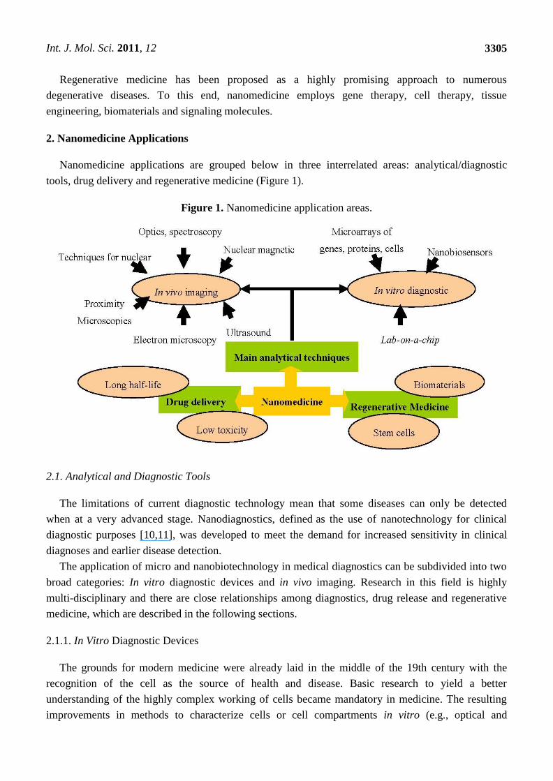
2. Nanomedicine Applications
Nanomedicine applications are grouped below in three interrelated areas: analytical/diagnostic
tools, drug delivery and regenerative medicine (Figure 1).
Figure 1. Nanomedicine application areas.
2.1. Analytical and Diagnostic Tools
The limitations of current diagnostic technology mean that some diseases can only be detected
when at a very advanced stage. Nanodiagnostics, defined as the use of nanotechnology for clinical
diagnostic purposes [10,11], was developed to meet the demand for increased sensitivity in clinical
diagnoses and earlier disease detection.
The application of micro and nanobiotechnology in medical diagnostics can be subdivided into two
broad categories: In vitro diagnostic devices and in vivo imaging. Research in this field is highly
multi-disciplinary and there are close relationships among diagnostics, drug release and regenerative
medicine, which are described in the following sections.
2.1.1. In Vitro Diagnostic Devices
The grounds for modern medicine were already laid in the middle of the 19th century with the
recognition of the cell as the source of health and disease. Basic research to yield a better
understanding of the highly complex working of cells became mandatory in medicine. The resulting
improvements in methods to characterize cells or cell compartments in vitro (e.g., optical and
Int. J. Mol. Sci. 2011, 12
3306
luminescence microscopy, scanning probe microscopy, electron microscopy and imaging
mass-spectrometry) have been critically important for the development of nanomedicine.
The miniaturization and integration of different functions in a single device, based on
nanotechnology-derived techniques, have led to a new generation of devices that are smaller, faster
and cheaper, require no special skills and give accurate readings. They require much smaller samples,
implying less invasive and traumatic sample extraction methods, and deliver more complete and more
accurate biological data from a single measurement. The use of these devices in research has become
routine and has improved our understanding of the molecular basis of disease and helped to identify
new therapeutic targets. In vitro diagnostic devices include nanobiosensors, microarrays, biochips of
different elements (DNA, proteins or cells) and lab-on-a-chip devices.
2.1.1.1. Nanobiosensor
A nanobiosensor is defined as a compact analysis device that incorporates biological (nucleic acid,
enzyme, antibody, receptor, tissue, cell) or biomimetic (macrophage-inflammatory proteins, aptamers,
peptide nucleic acids) recognition elements [11,12]. Interaction between the compound or
microorganism of interest and the recognition element produces a variation in one or more
physical-chemical properties (e.g., pH, electron transfer, heat, potential, mass, optical properties, etc.)
that are detected by the transducer. The resulting electronic signal indicates the presence of the analyte
of interest and its concentration in the sample. These sensors can be electronically gated to respond to
the binding of a single molecule. Prototype sensors have been successfully used to detect nucleic acids,
proteins and ions. They can operate in liquid or gas phase, opening up an enormous variety of
downstream applications. These detection systems use inexpensive low-voltage measurement methods
and detect binding events directly, so there is no need for costly, complicated and time-consuming
chemical labeling, e.g., with fluorescent dyes, or for bulky and expensive optical detection systems. As
a result, these sensors are inexpensive to manufacture and portable [10]. Hence, nanobiosensors are
revolutionizing the in vitro diagnosis of diseases and have major implications for human health. They
allow healthcare professionals to simultaneously measure multiple clinical parameters using a simple,
effective and accurate test. These devices are also ideal for high-throughput screening and for the
detection of a single disease in various samples or of various diseases in a single sample [13].
2.1.1.2. Microarrays
The microarray is another diagnostic device that is becoming a standard technology in research
laboratories worldwide. Since their first application, microarray technologies have proven productively
functional in almost all areas of biomedical research [14–21]. The emergence of this new tool has
allowed investigators to address previously intractable problems and identify novel potential
therapeutic targets. They are using microarray technology to identify cardinal aspects of growth and
development and explore the underlying genetic causes of numerous human diseases [14].
Microarray-based studies have enormous potential in the exploration of diseases such as cancer [15]
and in the design and development of new drugs [16]. Microarrays have been widely applied in the
study of various pathological conditions, including inflammation [17], atherosclerosis [18], breast
Int. J. Mol. Sci. 2011, 12
3307
cancer [19,20], colon cancer [21] and pulmonary fibrosis [22]. As a result, functions have been assigned
to previously unannotated genes, and genes have been grouped into functional pathways [23,24].
Several types of microarray have been developed for different target materials, which can be DNA,
cDNA, mRNA, protein, small molecules, tissues or any other material that can be quantitatively
analyzed. DNA microarray technology has progressed rapidly over the past 10 years and allows the
large-scale quantification of gene expression. A DNA array consists of a large number of DNA
molecules orderly arranged on a solid substrate to form a matrix of sequences in two dimensions [25].
cDNA microarrays and oligonucleotide microarrays, called ‗expression chips‘, are used for microarray
expression analysis, i.e., to determine the level or volume of expression of a given gene. Single
nucleotide polymorphism (SNP) microarrays detect mutations or polymorphisms in a gene
sequence [26]. This technology is used to test an individual for disease expression patterns and to
determine whether or not individuals are susceptible to a disease. Microarray comparative genomic
hybridisation [27] is employed to seek genomic gains and losses or changes in the number of copies of
a gene implicated in a disease. Protein microarrays are comprised of protein detectors (usually
antibodies) arranged systematically over a glass slide and allow the investigation of expression profiles
and the precise definition of protein functions in relation to biological processes [28]. Tissue
microarrays represent one of the most valuable approaches in the microarray field, because they allow
the simultaneous analysis of protein, RNA and DNA expression in multiple tissue samples [29].
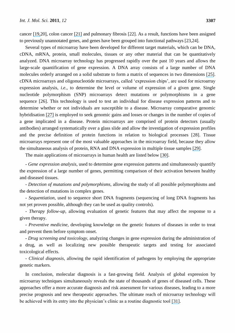
The main applications of microarrays in human health are listed below [30].
- Gene expression analysis, used to determine gene expression patterns and simultaneously quantify
the expression of a large number of genes, permitting comparison of their activation between healthy
and diseased tissues.
- Detection of mutations and polymorphisms, allowing the study of all possible polymorphisms and
the detection of mutations in complex genes.
- Sequentiation, used to sequence short DNA fragments (sequencing of long DNA fragments has
not yet proven possible, although they can be used as quality controls).
- Therapy follow-up, allowing evaluation of genetic features that may affect the response to a
given therapy.
- Preventive medicine, developing knowledge on the genetic features of diseases in order to treat
and prevent them before symptom onset.
- Drug screening and toxicology, analyzing changes in gene expression during the administration of
a drug, as well as localizing new possible therapeutic targets and testing for associated
toxicological effects.
- Clinical diagnosis, allowing the rapid identification of pathogens by employing the appropriate
genetic markers.
In conclusion, molecular diagnosis is a fast-growing field. Analysis of global expression by
microarray techniques simultaneously reveals the state of thousands of genes of diseased cells. These
approaches offer a more accurate diagnosis and risk assessment for various diseases, leading to a more
precise prognosis and new therapeutic approaches. The ultimate reach of microarray technology will
be achieved with its entry into the physician‘s clinic as a routine diagnostic tool [31].
Int. J. Mol. Sci. 2011, 12
3308
2.1.1.3. Lab-on-a-Chip
The latest in vitro diagnostic development derives from the integration of several functions in a
single device. Lab-on-a-chip integrates one or several laboratory functions on a single chip ranging
from only a few millimeters to a square centimeter in size [32] and incorporates sample preparation,
purification, storage, mixing, detection and other functions. Its development was based on advances in
microsystem technologies and in the field of microfluidics on the design of devices that use
microscopic volumes of sample. The chips use a combination of phenomena, including pressure,
electroosmosis, electrophoresis and other mechanisms to move samples and reagents through
microscopic channels and capillaries, some as small as a few dozen nanometers.
Lab-on-a-chip has many applications in medicine and biology. These devices are likely to have a
significant socio-economic impact, bringing sophisticated analytical tools to Third World countries,
rural areas and resource-poor regions. Advantages of their use include the extremely rapid analysis of
samples containing fluid volumes that can be less than a picoliter, the high degree of automation, cost
savings due to the low consumption of reagents and samples and their portable and disposable nature.
Lab-on-a-chip is used in real-time polymerase chain reaction [33] and immunoassays [34] to detect
bacteria, viruses and cancers. It can also be used in blood sample preparation to crack cells and extract
their DNA [32]. Lab-on-a-chip may soon play an important role in efforts to improve global health,
especially with the development of point-of-care testing devices. Infectious diseases that are treatable
in developed nations are often deadly in countries with limited healthcare resources. In some cases,
healthcare clinics possess a drug to treat a given disease but lack the diagnostic tool to identify the
patients who should receive it. Many researchers believe that lab-on-a-chip technology will be the key
to powerful new diagnostic instruments. The goal is to create microfluidic chips that will allow
healthcare providers in poorly-equipped clinics to perform diagnostic tests (e.g., immunoassays and
nucleic acid assays) with no laboratory support. One active research line on the lab-on-a-chip
addresses the diagnosis and management of HIV infections [35]. Around 40 million people are
infected with HIV in the world, yet only 1.3 million receive antiretroviral treatment and around 90% of
HIV-infected individuals have never been tested for the disease. This is largely because its diagnosis
requires measurement of the number of CD4+ T lymphocytes in the blood by means of flow
cytometry, a complicated technique that requires trained technicians and expensive equipment that are
not available in most developing regions.
2.1.2. In Vivo Imaging
Imaging systems can be grouped according to the energy used to derive the visual information
(X-rays, positrons, photons or sound waves), the spatial resolution (macroscopic, mesoscopic or
microscopic) or the type of information obtained (anatomical, physiological, cellular or molecular).
Unlike classic imaging diagnosis with computed tomography (CT), magnetic resonance imaging
(MRI) or ultrasounds, nano-imaging or molecular imaging includes techniques designed to obtain
molecular data to identify the causes of the disease in vivo rather than its eventual consequences.
Nanotechnology has produced advances in imaging diagnosis, developing novel methods and increasing
the resolution and sensitivity of existing techniques. Although these systems have emerging recently and
Int. J. Mol. Sci. 2011, 12
3309
only some of them are in clinical and preclinical use, they have made it possible to study human
biochemical processes in different organs in vivo, opening up new horizons in instrumental diagnostic
medicine. These systems include positron-emission tomography (PET), single-photon-emission
CT (SPECT), fluorescence reflectance imaging, fluorescence-mediated tomography (FMT), fiber-optic
microscopy, optical frequency-domain imaging, bioluminescence imaging, laser-scanning confocal
microscopy and multiphoton microscopy [36].
Imaging diagnosis has gained importance over the years and is now an indispensable diagnostic tool
for numerous diseases, including cancer, cardiovascular diseases and neurological syndromes. The
main benefits of molecular imaging for in vivo diagnosis lie in the early detection of disease and the
monitoring of disease stages, e.g., in cancer metastasis [37], supporting the development of
individualized medicine and the real-time assessment of therapeutic and surgical efficacy. An ideal
imaging modality should be non-invasive, sensitive, and provide objective information on cell
survival, function and localization. MRI, CT, PET) and SPECT are the most widely used and studied
modalities in cancer patients [36,38,39] Overall, nuclear imaging by PET or SPECT offers greater
sensitivity (>5 × 10³ cells) [40] but is limited by the lack of anatomical context [41], whereas MRI
provides accurate anatomical detail but no data on cell viability and shows poor sensitivity
(>105 cells) [42]. Although none of these modalities is ideal, MRI is the preferred option for cellular
tracking [39,43,44] By detecting proton relaxations in the presence of a magnetic field (1.5 Tesla [T]-3
T for clinical imaging), it yields tomographic images with excellent soft tissue contrast and can locate
the cells of interest in the context of the surrounding milieu (oedema or inflammation) [45,46] without
the use of harmful ionizing radiations (the case with CT, PET or SPECT). In addition, MRI offers a
longer tracking window in comparison to PET and SPECT, which are limited by the decay of the
short-lived radioactive isotopes.
In parallel to the development of imaging techniques, intense research has been fuelled by the need
for practical, robust and highly sensitive and selective detection agents that can address the
deficiencies of conventional technologies. New contrast agents, used to increase the sensitivity and
contrast of imaging techniques are increasingly complex and formed by synthetic and biological
nanoparticles. Nanoparticles possess certain size-dependent properties, particularly with respect to
optical and magnetic parameters, which can be manipulated to achieve a detectable signal [47]. The
primary event in most nanoparticle-based assays is the binding of a nanoparticle label or probe to the
target biomolecule that will produce a measurable signal characteristic of the target biomolecule. A
probe that is to function in a biological system must be water-soluble and stable and have minimal
interaction with the surrounding environment. For fluorescence readouts, the probe should ideally have
a high fluorescence quantum yield and minimal photobleaching rates in order to generate a detectable
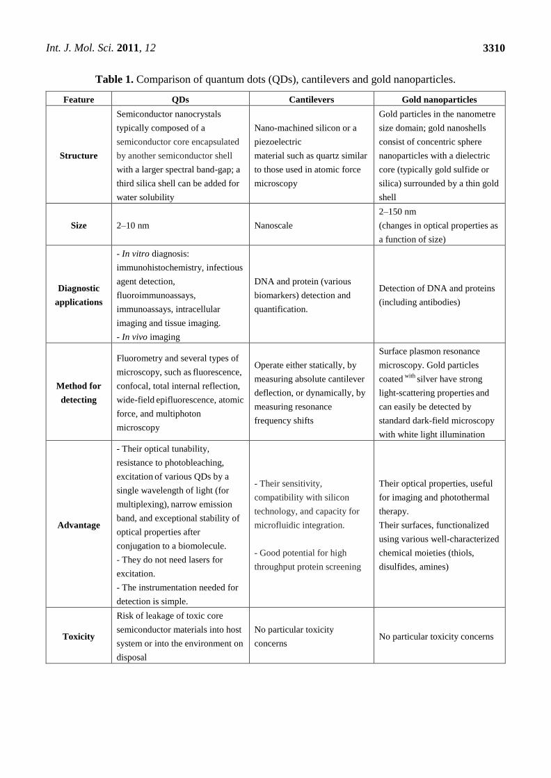
signal [48]. The most promising nanotechnologies for clinical diagnosis include quantum dots (QDs),
gold nanoparticles and cantilevers, whose main features are summarized in Table 1.
Int. J. Mol. Sci. 2011, 12
3310
Table 1. Comparison of quantum dots (QDs), cantilevers and gold nanoparticles.
Feature QDs Cantilevers Gold nanoparticles
Structure
Semiconductor nanocrystals
typically composed of a
semiconductor core encapsulated
by another semiconductor shell
with a larger spectral band-gap; a
third silica shell can be added for
water solubility
Nano-machined silicon or a
piezoelectric
material such as quartz similar
to those used in atomic force
microscopy
Gold particles in the nanometre
size domain; gold nanoshells
consist of concentric sphere
nanoparticles with a dielectric
core (typically gold sulfide or
silica) surrounded by a thin gold
shell
Size 2–10 nm Nanoscale
2–150 nm
(changes in optical properties as
a function of size)
Diagnostic
applications
- In vitro diagnosis:
immunohistochemistry, infectious
agent detection,
fluoroimmunoassays,
immunoassays, intracellular
imaging and tissue imaging.
- In vivo imaging
DNA and protein (various
biomarkers) detection and
quantification.
Detection of DNA and proteins
(including antibodies)
Method for
detecting
Fluorometry and several types of
microscopy, such as fluorescence,
confocal, total internal reflection,
wide-field epifluorescence, atomic
force, and multiphoton
microscopy
Operate either statically, by
measuring absolute cantilever
deflection, or dynamically, by
measuring resonance
frequency shifts
Surface plasmon resonance
microscopy. Gold particles
coated with
silver have strong
light-scattering properties and
can easily be detected by
standard dark-field microscopy
with white light illumination
Advantage
- Their optical tunability,
resistance to photobleaching,
excitation of various QDs by a
single wavelength of light (for
multiplexing), narrow emission
band, and exceptional stability of
optical properties after
conjugation to a biomolecule.
- They do not need lasers for
excitation.
- The instrumentation needed for
detection is simple.
- Their sensitivity,
compatibility with silicon
technology, and capacity for
microfluidic integration.
- Good potential for high
throughput protein screening
Their optical properties, useful
for imaging and photothermal
therapy.
Their surfaces, functionalized
using various well-characterized
chemical moieties (thiols,
disulfides, amines)
Toxicity
Risk of leakage of toxic core
semiconductor materials into host
system or into the environment on
disposal
No particular toxicity
concerns No particular toxicity concerns
Int. J. Mol. Sci. 2011, 12
3311
2.2. Drug Delivery
One of the most important nanotechnology applications developed over the past decade have been
nanovehicles, nanoscale compounds used as a therapeutic tool and designed to specifically accumulate
in the sites of the body where they are needed in order to improve pharmacotherapeutic outcomes. The
main objective of this application is to increase therapeutic effectiveness while obtaining lower toxicity
rates. Hence, nanodrugs and nanodiagnostics have been developed to increase bioavailability profiles,
enabling the administration of lower doses and thereby minimizing the adverse reactions found with
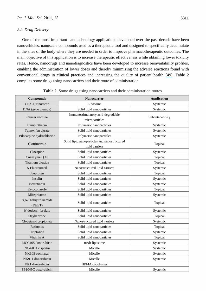
conventional drugs in clinical practices and increasing the quality of patient health [49]. Table 2
compiles some drugs using nanocarriers and their route of administration.
Table 2. Some drugs using nanocarriers and their administration routes.
Compounds Nanocarrier Application
CPX-1 irinotecan Liposome Systemic
DNA (gene therapy) Solid lipid nanoparticles Systemic
Cancer vaccine Immunostimulatory acid-degradable
microparticles Subcutaneously
Camptothecin Polymeric nanoparticles Systemic
Tamoxifen citrate Solid lipid nanoparticles Systemic
Pilocarpine hydrochloride Polymeric nanoparticles Systemic
Clotrimazole Solid lipid nanoparticles and nanostructured
lipid carriers Topical
Clozapine Solid lipid nanoparticles Systemic
Coenzyme Q 10 Solid lipid nanoparticles Topical
Titanium dioxide Solid lipid nanoparticles Topical
5-Fluorouracil Nanostructured lipid carriers Systemic
Ibuprofen Solid lipid nanoparticles Topical
Insulin Solid lipid nanoparticles Systemic
Isotretinoin Solid lipid nanoparticles Systemic
Ketoconazole Solid lipid nanoparticles Topical
Mifepristone Solid lipid nanoparticles Systemic
N,N-Diethyltoluamide
(DEET) Solid lipid nanoparticles Topical
N-dodecyl-ferulate Solid lipid nanoparticles Systemic
Oxybenzone Solid lipid nanoparticles Topical
Clobetasol propionate Nanostructured lipid carriers Systemic
Retinoids Solid lipid nanoparticles Topical
Triptolide Solid lipid nanoparticles Systemic
Vitamin A Solid lipid nanoparticles Topical
MCC465 doxorubicin mAb-liposome Systemic
NC-6004 cisplatin Micelle Systemic
NK105 paclitaxel Micelle Systemic
NK911 doxorubicin Micelle Systemic
PK1 doxorubicin HPMA copolymer
SP1049C doxorubicin Micelle Systemic
Int. J. Mol. Sci. 2011, 12
3312
Table 2. Cont.
Compounds Nanocarrier Application
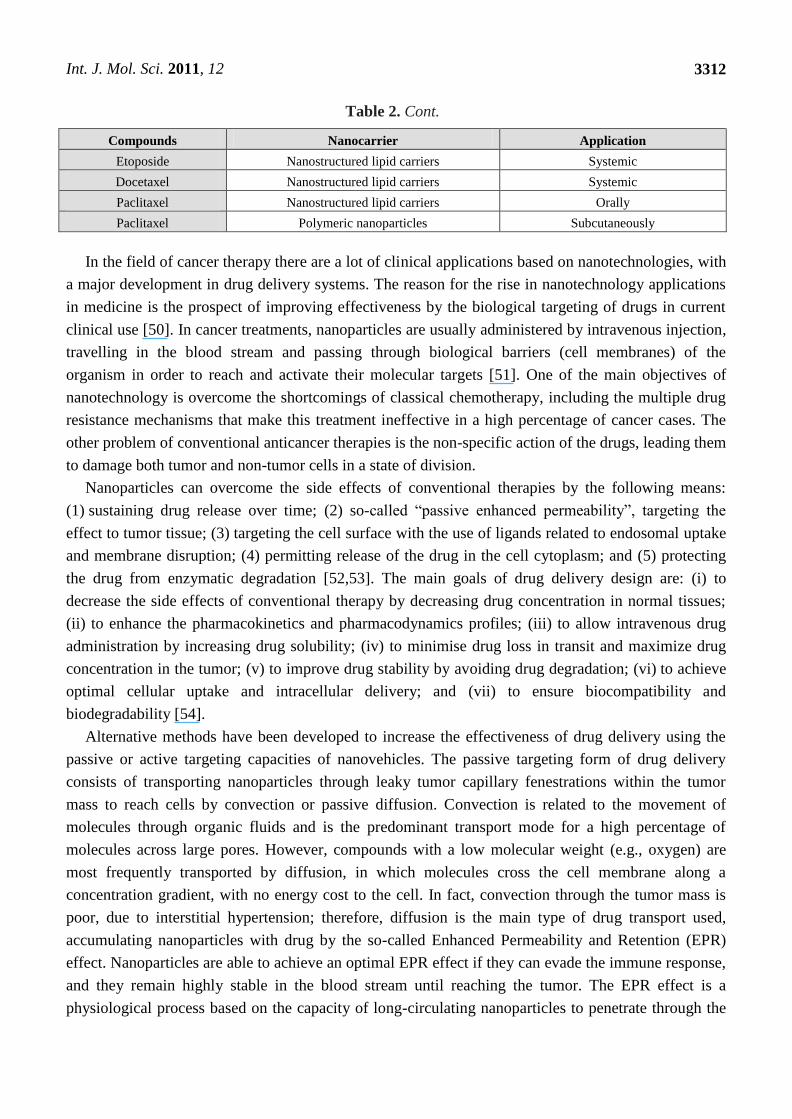
Etoposide Nanostructured lipid carriers Systemic
Docetaxel Nanostructured lipid carriers Systemic
Paclitaxel Nanostructured lipid carriers Orally
Paclitaxel Polymeric nanoparticles Subcutaneously
In the field of cancer therapy there are a lot of clinical applications based on nanotechnologies, with
a major development in drug delivery systems. The reason for the rise in nanotechnology applications
in medicine is the prospect of improving effectiveness by the biological targeting of drugs in current
clinical use [50]. In cancer treatments, nanoparticles are usually administered by intravenous injection,
travelling in the blood stream and passing through biological barriers (cell membranes) of the
organism in order to reach and activate their molecular targets [51]. One of the main objectives of
nanotechnology is overcome the shortcomings of classical chemotherapy, including the multiple drug
resistance mechanisms that make this treatment ineffective in a high percentage of cancer cases. The
other problem of conventional anticancer therapies is the non-specific action of the drugs, leading them
to damage both tumor and non-tumor cells in a state of division.
Nanoparticles can overcome the side effects of conventional therapies by the following means:
(1) sustaining drug release over time; (2) so-called ―passive enhanced permeability‖, targeting the
effect to tumor tissue; (3) targeting the cell surface with the use of ligands related to endosomal uptake
and membrane disruption; (4) permitting release of the drug in the cell cytoplasm; and (5) protecting
the drug from enzymatic degradation [52,53]. The main goals of drug delivery design are: (i) to
decrease the side effects of conventional therapy by decreasing drug concentration in normal tissues;
(ii) to enhance the pharmacokinetics and pharmacodynamics profiles; (iii) to allow intravenous drug
administration by increasing drug solubility; (iv) to minimise drug loss in transit and maximize drug
concentration in the tumor; (v) to improve drug stability by avoiding drug degradation; (vi) to achieve
optimal cellular uptake and intracellular delivery; and (vii) to ensure biocompatibility and
biodegradability [54].
Alternative methods have been developed to increase the effectiveness of drug delivery using the
passive or active targeting capacities of nanovehicles. The passive targeting form of drug delivery
consists of transporting nanoparticles through leaky tumor capillary fenestrations within the tumor
mass to reach cells by convection or passive diffusion. Convection is related to the movement of
molecules through organic fluids and is the predominant transport mode for a high percentage of
molecules across large pores. However, compounds with a low molecular weight (e.g., oxygen) are
most frequently transported by diffusion, in which molecules cross the cell membrane along a
concentration gradient, with no energy cost to the cell. In fact, convection through the tumor mass is
poor, due to interstitial hypertension; therefore, diffusion is the main type of drug transport used,
accumulating nanoparticles with drug by the so-called Enhanced Permeability and Retention (EPR)
effect. Nanoparticles are able to achieve an optimal EPR effect if they can evade the immune response,
and they remain highly stable in the blood stream until reaching the tumor. The EPR effect is a
physiological process based on the capacity of long-circulating nanoparticles to penetrate through the
Int. J. Mol. Sci. 2011, 12
3313
leaky tumor-formed vasculature and degrade within the tumor, releasing the drug and achieving a local
concentration in solid tumors that is several-fold higher than that obtained with free drugs [55].
Very high local concentrations of drug-loaded nanoparticles have been obtained within tumors,
from 10- to 50-fold higher in comparison to normal tissues in a time period of 1–2 days. The
achievement of these drug concentrations in the tumor requires nanoparticles to possess the following
characteristics: (a) nanoparticle size between 10 and 100 nm; (b) a neutral or anionic nanoparticle
surface charge to prevent elimination by the kidneys; and (c) the ability to avoid opsonization and
phagocytosis, which destroy foreign material via the reticuloendothelial system [54,56].
Active targeting using nanoparticles as the delivery system allows a specific area of the body to be
targeted, avoiding one of the drawbacks of current chemotherapy, i.e., toxic effects in non-malignant
organs [57]. Studies are being carried out on the attachment of targeting ligands on the nanoparticle
surface, enabling specific binding of the nanoparticle to receptors on the tumor cell surface. Ligands
that bind to specific overexpressed receptors are selected. The types of ligand used include antibodies,
antibody fragments or non-antibody (peptidic) ligands. Over the past few years, considerable interest
has developed in the numerous antigens present on tumor cells. For instance, HER2 receptor has been
proposed as a therapeutic target. HER2 gene, also known as c-erbB2 and neu, encodes a 185-kDa
transmembrane glycoprotein receptor that belongs to the ErbB family of growth factor receptors with
intrinsic tyrosine kinase activity, whose members exist in homodimer and heterodimer form when
activated [58]. Trastuzumab is a humanized antibody designed to specifically recognize the HER-2
receptor and was approved by the FDA in 1998 for the treatment of metastatic breast cancer. Different
studies have shown that normal cells can express moderate amounts of HER2 target antigen, while it is
overexpressed in tumors in certain patients, and this difference between healthy tissues and tumors
allows the effective use of this antibody in patients [59].
Fibroblast growth factor receptors (FGFRs) have also become potential targets for drug delivery
and cancer therapy. Fibroblast growth factors (FGFs) are small peptide growth factors that play an
important role in tumor growth and angiogenesis because of their high affinity to heparin. FGFRs are
upregulated versus normal tissues in numerous tumor cells and tumor neovasculature in situ [60]. One
candidate as a targeting ligand for tumor cells is the peptide KRTGQYKLC (bFGFp), which could
interact with FGFR1 by binding to bFGF. The epidermal growth factor receptor (EGFR) is a receptor
tyrosine kinase over-expressed on many human cancer cells surface, making it a target for anticancer
drug delivery. Liposomes targeting this receptor promoted the effective intracellular delivery of
doxorubicin to tumor cells, yielding superior anti-tumor effects in different in vivo assays. In previous
studies, the peptide GE11 was identified as a novel ligand with high affinity towards EGFR and proved
effective to mediate targeted liposome delivery to EGFR-positive tumors in vivo [61]. Investigations
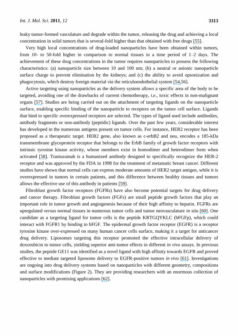
are ongoing into drug delivery systems based on nanoparticles with different geometry, compositions
and surface modifications (Figure 2). They are providing researchers with an enormous collection of
nanoparticles with promising applications [62].
Int. J. Mol. Sci. 2011, 12
3314
Figure 2. Some drug delivery systems.
2.2.1. Micelles
A micelle is an aggregate of amphiphilic surfactant molecules spontaneously created on immersion
in water, usually forming spherical vesicles. They contain a hydrophobic core in which hydrophobic
substances, such as pharmacological compounds, can be introduced for release in different parts of the
organism. A micelle typically comprises a hydrophilic or polar charged ―head‖ group and a
hydrophobic ―tail‖, normally composed of a hydrocarbonated portion of long fatty acids. The
characteristics of the surfactants that make up the micelle determine its size [63].
2.2.2. Nanoemulsions
Nanosized emulsions are composed of a mixture of two-phase insoluble liquids, in which vesicles
in the dispersed phase are surrounded by the continuous phase. Different types of surfactants are used
to stabilize the emulsion, preventing the dispersed phase from coalescing into a macroscopic
phase [64]. Many emulsions used in drug delivery systems can be formulated on a nanosize scale. In
these cases, the aqueous phase conform the continuous phase, and the drug is often carried in (or is
itself) the non-aqueous liquid phase of the emulsion. Generally, the surfactant molecules used to
stabilize micelles are the same as those forming the structural part of the nanoemulsions [65].
Int. J. Mol. Sci. 2011, 12
3315
2.2.3. Solid Nanoparticles
This structure presents different characteristic features such as: (a) the material present in the centre
of the particle forms a solid core at least at room temperature; (b) Solid nanoparticles not necessarily
present in a spherical form. The geometry may change, presenting angular forms. This characteristic is
particularly visible if the nanoparticle consists of crystals of a protein or another therapeutic agent. To
avoid flocculation a surfactant is usually required to stabilize the nanoparticle [65].
2.2.4. Dendrimers
These are usually regularly-branched three-dimensional structures with a treelike form and a
molecule as the central core. Branching units emerge from the central molecule by polymerization or
are synthesized from the periphery and terminate at the central molecule. Branch lengths have steric
limitations and dendrimer forms are sphere-shaped, with low molecular size but high molecular
weight. These structures are used to transport drugs in two ways: (a) by attaching drug molecules to
functional groups on the dendrimer surface; or (b) by enclosing the drugs in the dendritic channels
within the sphere. The well-defined structure, size monodispersity, surface functionalization capability
and stability of these nanoparticles make them a promising vehicle system for various agents (e.g.,
genes and anticancer drugs) by complexation or encapsulation [66,67]. One example is the formation
of dendrimers with polymers such as polyamidoamine (PAMAM), which confer stability, availability
and tolerability. The drug is contained in a central cavity and can be entrapped in channels between
dendrons [23].
2.2.5. Liposomes
The effectiveness of liposomes as drug vehicles is related to their pharmokinetics and depends on
the physicochemical conditions, e.g., size, surface charge, membrane lipid packing, steric stabilization,
dose, and administration route [68]. A major advantage of liposomes is their long persistence in the
blood, favoring the delivery of their contents to target tissues. Liposomes with diameters of
100–300 µm accumulate around tumors and are not subject to rapid clearance by the
reticuloendothelial system [69]. Liposomes are vesicular nanostructures formed by a lipid bilayer
composed of phospholipid and cholesterol molecules (structural components of cell membranes)
characterized by extended, two-dimensional and clearly separated hydrophilic and hydrophobic
regions. The hydrophilic portions of bilayer lipids are directed towards aqueous phases (external and
internal), whereas the hydrophobic portions of both lipid layers are directed towards one another,
forming an internal hydrophilic compartment that can encapsulate water-soluble drugs [70]. Actually
there is a type of liposome well studied in clinical, the pegylated liposomes, which forms a water shell
on the liposomal surface and provides a steric barrier to the liposomes for avoiding interactions with
plasma proteins, resulting in escape from trapping by the reticuloendothelial system [71]. One example
is liposomal-encapsulated doxorubicin, which has an elimination half-life of 55 h in comparison to
only 0.2 h for doxorubicin in free form. The drugs are solubilized and protected from enzymatic
degradation and inactivation either by physical entrapment within the nanoparticle or by conjugation
Int. J. Mol. Sci. 2011, 12
3316
with constituent components. Encapsulation into the liposomal carrier also causes a significant
reduction in the most significant adverse side effect of doxorubicin, such as cardiotoxicity [72].
2.3. Regenerative Medicine
Tissue engineering brings together principles and innovations from engineering and the life sciences
for the improvement, repair or replacement of tissue/organ function. Since its inception, this
multidisciplinary field has been governed by the generic concept of combining cell, scaffold (artificial
extracellular matrix) and bioreactor technologies in the design and fabrication of neo-tissues/organs [73].
Every tissue or organ in our body is composed of parenchymal cells (functional cells) and
mesenchymal cells (support cells) contained within an extracellular matrix to form a
microenvironment, and these microenvironments collectively form our tissues and organs. In terms of
the development and maintenance of tissues and organs, our body is the ―bioreactor‖, exposing the
microenvironment of the cell and extracellular matrix to biomechanical forces and biochemical signals.
The ultimate goal is to enable the body (cellular components) to heal itself by introducing a tissue
engineered scaffold that the body recognizes as ―self‖ and uses to regenerate ―neo-native‖ functional
tissues [74]. Furthermore, the demand for organs for transplantation far exceeds the supply, and the
construction of organs by regenerative therapy has been presented as a promising option to address this
deficit. Nanotechnology has the potential to provide instruments that can accelerate progress in the
engineering of organs [75]. Achievement of the more ambitious goals of regenerative medicine
requires control over the underlying nanostructures of the cell and extracellular matrix. Cells, typically
microns in diameter, are composed of numerous nanosized components that all work together to create
a highly organized, self-regulating machine. Cell-based therapies, especially those based on stem cells,
have generated considerable excitement in the media and scientific communities and are among the
most promising and active areas of research in regenerative medicine [76]. The pace of research could
be accelerated by the creation of multi-functional tools to improve the monitoring and modification of
cell behavior. While nanomedicine is primarily focused on cancer-related research, the application of
nanotechnology has considerable potential in cell-based therapies for regenerative medicine, e.g., in
localizing, recruiting and labeling stem cells to begin the regeneration process [75].
3. Conclusions
Nanotechnology is an emerging interdisciplinary field that combines biology, chemistry and
engineering. It is expected to lead to major advances towards individualized medicine, improving the
sensitivity and specificity of existing techniques to discover and detect biomarkers and developing
novel nanodiagnostic instruments. This would allow earlier and more personalized diagnosis and
therapy, improving the effectiveness of drug treatments and reducing side effects. In addition,
nanoparticles are a promising platform technology for the synthesis of molecular-specific
contrast agents.
Int. J. Mol. Sci. 2011, 12
3317
Acknowledgements
The study was supported by the Science and Innovation Ministry (Project no. SAF2009-12295).
The authors are grateful to ―Younes Taferssiti Zarouila‖ for the excellent linguistic assistance.
References
1. Appenzeller, T. The man who dared to think small. Science 1991, 254, 1300–1301.
2. Kang, Y.S.; Risbud, S.; Rabolt, J.F.; Stroeve, P. Synthesis and characterization of nanometer-size
Fe3O4 and g-Fe2O3 particles. Chem. Mater. 1996, 8, 2209–2211.
3. Pankhurst, Q.A.; Connoly, J.; Jones, S.K. Applications of magnetic nanoparticles in biomedicine.
J. Phys. D. Appl. Phys. 2003, 36, 167.
4. Dobson, J. Gene therapy progress and prospects: magnetic nanoparticle-based gene delivery.
Gene Ther. 2006, 13, 283–287.
5. Rudge, S.; Peterson, C.; Vessely, C.; Koda, J.; Stevens, S.; Catterall, L. Adsorption and
desorption of chemotherapeutic drugs from a magnetically targeted carrier (MTC). J. Control.
Release 2001, 74, 335–340.
6. Mody, V.V.; Siwale, R.; Singh, A.; Mody, H.R. Introduction to metallic nanoparticles. J. Pharm.
Bioallied. Sci. 2010, 2, 282–289.
7. Zhang, L.; Gu, F.X.; Chan, J.M.; Wang, A.Z.; Langer, R.S.; Farokhzad, O.C. Nanoparticles in
medicine: Therapeutic applications and developments. Clin. Pharmacol. Ther. 2008, 83, 761–769.
8. Surendiran, A.; Sandhiya, S.; Pradhan, S.C.; Adithan, C. Novel applications of nanotechnology in
medicine. Indian J. Med. Res. 2009, 130, 689–701.
9. Zarbin, M.A.; Montemagno, C.; Leary, J.F.; Ritch, R. Nanotechnology in ophthalmology. Can. J.
Ophthalmol. 2010, 45, 457–476.
10. Jain, K.K. Nanotechnology in clinical laboratory diagnostics. Clin. Chim. Acta 2005, 358, 37–54.
11. Jain, K.K. Nanodiagnostics: Application of nanotechnology in molecular diagnostics. Expert Rev.
Mol. Diagn. 2003, 3, 153–161.
12. Singh, R.P.; Oh, B.K.; Ch, J.W. Application of peptide nucleic acid towards development of
nanobiosensor arrays. Bioelectrochemisty 2010, 79, 153–161.
13. European Technology Platform on Nanomedicine. Nanotechnology for Health. Vision Paper and
Basis for a Strategic Research Agenda for Nanomedicine. Eur. Comm. 2005, 1, 1–39.
14. Debouck, C.; Goodfellow, P.N. DNA microarrays in drug discovery and development. Nat.
Genet. 1999, 21, 48–50.
15. Cole, K.A.; Krizman, D.B.; Emmert-Buck, M.R. Chuaqui, R.F.; Bonner, R.F.; Best, C.J. The
genetics of cancer: A 3D model. Nat. Genet.1999, 21, 38–41.
16. Gerhold, D.L.; Jensen, R.V.; Gullans, S.R. Better therapeutics through microarrays. Nat. Genet.
2002, 32, 547–551.
17. Heller, R.A.; Schena, M.; Chai, A.; Shalon, D.; Bedilion, T.; Gilmore, J.; Woolley, D.E.; Davis, R.W.
Discovery and analysis of inflammatory disease-related genes using cDNA microarrays. Proc.
Natl. Acad. Sci. USA 1997, 94, 2150–2155.
Int. J. Mol. Sci. 2011, 12
3318
18. McCaffrey, T.A.; Fu, C.; Du, B.; Eksinar, S.; Kent, K.C.; Bush, H., Jr.; Kreiger, K.; Rosengart, T.;
Cybulsky, M.I.; Silverman, E.S.; Collins, T. High-level expression of Egr-1 and Egr-1-inducible
genes in mouse and human atherosclerosis. J. Clin. Invest. 2000, 105, 653–662.
19. Perou, C.M.; Jeffrey, S.S.; Van de Rijn, M.; Rees, C.A.; Eisen, M.B.; Ross, D.T;
Pergamenschikov, A.; Williams, C.F.; Zhu, S.X.; Lee, J.C.; Lashkari, D.; Shalon, D.;
Brown, P.O.; Botstein, D. Distinctive gene expression patterns in human mammary epithelial cells
and breast cancers. Proc. Natl. Acad. Sci. USA 1999, 96, 9212–9217.
20. Perou, C.M.; Sørlie, T.; Eisen, M.B.; Rijn, M.; Jeffrey, S.S.; Rees, C.A.; Pollack, J.R.; Ross, D.T.;
Johnsen, H.; Akslen, L.A.; Fluge, O.; Pergamenschikov, A.; Williams, C.; Zhu, S.X.; Lønning, P.E.;
Børresen-Dale, A.L.; Brown, P.O.; Botstein, D. Molecular portraits of breast tumours. Nature
2000, 406, 747–752.
21. Alon, U.; Barkai, N.; Notterman, D.A.; Gish, K.; Ybarra, S.; Mack, D.; Levine, A.J. Broad
patterns of gene expression revealed by clustering analysis of tumor and normal colon tissues
probed by oligonucleotide arrays. Proc. Natl. Acad. Sci. USA 1999, 96, 6745–6750.
22. Kaminski, N.; Allard, J.D.; Pittet, J.F.; Zuo, F.; Griffiths, M.J.; Morris, D.; Huang, X.; Sheppard, D.;
Heller, R.A. Global analysis of gene expression in pulmonary fibrosis reveals distinct programs
regulating lung inflammation and fibrosis. Proc. Natl. Acad. Sci. USA 2000, 97, 1778–1783.
23. Stoughton, R.B. Applications of DNA microarrays in biology. Annu. Rev. Biochem. 2005, 74,
53–82.
24. Plomin, R.; Schalkwyk, L.C. Microarrays. Dev. Sci. 2007, 10, 19–23.
25. Schulze, A.; Downward, J. Navigating gene expression using microarrays—a technology review.
Nat. Cell Biol. 2001, 3, 190–195.
26. Kennedy, G.C.; Matsuzaki, H.; Dong, S.; Liu, W.M.; Huang, J.; Liu, G.; Su, X.; Cao, M.;
Chen, W.; Zhang, J.; Liu, W.; Yang, G.; Di, X.; Ryder, T.; He, Z.; Surti, U.; Phillips, M.S.;
Boyce-Jacino, M.T.; Fodor, S.P.; Jones, K.W. Large-scale genotyping of complex DNA. Nat.
Biotechnol. 2003, 21, 1233–1237.
27. Ulger, C.; Toruner, G.A.; Alkan, M.; Mohammed, M.; Damani, S.; Kang, J.; Galante, A.;
Aviv, H.; Soteropoulos, P.; Tolias, P.P.; Schwalb, M.N.; Dermody, J.J. Comprehensive
genome-wide comparison of DNA and RNA level scan using microarray technology for
identification of candidate cancer-related genes in the HL-60 cell line. Cancer Genet. Cytogenet.
2003, 147, 28–35.
28. MacBeath, G. Protein microarrays and proteomics. Nat. Genet. 2002, 32, 526–532.
29. Braunschweig, T.; Chung, J.Y.; Hewitt, S.M. Tissue microarrays: Bridging the gap between
research and the clinic. Expert Rev. Proteomics 2005, 2, 325–336.
30. Clarke, P.A.; Poele, R.; Wooster, R.; Workman, P. Gene expression microarray analysis in cancer
biology, pharmacology, and drug development: Progress and potential. Biochem. Pharmacol.
2001, 62, 1311–1336.
31. Jayapal, M.; Melendez, J.A. DNA microarray technology for target identification and validation.
Clin. Exp. Pharmacol. Physiol. 2006, 33, 496–503.
32. Ghallab, Y.H.; Badawy, W. Lab-on-a-Chip: Techniques, Circuits, and Biomedical Applications;
Artech House: Norwood, MA, USA, 2010; pp. 1-220.
Int. J. Mol. Sci. 2011, 12
3319
33. Kim, J.; Byun, D.; Mauk, M.G.; Bau, H.H. A disposable, self-contained PCR chip. Lab Chip
2009, 9, 606–612.
34. Lab-on-a-Chip: Miniaturized Systems for (Bio)Chemical Analysis and Synthesis, 2nd ed.;
Oosterbroek, E., Van den Berg, A., Eds.; Elsevier Science: Amsterdam, The Netherlands, 2003;
pp. 1-402.
35. Stybayeva, G.; Mudanyali, O.; Seo, S.; Silangcruz, J.; Macal, M.; Ramanculov, E.; Dandekar, S.;
Erlinger, A.; Ozcan, A.; Revzin, A. Lensfree holographic imaging of antibody microarrays for
high-throughput detection of Leukocyte numbers and function. Anal. Chem. 2010, 82, 3736–3744.
36. Weissleder, R.; Pittet, M.J. Imaging in the era of molecular oncology. Nature 2008, 452, 580–589.
37. Wang, W.; Eddy, R.; Condeelis, J. The cofilin pathway in breast cancer invasion and metastasis.
Nat. Rev. Cancer 2007, 7, 429–440.
38. Weissleder, R. Molecular imaging in cancer. Science 2006, 312, 1168–1171.
39. Jaffer, F.A.; Weissleder, R. Molecular imaging in the clinical arena. JAMA 2005, 293, 855–862.
40. Love, Z.; Wang, F.; Dennis, J.; Awadallah, A.; Salem, N.; Lin, Y.; Weisenberger, A.; Majewski, S.;
Gerson, S.; Lee, Z. Imaging of mesenchymal stem cell transplant by bioluminescence and PET.
J. Nucl. Med. 2007, 48, 2011–2020.
41. MacLaren, D.C.; Toyokuni, T.; Cherry, S.R.; Barrio, J.R; Phelps, M.E.; Herschman, H.R.;
Gambhir, S.S. PET imaging of transgene expression. Biol. Psychiatry 2000, 48, 337–348.
42. Kraitchman, D.L.; Heldman, A.W.; Atalar, E.; Amado, L.C.; Martin, B.J; Pittenger, M.F.;
Hare, J.M.; Bulte, J.W. In vivo magnetic resonance imaging of mesenchymal stem cells in
myocardial infarction. Circulation 2003, 107, 2290–2293.
43. Long, C.M.; Bulte, J.W. In vivo tracking of cellular therapeutics using magnetic resonance
imaging. Expert Opin. Biol. Ther. 2009, 9, 293–306.
44. Kraitchman, D.L.; Bulte, J.W. Imaging of stem cells using MRI. Basic Res. Cardiol. 2008, 103,
105–113.
45. Kalish, H.; Arbab, A.S.; Miller, B.R.; Lewis, B.K.; Zywicke, H.A.; Bulte, J.W.; Bryant, L.H., Jr;
Frank, J.A. Combination of transfection agents and magnetic resonance contrast agents for
cellular imaging: Relationship between relaxivities, electrostatic forces, and chemical
composition. Magn. Reson. Med. 2003, 50, 275–282.
46. Muja, N.; Bulte, J. Magnetic resonance imaging of cells in experimental models. Prog. Nucl.
Reson. Spectrosc. 2009, 55, 61–77.
47. Salata, O. Applications of nanoparticles in biology and medicine. J. Nanobiotechnol. 2004, 2, 3–9.
48. Bruchez, M.J.; Moronne, M.; Gin, P.; Weiss, S.; Alivisatos, A.P. Semiconductor nanocrystals as
fluorescent biological labels. Science 1998, 281, 2013–2016.
49. Vizirianakis, I.S. Nanomedicine and personalized medicine toward the application of
pharmacotyping in clinical practice to improve drug-delivery outcomes. Nanomedicine 2011, 7,
11–17.
50. Kawasaki, E.S.; Player, A. Nanotechnology, nanomedicine, and the development of new,
effective therapies for cancer. Nanomedicine 2005, 1, 101–109.
51. Veiseh, O.; Kievit, F.; Ellenbogen, R.G.; Zhang, M. Cancer Cell Invasion: Treatment and
Monitoring Opportunities in Nanomedicine. Adv. Drug. Deliv. Rev. 2011, in press.
Int. J. Mol. Sci. 2011, 12
3320
52. Maeda, H.; Wu, J.; Sawa, T.; Matsumura, Y.; Hori, K. Tumor vascular permeability and the EPR
effect in macromolecular therapeutics: A review. J. Control. Release 2000, 65, 271–284.
53. Farokhzad, O.C.; Langer, R. Impact of nanotechnology on drug delivery. ACS Nano 2009, 3, 16–20.
54. Danhier, F.; Feron, O.; Préat, V. To exploit the tumor microenvironment: Passive and active tumor
targeting of nanocarriers for anti-cancer drug delivery. J. Control. Release 2010, 148, 135–146.
55. Tamilvanan, S. Formulation of multifunctional oil-in-water nanosized emulsions for active and
passive targeting of drugs to otherwise inaccessible internal organs of the human body. Int. J.
Pharm. 2009, 381, 62–76.
56. Maruyama, K. Intracellular targeting delivery of liposomal drugs to solid tumors based on EPR
effects. Adv. Drug. Deliv. Rev. 2011, in press.
57. Arias, J.L.; Gallardo, V.; Ruiz, M.A.; Delgado, A.V. Magnetite/poly(alkylcyanoacrylate)
(core/shell) nanoparticles as 5-Fluorouracil delivery systems for active targeting. Eur. J. Pharm.
Biopharm. 2008, 69, 54–63.
58. Murphy, C.G.; Modi, S. HER2 breast cancer therapies: A review. Biologics Targets Ther. 2009, 3,
289–301.
59. Cirstoiu-Hapca, A.; Bossy-Nobs, L.; Buchegger, F.; Gurny, R.; Delie, F. Differential tumor cell
targeting of anti-HER2 (Herceptin®) and anti-CD20 (Mabthera®) coupled nanoparticles. Int. J.
Pharm. 2007, 331, 190–196.
60. Li, D.; Yu, H.; Huang, H.; Shen, F.; Wu, X.; Li, J.; Wang, J.; Cao, X.; Wang, Q.; Tang, G. FGF
receptor-mediated gene delivery using ligands coupled to polyethylenimine. J. Biomater. Appl.
2007, 22, 163–180.
61. Song, S.; Liu, D.; Peng, J.; Sun, Y.; Li, Z.; Gu, J.; Xu, Y. Peptide ligand-mediated liposome
distribution and targeting to EGFR expressing tumor in vivo. Int. J. Pharm. 2008, 363, 155–161.
62. Serda, R.E.; Godin, B.; Blanco, E.; Chiappini, C.; Ferrari, M. Multi-stage delivery nano-particle
systems for therapeutic applications. Biochim. Biophys. Acta 2011, 1810, 317–329.
63. Rangel-Yagui, C.O.; Pessoa, A.; Tavares, L.C. Micellar solubilization of drugs. J. Pharm. Pharm.
Sci. 2005, 8, 147–163.
64. Sarker, D.K. Engineering of nanoemulsions for drug delivery. Curr. Drug Deliv. 2005, 2,
297–310.
65. Husseini, G.A.; PittMicelles, W.G. Nanoparticles for ultrasonic drug and gene delivery. Adv.
Drug. Deliv. Rev. 2008, 60, 1137–1152.
66. Haley, B.; Frenkel, E. Nanoparticles for drug delivery in cancer treatment. Urol Oncol. 2008, 26,
57–64.
67. Hughes, G.A. Nanostructure-mediated drug delivery. Nanomedicine 2005, 1, 22–30.
68. Maruyama, K. Intracellular targeting delivery of liposomal drugs to solid tumors based on EPR
effects. Adv. Drug. Deliv. Rev. 2011, 63, 161–169.
69 Obata, Y.; Tajima, S.; Takeoka, S. Evaluation of pH-responsive liposomes containing amino
acid-based zwitterionic lipids for improving intracellular drug delivery in vitro and in vivo.
J. Control. Release 2010, 142, 267–276.
70. Huang, S. Liposomes in ultrasonic drug and gene delivery. Adv. Drug. Deliv. Rev. 2008, 60,
1167–1176.
Int. J. Mol. Sci. 2011, 12
3321
71. Koide, H.; Asai, T.; Hatanaka, K.; Akai, S.; Ishii, T.; Kenjo, E.; Ishida, T.; Kiwada, H.;
Tsukada, H.; Oku, N. T cell-independent B cell response is responsible for ABC phenomenon
induced by repeated injection of PEGylated liposomes. Int. J. Pharm. 2010, 392, 218–223.
72. Sakamoto, J.H.; Van de Ven, A.L.; Godin, B.; Blanco, E.; Serda, R.E.; Grattoni, A.; Ziemys, A.;
Bouamrani, A.; Hu, T.; Ranganathan, S.I.; De Rosa, E.; Martinez, J.O.; Smid, C.A.;
Buchanan, R.M.; Lee, S.; Srinivasan, S.; Landry, M.; Meyn, A.; Tasciotti, E.; Liu, X.;
Decuzzi, P.; Ferrari, M. Enabling individualized therapy through nanotechnology. Pharmacol.
Res. 2010, 62, 57–89.
73. Shekaran, A.; Garcia, A.J. Nanoscale engineering of extracellular matrix-mimetic bioadhesive
surfaces and implants for tissue engineering. Biochim. Biophys. Acta 2011, 1810, 350–360.
74. Barnes, C.P.; Sell, S.A.; Boland, E.D.; Simpson, D.G.; Bowlin, G.L. Nanofiber technology:
Designing the next generation of tissue engineering scaffolds. Adv. Drug. Deliv. Rev. 2007, 59,
1413–1433.
75. Atala, A.; Lanza, R.; Nerem, R.; Thomson, J.A. Principles of Regenerative Medicine, 2nd ed.;
Academic Press: London, UK, 2011; pp. 733–1105.
76. Prakash, S.; Khan, A.; Paul, A. Nanoscaffold based stem cell regeneration therapy: Recent
advancement and future potential. Expert Opin. Biol. Ther. 2010, 10, 1649–1661.
© 2011 by the authors; licensee MDPI, Basel, Switzerland. This article is an open access article
distributed under the terms and conditions of the Creative Commons Attribution license
(http://creativecommons.org/licenses/by/3.0/).
Related Documents