Nanomedicine and Protein Misfolding Diseases Alexey V. Kransnoslobodtsev 1,♦ , Luda S. Shlyakhtenko 1,♦ , Egor Ukraintsev 1 , Tatiana O. Zaikova 2 , John F.W. Keana 2 , and Yuri L. Lyubchenko 1,* 1 Department of Pharmaceutical Sciences, University of Nebraska Medical Center, Omaha, NE, 68198 2 Department of Chemistry, 1253 University of Oregon, Eugene, OR 97403-1253 Abstract Misfolding and self assembly of proteins in nano-aggregates of different sizes and morphologies (nano-ensembles, primarily nanofilaments and nano-rings) is a complex phenomenon that can be facilitated, impeded, or prevented, by interactions with various intracellular metabolites, intracellular nanomachines controlling protein folding and interactions with other proteins. A fundamental understanding of molecular processes leading to misfolding and self-aggregation of proteins involved in various neurodegenerative diseases will provide critical information to help identify appropriate therapeutic routes to control these processes. An elevated propensity of misfolded protein conformation in solution to aggregate with the formation of various morphologies impedes the use of traditional physical chemical approaches for studies of misfolded conformations of proteins. In our recent alternative approach, the protein molecules were tethered to surfaces to prevent aggregation and AFM force spectroscopy was used to probe the interaction between protein molecules depending on their conformations. It was shown that formation of filamentous aggregates is facilitated at pH values corresponding to the maximum of rupture forces. In this paper, a novel surface chemistry was developed for anchoring of amyloid β (Aβ) peptides at their N-terminal moieties. The use of the site specific immobilization procedure allowed to measure the rupture of Aβ - Aβ contacts at single molecule level. The rupture of these contacts is accompanied by the extension of the peptide chain detected by a characteristic elasto-mechanical component of the force- distance curves. Potential applications of the nanomechanical studies to understanding the mechanisms of development of protein misfolding diseases are discussed. Keywords Nanotechnology; amyloids; neurodegenerative diseases; protein aggregation; protein folding; intermolecular forces; AFM; Alzheimer’s disease 1. Introduction Misfolding and self assembly of proteins in nano-aggregates of different sizes and morphologies (nano-ensembles, primarily nanofilaments and nano-rings) is a common thread linking a number of important human health problems. Particularly, recent studies highlighted increasing recognition of the public health importance of protein deposition diseases, including neurodegenerative disorders such as Parkinson’s disease, Down’s syndrome, Alzheimer’s and Huntington’s diseases, systemic and localized amyloidoses, transmissible encephalopathies *Corresponding author: Yuri Lyubchenko, Department of Pharmaceutical Sciences, University of Nebraska Medical Center, 986025 Nebraska Medical Center, Omaha, NE 68198-6025, Phone: 402-559-1971 – office, 402-559-1973 - lab 1, 402-559-1974 - lab 2, Fax: 402-559-9543, E-mail:[email protected]. ♦ These authors contributed equally to this work NIH Public Access Author Manuscript Nanomedicine. Author manuscript; available in PMC 2006 January 25. Published in final edited form as: Nanomedicine. 2005 December ; 1(4): 300–305. NIH-PA Author Manuscript NIH-PA Author Manuscript NIH-PA Author Manuscript

Welcome message from author
This document is posted to help you gain knowledge. Please leave a comment to let me know what you think about it! Share it to your friends and learn new things together.
Transcript

Nanomedicine and Protein Misfolding Diseases
Alexey V. Kransnoslobodtsev1,♦, Luda S. Shlyakhtenko1,♦, Egor Ukraintsev1, Tatiana O.Zaikova2, John F.W. Keana2, and Yuri L. Lyubchenko1,*1 Department of Pharmaceutical Sciences, University of Nebraska Medical Center, Omaha, NE,68198
2 Department of Chemistry, 1253 University of Oregon, Eugene, OR 97403-1253
AbstractMisfolding and self assembly of proteins in nano-aggregates of different sizes and morphologies(nano-ensembles, primarily nanofilaments and nano-rings) is a complex phenomenon that can befacilitated, impeded, or prevented, by interactions with various intracellular metabolites, intracellularnanomachines controlling protein folding and interactions with other proteins. A fundamentalunderstanding of molecular processes leading to misfolding and self-aggregation of proteins involvedin various neurodegenerative diseases will provide critical information to help identify appropriatetherapeutic routes to control these processes. An elevated propensity of misfolded proteinconformation in solution to aggregate with the formation of various morphologies impedes the useof traditional physical chemical approaches for studies of misfolded conformations of proteins. Inour recent alternative approach, the protein molecules were tethered to surfaces to preventaggregation and AFM force spectroscopy was used to probe the interaction between proteinmolecules depending on their conformations. It was shown that formation of filamentous aggregatesis facilitated at pH values corresponding to the maximum of rupture forces. In this paper, a novelsurface chemistry was developed for anchoring of amyloid β (Aβ) peptides at their N-terminalmoieties. The use of the site specific immobilization procedure allowed to measure the rupture ofAβ - Aβ contacts at single molecule level. The rupture of these contacts is accompanied by theextension of the peptide chain detected by a characteristic elasto-mechanical component of the force-distance curves. Potential applications of the nanomechanical studies to understanding themechanisms of development of protein misfolding diseases are discussed.
KeywordsNanotechnology; amyloids; neurodegenerative diseases; protein aggregation; protein folding;intermolecular forces; AFM; Alzheimer’s disease
1. IntroductionMisfolding and self assembly of proteins in nano-aggregates of different sizes andmorphologies (nano-ensembles, primarily nanofilaments and nano-rings) is a common threadlinking a number of important human health problems. Particularly, recent studies highlightedincreasing recognition of the public health importance of protein deposition diseases, includingneurodegenerative disorders such as Parkinson’s disease, Down’s syndrome, Alzheimer’s andHuntington’s diseases, systemic and localized amyloidoses, transmissible encephalopathies
*Corresponding author: Yuri Lyubchenko, Department of Pharmaceutical Sciences, University of Nebraska Medical Center, 986025Nebraska Medical Center, Omaha, NE 68198-6025, Phone: 402-559-1971 – office, 402-559-1973 - lab 1, 402-559-1974 - lab 2, Fax:402-559-9543, E-mail:[email protected].♦These authors contributed equally to this work
NIH Public AccessAuthor ManuscriptNanomedicine. Author manuscript; available in PMC 2006 January 25.
Published in final edited form as:Nanomedicine. 2005 December ; 1(4): 300–305.
NIH
-PA Author Manuscript
NIH
-PA Author Manuscript
NIH
-PA Author Manuscript

and several other neurodegenerative disorders (1). The first and perhaps most importantelements in most neurodegenerative processes are misfolded and aggregated proteins. Theseare inducers of cellular stress and activators of immunity in neurodegenerative diseases andaffect neuronal dysfunction and loss. Altogether, the accumulation of abnormal protein nano-ensembles exerts toxicity by disrupting intracellular transport, overwhelming proteindegradation pathways, and/or disturbing vital cell functions. In addition, the formation ofinclusion bodies is known to represent a major problem in the production of recombinanttherapeutic proteins (2). Formulation of these therapeutic proteins into delivery systems andtheir in vivo delivery are often complicated by protein association (3). Finally, since proteinrefolding is frequently accompanied by the transient association of partially foldedintermediates, the propensity to self-assemble in nano-ensembles is considered a generalcharacteristic of the majority of partially folded proteins (4-7). Thus, protein foldingabnormalities and subsequent events underlie a multitude of pathologies and difficulties withprotein therapeutic applications. Current demographic trends indicate that need formacromolecule therapeutics for age-related and other degenerative disorders and will be at theforefront of future medical developments. The field of medicine therefore can be dramaticallyadvanced by establishing a fundamental understanding of key factors leading to misfoldingand self-assembly of the various protein folding pathologies.
Misfolded conformations of proteins differ from folded and other aberrant proteinconformations by their increased propensity to interact with each other leading to the formationof nano-aggregates. The structure of individual protein molecules within well orderedaggregates can be partially elucidated by traditional structural techniques, including X-raycrystallography, NMR, circular dichroism, fluorescence, and IR spectroscopies (reviewed in(8,9)). However, none of these techniques is capable of sensing the misfolded conformationof the protein prior to aggregation. Apparently, the conformation of misfolded proteinpreceding the aggregation is different from what it is in aggregates, but to what extent it isdifferent causing the disease is not clear. The vast majority of current experimental approachesto analyze protein misfolding and aggregation are based on traditional, ensemble techniquesthat do not allow investigators to distinguish between conformational changes in individualprotein molecules and the changes induced by protein-protein interaction. This leaves open thequestion of the effect of these factors on folding/misfolding of an individual protein molecule.Current tools do not allow for the measurement of protein interaction forces or the kinetics ofinterconversion among different protein conformations. The ability to measure theseparameters is critical to achieve a quantitative understanding of protein misfolding andaggregation at the nanoscale level. Thus, new experimental tools and approaches are crucialfor understanding the protein misfolding phenomenon.
It is well established that AFM force spectroscopy is an important method for probing thestability of protein conformations (10) and can provide high resolution structural information(11,12). In this context, we have recently demonstrated that force spectroscopy is capable ofprobing misfolded protein conformations (13). To measure single molecule interactionsproteins were anchored on the mica surface and the AFM probe, and the interaction betweenthe tethered molecules was measured after bringing them together by approaching the tip tothe surface. The major advantage of probing single molecules is to enable intimate detectionand quantitative characterization of conformational states without interference fromneighboring molecules. Using this approach, we were able to monitor directly the strength ofthe interprotein interaction depending on the protein conformation. The data obtained showthat the interaction between all the proteins increases sharply with a decrease in pH. AFMimaging showed that at pH values corresponding to maximum interprotein interaction, the rateof protein aggregation increases dramatically.
Kransnoslobodtsev et al. Page 2
Nanomedicine. Author manuscript; available in PMC 2006 January 25.
NIH
-PA Author Manuscript
NIH
-PA Author Manuscript
NIH
-PA Author Manuscript

We extended these studies by introducing a novel surface chemistry that allowed us to attachamyloid β peptides selectively at N-termini and in so doing to detect the interactions betweenindividual peptide molecules. These studies illustrate the power of nanomechanical tools forunraveling molecular mechanisms of protein misfolding and fibrils formation.
2. Materials and MethodsProcedure for the Synthesis of Maleimide-Pentaethylene Glycol-Silatrane (Maleimide-PEG-Silatrane)
Maleimide-PEG-silatrane 8 was prepared as shown in Scheme 1. 3-Cyclopentadienylpropyltrimethoxysilane 2 was prepared as an inseparable mixture of twodouble bond isomers, 2a and 2b by reaction of one equivalent 3-iodopropyltrimethoxysilane1 (Gelest) with one equivalent of sodium cyclopentadienide in tetrahydrofuran (Aldrich) atroom temperature (14). For convenience, only isomer 2a and its derivatives are shown insubsequent reactions. Heating a solution of 2 and one equivalent of triethanolamine 3 in sodiummethoxide-methanol followed by evaporation of the solvent led to cyclopentadiene silatrane4 (15). Separately, bis-maleimide 7 was prepared from chemically pure pentaethylene glycol5 by way of a bis Mitsunobu reaction with maleimide (6) (16). Silatrane 4 undergoes a rapidDiels-Alder reaction when treated with a 2-fold excess of bis-maleimide 7 giving the desiredmaleimide-PEG-silatrane 8. Unlike the lower trialkoxysilanes, silatrane 8 is reasonably stablein water solution. 1H NMR spectra of maleimide-PEG-silatrane 8 in D2O at ambienttemperature with pD values ranging from 2 to 7 indicate that 90-95% of the starting 8 remainsafter 48 h.
Amyloid β immobilization.Aβ peptide modified with additional cysteine at N-terminus was purchased from AnaSpec(SanJose, CA) and was used without further purification. The peptide powder was dissolvedin 1 ml of dd water and 20 μl aliquots were stored at −80°C until use. Buffered solution(Na2CO3, pH 9.8) of the protein was treated with 10 mM solution of TCEP-HCl (Tris(2-carboxyethyl)phosphine- hydrochloride) at 25°C for 10 min to reduce thiols of the protein. Inorder to immobilize protein the functionalization of mica surface and AFM tips was performedprior to the treatment with peptide. The immobilization chemistry is shown in Scheme 2. Afreshly cleaved mica surface (cartoon 9) or UV cleaned AFM tips were immersed in an aqueoussolution of silatrane 8 (167 μM) for 3 hours and then rinsed with dd water, leading to a surfacecontaining immobilized maleimide units as depicted in cartoon 10. Surface 10 was then treatedwith buffered solution of the amyloid peptide-cys (11.3 μM) for 1 hour after reduction of thiols.The terminal cysteine residue reacts with the maleimide units on the surface leading to thecovalently immobilized peptide as shown in cartoon 11. Unreacted maleimide moieties werequenched with buffered (Na2CO3, pH 9.8) 10 mM β -mercaptoethanol solution by 10 mintreatment. Mica and tips with attached Aβ peptide were left overnight in Na2CO3 buffer, pH9.8 to prevent peptide aggregation and dissociate aggregates which possibly formed at earlystages of surface modification (13).
Measurements of Aβ - Aβ interactions.Force curves were collected on Nanoscope Multimode AFM with IIIa controller equipped withthe Picoforce controller (Veeco Metrology Inc., Santa Barbara, CA) and on Molecular ForceProbe 3D instrument (MFP 3D, Asylum Research Inc., Santa Barbara, CA). The ramp sizeused was 300 nm with a 0.25 Hz frequency, an application force of 50 pN for both instruments.The noise level for force curves was obtained by using the averaging procedure in which themean force value was calculated over 10 consecutive data points of the force-distance curvethat was subtracted from the value obtained for the next set calculated after shifting the 10 datapoint interval to another data point. Silicon nitride cantilevers with nominal values of spring
Kransnoslobodtsev et al. Page 3
Nanomedicine. Author manuscript; available in PMC 2006 January 25.
NIH
-PA Author Manuscript
NIH
-PA Author Manuscript
NIH
-PA Author Manuscript

constants in the range of 0.01-0.04 N/m were used. Spring constants for each cantilever wereobtained using thermal method (17) with the MFP-3D instrument (Asylum Research, CA).Extension and retraction curves were acquired initially in Na2CO3 buffer with 180mM NaClpH 9.8. The buffers were exchanged to lower pH each time starting with the buffer at pH 9.8.The other buffers, appearing in the order used, were pH 5.6: 20mM phosphate buffer saline(PBS), pH 3.7: 10mM sodium acetate, and pH 2.0: 50mM KCl and 13mM HCl. NaCl wasadded to each buffer to keep constant the ionic strength, 180mM.
3. RESULTS AND DISCUSSIONTo achieve this goal and to measure the interprotein interactions, we used the AFM forcespectroscopy approach, in which proteins to be anchored to the substrate surface and the AFMtip (Fig. 1). We have recently tested this idea using the immobilization approach in whichprotein was covalently linked to amino-functionalized mica and Si3N4 AFM probes viaglutaraldehyde crosslinking (13). Although this procedure allowed us to study various proteinsusing the same immobilization strategy, glutaraldehyde reaction takes place at all amine-containing moieties (e.g., lysine and arginine) in addition to the protein N-termini. To avoidthis ambiguity in the protein tethering that can complicate the single molecule analysis ofrupture events, in this paper we used an alternative approach, in which Aβ peptide was boundto the surface at N-terminal only. We took advantage of the fact that Aβ peptide does not containcysteins and attached this aminoacid to the N-terminus of the peptide allowing for the use ofthiol-specific chemistry for the protein immobilization. We selected N-terminus because N-terminal part of the peptide is not critically involved in various conformational transitions ofthis peptide including the formation of β-sheet conformations within amyloid fibrils (e.g.,(17)). We have recently proposed the surface chemistry based on the use of silatrane forfunctionalization of mica for imaging various biological samples (15,18). An important featureof the silatrane chemistry is that immobilization of such a large molecule as an oligonucleotidecan be performed in one step if silatrane with attached oligonucleotide is used. We synthesizedmaleimide-PEG-silatrane (MAS; see Methods section, Scheme 1) to functionalize mica (MASmica) and the AFM tip surfaces with maleimide capable of binding SH-terminated peptide(Methods section, Scheme 2).
Aβ peptide was immobilized on both MAS mica and the tip surfaces and the interaction betweenthe peptide molecules was measured by a series of approach-retraction cycles. The results formeasuring interprotein interaction at pH 9.8 and 5.6 did not reveal clear rupture events. Thisfinding is consistent with our earlier observations (13). One of the typical force-distance curvesfor the experiments performed at pH5.6 is shown in Fig. 2A. There are some variations (steps)along the curves that were measured as described in Methods. The maximal steps for eachforce-distance curve were plotted as histogram shown as an insert in Fig. 2A. The force rupturevalues are centered around the value 10±2 pN that is close to the noise level for the force-distance curves for the cantilevers with the spring constants used in these experiments. Similaranalysis for the data obtained at pH 9.8 provided the value 9±2 pN.
The data for pH3.7 are dramatically different. One of the force-distance curves is shown inFig. 2B. An intensive peak at the beginning of the force curve (a green arrow) correspondingto the short-range adhesion forces between the tip and mica surfaces typically observed atacidic pH values (13,19) is accompanied by a rupture event (indicated with a red arrowhead)with the step size ~70 pN that is substantially larger the noise level. The histogram for the dataobtained from the analysis of series of such events is shown as insert (i) in Fig. 2B. The meanvalue for this distribution is 45±2 pN. Note that control experiments with Aβ-peptide at themica surface or the tip only did not show these types of interactions. Given a finite size of theMAS-PEG linkers and similar size of the peptide (ca. 15 nm total), it is reasonable to attributethe second peak to the rupture of a single Aβ-Aβ pair. Additional evidence supporting this
Kransnoslobodtsev et al. Page 4
Nanomedicine. Author manuscript; available in PMC 2006 January 25.
NIH
-PA Author Manuscript
NIH
-PA Author Manuscript
NIH
-PA Author Manuscript

interpretation comes from the analysis of this particular part of the force-distance curve. Theprofile of this curve is typical for stretching of polymer of various kinds, and this section canbe fitted by the worm like chain (WLC). The WLC fit of this force distance curve is shown asblack smooth curve to the left of the rupture event above the arrowhead. Such an analysisallowed us to measure the entire stretching range. The value 15.8 nm was obtained for theexperiment shown in Fig. 2B. The histogram for the measurements of this value performedover ~50 various rupture events is shown in Fig. 2C. The mean value is 18±1 nm. Note thatthis number is close to the expected value (15 nm) for a full extension of the N-terminus ofAβ peptide that is not involved in the peptide folding and 5 PEG moieties of the MAS linker.Additional proofs for these estimates were obtained from the experiments with stretching ofthe surface immobilized Aβ peptide via MAS chemistry with the tip terminated withglutaraldehyde. During the tip-surface contact covalent bonds between the residues with freeamino groups of the peptide (the closest to the N-terminal is Lys-16) and the tip immobilizedglutaraldehyde can be formed allowing the stretching the Aβ peptide molecule between theanchors. The stretching experiments showed the elastic-type profile of the force curve with thestretch value 7.8-8.5 nm; this value is very close to the half of the stretching effect observedfor stretching Aβ-Aβ pairs.
Similar force spectroscopy data were obtained for pulling experiments for pH2.0 and the meanvalue for the rupture forces was 41±1 pN (Fig. 2C) that is very close to the value obtained atpH 3.7. Overall, the data on the pH-dependent interaction obtained are consistent with ourearlier data (13) suggesting the Aβ-peptide misfolding at acidic pH, but here we were able tomeasure the interactions between individual Aβ-Aβ pairs.
According to the solid state NMR data (17,20), 1-40 Aβ peptides within the fibril adopt parallel,in-register β structures in which residues 12–24 and 30–40 adopt β-strand backboneconformations. Parallel orientation of these monomer units provides the stability of the amyloidfibrils. If misfolded conformation of Aβ peptides is close to the peptide conformation in fibrils,the force spectroscopy data provide the quantitative measure for the intermolecular interactionstabilizing the fibrils. Recently Kellermayer et al (21) applied AFM force spectroscopy toanalysis forces between amyloid β molecules within amyloid fibrils. In this approach, the fibrilis firmly attached to the surface and the AFM tip pulls the filament from the fibril after makinga strong contact to the fibril. These experiments provided a minimal force ~30 pN for unzippingone 1-40 Aβ- peptide from the fibril. This value is very close to 45 pN rupture force obtainedin our single molecule force spectroscopy experiments suggesting that misfolded Aβ-peptideconformations is close to one within the amyloid fibril. It is instructive to compare the ruptureforces measured in this paper values with the stability of other systems in order to obtain abetter understanding about the nature of forces stabilizing Aβ-Aβ pairs. The value ~40pN wasobtained in the paper (22) for the 15 bp DNA duplex rupture in the pulling experiments witha very similar approach-retraction rate. This force is rather close to ~45 pN value for the ruptureof Aβ-Aβ interaction, opening a speculation on the similarities of forces for stabilization ofDNA helix and the Aβ-Aβ duplex.
ConclusionsThe principal unanswered question for all protein misfolding disorders including Alzheimer’sdisease is why natively soluble proteins (some of them with a defined tertiary structure)undergo refolding in conformations dramatically changing their interactions. Available dataon the structure of nano-particles formed by various misfolded proteins suggest that thepathways for their misfolding can be different. For example, our recent data (23) showed thatnanofibrils formed by Exon 1 htt in contrast to the majority of other nanofibrils have extensivebranched morphology. At the same time, self-assembly patterns for Aβ peptide and α synucleinprotein have many similarities despite the 3-fold difference in size. Neither protein is folded
Kransnoslobodtsev et al. Page 5
Nanomedicine. Author manuscript; available in PMC 2006 January 25.
NIH
-PA Author Manuscript
NIH
-PA Author Manuscript
NIH
-PA Author Manuscript

under native conditions. Lysozyme belongs to the family of proteins with well-definedsecondary and tertiary structures, but forms nanofibrils with morphology similar to Aβ peptideand α synuclein. What does account for this? We believe that understanding the misfoldedstructures and properties of these proteins and pathways of their formation will answer thisfundamental question. Given the widespread nature of protein misfolding and the structuralsimilarity of protein nano-aggregates, one can hypothesize that mechanisms for the formationof misfolded conformations and/or pathways leading to misfolding within cells have commonfeatures.
The tools currently available to researchers and clinicians essentially provide a population-based picture of a living-dynamical system. However, it is evident that the mechanisms of lifeand of disease involve dynamic molecular-scale interactions—most of which occurs undersome level of physical stress. An apparently simple process, as examined by existingtechniques, may actually have many steps and a variety of important intermediate states—eachwith their own unique dynamics and response to stress. The intermediate states can be veryimportant as control mechanisms that involve several pathways. Thus, it is vitally importantto incorporate available nanoscience methods capable of probing protein conformations andweak intermolecular interactions into biomedical studies. The current work illustrates thatsingle molecule biophysics techniques are capable of probing transiently existingintermolecular interactions. We believe that further exploiting new and established singlemolecule methods, in conjunction with complementary ensemble techniques will provide thescientific resources needed to develop innovative nanomedicine approaches for understanding,diagnosis, prevention, and cure of protein misfolding diseases.
Acknowledgements
We thank O Kiselyova for the help at the initial stage of this work. Supported by grants from M. J. Fox Parkinson’sFoundation, NIH (GM 62235, 1 PN1 EY016593-01) and NATO (LST.CLG.980194) to YLL.
References1. Dobson CM. Principles of protein folding, misfolding and aggregation. Semin Cell Dev Biol
2004;15:3–16. [PubMed: 15036202]2. Fink AL. Protein aggregation: folding aggregates, inclusion bodies and amyloid. Fold Des 1998;3:R9–
23. [PubMed: 9502314]3. Demidov VV. Proper Refolding helps express “difficult” proteins. Drug Discovery and Development
2004;7:41–47.4. Ptitsyn OB. Molten globule and protein folding. Adv Protein Chem 1995;47:83–229. [PubMed:
8561052]5. Uversky VN, Karnoup AS, Khurana R, Segel DJ, Doniach S, Fink AL. Association of partially-folded
intermediates of staphylococcal nuclease induces structure and stability. Protein Sci 1999;8:161–73.[PubMed: 10210194]
6. Segel DJ, Eliezer D, Uversky V, Fink AL, Hodgson KO, Doniach S. Transient dimer in the refoldingkinetics of cytochrome c characterized by small-angle X-ray scattering. Biochemistry 1999;38:15352–9. [PubMed: 10563821]
7. Dobson CM. Protein folding and misfolding. Nature 2003;426:884–90. [PubMed: 14685248]8. Dobson CM. Experimental investigation of protein folding and misfolding. Methods 2004;34:4–14.
[PubMed: 15283911]9. Dobson CM. Structural biology: prying into prions. Nature 2005;435:747–9. [PubMed: 15944684]10. Rief M, Gautel M, Oesterhelt F, Fernandez JM, Gaub HE. Reversible unfolding of individual titin
immunoglobulin domains by AFM. Science 1997;276:1109–12. [PubMed: 9148804]11. Engel A, Lyubchenko Y, Muller D. Atomic force microscopy: a powerful tool to observe biomolecules
at work. Trends Cell Biol 1999;9:77–80. [PubMed: 10087624]
Kransnoslobodtsev et al. Page 6
Nanomedicine. Author manuscript; available in PMC 2006 January 25.
NIH
-PA Author Manuscript
NIH
-PA Author Manuscript
NIH
-PA Author Manuscript
12. Muller DJ, Baumeister W, Engel A. Controlled unzipping of a bacterial surface layer with atomicforce microscopy. Proc Natl Acad Sci U S A 1999;96:13170–4. [PubMed: 10557292]
13. McAllister C, Karymov M, Kawano Y, Lushnikov AY, Mikheikin A, Uversky VN, et al. ProteinInteractions and Misfolding Analyzed by AFM Force Spectroscopy. J mol Biol. 2005; in press:
14. Booth BL, Ofunne GC, Stacey C, Tai PJT. Silica-supported cyclopentadienyl-Rodium(I), -Cobalt(I),and -Titanium (IV) complexes. Journal of Organometallic Chemistry 1986;315:143–156.
15. Shlyakhtenko LS, Gall AA, Filonov A, Cerovac Z, Lushnikov A, Lyubchenko YL. Silatrane-basedsurface chemistry for immobilization of DNA, protein-DNA complexes and other biologicalmaterials. Ultramicroscopy 2003;97:279–87. [PubMed: 12801681]
16. Wang X, Zhou D, Rayment T, Abell C. Systematic nanoscale manipulation of surface reactions: novelapproach for constructing three-demensional nanostructure. Chemical Communications 2003;4:474–475. [PubMed: 12638956]
17. Tycko R. Insights into the amyloid folding problem from solid-state NMR. Biochemistry2003;42:3151–9. [PubMed: 12641446]
18. Lyubchenko YL. DNA structure and dynamics: an atomic force microscopy study. Cell BiochemBiophys 2004;41:75–98. [PubMed: 15371641]
19. Vezenov DV, Aleksandr Noy, Lawrence F. Rozsnyai, and Charles M. Lieber. Force Titrations andIonization State Sensitive Imaging of Functional Groups in Aqueous Solutions by Chemical ForceMicroscopy. J Am Chem Soc 1997;119:2006–2015.
20. Tycko R, Ishii Y. Constraints on supramolecular structure in amyloid fibrils from two-dimensionalsolid-state NMR spectroscopy with uniform isotopic labeling. J Am Chem Soc 2003;125:6606–7.[PubMed: 12769550]
21. Kellermayer MS, Grama L, Karsai A, Nagy A, Kahn A, Datki ZL, et al. Reversible mechanicalunzipping of amyloid beta-fibrils. J Biol Chem 2005;280:8464–70. [PubMed: 15596431]
22. Strunz T, Oroszlan K, Schafer R, Guntherodt HJ. Dynamic force spectroscopy of single DNAmolecules. Proc Natl Acad Sci U S A 1999;96:11277–82. [PubMed: 10500167]
23. Dahlgren PR, Bankston J, Holden T, Karymov MA, Thumfort P, Ingram VM, et al. Atomic ForceMicroscopy Analysis of the Huntington Protein Nanofibril Formation. Nanomedicine:Nanotechnology, Biology and Medicine 2005;1:52–57.
Kransnoslobodtsev et al. Page 7
Nanomedicine. Author manuscript; available in PMC 2006 January 25.
NIH
-PA Author Manuscript
NIH
-PA Author Manuscript
NIH
-PA Author Manuscript

Figure 1.Schemes illustrating the rupture of the protein in folded (low affinity, green arrows) andmisfolded conformations (high affinity with multiple contact points-red wave shape).
Kransnoslobodtsev et al. Page 8
Nanomedicine. Author manuscript; available in PMC 2006 January 25.
NIH
-PA Author Manuscript
NIH
-PA Author Manuscript
NIH
-PA Author Manuscript

Figure 2A.A typical force curve obtained for probing the Aβ-Aβ interaction at pH 5.6. The histogramsummarizing the rupture force measurements is inserted into the figure.
Kransnoslobodtsev et al. Page 9
Nanomedicine. Author manuscript; available in PMC 2006 January 25.
NIH
-PA Author Manuscript
NIH
-PA Author Manuscript
NIH
-PA Author Manuscript

Figure 2B.A force curve for the Aβ-Aβ interaction at pH 3.7. The arrowhead (red) points to the Aβ-Aβrupture event. Insert (i) shows the histogram for the Aβ-Aβ rupture force values. Black lineabove the arrowhead shows the WLC approximation fit.
Kransnoslobodtsev et al. Page 10
Nanomedicine. Author manuscript; available in PMC 2006 January 25.
NIH
-PA Author Manuscript
NIH
-PA Author Manuscript
NIH
-PA Author Manuscript
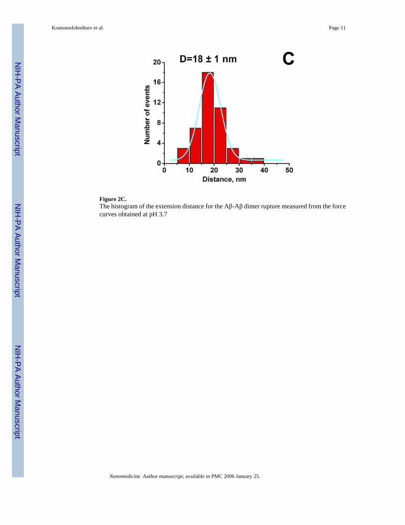
Figure 2C.The histogram of the extension distance for the Aβ-Aβ dimer rupture measured from the forcecurves obtained at pH 3.7
Kransnoslobodtsev et al. Page 11
Nanomedicine. Author manuscript; available in PMC 2006 January 25.
NIH
-PA Author Manuscript
NIH
-PA Author Manuscript
NIH
-PA Author Manuscript

Figure 2D.The histogram of the force rupture values for the Aβ-Aβ dimer measured from the force curvesobtained at pH.2.
Kransnoslobodtsev et al. Page 12
Nanomedicine. Author manuscript; available in PMC 2006 January 25.
NIH
-PA Author Manuscript
NIH
-PA Author Manuscript
NIH
-PA Author Manuscript

Scheme 1.Synthesis of maleimide-PEG-silatrane 8
Kransnoslobodtsev et al. Page 13
Nanomedicine. Author manuscript; available in PMC 2006 January 25.
NIH
-PA Author Manuscript
NIH
-PA Author Manuscript
NIH
-PA Author Manuscript

Scheme 2.Immobilization of amyloid peptide-cys on a functionalized mica surface
Kransnoslobodtsev et al. Page 14
Nanomedicine. Author manuscript; available in PMC 2006 January 25.
NIH
-PA Author Manuscript
NIH
-PA Author Manuscript
NIH
-PA Author Manuscript
Related Documents