Small Molecule Therapeutics Nanolipolee-007, a Novel Nanoparticle-Based Drug Containing Leelamine for the Treatment of Melanoma Raghavendra Gowda 1,2,3,4 , SubbaRao V. Madhunapantula 1 , Arati Sharma 1,2,3,4 , Omer F. Kuzu 1,2 , and Gavin P. Robertson 1,2,3,4,5,6,7 Abstract Malignant melanoma is a difficult cancer to treat due to the rapid development of resistance to drugs targeting single proteins. One response to this observation is to identify single pharmacologic agents that, due to a unique mechanism of action, simultaneously target multiple key pathways involved in melanoma development. Leelamine has been identified as functioning in this manner but has poor bioavailability in animals and causes lethality when administered intravenously. Therefore, a nano- liposomal-based delivery system has been developed, called Nanolipolee-007, which stably loads 60% of the compound. The nanoparticle was as effective at killing melanoma cells as leelamine dissolved in DMSO and was more effective at killing cultured melanoma compared with normal cells. Mechanis- tically, Nanolipolee-007 inhibited PI3K/Akt, STAT3, and MAPK signaling mediated through inhibition of cholesterol transport. Nanolipolee-007 inhibited the growth of preexisting xenografted melanoma tumors by an average of 64% by decreasing cellular proliferation, reducing tumor vascularization, and increasing cellular apoptosis, with negligible toxicity. Thus, a unique clinically viable nanoparticle-based drug has been developed containing leelamine for the treatment of melanoma that acts by inhibiting the activity of major signaling pathways regulating the development of this disease. Mol Cancer Ther; 13(10); 2328–40. Ó2014 AACR. Introduction Malignant melanoma is one of the most difficult cancers to treat due to the development of recurrent resistant disease (1). Although encouraging recent studies showed promising results with Zelboraf, a selective V600E B-Raf kinase inhibitor, development of drug resistance remains a problem (2). To address this concern, one approach has been to develop liposomes containing novel agents spe- cifically targeting multiple key pathways or signaling cascades, leading to recurrent resistance disease develop- ment (3). Loading of drugs into nanoparticles to circumvent bioavailability, toxicity, or lethality can be used to over- come limitations of standard drug delivery systems, which involve nonspecific targeting and biodistribution with low solubility at a modest therapeutic index (4, 5). Nanoparticles can be designed with optimal size and surface characteristics to increase the circulation time and biodistribution (6). One advantage of nanoparticles is the enhanced permeability and retention (EPR) effect that enables nanoparticles to accumulate in tumors at much higher concentrations than in normal tissue (6). The EPR effect occurs because the vasculature of tumors is poorly developed and leaky, enabling nanoparticles and macro- molecules to preferentially accumulate and concentrate in tumors (7). Natural or synthetic polymers or lipids can be used as drug delivery vectors (7). Lipid-based delivery vehicles have established track records, and have suitable biological properties, including biocompatibility, biode- gradability, and the ability to accommodate both hydro- philic and hydrophobic drugs (8). Liposomes primarily consist of phospholipid amphi- philes that self assemble to form bilayer membranes. Liposomes have been extensively studied as drug-deliv- ery systems and are used as biocompatible carriers of drugs, enzymes, peptides, vaccines, imaging agents, and/or genetic material (9). Depending on the proper- ties of the drugs, they can be loaded into the aqueous or lipid compartments of liposomes and can be effective for delivering the therapeutic cargo into tumors in 1 Department of Pharmacology, The Pennsylvania State University College of Medicine, Hershey, Pennsylvania. 2 The Penn State Melanoma Center, The Pennsylvania State University College of Medicine, Hershey, Penn- sylvania. 3 Penn State Melanoma Therapeutics Program, The Pennsylvania State University College of Medicine, Hershey, Pennsylvania. 4 Foreman Foundation for Melanoma Research, The Pennsylvania State University College of Medicine, Hershey, Pennsylvania. 5 Department of Pathology, The Pennsylvania State University College of Medicine, Hershey, Penn- sylvania. 6 Department of Dermatology, The Pennsylvania State University College of Medicine, Hershey, Pennsylvania. 7 Department of Surgery, The Pennsylvania State University College of Medicine, Hershey, Pennsylvania. Note: Supplementary data for this article are available at Molecular Cancer Therapeutics Online (http://mct.aacrjournals.org/). Corresponding Author: Gavin P. Robertson, Department of Pharmacol- ogy, The Pennsylvania State University College of Medicine, 500 University Drive, Hershey, PA 17033. Phone: 717-531-8098; Fax: 717-531-0480; E-mail: [email protected] doi: 10.1158/1535-7163.MCT-14-0357 Ó2014 American Association for Cancer Research. Molecular Cancer Therapeutics Mol Cancer Ther; 13(10) October 2014 2328

Welcome message from author
This document is posted to help you gain knowledge. Please leave a comment to let me know what you think about it! Share it to your friends and learn new things together.
Transcript

Small Molecule Therapeutics
Nanolipolee-007, a Novel Nanoparticle-Based DrugContaining Leelamine for the Treatment of Melanoma
Raghavendra Gowda1,2,3,4, SubbaRao V. Madhunapantula1, Arati Sharma1,2,3,4, Omer F. Kuzu1,2, andGavin P. Robertson1,2,3,4,5,6,7
AbstractMalignant melanoma is a difficult cancer to treat due to the rapid development of resistance to drugs
targeting single proteins. One response to this observation is to identify single pharmacologic agents
that, due to a unique mechanism of action, simultaneously target multiple key pathways involved in
melanoma development. Leelamine has been identified as functioning in this manner but has poor
bioavailability in animals and causes lethality when administered intravenously. Therefore, a nano-
liposomal-based delivery system has been developed, called Nanolipolee-007, which stably loads 60% of
the compound. The nanoparticle was as effective at killing melanoma cells as leelamine dissolved in
DMSO and was more effective at killing cultured melanoma compared with normal cells. Mechanis-
tically, Nanolipolee-007 inhibited PI3K/Akt, STAT3, and MAPK signaling mediated through inhibition
of cholesterol transport. Nanolipolee-007 inhibited the growth of preexisting xenografted melanoma
tumors by an average of 64% by decreasing cellular proliferation, reducing tumor vascularization, and
increasing cellular apoptosis, with negligible toxicity. Thus, a unique clinically viable nanoparticle-based
drug has been developed containing leelamine for the treatment of melanoma that acts by inhibiting the
activity of major signaling pathways regulating the development of this disease. Mol Cancer Ther; 13(10);
2328–40. �2014 AACR.
IntroductionMalignantmelanoma is one of themost difficult cancers
to treat due to the development of recurrent resistantdisease (1). Although encouraging recent studies showedpromising results with Zelboraf, a selective V600EB-Rafkinase inhibitor, development of drug resistance remainsa problem (2). To address this concern, one approach hasbeen to develop liposomes containing novel agents spe-cifically targeting multiple key pathways or signalingcascades, leading to recurrent resistance disease develop-ment (3).
Loading of drugs into nanoparticles to circumventbioavailability, toxicity, or lethality can be used to over-come limitations of standard drug delivery systems,which involve nonspecific targeting and biodistributionwith low solubility at a modest therapeutic index (4, 5).Nanoparticles can be designed with optimal size andsurface characteristics to increase the circulation time andbiodistribution (6). One advantage of nanoparticles is theenhanced permeability and retention (EPR) effect thatenables nanoparticles to accumulate in tumors at muchhigher concentrations than in normal tissue (6). The EPReffect occurs because the vasculature of tumors is poorlydeveloped and leaky, enabling nanoparticles and macro-molecules to preferentially accumulate and concentrate intumors (7). Natural or synthetic polymers or lipids can beused as drug delivery vectors (7). Lipid-based deliveryvehicles have established track records, and have suitablebiological properties, including biocompatibility, biode-gradability, and the ability to accommodate both hydro-philic and hydrophobic drugs (8).
Liposomes primarily consist of phospholipid amphi-philes that self assemble to form bilayer membranes.Liposomes have been extensively studied as drug-deliv-ery systems and are used as biocompatible carriers ofdrugs, enzymes, peptides, vaccines, imaging agents,and/or genetic material (9). Depending on the proper-ties of the drugs, they can be loaded into the aqueous orlipid compartments of liposomes and can be effectivefor delivering the therapeutic cargo into tumors in
1Department of Pharmacology, The Pennsylvania State University Collegeof Medicine, Hershey, Pennsylvania. 2The Penn State Melanoma Center,The Pennsylvania State University College of Medicine, Hershey, Penn-sylvania. 3Penn State Melanoma Therapeutics Program, The PennsylvaniaState University College of Medicine, Hershey, Pennsylvania. 4ForemanFoundation for Melanoma Research, The Pennsylvania State UniversityCollege of Medicine, Hershey, Pennsylvania. 5Department of Pathology,The Pennsylvania State University College of Medicine, Hershey, Penn-sylvania. 6Department of Dermatology, The Pennsylvania State UniversityCollege of Medicine, Hershey, Pennsylvania. 7Department of Surgery, ThePennsylvania StateUniversityCollege ofMedicine, Hershey, Pennsylvania.
Note: Supplementary data for this article are available at Molecular CancerTherapeutics Online (http://mct.aacrjournals.org/).
Corresponding Author: Gavin P. Robertson, Department of Pharmacol-ogy, The Pennsylvania State University College ofMedicine, 500UniversityDrive, Hershey, PA 17033. Phone: 717-531-8098; Fax: 717-531-0480;E-mail: [email protected]
doi: 10.1158/1535-7163.MCT-14-0357
�2014 American Association for Cancer Research.
MolecularCancer
Therapeutics
Mol Cancer Ther; 13(10) October 20142328

animals (9). In addition, liposomes can be used asmodels for artificial cells that are able to mimic cellmembrane trafficking and studying drug movement(10). Currently, nanoliposomal-based therapeutics areused clinically and many are at various stages of clinicaldevelopment (3, 11).Recently, a cell-based screen of a natural product library
was undertaken to identify a pharmacologic agent thatcan decrease melanoma development by targeting thePI3K, STAT3, and MAPK signaling cascade, which isdetailed in the article by Gowda and colleagues (12).Leelamine-mediated inhibition of these cascades wasattributed to the inhibition of receptor-mediated endocy-tosis of receptor tyrosine kinases (RTK) causing aberrantaccumulationof theseproteins in theperinuclear region ofcells, which is detailed in the article by Kuzu and collea-gues (13). Using a combination of protein arrays andsystems biology followed by validation studies, leelaminewas found to inhibit the PI3K/Akt, STAT3, and MAPKpathways by disrupting cancer cell cholesterol transport.These are key driver pathways in melanoma cells consti-tutively activated in 50% to 70% of melanomas, function-ing to reduce cellular apoptosis, increase proliferation andaid the invasive processes, promotingmelanomaprogres-sion (14–16).Leelamine dissolved in DMSO and administered by
intraperitoneal injection, inhibited the growth of preexist-ing xenografted melanoma tumors by 60% without sig-nificant toxicity. However, due to poor bioavailability,toxicity and lethality of this agent when administeredintravenously, a stable PEGylated nanoliposomal deliv-ery system has been developed called Nanolipolee-007.The nanoparticle has an average size of 80 nmand is stablein saline for 1 year at 4�C. Nanolipolee-007 was 5.69-foldmore effective at killing melanoma than normal cells,decreased cellular proliferation, and triggered apoptosisthrough a G0–G1 block, resulting in fewer cells in theS-phase. Intravenously administered Nanolipolee-007had negligible toxicity and retarded existing xenograftmelanoma tumor growth by up to 60% without affectinganimalweight or organ function by decreasingmelanomacell proliferation, increasing apoptosis, and decreasingvascular development. Therefore, Nanolipolee-007 is apotentially clinically viable intravenous nanoparticle for-mulation that can be used to treat melanoma in mice andhas potential for use in humans.
Materials and MethodsCell lines and culture conditionsNormal human primary melanocytes FOM103 and
wild-type B-Raf containing SbCl2 (provided by Dr. Her-lyn between 2003 and 2005;Wistar Institute, Philadelphia,PA) were cultured as described (14). Human fibroblastFF2441 cells were provided by Dr. Craig Myers labbetween 2005 and 2006, Penn State College of Medicine,Hershey, PA. Mutant type B-Raf melanoma cell linesUACC 903 were provided from Mark Nelson between
1995 and 1999, University of Arizona, Tucson, AZ and1205 Lu provided by Dr. Herlyn between 2005 and 2006,Wistar Institute, Philadelphia, PA. Wild type B-Raf mel-anoma cell line containingC8161.Cl9was provided byDr.Danny Welch (2003), University of Kansas, Kansas City,KS andMelJuSo provided byDr. Judith Johnson (between1995 and 1999), Institute for Immunology, Germany. Celllines were maintained in a 37�C humidified 5% CO2
atmosphere incubator and periodically monitored forgenotypic characteristics, phenotypic behavior, andtumorigenic potential to confirm the cell line indentity(14).
Intravenous administration of free leelamine andliposomal leelamine
Four- to six-week-old female Athymic-Foxn1nu nudemice (Harlan) were injected intravenouslywith 30mg/kgbody weight leelamine dissolved in DMSO or Nanolipo-lee-007. Vehicle control animals were treated with DMSOor empty liposome. Animal mortality or hemolysis andcoagulation was recorded after 1 hour (n ¼ 3).
Generation of Nanolipolee-007Leelamine hydrochloride (Tocris Biosciences) was
encapsulated into a nanoliposome (called Nanolipo-lee-007) prepared by combining egg L-a-Phosphatidyl-choline (ePC) and 1,2-Dipalmitoyl-sn-Glycero-3-Phos-phoethanolamine-N- [Methoxy(Polyethylene glycol)-2000] ammonium salt (DPPE-PEG-2000) in chloroformat 80:20mol% and a final lipid concentration of 25mg/mLin buffer solution (Avanti Polar Lipids Inc). The samelipid formulation was found to load Abietic acid (SigmaChemical Co.) and an empty control nanoliposome wasalso made. 7.5 mg of leelamine hydrochloride and/orabietic acid (in methanol) was added to 1.0 mL of nano-liposome solution, resulting in a 22.6 mmol/L concen-tration. Mixture was then dried under nitrogen gas andresuspended in 0.9% saline at 60�C. Following rehydra-tion, the mixture was sonicated at 60�C for 30 minutesfollowed by extrusion at 60�C through a 100-nm poly-carbonate membrane using Avanti Mini Extruder(Avanti Polar Lipids Inc). The particle size and chargecharacteristics were determined using a Malvern Zeta-sizer (Malvern Instruments).
Characterization of Nanolipolee-007Loading efficacy. Amount of leelamine encapsulated
into the nanoliposome was measured by calculating theamount of tritium-labeled leelamine incorporated into thenanoparticle. Tritium-labeled leelamine was synthesizedby bromination, followed by replacing the bromine atomwith tritiumwith a specific activity of 25Ci/mmol (Amer-ican Radio Chemicals Inc; http://www.arc-inc.com/index.php). Dialysis and size exclusion chromatographywere used to calculate tritium-labeled leelamine loadingefficacy (17, 18). One milliliter of Nanolipolee-007 madewith tritium-labeled leelamine was placed into a 1� 5 cmlong dialysis membrane bag (molecular weight cutoff: 25
Targeting Melanoma with Nanolipolee-007
www.aacrjournals.org Mol Cancer Ther; 13(10) October 2014 2329
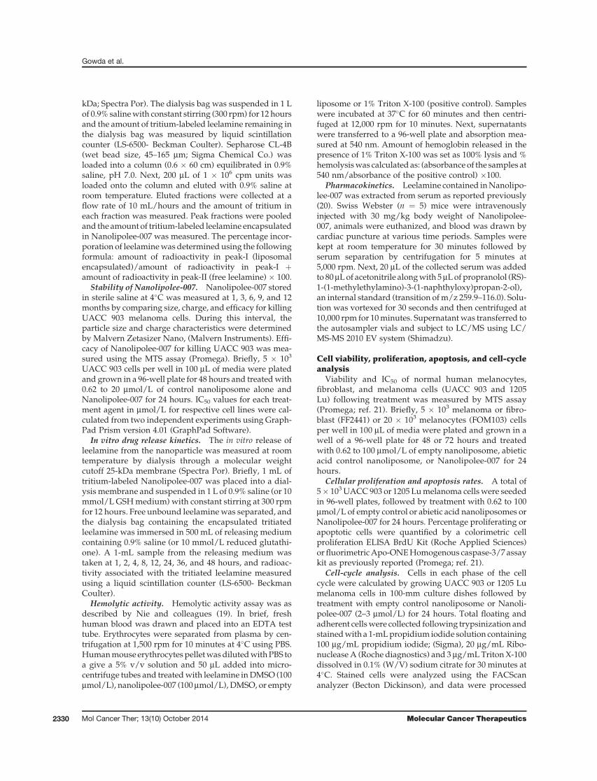
kDa; Spectra Por). The dialysis bag was suspended in 1 Lof 0.9% salinewith constant stirring (300 rpm) for 12 hoursand the amount of tritium-labeled leelamine remaining inthe dialysis bag was measured by liquid scintillationcounter (LS-6500- Beckman Coulter). Sepharose CL-4B(wet bead size, 45–165 mm; Sigma Chemical Co.) wasloaded into a column (0.6 � 60 cm) equilibrated in 0.9%saline, pH 7.0. Next, 200 mL of 1 � 106 cpm units wasloaded onto the column and eluted with 0.9% saline atroom temperature. Eluted fractions were collected at aflow rate of 10 mL/hours and the amount of tritium ineach fraction was measured. Peak fractions were pooledand the amount of tritium-labeled leelamine encapsulatedin Nanolipolee-007 was measured. The percentage incor-poration of leelaminewas determined using the followingformula: amount of radioactivity in peak-I (liposomalencapsulated)/amount of radioactivity in peak-I þamount of radioactivity in peak-II (free leelamine) � 100.
Stability of Nanolipolee-007. Nanolipolee-007 storedin sterile saline at 4�C was measured at 1, 3, 6, 9, and 12months by comparing size, charge, and efficacy for killingUACC 903 melanoma cells. During this interval, theparticle size and charge characteristics were determinedby Malvern Zetasizer Nano, (Malvern Instruments). Effi-cacy of Nanolipolee-007 for killing UACC 903 was mea-sured using the MTS assay (Promega). Briefly, 5 � 103
UACC 903 cells per well in 100 mL of media were platedand grown in a 96-well plate for 48 hours and treatedwith0.62 to 20 mmol/L of control nanoliposome alone andNanolipolee-007 for 24 hours. IC50 values for each treat-ment agent in mmol/L for respective cell lines were cal-culated from two independent experiments using Graph-Pad Prism version 4.01 (GraphPad Software).
In vitro drug release kinetics. The in vitro release ofleelamine from the nanoparticle was measured at roomtemperature by dialysis through a molecular weightcutoff 25-kDa membrane (Spectra Por). Briefly, 1 mL oftritium-labeled Nanolipolee-007 was placed into a dial-ysis membrane and suspended in 1 L of 0.9% saline (or 10mmol/L GSHmedium) with constant stirring at 300 rpmfor 12 hours. Free unbound leelaminewas separated, andthe dialysis bag containing the encapsulated tritiatedleelamine was immersed in 500 mL of releasing mediumcontaining 0.9% saline (or 10 mmol/L reduced glutathi-one). A 1-mL sample from the releasing medium wastaken at 1, 2, 4, 8, 12, 24, 36, and 48 hours, and radioac-tivity associated with the tritiated leelamine measuredusing a liquid scintillation counter (LS-6500- BeckmanCoulter).
Hemolytic activity. Hemolytic activity assay was asdescribed by Nie and colleagues (19). In brief, freshhuman blood was drawn and placed into an EDTA testtube. Erythrocytes were separated from plasma by cen-trifugation at 1,500 rpm for 10 minutes at 4�C using PBS.Humanmouse erythrocytes pelletwasdilutedwithPBS toa give a 5% v/v solution and 50 mL added into micro-centrifuge tubes and treatedwith leelamine inDMSO (100mmol/L), nanolipolee-007 (100 mmol/L), DMSO, or empty
liposome or 1% Triton X-100 (positive control). Sampleswere incubated at 37�C for 60 minutes and then centri-fuged at 12,000 rpm for 10 minutes. Next, supernatantswere transferred to a 96-well plate and absorption mea-sured at 540 nm. Amount of hemoglobin released in thepresence of 1% Triton X-100 was set as 100% lysis and %hemolysiswas calculatedas: (absorbanceof the samples at540 nm/absorbance of the positive control) �100.
Pharmacokinetics. Leelamine contained inNanolipo-lee-007 was extracted from serum as reported previously(20). Swiss Webster (n ¼ 5) mice were intravenouslyinjected with 30 mg/kg body weight of Nanolipolee-007, animals were euthanized, and blood was drawn bycardiac puncture at various time periods. Samples werekept at room temperature for 30 minutes followed byserum separation by centrifugation for 5 minutes at5,000 rpm. Next, 20 mL of the collected serum was addedto 80mLof acetonitrile alongwith 5mLof propranolol (RS)-1-(1-methylethylamino)-3-(1-naphthyloxy)propan-2-ol),an internal standard (transition ofm/z 259.9–116.0). Solu-tion was vortexed for 30 seconds and then centrifuged at10,000 rpm for 10minutes. Supernatantwas transferred tothe autosampler vials and subject to LC/MS using LC/MS-MS 2010 EV system (Shimadzu).
Cell viability, proliferation, apoptosis, and cell-cycleanalysis
Viability and IC50 of normal human melanocytes,fibroblast, and melanoma cells (UACC 903 and 1205Lu) following treatment was measured by MTS assay(Promega; ref. 21). Briefly, 5 � 103 melanoma or fibro-blast (FF2441) or 20 � 103 melanocytes (FOM103) cellsper well in 100 mL of media were plated and grown in awell of a 96-well plate for 48 or 72 hours and treatedwith 0.62 to 100 mmol/L of empty nanoliposome, abieticacid control nanoliposome, or Nanolipolee-007 for 24hours.
Cellular proliferation and apoptosis rates. A total of5� 103UACC903 or 1205 Lumelanoma cellswere seededin 96-well plates, followed by treatment with 0.62 to 100mmol/L of empty control or abietic acid nanoliposomes orNanolipolee-007 for 24 hours. Percentage proliferating orapoptotic cells were quantified by a colorimetric cellproliferation ELISA BrdU Kit (Roche Applied Sciences)or fluorimetricApo-ONEHomogenous caspase-3/7 assaykit as previously reported (Promega; ref. 21).
Cell-cycle analysis. Cells in each phase of the cellcycle were calculated by growing UACC 903 or 1205 Lumelanoma cells in 100-mm culture dishes followed bytreatment with empty control nanoliposome or Nanoli-polee-007 (2–3 mmol/L) for 24 hours. Total floating andadherent cellswere collected following trypsinization andstainedwith a 1-mLpropidium iodide solution containing100 mg/mL propidium iodide; (Sigma), 20 mg/mL Ribo-nucleaseA (Roche diagnostics) and 3 mg/mLTriton X-100dissolved in 0.1% (W/V) sodium citrate for 30 minutes at4�C. Stained cells were analyzed using the FACScananalyzer (Becton Dickinson), and data were processed
Gowda et al.
Mol Cancer Ther; 13(10) October 2014 Molecular Cancer Therapeutics2330
utilizing ModFit LT software (Verity Software House;ref. 21).
Western blot analysisCell lysates treated with empty control nanoliposome
or Nanolipolee-007 (3–5 mmol/L) for 3 to 24 hours wereharvested in RIPA lysis buffer containing protease andphosphatase inhibitors (Pierce Biotechnology; ref. 21).Blots were probed with antibodies according to eachsupplier’s recommendations: antibodies to total Akt,phospho-Akt (Ser473), phospho-PRAS40 (Thr246), totalBad, pBad (Ser 112), total Erk1/2, phospho-Erk1/2(Thr202/Tyr 204), total CDK2, phospho-CDK2 (Thr160),phospho-Rb (Ser807), total Stat, phospho-Stat3 (Tyr705),caspase-3, and cleaved PARP from Cell Signaling Tech-nology; total PRAS40 from Invitrogen; cyclin D1, a-eno-lase, and secondary antibodies conjugatedwith horserad-ish peroxidase from Santa Cruz Biotechnology.
Tumorigenicity assessmentsTumor kinetics were measured by subcutaneous injec-
tion of 1.5 � 106 UACC 903 or 1205 Lu cells in 0.2 mL ofDMEM supplemented with 10% FBS. Cells were injectedabove both left and right rib cages of 3- to 4 week-oldfemale Athymic-Foxn1nu nude mice (Harlan). Six dayslater, when a fully vascularized 50 to 75 mm3 tumor hadformed, mice were randomly divided into vehicle controlor experimental groups (5mice/group; 2 tumors/mouse)and treated intravenously everyday for 3 to 4 weeks with30-mg/kg body weight of Nanolipolee-007 or controlabietic acid or empty nanoliposome. Body weight ingrams anddimensions of developing tumors inmm3weremeasured on alternate days (21).
Size and time match tumors for analysis oftumorigenic processes regulating tumordevelopmentMechanism by which Nanolipolee-007 delayed tumor
growth was determined by comparing size and timematched xenografted melanoma tumors treated withNanolipolee-007 or empty control nanoliposome. UACC903 cells (2.5 � 106) were injected subcutaneously intonudemice, generating tumors of the same size developingat parallel time points. Six days later, mice were treatedintravenously with empty nanoliposome or Nanolipolee-007 (30 mg/kg body weight) daily upto day 15. Tumorswere harvested at 11, 13, and 15 days for comparison ofrates of cellular proliferation, apoptosis, and vessel den-sity by IHC (22, 23).
Toxicity assessmentsFour to six weeks old female Athymic-Foxn1nu nude
mice (Harlan) were treated intravenously either empty orabietic acid control nanoliposomes or Nanolipolee-007(n ¼ 5). Animals were weighed daily to ascertain toxicityleading to changes in body weight. At the end of treat-ment, blood was collected from each of the euthanizedanimals in a serum separator tube with lithium heparin
(BD Microtainer) following cardiac puncture and ana-lyzed for bloodmakers ofmajor organ function indicativeof toxicity (24). A portion of liver, heart, kidney, pancreas,and spleen tissue from each animal was formalin fixedand paraffin embedded to examine changes in cell mor-phology and tissue organization following hematoxylinand eosin (H&E) staining (24).
Statistical analysisStatistical analysis was performed using Prism 4.0
GraphPad Software. One-way or two-way ANOVA wasused for groupwise comparisons (24). Results represent atleast three to four independent experiments and areshown as averages � SEM. The number of asterisks inthe figures indicates the level of statistical significance asfollows: �, P < 0.05; ��, P < 0.01; ���, P < 0.001.
ResultsDevelopment of Nanolipolee-007 and its efficacy forkilling melanoma cells
Natural Product Library (NPL-480) consisting of 480compounds derived from plants, animal, bacteria, andfungal sources was used to identify the agents effective atkilling melanoma cells, which is detailed in the manu-script by Gowda and colleagues (12). Leelamine wasfound to be the most potent of the agents identified.Leelamine simultaneously inhibits the PI3K/Akt, STAT3,andMAPKcascades to inhibitmelanomadevelopment bydisrupting cholesterol transport and endosomal traffick-ing, which are detailed in the article by Kuzu and collea-gues (13). Leelamine and a structurally similar inactivecontrol compoundcalled abietic acid are shown in Fig. 1A.The IC50 of leelamine dissolved in DMSO for killingUACC 903 or 1205 Lu melanoma cells are 1.78 and 2.49mmol/L, respectively (Fig. 1A). In contrast, control abieticacid had no effect on cell viability at concentrations >100mmol/L. Therefore, abietic acid that shares structuralsimilarity with leelamine was used as a control (Fig. 1A).
Use of leelamine in animals or humans is limited by itspoor bioavailability and insolubility in saline (Supple-mentary Fig. S1). It had previously been dissolved inDMSO for use in animals, which is not useful for clinicalapplications requiring intravenous administration (25,26). Intravenously administered DMSO is lethal to micenecessitating the development of a clinically viable for-mulation (27). Toxicity limiting the potential clinical util-ity of free leelamine in DMSO, but not liposomal leela-mine,was corroborated in animal studies (SupplementaryTable S1). Free leelamine in DMSO at 30 mg/kg bodyweight led to death of all animals within 1 hour oftreatment, whereas liposomal leelamine at this concen-tration exhibited no mortality. Because of the toxicityassociated with leelamine, a clinical viable delivery sys-temwas developed. A novel PEGylated neutral leelamineloaded liposomal system (80:20 mol% of ePC: DPPE PEG-2000) called Nanolipolee-007 was developed (Fig. 1B),which was based on the size, zeta potential, efficacy,
Targeting Melanoma with Nanolipolee-007
www.aacrjournals.org Mol Cancer Ther; 13(10) October 2014 2331

stability, and loading efficiency of leelamine (Supplemen-tary Table S2). The same lipid formulation also loadedabietic acid. An empty nanoliposome served as a vehiclecontrol.
Nanolipolee-007, the control abietic acid nanoliposome,and the empty nanoliposomewere 70 to 80 nm in sizewitha surface charge close to zero in saline (Fig. 1C). Nanoli-polee-007 retained the growth inhibitory activity of lee-lamine dissolved in DMSO as evidenced by decreasedviability of melanoma cells in culture, killing these cells5.69-fold more effectively than that normal cells (Fig. 1D).The average IC50 of Nanolipolee-007 for killing normalFOM103 human melanocytes and FF2441 human fibro-blast cells was 12.86 mmol/L compared with 2.26 mmol/Lformelanoma cells. Thiswas a concentration similar to thekilling efficacy previously found for the leelamine dis-solved in DMSO reported in the article by Gowda andcolleagues (12). Furthermore, the efficacy of Nanolipolee-007 for wild-type B-RAF containing melanoma cell linesSbCl2, C8161.Cl9, and MelJuSo was examined and foundto range from 7 to 8 mmol/L compared with cell linescontaining V600EB-RAF (SK-Mel-24, 1205 Lu, UACC 903),having 2 to 2.3 mmol/L (Supplementary Table S3).
Nanolipolee-007 stably loaded leelamineParameters to examine the stability of Nanolipolee-007
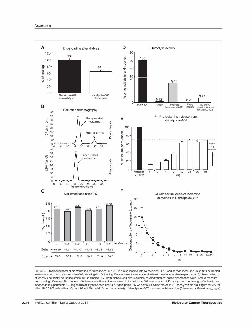
such as size, charge, and melanoma cell killing efficacywere analyzed for multiple batches (28). Loading of lee-lamine into the nanoliposomal formulation was assessedusing tritium-labeled drug during the manufacture ofNanolipolee-007, followed by dialysis to remove freecompound. Tritium-labeled leelamine was synthesizedby bromination, followed by replacing the bromine atomwith tritium with a specific activity of 25 Ci/mmol andpurity of the compound was determined by HPLC (Sup-plementary Fig. S2A). Biological efficacy of tritiated lee-lamine for killing melanoma cells was compared withleelamine by MTS assay (Supplementary Fig. S2B). Sizeexclusion chromatography was used to measure removalof loosely bound leelamine from that contained moretightly in the nanoliposome. Leelamine loading afterdialysis showed that 64.1% of the drug was incorporatedinto the nanoliposomal formulation (Fig. 2A). Columnchromatography of samples before and after dialysisshowed the disappearance of the loosely bound leelamineafter dialysis (Fig. 2B). Loading efficiency was also mea-sured by UV-visible spectrophotometry following
Structure and activity of leelamine andabietic acid
A B
C D
Schematic of the leelamine-loaded liposome(Nanolipolee-007)
Hydrophilic head
Hydrophobic tail
Phospholipid bilayer[ePC:DPPE (80:20 mol %)]
Leelamine
PEG-2000
Internalaqueous core
Leelamine hydrochloride
IC50 (µmol/L) IC50 (µmol/L)UACC 903: >100UACC 903: 1.78 ± 0.11
1205 Lu: >1001205 Lu: 2.49 ± 0.30
Str
uctu
reA
ctiv
ity
Abietic acid
Size and charge of Nanolipolee-007
Empty liposome Abietic acid liposome Leelamine liposome
Nanolipolee-007 has greater efficacykilling melanoma than normal cells
12.8615
12
9
IC50
(µm
ol/L
)
6
3
0
2.26
Average of melanoma cells
(1205 Lu + UACC 903)
Average of normal cells(FOM103 + FF2441)
The
rape
utic
inde
x5.
69 fo
ld
Me
MeMe
HCI
HMe
HN2
Me H
HMe
HO
16
14
12
10
8
6
4
2
0 100 1,000
100
80
60
Ove
rsiz
e
Inte
nsity
(%
)
40
20
0
16
14
12
10
8
6
4
2
0 100Size (d.nm)
1,000
100
80
60
Ove
rsiz
e
Inte
nsity
(%
)
40
20
0
16
14
12
10
8
6
4
2
0 100Size (d.nm)
1,000
100
80
60
Ove
rsiz
e
Inte
nsity
(%
)
40
20
0
Size: 85.60 ± 7.32
Zeta: +0.40 ± 0.02
PDI: 0.23 ± 0.01
Size: 81.26 ± 5.85
Zeta: –1.30 ± 0.07
PDI: 0.23 ± 0.01
Size: 73.50 ± 4.42
Zeta: +1.20 ± 0.04
PDI: 0.23 ± 0.01
O
Size (d.nm)
Figure 1. Development of Nanolipolee-007 and comparison of its killing efficacy on normal as well as on melanoma cells. A, to identify a novel drug targetingmultiple key pathways in melanoma, a cell-based screen was undertaken of a natural product library, identifying leelamine (A, left). Abietic acid, structurallysimilar to leelamine, hadnoeffect onmelanomacell viability (A, right). B, schematic ofNanolipolee-007.C, size and charge ofNanolipolee-007was70 to 80nmin size with a neutral surface charge in saline. The particle size and charge characteristics were established using a Malvern Zetasizer. D, efficacy ofNanolipolee-007 for killing normal and melanoma cells. Nanolipolee-007 was 5.69-fold more effective at killing metastatic melanoma than normal cells,suggesting potential cancer therapeutic utility at concentrations <2.26 mmol/L. Data represent an average of at least three independent experiments.
Gowda et al.
Mol Cancer Ther; 13(10) October 2014 Molecular Cancer Therapeutics2332
centrifugation using 10-kDa Centricon filters to removefree drugs from the nanoparticle. The loading efficiency ofleelaminewas found to be 70.6% (Supplementary Fig. S3).Stability of Nanolipolee-007 stored in sterile saline at 4�Cwasmeasured at 1, 3, 6, 9, and 12months, and size, chargeas well as efficacy for killing UACC 903 melanoma cellswere compared (Fig. 2C). During this period, the nanoli-posomes retained similar size and charge distributions aswell as efficacy for killingUACC 903melanoma cells withan IC50 ranging from 1.96 to 2.60 mmol/L (Fig. 2C). Noaggregation or precipitation of the nanoparticles occurredduring this period.
Nanolipolee-007 decreased the hemolytic activity ofleelamineLiposomal formulations of a hydrophobic drug can
overcome solubility and hemolysis occurring with thefree drug (29). A hemolytic assay was performed toexamine whether Nanolipolee-007 caused the same levelof red blood cell lysis occurringwith leelamine. Leelaminein DMSO induced 15.81% hemolysis, which is significantcompared with 3.29% occurring with Nanolipolee-007(Fig. 2D).
Leelamine released from Nanolipollee-007 waspresent in the serum of animalsLeelamine release fromNanolipolee-007wasmeasured
in vitro by dialysis in 0.9% saline for 24 hours, whichshowed that approximately 69.1% of leelamine wasreleased. Release initially occurred slowly for the firstfour hours and reached a maximum level 12 hours later(Fig. 2E). Leelamine release was also examined with 10mmol/L glutathione, leading to similar results, suggest-ing that unencapsulated drug is not observed at theconcentrations released from the individual nanolipo-somes (Supplementary Fig. S4). Next, presence of leela-mine in the serum of mice was measured followingintravenous injection of 30 mg/kg body weight of Nano-lipolee-007 and serum analyzed for the presence of thedrug over a 24-hour period by LC/MS-MS. Leelaminecontained in Nanolipolee-007 was present in the serumof mice for >10 hours after intravenous administration(Fig. 2F).
Nanolipolee-007 inhibited melanoma tumordevelopment with negligible major organ-relatedtoxicityThe study by Gowda and colleagues (12) showed that
intraperitoneal injection of leelamine dissolved in DMSOat 7.5 mg/kg body weight retarded existing xenograftmelanoma tumor growth by up to 60%. To determinewhether intravenously administered Nanolipolee-007would function in a similar manner, mice were injectedsubcutaneously with 1.5 million UACC 903 or 1205 Lumelanomacells and tumors let develop for 6days atwhichtime fully vascularized tumors had formed.Animalswerethen treated by daily intravenous injections of 30 mg/kgbody weight of Nanolipolee-007 and compared with con-
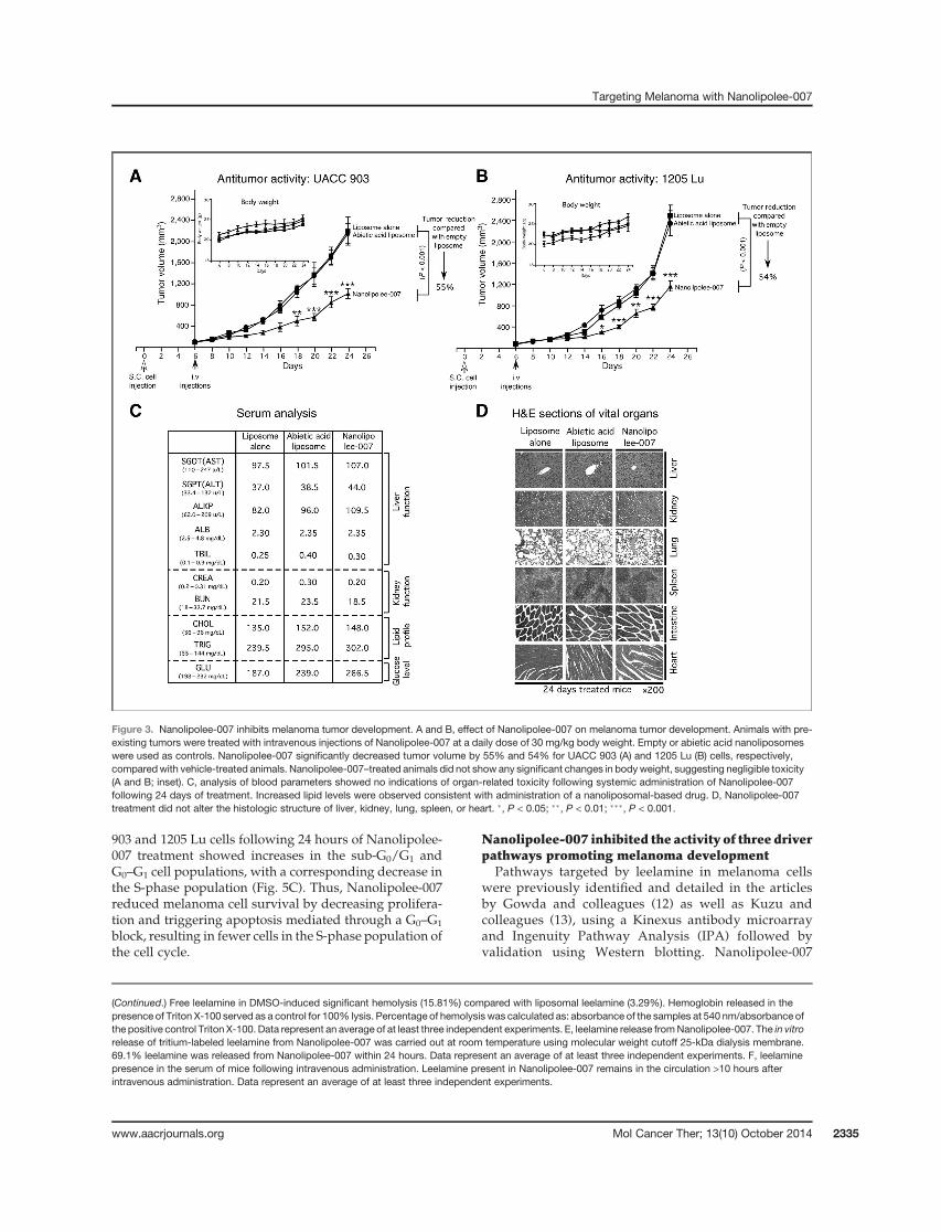
trols. Nanolipolee-007 decreased tumor volume byapproximately 55% for UACC 903 (Fig. 3A) and 1205 Lu(Fig. 3B) cells compared with controls. Nanolipolee-007treated animals did not show any significant changes inbodyweight, suggestingnegligible toxicity (Fig. 3AandB;inset). No noticeable changes in the serum parametersindicative of vital organ toxicity were observed (Fig. 3C),but an increase in cholesterol and triglyceride levels wasseen following nanoliposomal treatment after 24 days.This is expected following daily administration of lipid-based nanoparticles. However, analysis of the H&E-stained liver from vehicle or Nanolipolee-007–treatedmice showed no changes in the morphology or histologicarchitecture of the organ (Fig. 3D). Furthermore, nochanges were detected in histologic sections from heart,lung, kidney, or spleen (Fig. 3D). Thesedatademonstratedthat Nanolipolee-007 effectively inhibited xenograftedmelanoma tumor development without significantorgan-related toxicity other than increased lipid levelsthat could be controlled pharmacologically or by admin-istering the agent on alternate days.
In vivo mechanistic study of Nanolipolee-007 in sizeand time matched xenograft tumors
To investigate the mechanism by which Nanolipolee-007 delayed tumor growth, the rates of cell proliferation,apoptosis, and tumor angiogenesis occurring in timeand size-matched xenograft tumors treated with Nano-lipolee-007 were compared with empty control nanoli-posome-treated animals (22, 23). Size and time matchedtumors at days 11, 13, and 15 were compared to identifystatistically quantifiable differences in cell proliferation,apoptosis, or vascular development affected by Nano-lipolee-007 treatment. At day 11, a statistically signifi-cant 60% reduction in proliferating cells was observedas well as an increased number of cells undergoingapoptosis compared with control-treated animals(Fig. 4A and B). Furthermore, differences in vasculardevelopment were also detected in all tumors comparedwith vehicle controls. Thus, intravenously developedNanolipolee-007 at 30 mg/kg inhibited melanomatumor development by decreasing proliferation, trig-gering apoptosis, and decreasing vascular development(Fig. 4A–C).
Nanolipolee-007 decreased cellular proliferation,triggered apoptosis, and arrested melanoma cells inthe G0–G1 phase of the cell cycle
To further unravel the mechanisms leading to cellgrowth inhibition after treatment of mice with Nanolipo-lee-007, the rates of cellular proliferation, apoptosis, andthe percentage of cells in the various phases of the cellcycle were measured. Increasing concentrations of Nano-lipolee-007 from 0.62 to 10 mmol/L decreased the cellularproliferative potential as measured by bromodeoxyuri-dine (BrdUrd) incorporation (Fig. 5A) and increased cel-lular apoptosismeasuredby caspase-3/7 activity (Fig. 5B).Cell-cycle analysis of propidium iodide–stained UACC
Targeting Melanoma with Nanolipolee-007
www.aacrjournals.org Mol Cancer Ther; 13(10) October 2014 2333
Drug loading after dialysisA
B
C
E
F
D
Column chromatography
Hemolytic activity
100
64.1
100
% o
f hem
olys
is in
ery
thro
cyte
s
% o
f loa
ding
120
100
80
6020
0TritonX-100 DMSO 100 µmol/L
Leelamine in DMSO100 µmol/L
Leelamine liposome(Nanolipolee-007)
69.1%
Drug
released
Emptyliposome
15.81
In vitro leelamine release fromNanolipolee-007
In vivo serum levels of leelaminecontained in Nanolipolee-007
% o
f lee
lam
ine
rele
ased
0.233.29
0.72
120
100
80
60
40
20
0Nanolipolee-007before dialysis
Nanolipolee-007after dialysis
Nanolipolee-007
30
25
20
15
10
5
0
1 2 4 8(h)
12 24 36 48
Encapsulatedleelamine
Encapsulatedleelamine
Free leelamine
Fractions numbers
Stability of Nanolipolee-007
2.131.96
2.202.09
2.132.60
Bef
ore
dial
ysis
Afte
r di
alys
is40
3530
25
20
15
105
0
40
3530
25
20
15
105
0
0 5 10 15 20 25 30 35
0 5 10 15 20 25 30 35
CP
M (
1×10
4 )C
PM
(1×
104 )
100
80
60
40
20
0
0 1 2 4 6 8 10 12
(h)
Con
cent
ratio
ns o
f lee
lam
ine
(µg/
mL)
14 16 18 20 22 24
2.5
2.0
1.5
IC50
(µm
ol/L
)
1.0
0.5
00
+0.89 +1.27 +1.16 +1.43 +2.21 +4.13
66.371.668.379.388.289.3
Zeta
Size
1.0 3.0 6.0 9.0 12.0 Months
Figure 2. Physicochemical characterization of Nanolipolee-007. A, leelamine loading into Nanolipolee-007. Loading was measured using tritium-labeledleelamine when making Nanolipolee-007, showing 64.1% loading. Data represent an average of at least three independent experiments. B, characterizationof loosely and tightly bound leelamine in Nanolipolee-007. Both dialysis and size exclusion chromatography-based approaches were used to measuredrug loading efficiency. The amount of tritium-labeled leelamine remaining in Nanolipolee-007 was measured. Data represent an average of at least threeindependent experiments. C, long-term stability of Nanolipolee-007. Nanolipolee-007 was stable in saline stored at 4�C for a year; maintaining its activity forkillingUACC903cellswith an IC50of 1.96 to 2.60mmol/L.D, hemolytic activityofNanolipolee-007comparedwith leelamine. (Continuedon the followingpage.)
Gowda et al.
Mol Cancer Ther; 13(10) October 2014 Molecular Cancer Therapeutics2334
903 and 1205 Lu cells following 24 hours of Nanolipolee-007 treatment showed increases in the sub-G0/G1 andG0–G1 cell populations, with a corresponding decrease inthe S-phase population (Fig. 5C). Thus, Nanolipolee-007reduced melanoma cell survival by decreasing prolifera-tion and triggering apoptosis mediated through a G0–G1
block, resulting in fewer cells in the S-phase population ofthe cell cycle.
Nanolipolee-007 inhibited the activity of three driverpathways promoting melanoma development
Pathways targeted by leelamine in melanoma cellswere previously identified and detailed in the articlesby Gowda and colleagues (12) as well as Kuzu andcolleagues (13), using a Kinexus antibody microarrayand Ingenuity Pathway Analysis (IPA) followed byvalidation using Western blotting. Nanolipolee-007
Figure 3. Nanolipolee-007 inhibits melanoma tumor development. A and B, effect of Nanolipolee-007 on melanoma tumor development. Animals with pre-existing tumors were treated with intravenous injections of Nanolipolee-007 at a daily dose of 30 mg/kg body weight. Empty or abietic acid nanoliposomeswere used as controls. Nanolipolee-007 significantly decreased tumor volume by 55% and 54% for UACC 903 (A) and 1205 Lu (B) cells, respectively,comparedwith vehicle-treated animals. Nanolipolee-007–treated animals did not show any significant changes in bodyweight, suggesting negligible toxicity(A and B; inset). C, analysis of blood parameters showed no indications of organ-related toxicity following systemic administration of Nanolipolee-007following 24 days of treatment. Increased lipid levels were observed consistent with administration of a nanoliposomal-based drug. D, Nanolipolee-007treatment did not alter the histologic structure of liver, kidney, lung, spleen, or heart. �, P < 0.05; ��, P < 0.01; ���, P < 0.001.
(Continued.) Free leelamine in DMSO-induced significant hemolysis (15.81%) compared with liposomal leelamine (3.29%). Hemoglobin released in thepresence of Triton X-100 served as a control for 100% lysis. Percentage of hemolysis was calculated as: absorbance of the samples at 540 nm/absorbance ofthe positive control Triton X-100. Data represent an average of at least three independent experiments. E, leelamine release fromNanolipolee-007. The in vitrorelease of tritium-labeled leelamine from Nanolipolee-007 was carried out at room temperature using molecular weight cutoff 25-kDa dialysis membrane.69.1% leelamine was released from Nanolipolee-007 within 24 hours. Data represent an average of at least three independent experiments. F, leelaminepresence in the serum of mice following intravenous administration. Leelamine present in Nanolipolee-007 remains in the circulation >10 hours afterintravenous administration. Data represent an average of at least three independent experiments.
Targeting Melanoma with Nanolipolee-007
www.aacrjournals.org Mol Cancer Ther; 13(10) October 2014 2335

Figure 4. Mechanistic basis for tumor inhibition mediated by Nanolipolee-007. Formalin-fixed paraffin-embedded size- and time-matched tumorssections were subjected to Ki67 staining for cell proliferation (A), TUNEL staining for apoptosis (B), CD31 staining for tumor angiogenesis (C), andcompared with empty nanoliposome-treated animals. From day 11, a statistically significant decrease in proliferating tumor cells, increase in numberof cells undergoing apoptosis, and reduction in vascular development were identified compared with vehicle-treated animals (A–C). ��, P < 0.01;���, P < 0.001.
Gowda et al.
Mol Cancer Ther; 13(10) October 2014 Molecular Cancer Therapeutics2336
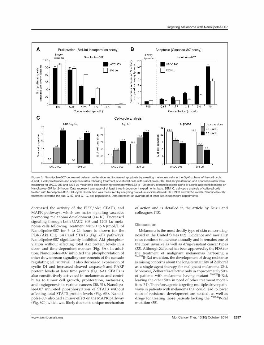
decreased the activity of the PI3K/Akt, STAT3, andMAPK pathways, which are major signaling cascadespromoting melanoma development (14–16). Decreasedsignaling through both UACC 903 and 1205 Lu mela-noma cells following treatment with 3 to 6 mmol/L ofNanolipolee-007 for 3 to 24 hours is shown for thePI3K/Akt (Fig. 6A) and STAT3 (Fig. 6B) pathways.Nanolipolee-007 significantly inhibited Akt phosphor-ylation without affecting total Akt protein levels in adose- and time-dependent manner (Fig. 6A). In addi-tion, Nanolipolee-007 inhibited the phosphorylation ofother downstream signaling components of the cascaderegulating cell survival. It also decreased expression ofcyclin D1 and increased cleaved caspase-3 and PARPprotein levels at later time points (Fig. 6A). STAT3 isalso constitutively activated in melanomas and contri-butes to tumor cell growth, proliferation, metastasis,and angiogenesis in various cancers (30, 31). Nanolipo-lee-007 inhibited phosphorylation of STAT3 withoutaffecting total STAT3 protein levels (Fig. 6B). Nanoli-polee-007 also had a minor effect on the MAPK pathway(Fig. 6C), which was likely due to its unique mechanism
of action and is detailed in the article by Kuzu andcolleagues (13).
DiscussionMelanoma is the most deadly type of skin cancer diag-
nosed in the United States (32). Incidence and mortalityrates continue to increase annually and it remains one ofthe most invasive as well as drug-resistant cancer types(33).AlthoughZelboraf has been approvedby the FDA forthe treatment of malignant melanomas harboring aV600EB-Raf mutation, the development of drug resistanceis raising concerns about the long-term utility of Zelborafas a single-agent therapy for malignant melanoma (34).Moreover, Zelboraf is effective only in approximately 50%of patients with melanoma having mutant V600EB-Raf,leaving the other 50% in need of other treatment modal-ities (34). Therefore, agents targetingmultiple driver path-ways in patients with melanoma that could lead to lowerrates of resistance development are needed, as well asdrugs for treating those patients lacking the V600EB-Rafmutation (35).
Figure 5. Nanolipolee-007 decreased cellular proliferation and increased apoptosis by arresting melanoma cells in the G0–G1 phase of the cell cycle.A and B, cell proliferation and apoptosis rates following treatment of cultured cells with Nanolipolee-007. Cellular proliferation and apoptosis rates weremeasured for UACC 903 and 1205 Lu melanoma cells following treatment with 0.62 to 100 mmol/L of nanoliposome alone or abietic acid nanoliposome orNanolipolee-007 for 24 hours. Data represent averages of at least three independent experiments; bars; SEM. C, cell-cycle analysis of cultured cellstreated with Nanolipolee-007. Cell-cycle distribution was measured by analyzing propidium iodide–stained UACC 903 and 1205 Lu cells. Nanolipolee-007treatment elevated the sub-G0/G1 and G0–G1 cell populations. Data represent an average of at least two independent experiments.
Targeting Melanoma with Nanolipolee-007
www.aacrjournals.org Mol Cancer Ther; 13(10) October 2014 2337

Natural products have played an important role in thedevelopment of new anticancer drugs and currently con-stitute more than 60% of cancer therapeutics in the clinic(36, 37). Leelamine was identified from a natural productlibrary screen, to inhibit melanoma cells growth but intra-venous administration was limited by hemolysis andanimal death. Liposomal formulation of drugs such asleelamine can be used to moderate these concerns (29).Leelamine in DMSO induced 15.81% hemolysis, which isconsidered high, compared with 3.29% observed withNanolipolee-007. This difference might be due to therigidity of the liposome and electrostatic repulsion ofanionic RBCs (29). Rigid molecules such as DPPE PEG-2000 are less prone to attach to the RBC membranecompared with flexible molecules (29).
Nanotechnology can be used to improve solubility,pharmacokinetics, and reduce side effects associatedwithvarious drugs (38, 39). Among different nanoparticles,liposomes are well-studied colloidal particles deliveringdrugs to tumors and increasing the solubility of amphi-
philic agents (40, 41). Liposomes less than 100 nm canenter tumors due to the leaky vasculature, which does notoccur in the normal vasculature due to the EPR effect (4).Nanolipolee-007 fulfills all these criteria having an aver-age size of 70 to 80 nm and a neutral charge. The mostcommon surface modification of nanoparticles is PEGyla-tion, in which polyethylene glycol is covalently linkedthrough lipids to the surface of the liposome (42). PEGy-lated liposomes tend to be stable, have enhanced circu-lation time, avoid clearance by the reticulo-endothelialsystem, and have minimal toxicity (43). In this report,leelamine was loaded into a PEGylated neutral liposomalformulation that was stable at 4�C for a year and had anincreased circulating half-life, promoting accumulation atthe tumor site.
In clinical studies, liposomes have improved the phar-macokinetics and bio-distribution properties of therapeu-tic agents as well as an ability to reduce toxicity byaccumulation in tumors due to the EPR effect (44). Cur-rently, there are 12 liposome-based drugs approved for
Figure 6. Nanolipolee-007 inhibits key signaling pathways regulating melanoma development. A–C, Western blot analysis of cultured cells treated withNanolipolee-007. UACC 903 and 1205 Lu melanoma cells were treated with 3 to 6 mmol/L of Nanolipolee-007 for 3 to 24 hours and cell lysates analyzed todetermine the expression as well as activity of PI3K (A), STAT3 (B), and MAPK pathways (C). a-enolase served as a control for equal protein loading.
Gowda et al.
Mol Cancer Ther; 13(10) October 2014 Molecular Cancer Therapeutics2338
clinical use and others are in various stages of clinicaldevelopment (3, 5, 11). For example, PEGylated liposomalformulations of doxorubicin such as Doxil and Lipo-doxare approved for intravenous application with minordose-limiting toxicity compared with doxorubicin (45,46). Nab-paclitaxel, a nanoparticle formulation of pacli-taxel, has also demonstrated higher therapeutic efficacyagainst breast cancer than paclitaxel and several othernab-based chemotherapeutics are currently under clinicalevaluation (47, 48).Mechanism of leelamine-mediated cell death has been
investigated and reported in themanuscript by Kuzu andcolleagues (13). Briefly, leelamine is a lysosomotropiccompound accumulating inside acidic cell compartmentssuch as lysosomes and endosomes.Accumulation leads todisruption of intracellular cholesterol homeostasis andinterferes with autophagic flux as well as receptor-medi-ated endocytosis. Inhibition of receptor-mediated endo-cytosis shut downs RTK signaling and inhibits the acti-vation of downstream PI3K/Akt, STAT3, and MAPKsignaling cascades. B-Raf mutation is not able to triggermelanoma development alone and requires cooperationwith other cellular alterations inRTK signaling such as theAkt pathway (49) and leelamine has the potential to targetthese pathways as well.Nanolipolee-007 inhibited the phosphorylation of Akt
without affecting total Akt protein levels in a dose- andtime-dependent manner. This, in turn, decreased down-stream levels of active PRAS40 and BAD proteins. Inaddition, Nanolipolee-007 decreased expression of pro-liferation marker cyclin D1 as well as increasing levels ofapoptosis markers cleaved caspase-3 and PARP proteins(14). Nanolipolee-007 also inhibited Stat3 signaling inmelanomas. Targeted inhibition of Stat3 is known toretard melanoma development and other studies alsodemonstrated that targeting Stat3 in conjunctionwithAktsynergistically inhibits melanomas (16, 50).In conclusion, Nanolipolee-007 retained the tumor
inhibitory activity of leelamine dissolved in DMSO andimproved the solubility of the drug with negligible tox-
icity in mice, suggesting its potential as a therapeuticagent for the treatment of melanoma or other cancers inwhich the PI3/Akt kinase, and STAT3, and to a lesserextent MAPK pathways are deregulated.
Disclosure of Potential Conflicts of InterestThe Pennsylvania State University has patented this discovery and
licensed it toMelanovusOncology, Inc. for commercialization. Dr. Robert-son has a financial interest (equity ownership) and business interests(Chief Scientific Officer and member of the Board of Directors) in Mela-novus, Oncology, Inc. The Pennsylvania State University also has equityand royalty interests in Melanovus, Oncology, Inc. These interests andpositions have been reviewed and managed by the University in accor-dance with its Conflict of Interest policies.
Authors' ContributionsConception and design: R. Gowda, S.R.V. Madhunapantula, A. Sharma,G.P. RobertsonDevelopment of methodology: R. Gowda, S.R.V. Madhunapantula,A. Sharma, G.P. RobertsonAcquisition of data (provided animals, acquired and managed patients,provided facilities, etc.): R. Gowda, S.R.V. Madhunapantula, O.F. Kuzu,G.P. RobertsonAnalysis and interpretation of data (e.g., statistical analysis, biostatis-tics, computational analysis): R. Gowda, S.R.V. Madhunapantula,O.F. Kuzu, G.P. RobertsonWriting, review, and/or revision of the manuscript: R. Gowda,S.R.V. Madhunapantula, A. Sharma, O.F. Kuzu, G.P. RobertsonAdministrative, technical, or material support (i.e., reporting or orga-nizing data, constructing databases):R. Gowda, S.R.V.Madhunapantula,G.P. RobertsonStudy supervision: R. Gowda, G.P. Robertson
AcknowledgmentsThe authors thank Katie Huber for technical assistance.
Grant SupportThis work was supported by NIH grants R01 CA-136667-02, RO1 CA-
1138634-02, RO1 CA-127892-01A (to G.P. Robertson), The Foreman Foun-dation for Melanoma Research (to G.P. Robertson), and the H.G. Barsu-mian, M.D. Memorial Fund (to A. Sharma).
The costs of publication of this article were defrayed in part by thepayment of page charges. This article must therefore be hereby markedadvertisement in accordance with 18 U.S.C. Section 1734 solely to indicatethis fact.
Received April 24, 2014; revised May 7, 2014; accepted July 21, 2014;published OnlineFirst July 31, 2014.
References1. Bagrodia S, Smeal T, Abraham RT. Mechanisms of intrinsic and
acquired resistance to kinase-targeted therapies. Pigment Cell Mel-anoma Res 2012;25:819–31.
2. Solit DB,RosenN.Resistance toBRAF inhibition inmelanomas.NEnglJ Med 2011;364:772–4.
3. Schroeder A, Heller DA, Winslow MM, Dahlman JE, Pratt GW, LangerR, et al. Treating metastatic cancer with nanotechnology. Nat RevCancer 2011;12:39–50.
4. Petros RA, DeSimone JM. Strategies in the design of nanopar-ticles for therapeutic applications. Nat Rev Drug Discov 2010;9:615–27.
5. Thierry B.Drug nanocarriers and functional nanoparticles: applicationsin cancer therapy. Curr Drug Deliv 2009;6:391–403.
6. Drummond DC, Meyer O, Hong K, Kirpotin DB, Papahadjopoulos D.Optimizing liposomes for delivery of chemotherapeutic agents to solidtumors. Pharmacol Rev 1999;51:691–743.
7. Maeda H, Nakamura H, Fang J. The EPR effect for macromoleculardrug delivery to solid tumors: Improvement of tumor uptake, lowering
of systemic toxicity, and distinct tumor imaging in vivo. Adv Drug DelivRev 2013;65:71–9.
8. Peer D, Karp JM, Hong S, Farokhzad OC, Margalit R, Langer R.Nanocarriers as an emerging platform for cancer therapy. Nat Nano-technol 2007;2:751–60.
9. Immordino ML, Dosio F, Cattel L. Stealth liposomes: review of thebasic science, rationale, and clinical applications, existing and poten-tial. Int J Nanomedicine 2006;1:297–315.
10. Szostak JW, Bartel DP, Luisi PL. Synthesizing life. Nature 2001;409:387–90.
11. ZhaoW, Karp JM, Ferrari M, Serda R. Bioengineering nanotechnology:towards the clinic. Nanotechnology 2012;22:490201.
12. Gowda R, Madhanupantula SV, Kuzu OF, Sharma A, Robertson GPD.Targeting multiple key signaling pathways in melanoma using leela-mine. Mol Cancer Ther 2014;13:1679–89.
13. Kuzu OF, Gowda R, Sharma A, Robertson GPD. Leelamine mediatescancer cell death through inhibition of intracellular cholesterol trans-port. Mol Cancer Ther 2014;13:1690–703.
www.aacrjournals.org Mol Cancer Ther; 13(10) October 2014 2339
Targeting Melanoma with Nanolipolee-007
14. Gowda R, Madhunapantula SV, Desai D, Amin S, Robertson GP.Simultaneous targeting of COX-2 and AKT using selenocoxib-1-GSHto inhibit melanoma. Mol Cancer Ther 2012;12:3–15.
15. Madhunapantula SV, Robertson GP. Is B-Raf a good therapeutictarget for melanoma and other malignancies? Cancer Res 2008;68:5–8.
16. Kortylewski M, Jove R, Yu H. Targeting STAT3 affects melanoma onmultiple fronts. Cancer Metastasis Rev 2005;24:315–27.
17. Ruysschaert T, Marque A, Duteyrat JL, Lesieur S, Winterhalter M,Fournier D. Liposome retention in size exclusion chromatography.BMC Biotechnol 2005;5:11.
18. Zheng S, Chang S, Lu J, Chen Z, Xie L, Nie Y, et al. Characterization of9-nitrocamptothecin liposomes: anticancer properties and mechan-isms on hepatocellular carcinoma in vitro and in vivo. PLoS ONE2011;6:e21064.
19. Nie Y, Ji L, Ding H, Xie L, Li L, He B, et al. Cholesterol derivatives basedcharged liposomes for doxorubicin delivery: preparation, in vitro and invivo characterization. Theranostics 2012;2:1092–103.
20. Song M, Lee D, Lee T, Lee S. Determination of leelamine in mouseplasma by LC-MS/MS and its pharmacokinetics. J Chromatogr BAnalyt Technol Biomed Life Sci 2013;931:170–3.
21. Madhunapantula SV, Hengst J, Gowda R, Fox TE, Yun JK, RobertsonGP. Targeting sphingosine kinase-1 to inhibit melanoma. Pigment CellMelanoma Res 2013;25:259–74.
22. Sharma A, Tran MA, Liang S, Sharma AK, Amin S, Smith CD, et al.Targeting mitogen-activated protein kinase/extracellular signal-regu-lated kinase kinase in the mutant (V600E) B-Raf signaling cascadeeffectively inhibits melanoma lung metastases. Cancer Res2006;66:8200–9.
23. Stahl JM,CheungM,SharmaA, Trivedi NR, ShanmugamS,RobertsonGP. Loss of PTEN promotes tumor development in malignant mela-noma. Cancer Res 2003;63:2881–90.
24. Sharma A, Sharma AK, Madhunapantula SV, Desai D, Huh SJ, MoscaP, et al. Targeting Akt3 signaling in malignant melanoma using iso-selenocyanates. Clin Cancer Res 2009;15:1674–85.
25. Montaguti P, Melloni E, Cavalletti E. Acute intravenous toxicity ofdimethyl sulfoxide, polyethylene glycol 400, dimethylformamide,absolute ethanol, and benzyl alcohol in inbred mouse strains. Arznei-mittel-Forschung 1994;44:566–70.
26. Sharma A, Mayhew E, Bolcsak L, Cavanaugh C, Harmon P, Janoff A,et al. Activity of paclitaxel liposome formulations against humanovarian tumor xenografts. Int J Cancer 1997;71:103–7.
27. DiStefano V, Klahn JJ. Observations on the pharmacology and hemo-lytic activity of dimethyl sulfoxide. Toxicol Appl Pharmacol 1965;7:660–6.
28. Tran MA, Gowda R, Sharma A, Park EJ, Adair J, Kester M, et al.Targeting V600EB-Raf andAkt3 using nanoliposomal-small interferingRNA inhibits cutaneous melanocytic lesion development. Cancer Res2008;68:7638–49.
29. Mourtas S, Michanetzis GP, Missirlis YF, Antimisiaris SG. Haemolyticactivity of liposomes: effect of vesicle size, lipid concentration andpolyethylene glycol-lipid or arsonolipid incorporation. J BiomedNano-technol 2009;5:409–15.
30. Jackson DP,Watling D, Rogers NC, Banks RE, Kerr IM, Selby PJ, et al.The JAK/STAT pathway is not sufficient to sustain the antiproliferative
response in an interferon-resistant human melanoma cell line. Mela-noma Res 2003;13:219–29.
31. Liu F, Cao J, Wu J, Sullivan K, Shen J, Ryu B, et al. Stat3-targetedtherapies overcome the acquired resistance to vemurafenib in mela-nomas. J Invest Dermatol 2013;133:2041–9.
32. Roth BJ, Krilov L, Adams S, Aghajanian CA, Bach P, Braiteh F, et al.Clinical cancer advances 2012: annual report on progress againstcancer from the american society of clinical oncology. J Clin Oncol2013;31:131–61.
33. Villanueva J, Vultur A, Herlyn M. Resistance to BRAF inhibitors:unraveling mechanisms and future treatment options. Cancer Res2011;71:7137–40.
34. Sullivan RJ, Flaherty KT. Resistance to BRAF-targeted therapy inmelanoma. Eur J Cancer 2013;49:1297–304.
35. Tsao H, Chin L, Garraway LA, Fisher DE. Melanoma: frommutations tomedicine. Genes Dev 2012;26:1131–55.
36. Newman DJ, Cragg GM. Natural products as sources of new drugsover the last 25 years. J Nat Prod 2007;70:461–77.
37. Altmann KH, Gertsch J. Anticancer drugs from nature–natural pro-ducts as a unique source of new microtubule-stabilizing agents. NatProd Rep 2007;24:327–57.
38. Cho K, Wang X, Nie S, Chen ZG, Shin DM. Therapeutic nanoparticlesfor drug delivery in cancer. Clin Cancer Res 2008;14:1310–6.
39. Wang J, Sui M, FanW. Nanoparticles for tumor targeted therapies andtheir pharmacokinetics. Curr Drug Metab 2010;11:129–41.
40. YezhelyevMV, Gao X, Xing Y, Al-Hajj A, Nie S, O'Regan RM. Emerginguse of nanoparticles in diagnosis and treatment of breast cancer.Lancet Oncol 2006;7:657–67.
41. van Vlerken LE, Amiji MM.Multi-functional polymeric nanoparticles fortumour-targeted drug delivery. Expert Opin DrugDeliv 2006;3:205–16.
42. Maeda H, Wu J, Sawa T, Matsumura Y, Hori K. Tumor vascularpermeability and the EPR effect in macromolecular therapeutics: areview. J Control Release 2000;65:271–84.
43. Jain A, Jain SK. PEGylation: an approach for drug delivery. A review.Crit Rev Ther Drug Carrier Syst 2008;25:403–47.
44. Tran MA, Watts RJ, Robertson GP. Use of liposomes as drug deliveryvehicles for treatment of melanoma. Pigment Cell Melanoma Res2009;22:388–99.
45. Poh SB, Bai LY, Chen PM. Pegylated liposomal doxorubicin-basedcombination chemotherapy as salvage treatment in patients withadvanced hepatocellular carcinoma. Am J Clin Oncol 2005;28:540–6.
46. Green AE, Rose PG. Pegylated liposomal doxorubicin in ovariancancer. Int J Nanomedicine 2006;1:229–39.
47. Montero AJ, Adams B, Diaz-Montero CM, Gluck S. Nab-paclitaxel inthe treatment of metastatic breast cancer: a comprehensive review.Expert Rev Clin Pharmacol 2011;4:329–34.
48. Gradishar WJ. Albumin-bound paclitaxel: a next-generation taxane.Expert Opin Pharmacother 2006;7:1041–53.
49. Haluska F, Pemberton T, Ibrahim N, Kalinsky K. The RTK/RAS/BRAF/PI3K pathways in melanoma: biology, small molecule inhibitors, andpotential applications. Semin Oncol 2007;34:546–54.
50. Krasilnikov M, Ivanov VN, Dong J, Ronai Z. ERK and PI3K negativelyregulate STAT-transcriptional activities in human melanoma cells:implications towards sensitization to apoptosis. Oncogene 2003;22:4092–101.
Mol Cancer Ther; 13(10) October 2014 Molecular Cancer Therapeutics2340
Gowda et al.
Related Documents