Nanoengineering of Immune Cell Function Keyue Shen 1 , Michael C. Milone 2 , Michael L. Dustin 3 , and Lance C. Kam 1 1 Department of Biomedical Engineering, Columbia University, New York, NY 10027, U.S.A 2 Department of Pathology and Laboratory Medicine, University of Pennsylvania School of Medicine, Philadelphia, PA 19104, U.S.A 3 Skirball Institute of Biomolecular Medicine, New York University School of Medicine, New York, NY 10016, U.S.A Abstract T lymphocytes are a key regulatory component of the adaptive immune system. Understanding how the micro- and nano-scale details of the extracellular environment influence T cell activation may have wide impact on the use of T cells for therapeutic purposes. In this article, we examine how the micro- and nano-scale presentation of ligands to cell surface receptors, including microscale organization and nanoscale mobility, influences the activation of T cells. We extend these studies to include the role of cell-generated forces, and the rigidity of the microenvironment, on T cell activation. These approaches enable delivery of defined signals to T cells, a step toward understanding the cell-cell communication in the immune system, and developing micro/nano- and material- engineered systems for tailoring immune responses for adoptive T cell therapies. INTRODUCTION The immune system protects against pathogens and other agents. The adaptive immune system, a recently evolved part of the vertebrate immune system, forms a strong line of defense against biological challenges through its ability to recognize new antigens, develop an appropriate response, and rapidly recall this action on subsequent re-exposure. T lymphocytes are a key regulatory component of this system, coordinating the activity of other cells and directly carrying out specific immune functions. Given these roles, T cell manipulation has been proposed as a therapeutic treatment for many diseases, most notably cancer [1–5]. A key step in many of these treatments, collectively referred to as adoptive immunotherapy, is the ex vivo stimulation and expansion of T cells, with subsequent re- introduction of these cells into the patient. This process is most often carried out by presenting T cells with ligands to specific receptors present on the T cell surface. The T Cell Receptor (TCR) complex which provides the primary antigenic signal conferring specificity and the CD28, and LFA-1 receptors which provide costimulatory and adhesive signals have been the most intensively used. Additional co-receptors assist TCR signaling in responding to different types of major histocompatibility complex (MHC) antigen presenting molecules. CD4 assists with recognition of extracellular peptides on MHC class II and CD8 assists with intracellular peptides presented on MHC class I. Mature T cells express CD4 or CD8 and both types of cells are valued adoptive immunotherapy. In vivo, these ligands are presented to T cells on the surface of specialized Antigen Presenting Cells (APCs), and this association with the plasma membrane is required for optimal effect. While ideal for use in stimulating T cells in vivo, culture and growth of native APCs is difficult and inconsistent. As such, the growth of T cells for contemporary adoptive immunotherapy is often initiated by ligands presenting on engineered beads or artificial APCs [6–9] (Fig. 1B). These approaches allow production of sufficient quantities of T cells for clinical use, but have severe limitations including lack of control over the phenotype or performance of the resultant cells [4]. NIH Public Access Author Manuscript Mater Res Soc Symp Proc. Author manuscript; available in PMC 2011 May 9. Published in final edited form as: Mater Res Soc Symp Proc. 2009 January 1; 1209: . doi:10.1557/PROC-1209-YY03-01. NIH-PA Author Manuscript NIH-PA Author Manuscript NIH-PA Author Manuscript

Welcome message from author
This document is posted to help you gain knowledge. Please leave a comment to let me know what you think about it! Share it to your friends and learn new things together.
Transcript
Nanoengineering of Immune Cell Function
Keyue Shen1, Michael C. Milone2, Michael L. Dustin3, and Lance C. Kam1
1 Department of Biomedical Engineering, Columbia University, New York, NY 10027, U.S.A2 Department of Pathology and Laboratory Medicine, University of Pennsylvania School ofMedicine, Philadelphia, PA 19104, U.S.A3 Skirball Institute of Biomolecular Medicine, New York University School of Medicine, New York,NY 10016, U.S.A
AbstractT lymphocytes are a key regulatory component of the adaptive immune system. Understandinghow the micro- and nano-scale details of the extracellular environment influence T cell activationmay have wide impact on the use of T cells for therapeutic purposes. In this article, we examinehow the micro- and nano-scale presentation of ligands to cell surface receptors, includingmicroscale organization and nanoscale mobility, influences the activation of T cells. We extendthese studies to include the role of cell-generated forces, and the rigidity of the microenvironment,on T cell activation. These approaches enable delivery of defined signals to T cells, a step towardunderstanding the cell-cell communication in the immune system, and developing micro/nano-and material- engineered systems for tailoring immune responses for adoptive T cell therapies.
INTRODUCTIONThe immune system protects against pathogens and other agents. The adaptive immunesystem, a recently evolved part of the vertebrate immune system, forms a strong line ofdefense against biological challenges through its ability to recognize new antigens, developan appropriate response, and rapidly recall this action on subsequent re-exposure. Tlymphocytes are a key regulatory component of this system, coordinating the activity ofother cells and directly carrying out specific immune functions. Given these roles, T cellmanipulation has been proposed as a therapeutic treatment for many diseases, most notablycancer [1–5]. A key step in many of these treatments, collectively referred to as adoptiveimmunotherapy, is the ex vivo stimulation and expansion of T cells, with subsequent re-introduction of these cells into the patient. This process is most often carried out bypresenting T cells with ligands to specific receptors present on the T cell surface. The T CellReceptor (TCR) complex which provides the primary antigenic signal conferring specificityand the CD28, and LFA-1 receptors which provide costimulatory and adhesive signals havebeen the most intensively used. Additional co-receptors assist TCR signaling in respondingto different types of major histocompatibility complex (MHC) antigen presenting molecules.CD4 assists with recognition of extracellular peptides on MHC class II and CD8 assists withintracellular peptides presented on MHC class I. Mature T cells express CD4 or CD8 andboth types of cells are valued adoptive immunotherapy. In vivo, these ligands are presentedto T cells on the surface of specialized Antigen Presenting Cells (APCs), and this associationwith the plasma membrane is required for optimal effect. While ideal for use in stimulatingT cells in vivo, culture and growth of native APCs is difficult and inconsistent. As such, thegrowth of T cells for contemporary adoptive immunotherapy is often initiated by ligandspresenting on engineered beads or artificial APCs [6–9] (Fig. 1B). These approaches allowproduction of sufficient quantities of T cells for clinical use, but have severe limitationsincluding lack of control over the phenotype or performance of the resultant cells [4].
NIH Public AccessAuthor ManuscriptMater Res Soc Symp Proc. Author manuscript; available in PMC 2011 May 9.
Published in final edited form as:Mater Res Soc Symp Proc. 2009 January 1; 1209: . doi:10.1557/PROC-1209-YY03-01.
NIH
-PA Author Manuscript
NIH
-PA Author Manuscript
NIH
-PA Author Manuscript
We propose that the process of T cell expansion can be improved by designing biomaterialsto better capture the native T cell/APC interface, a small (70 μm2) region termed theimmunological synapse (IS), which serves as a major point of communication. The bindingof TCR to peptide-loaded MHCs (pMHC), CD28 by B7, and LFA-1 by ICAM-1, threemajor receptor-ligand complexes, occurs in this interface, directing a range of subsequentcell functions including polarization, cytokine secretion, signal integration, and asymmetriccell division [10–13]. An emerging picture is that the micro- and nano-scale details of thisstructure play important roles in coordinating the function of these cells. This reportdescribes recent progress towards understanding these aspects, which may lead to newdesign rules for biomaterials that when used in T cell expansion will improve adoptiveimmunotherapy.
1. MICRO-/NANO-SCALE PATTERNINGA distinguishing characteristic of the immune synapse is the presence of highly complex,micrometer-scale organizations of signaling complexes within this structure. The maturesynapse was initially described as a microscale, annular disc of LFA-1/ICAM-1supramolecular activation clusters (SMACs) surrounding centralized clusters of TCR/pMHC[14]. However, additional patterns of these and other receptor pairs, including CD28/B7,have been identified (Figure 2A) [15–18], raising the possibility that the different motifsmay convey specific instructions to the T cells, and conversely be used to control cellularactivation.
Recent years have seen the application of micro- and nano-patterning methods tounderstanding the immune synapse [19–22]. Most notably, Doh and Irvine [19] introduced alithographic approach to create complementary patterns of ICAM-1 and an activatingantibody to CD3 (a key signaling component of the TCR) on material surfaces, anddemonstrated that preactivated T cells recognize different microscale patterns of theseligands. Our group built upon this concept [21], introducing techniques to combine highernumbers of patterns on a single surface (Figure 2B and C). By applying multiple rounds ofmicrocontact printing to a single surface, we created independently-defined patterns ofactivating antibodies to CD3 and CD28, surrounded and separated by ICAM-1. Theseimmobilized proteins were highly effective in patterning the engagement of T cell receptors(Figure 2C).
Naïve CD4+ T cells isolated from mice were able to recognize different patterns ofactivating antibodies to CD3 and CD28, as measured by secretion of IL-2, a key cytokineinvolved in T cell activation. Figure 2D compares IL-2 secretion across a select set ofpatterns indicated along the bottom axis of that graph, revealing a set of design rules: 1)activation of naïve CD4+ T cells is enhanced by engaging CD28 in the periphery of the Tcell-substrate interface, as opposed to the center, 2) a similar change in CD3 engagementhad a minor influence on IL-2 secretion, and 3) segregation/colocalization of the CD3 andCD28 signaling had a very small impact on T cell function.
Together, these studies point to a powerful role of spatial organization in directing T cellactivation and, potentially, subsequent differentiation and function. Incorporation of thesepatterning concepts into biomaterial design may provide a similar, new level of control overT cell expansion.
2. MODULATING T CELL ACTIVATION THROUGH SUBSTRATE RIGIDITYThe mechanical rigidity of the extracellular environment is increasingly recognized as amodulator of numerous cell functions, including formation/disassembly of adhesioncomplexes (i.e., focal adhesions), cell spreading, gene transcription, and cell fate [23–25].
Shen et al. Page 2
Mater Res Soc Symp Proc. Author manuscript; available in PMC 2011 May 9.
NIH
-PA Author Manuscript
NIH
-PA Author Manuscript
NIH
-PA Author Manuscript
This rigidity-sensing is coupled with force generation by the acto-myosin cytoskeleton in thecell, which may alter the phosphorylation of key molecules and switch integrins betweenresting and active states [26,27].
In T cells, formation of an IS also involves a high level of acto-myosin activity, which isinitiated by TCR microclusters [28] and driven by continuous centripetal transportation ofthese microclusters [28–31]. Inhibiting actin polymerization abrogates the formation of TCRmicroclusters [28] and the activation processes [32,33]. Myosin IIA was found to be themotor protein for the centripetal actin flow and microcluster movement [34]. Actinpolymerization and myosin II-induced retraction at the periphery of the IS result in“contractile oscillation” [35], which may help stabilize integrin-mediated adhesion throughforce-mediated signaling [36]. Inhibition of myosin IIA activity significantly reducedphosphorylation of Zap70 and LAT, two pivotal signaling events in T cell activation [34],implying that the mechanical force generated inside T cell may play a role in elevatingactivation signals.
We tested this hypothesis by intervening with the mechanical contraction process usingantibody-coated substrates of varied rigidities. The bulk rigidity of planarpolydimethylsiloxane (PDMS, Sylgard 184) substrates, which were coated with activatingantibodies to CD3 and CD28 (Figure 3A) was controlled by varying the ratio of elastomerbase to curing agent, resulting in substrates ranging in Young’s modulus from tens ofkiloPascals to several MegaPascals (Figure 3B) [37]. PDMS was prepared as a thin(hundreds of micrometers) layer backed by glass. Cells can probe the first few micrometersof elastomer depth [23], so these preparations appear as a half-infinite slab to the cells, yetare thin enough to allow microscopy and handling. The amount of antibody adsorbed to eachsurface was measured using an ELISA-based approach, and was found to be similar acrossthis range of compositions/rigidities (P < 0.005), varying by approximately 5%.
Figure 3B compares IL-2 secretion by naïve mouse CD4+ T cells as a function of PDMSformulation; the Box plots in this figure illustrate one representative experiment of threerepetitions. T cell costimulation was sensitive to the rigidity of the underlying substrate,where cells on the stiffer substrate had higher IL-2 secretion. All samples were statisticallydifferent from each other by Kruskal-Wallis and ANOVA tests (α = 0.05, n > 2000 cells persurface).
Control over the mechanical properties of an activating surface may similarly modulatelong-term T cell function, including division, proliferation, and differentiation, providing anew level of control over ex vivo T cell expansion.
3. LATERAL MOBILITY OF SIGNALING CLUSTERSUnlike extracellular matrix proteins, ligands to proteins involved in cell-cell communication,such as those presented by an APC, exhibit lateral mobility across the cell surface, owing toits association with the plasma membrane. Supported lipid bilayers (SLBs) presentingmembrane proteins capture the lateral mobility of the natural cell surface, and have emergedas a powerful model for investigating cell signaling [38–46]. Using this system, Chan et al.[47] demonstrated that laterally mobile, GPI-tethered CD58 was much more potent than animmobile counterpart in promoting cell interaction. Subsequent implementations of thismodel provide a rare look into the organization of the immunological synapse [39,40]. Tcells cluster and reorganize various components of antigen-presenting cells that have beenisolated and tethered to planar supported lipid bilayer, a phenomenon not possible if theproteins were immobilized to the experimental surface.
Shen et al. Page 3
Mater Res Soc Symp Proc. Author manuscript; available in PMC 2011 May 9.
NIH
-PA Author Manuscript
NIH
-PA Author Manuscript
NIH
-PA Author Manuscript

It is increasingly recognized that the plasma membrane exhibits considerable micro- andnano-scale order. Sub-cellular patterning of a single type of SLB has been studied usingtechniques of e-beam lithography [41,48], micro-contact printing [49], photolithography andparylene peel-off [50]. The ability to pattern multiple bilayers of different composition on asingle surface is important for bridging between studies using uniform SLBs and micro-patterned immobilized proteins [19,22]. However, patterning at sub-cellular resolution hasbeen elusive, owing to the limitations of non-mixing laminar flow [51–54]. More recently,researchers have patterned SLBs with sub-micrometer precision and multiple compositionsusing AFM-related techniques [55–57], but these are not well-suited for covering therelatively large areas intended for cell-based experiments (millimeters to centimeters on aside).
As shown in Figure 4A, we introduced a SLB patterning strategy that takes advantage ofdiffusive transport in supported membranes after formation on a substrate [58]. A bilayer-compatible substrate is divided into multiple regions, consisting of two large open regionsseparated by a middle region which contains a continuous barrier that divides the entiresurface into two topologically distinct regions. A three-flow chamber system is used to formthree different types of lipid bilayer on this surface; bilayers containing two differenttethered proteins are deposited on the outer regions, while a plain bilayer is formed on themiddle zone. Over time, tethered biomolecules will diffuse from the outer regions into theinterdigitated middle region, leading to an interlacing of regions each separately containingdifferent components (Figure 4B). Importantly, the spatial resolution of the resultant bilayersis determined by the barrier, providing finer resolution than that provided by laminar flow,reaching potentially into the realm of tens of nanometers [59]. With this approach, multipleligands can be presented to cells, each confined to separate regions of the cell-surfaceinterface while retaining the mobility required for effective membrane protein function.Figure 4C shows the use of this platform in presenting spatially segregated, micropatternedligands to the T cell surface proteins TCR and LFA-1.
The supported lipid bilayer model has been extremely useful for contemporaryinvestigations into the impact of membrane protein mobility on cell signaling. Adaptation ofthese design rules into new biomaterials, however, poses several challenges, including thefragility and limited lifetime of supported lipid bilayer. Continued advances in artificialamphiphilic molecules capturing key properties of lipids may yield to more effectivetechniques for including lateral mobility into material systems. Conversely continued designof polymer structure may allow the capture of the nanoscale behavior of natural lipids intothese systems.
CONCLUSIONSThis article reviews recent advances in understanding the role of micro- and nano-scaleorganization and cellular biomechanics in directing T cell function. Together, theseprinciples may lead to biomaterials that provide significantly enhanced control over T cellexpansion, leading to improved implementation of adoptive immunotherapy.
AcknowledgmentsThis work is supported in part by the National Institutes of Health, EY016586 and EB008199.
References1. Finn OJ. Cancer immunology. New England Journal of Medicine. 2008; 358:2704–15. [PubMed:
18565863]
Shen et al. Page 4
Mater Res Soc Symp Proc. Author manuscript; available in PMC 2011 May 9.
NIH
-PA Author Manuscript
NIH
-PA Author Manuscript
NIH
-PA Author Manuscript
2. Hunder NN, Wallen H, Cao J, Hendricks DW, Reilly JZ, Rodmyre R, Jungbluth A, Gnjatic S,Thompson JA, Yee C. Treatment of metastatic melanoma with autologous CD4+ T cells againstNY-ESO-1. New England Journal of Medicine. 2008; 358:2698–703. [PubMed: 18565862]
3. June CH. Adoptive T cell therapy for cancer in the clinic. Journal of Clinical Investigation. 2007;117:1466–76. [PubMed: 17549249]
4. June CH. Principles of adoptive T cell cancer therapy. Journal of Clinical Investigation. 2007;117:1204–12. [PubMed: 17476350]
5. Weiner LM. Cancer immunotherapy--the endgame begins. New England Journal of Medicine. 2008;358:2664–5. [PubMed: 18565858]
6. Dudley ME, Wunderlich JR, Yang JC, Sherry RM, Topalian SL, Restifo NP, Royal RE, KammulaU, White DE, Mavroukakis SA, Rogers LJ, Gracia GJ, Jones SA, Mangiameli DP, Pelletier MM,Gea-Banacloche J, Robinson MR, Berman DM, Filie AC, Abati A, Rosenberg SA. Adoptive celltransfer therapy following non-myeloablative but lymphodepleting chemotherapy for the treatmentof patients with refractory metastatic melanoma. Journal of Clinical Oncology. 2005; 23:2346–57.[PubMed: 15800326]
7. Riddell SR, Greenberg PD. The use of anti-CD3 and anti-CD28 monoclonal antibodies to clone andexpand human antigen-specific T cells. Journal of Immunoogicall Methods. 1990; 128:189–201.
8. Rosenberg SA, Restifo NP, Yang JC, Morgan RA, Dudley ME. Adoptive cell transfer: a clinicalpath to effective cancer immunotherapy. Nature Reviews Cancer. 2008; 8:299–308.
9. Suhoski MM, Golovina TN, Aqui NA, Tai VC, Varela-Rohena A, Milone MC, Carroll RG, RileyJL, June CH. Engineering Artificial Antigen-presenting Cells to Express a Diverse Array of Co-stimulatory Molecules. Molecular Therapeutics. 2007; 15:981–988.
10. Chang JT, Palanivel VR, Kinjyo I, Schambach F, Intlekofer AM, Banerjee A, Longworth SA,Vinup KE, Mrass P, Oliaro J, Killeen N, Orange JS, Russell SM, Weninger W, Reiner SL.Asymmetric T lymphocyte division in the initiation of adaptive immune responses. Science. 2007;315:1687–91. [PubMed: 17332376]
11. Dustin ML. Hunter to gatherer and back: immunological synapses and kinapses as variations onthe theme of amoeboid locomotion. Annual Review of Cell and Developmental Biology. 2008;24:577–96.
12. Dustin ML, Colman DR. Neural and immunological synaptic relations. Science. 2002; 298:785–9.[PubMed: 12399580]
13. Dustin ML, Olszowy MW, Holdorf AD, Li J, Bromley S, Desai N, Widder P, Rosenberger F, vander Merwe PA, Allen PM, Shaw AS. A novel adaptor protein orchestrates receptor patterning andcytoskeletal polarity in T-cell contacts. Cell. 1998; 94:667–77. [PubMed: 9741631]
14. Monks CR, Freiberg BA, Kupfer H, Sciaky N, Kupfer A. Three-dimensional segregation ofsupramolecular activation clusters in T cells. Nature. 1998; 395:82–6. [PubMed: 9738502]
15. Thauland TJ, Koguchi Y, Wetzel SA, Dustin ML, Parker DC. Th1 and Th2 cells formmorphologically distinct immunological synapses. Journal of Immunology. 2008; 181:393–9.
16. Trautmann A, Valitutti S. The diversity of immunological synapses. Current Opinion inImmunology. 2003; 15:249–54. [PubMed: 12787748]
17. Tseng SY, Liu M, Dustin ML. CD80 cytoplasmic domain controls localization of CD28, CTLA-4,and protein kinase Ctheta in the immunological synapse. Journal of Immunology. 2005; 175:7829–36.
18. Tseng SY, Waite JC, Liu M, Vardhana S, Dustin ML. T cell-dendritic cell immunological synapsescontain TCR-dependent CD28-CD80 clusters that recruit protein kinase C theta. Journal ofImmunology. 2008; 181:4852–63.
19. Doh J, Irvine DJ. Immunological synapse arrays: patterned protein surfaces that modulateimmunological synapse structure formation in T cells. Proceedings of the National Academy ofSciences of the United States of America. 2006; 103:5700–5. [PubMed: 16585528]
20. Senaratne W, Sengupta P, Jakubek V, Holowka D, Ober CK, Baird B. Functionalized surfacearrays for spatial targeting of immune cell signaling. Journal of the American Chemical Society.2006; 128:5594–5. [PubMed: 16637600]
Shen et al. Page 5
Mater Res Soc Symp Proc. Author manuscript; available in PMC 2011 May 9.
NIH
-PA Author Manuscript
NIH
-PA Author Manuscript
NIH
-PA Author Manuscript
21. Shen K, Thomas VK, Dustin ML, Kam LC. Micropatterning of costimulatory ligands enhancesCD4+ T cell function. Proceedings of the National Academy of Sciences of the United States ofAmerica. 2008; 105:7791–6. [PubMed: 18505845]
22. Shen K, Qi J, Kam LC. Microcontact printing of proteins for cell biology. Journal of VisualizedExperiments. 2008
23. Engler AJ, Sen S, Sweeney HL, Discher DE. Matrix elasticity directs stem cell lineagespecification. Cell. 2006; 126:677–89. [PubMed: 16923388]
24. Giannone G, Sheetz MP. Substrate rigidity and force define form through tyrosine phosphatase andkinase pathways. Trends in Cell Biology. 2006; 16:213–23. [PubMed: 16529933]
25. Pelham RJ Jr, Wang Y. Cell locomotion and focal adhesions are regulated by substrate flexibility.Proceedings of the National Academy of Sciences of the United States of America. 1997;94:13661–5. [PubMed: 9391082]
26. Jiang G, Huang AH, Cai Y, Tanase M, Sheetz MP. Rigidity sensing at the leading edge throughalphavbeta3 integrins and RPTPalpha. Biophysical Journal. 2006; 90:1804–9. [PubMed:16339875]
27. Astrof NS, Salas A, Shimaoka M, Chen J, Springer TA. Importance of force linkage inmechanochemistry of adhesion receptors. Biochemistry. 2006; 45:15020–8. [PubMed: 17154539]
28. Campi G, Varma R, Dustin ML. Actin and agonist MHC-peptide complex-dependent T cellreceptor microclusters as scaffolds for signaling. Journal of Experimental Medicine. 2005;202:1031–1036. [PubMed: 16216891]
29. Freiberg BA, Kupfer H, Maslanik W, Delli J, Kappler J, Zaller DM, Kupfer A. Staging andresetting T cell activation in SMACs. Nature Immunology. 2002; 3:911–7. [PubMed: 12244310]
30. Krummel MF, Sjaastad MD, Wulfing C, Davis MM. Differential clustering of CD4 and CD3zetaduring T cell recognition. Science. 2000; 289:1349–52. [PubMed: 10958781]
31. Huppa JB, Gleimer M, Sumen C, Davis MM. Continuous T cell receptor signaling required forsynapse maintenance and full effector potential. Nature Immunology. 2003; 4:749–55. [PubMed:12858171]
32. Valitutti S, Dessing M, Aktories K, Gallati H, Lanzavecchia A. Sustained signaling leading to Tcell activation results from prolonged T cell receptor occupancy. Role of T cell actin cytoskeleton.Journal of Experimental Medicine. 1995; 181:577–84. [PubMed: 7836913]
33. Wulfing C, Davis MM. A receptor/cytoskeletal movement triggered by costimulation during T cellactivation. Science. 1998; 282:2266–9. [PubMed: 9856952]
34. Ilani T, Vasiliver-Shamis G, Vardhana S, Bretscher A, Dustin ML. T cell antigen receptorsignaling and immunological synapse stability require myosin IIA. Nature Immunology. 2009;10:531–9. [PubMed: 19349987]
35. Dobereiner HG, Dubin-Thaler BJ, Hofman JM, Xenias HS, Sims TN, Giannone G, Dustin ML,Wiggins CH, Sheetz MP. Lateral membrane waves constitute a universal dynamic pattern ofmotile cells. Physical Review Letters. 2006; 97:038102. [PubMed: 16907546]
36. Dustin ML. Cell adhesion molecules and actin cytoskeleton at immune synapses and kinapses.Current Opinion in Cell Biology. 2007; 19:529–33. [PubMed: 17923403]
37. Tsai J, Kam L. Rigidity-dependent cross talk between integrin and cadherin signaling. BiophysicalJournal. 2009; 96:L39–41. [PubMed: 19289031]
38. Dori Y, Bianco-Peled H, Satija SK, Fields GB, McCarthy JB, Tirrell M. Ligand accessibility asmeans to control cell response to bioactive bilayer membranes. Journal of Biomedical MaterialsResearch. 2000; 50:75–81. [PubMed: 10644966]
39. Grakoui A, Bromley SK, Sumen C, Davis MM, Shaw AS, Allen PM, Dustin ML. Theimmunological synapse: a molecular machine controlling T cell activation. Science. 1999;285:221–7. [PubMed: 10398592]
40. Groves JT, Dustin ML. Supported planar bilayers in studies on immune cell adhesion andcommunication. Journal of Immunological Methods. 2003; 278:19–32. [PubMed: 12957393]
41. Mossman KD, Campi G, Groves JT, Dustin ML. Altered TCR signaling from geometricallyrepatterned immunological synapses. Science. 2005; 310:1191–3. [PubMed: 16293763]
Shen et al. Page 6
Mater Res Soc Symp Proc. Author manuscript; available in PMC 2011 May 9.
NIH
-PA Author Manuscript
NIH
-PA Author Manuscript
NIH
-PA Author Manuscript
42. Oliver AE, Ngassam V, Dang P, Sanii B, Wu H, Yee CK, Yeh Y, Parikh AN. Cell AttachmentBehavior on Solid and Fluid Substrates Exhibiting Spatial Patterns of Physical Properties.Langmuir. 2009
43. Pautot S, Lee H, Isacoff EY, Groves JT. Neuronal synapse interaction reconstituted between livecells and supported lipid bilayers. Nature Chemical Biology. 2005; 1:283.
44. Perez TD, Nelson WJ, Boxer SG, Kam L. E-Cadherin Tethered to Micropatterned Supported LipidBilayers as a Model for Cell Adhesion. Langmuir. 2005; 21:11963–8. [PubMed: 16316139]
45. Stroumpoulis D, Zhang H, Rubalcava L, Gliem J, Tirrell M. Cell adhesion and growth to Peptide-patterned supported lipid membranes. Langmuir. 2007; 23:3849–56. [PubMed: 17335250]
46. Yokosuka T, Kobayashi W, Sakata-Sogawa K, Takamatsu M, Hashimoto-Tane A, Dustin ML,Tokunaga M, Saito T. Spatiotemporal regulation of T cell costimulation by TCR-CD28microclusters and protein kinase C theta translocation. Immunity. 2008; 29:589–601. [PubMed:18848472]
47. Chan PY, Lawrence MB, Dustin ML, Ferguson LM, Golan DE, Springer TA. Influence ofReceptor Lateral Mobility On Adhesion Strengthening Between Membranes Containing Lfa-3 andCd2. Journal of Cell Biology. 1991; 115:245–255. [PubMed: 1717480]
48. DeMond AL, Groves JT. Interrogating the T cell synapse with patterned surfaces andphotoactivated proteins. Current Opinion in Immunology. 2007; 19:722–7. [PubMed: 17703931]
49. Kam L, Boxer SG. Cell adhesion to protein-micropatterned-supported lipid bilayer membranes.Journal of Biomedical Materials Research. 2001; 55:487–95. [PubMed: 11288076]
50. Orth RN, Wu M, Holowka DA, Craighead HG, Baird BA. Mast Cell Activation on Patterned LipidBilayers of Subcellular Dimensions. Langmuir. 2003; 19:1599–1605.
51. Kam L, Boxer SG. Formation of supported lipid bilayer composition arrays by controlled mixingand surface capture. Journal of the American Chemical Society. 2000; 122:12901–2.
52. Kam L, Boxer SG. Spatially selective manipulation of supported lipid bilayers by laminar flow:steps towards biomembrane microfluidics. Langmuir. 2003; 19:1624–31.
53. Yang T, Simanek EE, Cremer P. Creating addressable aqueous microcompartments above solidsupported phospholipid bilayers using lithographically patterned poly(dimethylsiloxane) molds.Analytical Chemistry. 2000; 72:2587–9. [PubMed: 10857639]
54. Yoshina-Ishii C, Boxer SG. Arrays of mobile tethered vesicles on supported lipid bilayers. Journalof the American Chemical Society. 2003; 125:3696–7. [PubMed: 12656589]
55. Jackson BL, Groves JT. Scanning probe lithography on fluid lipid membranes. Journal of theAmerican Chemical Society. 2004; 126:13878–9. [PubMed: 15506721]
56. Lenhert S, Sun P, Wang Y, Fuchs H, Mirkin CA. Massively parallel dip-pen nanolithography ofheterogeneous supported phospholipid multilayer patterns. Small. 2007; 3:71–5. [PubMed:17294472]
57. Shi J, Chen J, Cremer PS. Sub-100 nm patterning of supported bilayers by nanoshavinglithography. Journal of the American Chemical Society. 2008; 130:2718–9. [PubMed: 18257567]
58. Shen K, Tsai J, Shi P, Kam LC. Self-aligned supported lipid bilayers for patterning the cell-substrate interface. Journal of the American Chemical Society. 2009; 131:13204–5. [PubMed:19708648]
59. Tsai J, Sun E, Gao Y, Hone JC, Kam LC. Non-Brownian diffusion of membrane molecules innanopatterned supported lipid bilayers. Nano Letters. 2008; 8:425–30. [PubMed: 18205424]
Shen et al. Page 7
Mater Res Soc Symp Proc. Author manuscript; available in PMC 2011 May 9.
NIH
-PA Author Manuscript
NIH
-PA Author Manuscript
NIH
-PA Author Manuscript

Figure 1. T cell activation(A) Activation of T cells in vivo is mediated in large part by contact-mediatedcommunication with Antigen Presenting Cells (APCs). (B) For therapeutic ex vivoexpansion, activation is commonly carried out by replacing the APC with either engineeredcells or beads that engage the same receptors involved in T cell/APC interaction.
Shen et al. Page 8
Mater Res Soc Symp Proc. Author manuscript; available in PMC 2011 May 9.
NIH
-PA Author Manuscript
NIH
-PA Author Manuscript
NIH
-PA Author Manuscript

Figure 2. Micropatterned activation of T cells(A) Signaling complexes for distinct patterns of organization within the immune synapse.Adapted from [14] and [18]. (B) Micropattering of activating ligands to cell surfacereceptors allows the study of how cells respond to specific organizations of signalingcomplexes. (C) Demonstration of the ability to control receptor organization usingactivating antibodies to CD3 (central 2 μm dot) and CD28 (satellite 1 μm features). (D)Comparison of IL-2 secretion by mouse naïve CD4+ T cells on specific microscale patternsof CD3 and CD28. Scale bars: 2 μm.
Shen et al. Page 9
Mater Res Soc Symp Proc. Author manuscript; available in PMC 2011 May 9.
NIH
-PA Author Manuscript
NIH
-PA Author Manuscript
NIH
-PA Author Manuscript
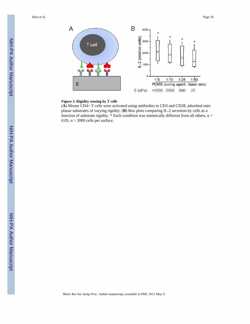
Figure 3. Rigidity sensing by T cells(A) Mouse CD4+ T cells were activated using antibodies to CD3 and CD28, adsorbed ontoplanar substrates of varying rigidity. (B) Box plots comparing IL-2 secretion by cells as afunction of substrate rigidity. * Each condition was statistically different from all others, α =0.05, n > 2000 cells per surface.
Shen et al. Page 10
Mater Res Soc Symp Proc. Author manuscript; available in PMC 2011 May 9.
NIH
-PA Author Manuscript
NIH
-PA Author Manuscript
NIH
-PA Author Manuscript

Figure 4. Multicomponent supported lipid bilayers(A) Microfluidic approaches allow patterning of supported lipid bilayers at subcellularlevels. (B) Example of a two-component lipid bilayer system. (C) Comparison of T cellreceptor organization on unpatterned (left) and segregated (right) lipid bilayers presentingligands to TCR and LFA-1; scale bar = 5 μm. Adapted from [58].
Shen et al. Page 11
Mater Res Soc Symp Proc. Author manuscript; available in PMC 2011 May 9.
NIH
-PA Author Manuscript
NIH
-PA Author Manuscript
NIH
-PA Author Manuscript
Related Documents