Nanocomposites of bacterial cellulose/hydroxyapatite for biomedical applications Cristian J. Grande a,b , Fernando G. Torres a, * , Clara M. Gomez b , M. Carmen Ban ˜o ´ c a Department of Mechanical Engineering, Catholic University of Peru, Lima 32, Peru b Departament de Quı ´mica Fı ´sica and Institut de Ciencia dels Materials, Universitat de Vale `ncia, E-46100 Burjassot, Spain c Departament de Bioquı ´mica i Biologia Molecular, Facultat de Biologia, Universitat de Vale ` ncia, E-46100 Burjassot, Spain Received 8 May 2008; received in revised form 19 November 2008; accepted 13 January 2009 Available online 31 January 2009 Abstract In the present work, a nanocomposite material formed by bacterial cellulose (BC) networks and calcium-deficient hydroxyapatite (HAp) powders was synthesized and characterized. The HAp nanoparticles were previously prepared by a wet chemical precipitation method, starting from aqueous solutions of calcium nitrate and di-ammonium phosphate salts. Energy-dispersive spectroscopy reveals that the prepared HAp corresponds to calcium-deficient hydroxyapatite. BC-HAp nanocomposites were prepared by introducing car- boxymethylcellulose (CMC) into the bacteria culture media. HAp nanoparticles were then introduced and remained suspended in the culture medium during the formation of cellulose nanofibrils. The maximum gel thickness was obtained after 21 days of bacteria culti- vation. X-ray diffractograms showed the difference of crystallinity among the materials involved in the formation of nanocomposites. The inorganic and organic bonds that corresponded to hydroxyapatite and bacterial cellulose respectively, were depicted by attenuated total reflectance Fourier transform infrared spectra. Scanning electron microscopy and atomic force microscopy measurements con- firmed the formation of networks and fibres with smaller diameter corresponding to BC synthesized in the presence of CMC. Image anal- ysis was also used to assess the orientation distributions and Feret diameters for networks of BC and BC-CMC. Thermogravimetric analysis showed that the amount of the mineral phase is 23.7% of the total weight of the nanocomposite. Moreover, HEK cells were cultivated and the biocompatibility of the materials and the cell viability was demonstrated. Ó 2009 Acta Materialia Inc. Published by Elsevier Ltd. All rights reserved. Keywords: Bacterial cellulose; Hydroxyapatite; Carboxymethylcellulose; Nanocomposites 1. Introduction Bacterial cellulose (BC) is a polysaccharide used tradi- tionally in the food industry [1,2], later in the fabrication of reinforced paper [3] and recently it was investigated as a material for medical applications. Studies carried out in vitro and in vivo have demonstrated its biocompatibility [4,5]. Due to its good mechanical properties, water sorption capacity, porosity, stability and conformability, BC has been used in tissue engineering of cartilage [6], replacement of blood vessels in rats [7] and in the wound healing process [8,9]. BC is pure cellulose with no other components [1]. Nanocomposites based on BC can be fabricated statically either by using the synthesized BC gel or modifying the cel- lulose biosynthesis. For instance, BC nanocomposites for biomedical applications with improved mechanical proper- ties were created by soaking BC on polyacrilamide and gel- atin solutions [10,11]. BC-hydroxyapatite scaffolds for bone regeneration have been developed by immersing the BC gel in simulated body fluid (SBF) or in both calcium and phosphate solutions [12–16]. Furthermore, BC-polyes- ter and BC-PVA nanocomposites were developed for potential applications as vascular implants [17,18]. 1742-7061/$ - see front matter Ó 2009 Acta Materialia Inc. Published by Elsevier Ltd. All rights reserved. doi:10.1016/j.actbio.2009.01.022 * Corresponding author. Tel.: +1 5116262000; fax: +1 5116262461. E-mail address: [email protected] (F.G. Torres). Available online at www.sciencedirect.com Acta Biomaterialia 5 (2009) 1605–1615 www.elsevier.com/locate/actabiomat

Welcome message from author
This document is posted to help you gain knowledge. Please leave a comment to let me know what you think about it! Share it to your friends and learn new things together.
Transcript
Available online at www.sciencedirect.com
Acta Biomaterialia 5 (2009) 1605–1615
www.elsevier.com/locate/actabiomat
Nanocomposites of bacterial cellulose/hydroxyapatitefor biomedical applications
Cristian J. Grande a,b, Fernando G. Torres a,*, Clara M. Gomez b, M. Carmen Bano c
a Department of Mechanical Engineering, Catholic University of Peru, Lima 32, Perub Departament de Quımica Fısica and Institut de Ciencia dels Materials, Universitat de Valencia, E-46100 Burjassot, Spainc Departament de Bioquımica i Biologia Molecular, Facultat de Biologia, Universitat de Valencia, E-46100 Burjassot, Spain
Received 8 May 2008; received in revised form 19 November 2008; accepted 13 January 2009Available online 31 January 2009
Abstract
In the present work, a nanocomposite material formed by bacterial cellulose (BC) networks and calcium-deficient hydroxyapatite(HAp) powders was synthesized and characterized. The HAp nanoparticles were previously prepared by a wet chemical precipitationmethod, starting from aqueous solutions of calcium nitrate and di-ammonium phosphate salts. Energy-dispersive spectroscopy revealsthat the prepared HAp corresponds to calcium-deficient hydroxyapatite. BC-HAp nanocomposites were prepared by introducing car-boxymethylcellulose (CMC) into the bacteria culture media. HAp nanoparticles were then introduced and remained suspended in theculture medium during the formation of cellulose nanofibrils. The maximum gel thickness was obtained after 21 days of bacteria culti-vation. X-ray diffractograms showed the difference of crystallinity among the materials involved in the formation of nanocomposites.The inorganic and organic bonds that corresponded to hydroxyapatite and bacterial cellulose respectively, were depicted by attenuatedtotal reflectance Fourier transform infrared spectra. Scanning electron microscopy and atomic force microscopy measurements con-firmed the formation of networks and fibres with smaller diameter corresponding to BC synthesized in the presence of CMC. Image anal-ysis was also used to assess the orientation distributions and Feret diameters for networks of BC and BC-CMC. Thermogravimetricanalysis showed that the amount of the mineral phase is 23.7% of the total weight of the nanocomposite. Moreover, HEK cells werecultivated and the biocompatibility of the materials and the cell viability was demonstrated.� 2009 Acta Materialia Inc. Published by Elsevier Ltd. All rights reserved.
Keywords: Bacterial cellulose; Hydroxyapatite; Carboxymethylcellulose; Nanocomposites
1. Introduction
Bacterial cellulose (BC) is a polysaccharide used tradi-tionally in the food industry [1,2], later in the fabricationof reinforced paper [3] and recently it was investigated asa material for medical applications. Studies carried outin vitro and in vivo have demonstrated its biocompatibility[4,5]. Due to its good mechanical properties, water sorptioncapacity, porosity, stability and conformability, BC hasbeen used in tissue engineering of cartilage [6], replacement
1742-7061/$ - see front matter � 2009 Acta Materialia Inc. Published by Else
doi:10.1016/j.actbio.2009.01.022
* Corresponding author. Tel.: +1 5116262000; fax: +1 5116262461.E-mail address: [email protected] (F.G. Torres).
of blood vessels in rats [7] and in the wound healing process[8,9].
BC is pure cellulose with no other components [1].Nanocomposites based on BC can be fabricated staticallyeither by using the synthesized BC gel or modifying the cel-lulose biosynthesis. For instance, BC nanocomposites forbiomedical applications with improved mechanical proper-ties were created by soaking BC on polyacrilamide and gel-atin solutions [10,11]. BC-hydroxyapatite scaffolds forbone regeneration have been developed by immersing theBC gel in simulated body fluid (SBF) or in both calciumand phosphate solutions [12–16]. Furthermore, BC-polyes-ter and BC-PVA nanocomposites were developed forpotential applications as vascular implants [17,18].
vier Ltd. All rights reserved.
1606 C.J. Grande et al. / Acta Biomaterialia 5 (2009) 1605–1615
Some researchers have introduced different materialsinto the culture media of BC. BC synthesized in the pres-ence of collagen [19] and chitosan [20] has improved prop-erties as wound dressing and for other biomedicalapplications. It has been reported that BC membranes pro-duced in the presence of carboxymethylcellulose (CMC)have better adsorption capacity of metal ions than mem-branes of pure BC [21–23].
However, the addition of some polymers can modifydrastically the cellulose biosynthesis. The addition ofCMC into the culture medium alters the crystallizationand assembly of the cellulose fibrils [24]. A similar effectoccurred when polyethylene oxide is added to the mediumin the process for obtaining BC based nanocomposites [25].
In agitated cultures, it has been demonstrated that BCcan be produced in the presence of solid particles (i.e. glassbeads, paper fibres and CaCO3) without affecting the rateof formation of the hydrogel [26]. Recent studies reportthe inclusion of silica particles of 10–20 nm and multi-walled carbon nanotubes (20–40 nm outer diameter, 10–50 lm length) into the culture medium to produce BC-nanocomposites in static cultures [27,28]. Hydroxyapatite(HAp) has been used in bone regeneration and as a substi-tute of bone and teeth because it is a biocompatible, bioac-tive, non-inflammatory, non-toxic and non-immunogenicmaterial [29,30].
The aim of this study was to fabricate BC-HAp nano-composites by the formation of cellulose nanofibrils inthe presence of a mineral phase in a static culture. In orderto suspend the HAp nanoparticles, the bacteria culturemedia were modified by the addition of CMC. Nanocom-posites were characterized by means of X-ray diffraction(XRD), Fourier transform infrared spectroscopy (ATR-FTIR), scanning electron microscopy (SEM), atomic forcemicroscopy (AFM), thermogravimetric analysis (TGA)and image analysis. In vitro biocompatibility and viabilitywas assessed using HEK cells.
2. Materials and methods
2.1. Preparation of HAp powders
Hydroxyapatite (HAp) powders were prepared in vitrousing a wet chemical precipitation method. Ca(NO3).4H2Oand (NH4)2HPO4 were used as Ca and P precursors respec-tively, following the next basic reaction:
5CaðNO3Þ:4H2Oþ 3ðNH4Þ2HPO4
! Ca5ðPO4Þ3OHþ 6NH4NO3 þ 19H2Oþ 4HNO3
Initially, 0.6 M (NH4)2HPO4 and 1.0 M Ca(NO3).4H2Osolutions were adjusted at pH 10.2 by the addition of con-centrated NH4OH. The phosphate solution was added indrops into the stirring calcium solution at 70 �C. Stirringat this temperature was carried out for 24 h and this pro-cess was followed by further stirring for 48 h at room tem-
perature. The resultant milky solution was filtered-washedfour times with distilled water. The precipitate was dried ina vacuum oven at 60 �C for 24 h. The resultant HAp pow-ders were milled in an agate mortar.
2.2. Preparation of BC and BC-HAp nanocomposite gels
The original culture medium for the growth of BC con-sisted of 1.0% (w/v) D-glucose, 1.5% (w/v) peptone, 0.8%(w/v) yeast extract and 0.3% (v/v) glacial acetic acid. ThepH of the solution was adjusted to 3.5 with hydrochloricacid. In order to maintain the medium free of the actionof microorganisms, it was autoclaved at 121 �C for20 min. After the medium had cooled down, 0.01% (w/v)cycloheximide and 0.5% (w/v) absolute ethanol wereadded. Cycloheximide was used in order to avoid the pres-ence of filaments while ethanol acts as an additional energysource for ATP generation enhancing thus the BC produc-tion in stationary cultures [31]. The described culture med-ium was used by Lisdiyanti et al. [32] and Yamada et al.[33] for the identification of acetic acid bacteria.
The strain Gluconacetobacter saccharivorans (LMG1582) isolated from a Kombucha tea mat [34] was inocu-lated and cultivated at 30 �C for 21 days. After this period,BC gels were removed and washed with deionized water. Inorder to remove bacteria and eliminate the remaining cul-ture medium, the cellulose pellicles were boiled in 1.0 MNaOH at 70 �C for 90 min followed by repetitive rinsingin deionized water.
For the formation of the new nanocomposite, HApnanoparticles were suspended in the culture medium. Inorder to avoid the settling of HAp nanoparticles, the vis-cosity of the solution was controlled using carboxymethyl-cellulose sodium salt (CMC) from Acros Organics (averageMW 25,0000, DS = 1.2). CMC was added to the mediumin concentrations of 1.0 and 2.0% (w/v) and stirring wascarried out until CMC dissolved. HAp powders wereadded in each vessel in concentrations of 1 and 2% (w/v)and the solutions remained under agitation overnight atroom temperature. The pH was adjusted to 3.5. Both cyclo-heximide and ethanol were added after the sterilizationprocess to prevent ethanol from reaching the boiling pointas well as the melting of cycloheximide. Finally, an inocu-lum of a previously cultivated BC was introduced in thecultures. The culture conditions and washing process werethe same as described above.
2.3. Samples preparation
In order to characterize the nanocomposite structures,water was removed from gels by either freeze drying, sol-vent exchange or hot pressing.
Pure BC, BC synthesized in the presence of 1% w/vCMC in the culture medium (BC-CMC), and nanocompos-ites of BC-CMC with HAp added to the culture medium inconcentration of 1% w/v (BC-CMC-HAp) were frozen inliquid nitrogen (�196 �C) and freeze-dried in a Telstar
C.J. Grande et al. / Acta Biomaterialia 5 (2009) 1605–1615 1607
Cryodos 80 at a subliming temperature of �58 �C and apressure of 0.18 mbar.
The solvent exchange drying method (water-ethanol-t-butyl alcohol) is suitable for preserving the original struc-ture of BC networks [35]. BC and CMC modified BCsamples were prepared using solvent exchange drying ofthe gels. Briefly, samples were introduced in ethanol for45 min and then in t-butyl alcohol for 45 min. After solventexchange, the samples were vacuum dried.
Gels of pure BC, BC-CMC (1% w/v) and nanocompos-ites of BC-CMC-HAp were dehydrated by hot pressing(0.015 MPa) at 105 �C during 5 min in order to obtainsheets for analysing their crystallinity and cell viability.
2.4. Characterization
A few milligrams of the freeze-dried samples and pureHAp powders were evaluated separately with a FTIR-ATR, Nicolet Nexus 470 equipped with a diamond probe.64 scans were used in the reflectance mode at a resolutionof 8 cm�1 in the range from 4000 to 400 cm�1. The datawere analysed with Omnic software.
In order to analyse the microstructure of the freeze-driedsamples, scanning electron microscopy (SEM) was used.BC-based samples and HAp powders were previously sput-ter coated with Au-Pd. Energy-dispersive spectroscopy(EDS) was used to determine the relation Ca/P of theHAp powders. RONTEC-Shell and RONTEC-Tool sys-tems were used to acquire and process the microanalysis,respectively. SEM and EDS were carried out using a fieldemission SEM Hitachi S4100 at 15 and 20 kV, respectively.
Image analysis of the SEM micrographs was performedby using the ImageJ software (Research Services Branch,National Institute of Mental Health, National Institutesof Health, NHI, USA). The diameter of the fibrils, theirorientation and the pore size of the BC surface in contactwith the culture medium [36] were assessed. In order todetermine the orientation of the fibrils, the fibrils werereplaced by segments connecting the junction points andthe angles between the segments and the x axis wererecorded. The Feret diameter (i.e. the maximum distancebetween two parallel tangents) was taken as the measureof the pore size.
Freeze-dried samples were analysed by thermogravimet-ric analysis (TGA) in order to determine the composition ofHAp in the BC-HAp nanocomposites and the rate ofchange in weight of BC based materials and HAp powders.Also, washed gels were analysed by TGA in order to deter-mine the amount of water. A Setaram Setsys 92-12 was usedin the range 50–900 �C with a heating rate of 10 �C min�1.
Measurements of the surfaces of BC and BC-CMC sam-ples in contact with the culture medium and obtained bythe solvent exchange drying method were carried out usingAFM (easyScan 2, Nanosurf AG, Switzerland) in thedynamic mode. A cantilever with a nominal spring con-stant of 42 N m�1, resonance frequency of 179 kHz and atip radius lower than 10 nm was used.
For X-ray diffraction (XRD) and cell seeding studies,hot pressed BC, BC-CMC and BC-CMC-HAp sampleswere prepared. Using a Seifert XRD 3003 TT diffractome-ter, Ni-filtered CuKa radiation (k = 0.1542 nm) was pro-duced at 40 kV and 40 mA. Scattered radiation wasdetected in the angular range of 2.5–70�(2h) in steps of0.08�(2h). The data were analysed using Analyse and DRX-Win softwares.
2.5. Cell culture
HEK cells were used to assess the biocompatibility andviability of the materials of the nanocomposites. Previousto cell seeding, samples were UV sterilized overnight. Thecells were cultured in Dulbecco’s Modified Eagle’s Medium(DMEM, Gibco) supplemented with 10% fetal bovineserum (FBS, Gibco), 1% penicillin-streptomicyn (P/S)and 0.1% fungizone in a humidified atmosphere at 37 �Cand 5% CO2. Medium was removed and cells were washedwith phosphate buffered saline (PBS, Gibco). Cells weretrypsnized in 0.2% trypsin solution and 5 ml of culturemedium was added. The cell suspension was centrifugedat 1500 rpm for 3 min and the medium was removed. Cellswere re-suspended with 6 ml of culture media. 1 ml of cellsuspension was seeded directly onto the materials insidethe tissue culture plate (TCP). Furthermore, 3 ml of freshculture medium was added covering all parts of the mate-rial. A TCP was used as a control.
After 1 day of culture, images were taken for analysingbiocompatibility. Furthermore, materials were removedfrom TCP and put into new ones with fresh culture med-ium for analysing if cells are stacked on the surfaces. Forthe viability analysis, cells of the original TCP were trypsin-ized with 300 ll trypsin solution. Also, 700 ll of fresh cul-ture medium were used to stop the reaction. 100 ll was putinto eppendorf tubes with the same volume of trypan blue.These solutions were put into a Neubauer camera and ana-lysed by optical microscopy.
3. Results and discussion
Morphological characterization was used in order tofurther understand the effect of the HAp phase on the for-mation of the BC network. Fig. 1a shows the morphologyof the HAp powders obtained by the described wet chem-ical process. Powders appeared free of other substancesand agglomerated in micrometric particles. Also, micro-metric particles are composed of particles at the nano level[37].
Fig. 1b shows a typical bacterial cellulose network of thesurface exposed to the culture medium. In the presence ofCMC at 1% w/v in the culture media, bacteria form cellu-lose fibrils with smaller diameters than those occurring inpure BC (Fig. 2). The average diameter of BC fibres wasdetermined in 117.76 nm ± 29.58 nm; and the averagediameter of BC fibres produced in the presence of CMC(1% w/v) was 60.90 nm ± 12.63 nm.

Fig. 1. SEM micrographs of: (a) surface of precipitated HAp powders; (b) original cellulose network; (c) HAp powders inside the cellulose network; (d)distribution of HAp powders in cellulose network.
1608 C.J. Grande et al. / Acta Biomaterialia 5 (2009) 1605–1615
The water extraction method influences the morphologyof BC networks. A previous study carried out in our labo-ratory indicates that the fibre diameter of pure BC com-pressed sheets is in the range 100–200 nm [34]. Thesolvent exchange drying method (water-ethanol-t-butylalcohol) was carried out in order to maintain the cellulosefibre structure [35]. AFM measurements of these samples(Fig. 3) confirmed SEM measurements. The average diam-eter of BC and BC-CMC (1% w/v) were 119.90 nm ±49.97 nm and 70.44 ± 30.04, respectively. Thus, unmodi-fied BC fibres are larger than BC-CMC fibres.
It should be noted that these results differ from otherreported in the literature where BC-CMC fibres are largerthan the unmodified BC ones due to the aggregation ofbundles produced by the presence of a compatible polymer[38,39]. The differences reported here could be attributed tothe effect of CMC on the strain of bacteria used in ourexperiments; however, further work would be needed toconfirm this.
Other BC network properties assessed were the pore sizeand the orientation of the fibres (angle between the x-axisand the fibre). The distribution of the angles found isdepicted in Fig. 4a. The average value of the angle is similarfor both BC and BC-CMC network and is equal to 84.30�
and 84.73�, respectively. In contrast, the pore size distribu-tion (Fig. 4b) and the average pore size are different. Theaverage pore size in BC and BC-CMC (1% w/v) networksare 0.5230 lm ± 0.2733 lm and 0.7733 lm ± 0.5238 lm,respectively. These results are in agreement with data forthe water holding capacity of BC and BC-CMC reportedpreviously by other authors [22,23,40].
The Ca/P ratio for the stoichiometric HAp is 1.67. Cal-cium-deficient HAp is of greater biological interest becausethis is the type of HAp almost always present in physiologicalmedia [41]. An EDS (energy-dispersive spectroscopy) spec-trum of the HAp produced is shown in Fig. 5. The Ca/P ratiodetermined by the RONTEC software was 1.60, indicatingthat the mineral phase is calcium-deficient HAp.
The diameter of the HAp particles present in the nano-composites was compared with the diameter of the synthe-sized HAp particles added to the culture medium (Fig. 6).The average diameter of the particles in the nanocompos-ites (2.59 lm ± 1.25 lm) was lower than the average diam-eter of the HAp particles added to the medium(3.34 lm ± 2.25 lm). This difference should be due to thefact that the larger HAp particles in the culture mediumsank to the bottom of the vessel in accordance with theStoke’s settling theory.

Fig. 2. Distribution of diameter of fibrils corresponding to pure (a) BCand (b) BC–CMC (1% w/v).
C.J. Grande et al. / Acta Biomaterialia 5 (2009) 1605–1615 1609
CMC increases the viscosity of the culture medium [42]and improves the capability of the solution to retain theHAp particles in suspension. Without CMC, the HAp par-ticles would fall to the bottom of the vessel where the BCgel is produced. The Stokes settling theory has been usedto estimate the settling velocity of the HAp particles inthe culture medium. The settling velocity is directly propor-tional to the particle size and reduces with the viscosity ofthe medium. Thus, the settling velocity of HAp in the cul-ture medium containing CMC (v2) can be related to the set-
Fig. 3. AFM images of: (a) BC; (b) BC pro
tling velocity of HAp in the culture medium without CMC(v1) by the Eq. (1):
v1
v2
¼ l2
l1
; ð1Þ
where l2 is the dynamic viscosity of the culture mediumcontaining CMC and l1 is the dynamic viscosity of the cul-ture medium without CMC. According to the technicalspecifications of the CMC provided, l2/l1 is around1500–3000 in a 2% solution at 25 �C. Thus, the settlingvelocity of HAp in the culture medium containing CMCis around 3.5 � 10�9 m s�1 (i.e. HAp particles would fall0.3 mm day�1). Thus, HAp powders can be trapped inthe cellulose network (Fig. 1c and d).
The addition of CMC at concentrations of 2% (w/v)produces BC-HAp nanocomposites of very thin thickness(0.5 mm). These gels were not integral structures and couldnot be removed without suffering damage. This structure issimilar to other ones based on CMC reported by Brown[40]. On the other hand, studies indicate that strains inthe presence of different amounts of CMC with differentdegree of substitution have increased yields, due probablyto the presence of the additional carbon sources in CMC.In this study, gels of about 8 mm and 99.15% of water con-tent were obtained. Since the volume of the gels is about afourth of the volume of the vessel containing the culturemedium and assuming that CMC was uniformly solubi-lized in the culture medium, it was estimated that about0.25% of the initial CMC was incorporated into the finalnanocomposites.
The ATR-FTIR spectra of the HAp powders, CMC-HAp, BC, BC-CMC, and nanocomposites of BC-CMC-HAp are depicted in Fig. 7. A usual characteristic of themodification of cellulose networks by the addition ofCMC is the decreasing intensity of the band correspondingto the cellulose type Ia (750 cm�1) [43].
In the analysed CMC-HAp freeze-dried suspension, thetypical band of CMC corresponding to (m COONa)appears at 1599 cm�1. In this spectrum there is a predom-inance of the chemical groups corresponding to HAp at1032, 602 and 563 cm�1. The interaction between CMCand HAp is confirmed by the fact that the peak at
duced in the presence of 1% w/v CMC.
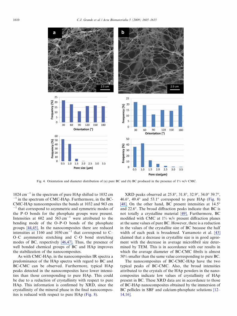
Fig. 4. Orientation and diameter distribution of (a) pure BC and (b) BC produced in the presence of 1% w/v CMC.
1610 C.J. Grande et al. / Acta Biomaterialia 5 (2009) 1605–1615
1024 cm�1 in the spectrum of pure HAp shifted to 1032 cm�1 in the spectrum of CMC-HAp. Furthermore, in the BC-CMC-HAp nanocomposites the bands at 1032 and 963 cm�1 that correspond to asymmetric and symmetric modes ofthe P–O bonds for the phosphate groups were present.Intensities at 602 and 563 cm�1 were attributed to thebending mode of the O–P–O bonds of the phosphategroups [44,45]. In the nanocomposites there are reducedintensities at 1160 and 1050 cm�1 that correspond to C–O–C asymmetric stretching and C–O bond stretchingmodes of BC, respectively [46,47]. Thus, the presence ofwell bonded chemical groups of BC and HAp improvesthe stabilization of the nanocomposites.
As with CMC-HAp, in the nanocomposites IR spectra apredominance of the HAp spectra with regard to BC andBC-CMC can be observed. Furthermore, typical HAppeaks detected in the nanocomposites have lower intensi-ties than those corresponding to pure HAp. This couldbe due to a reduction of crystallinity with respect to pureHAp. This information is confirmed by XRD, since thecrystallinity of the mineral phase in the final nanocompos-ites is reduced with respect to pure HAp (Fig. 8).
XRD peaks observed at 25.8�, 31.8�, 32.9�, 34.0� 39.7�,46.6�, 49.4� and 53.1� correspond to pure HAp (Fig. 8)[48]. On the other hand, BC present intensities at 14.5�and 22.6�. The broad diffraction peaks indicate that BC isnot totally a crystalline material [49]. Furthermore, BCmodified with CMC at 1% w/v present diffraction planesat the same values of pure BC. However, there is a reductionin the values of the crystallite size of BC because the halfwidth of each peak is broadened. Yamamoto et al. [43]claimed that a decrease in crystallite size is in good agree-ment with the decrease in average microfibril size deter-mined by TEM. This is in accordance with our results inwhich the average diameter of BC-CMC fibrils is almost50% smaller than the same value corresponding to pure BC.
The nanocomposites of BC-CMC-HAp have the twotypical peaks of BC-CMC. Also, the broad intensitiesattributed to the crystals of the HAp powders in the nano-composites indicate low values of crystallinity of HAppresent in BC. These XRD data are in accordance to thoseof BC-HAp nanocomposites obtained by the immersion ofBC pellicles in SBF and calcium-phosphate solutions [12–14,16].
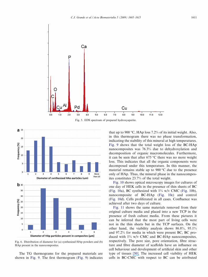
Fig. 5. EDS spectrum of prepared hydroxyapatite.
Fig. 6. Distribution of diameter for (a) synthesized HAp powders and (b)HAp present in the nanocomposites.
C.J. Grande et al. / Acta Biomaterialia 5 (2009) 1605–1615 1611
The TG thermograms for the prepared materials areshown in Fig. 9. The first thermogram (Fig. 9) indicates
that up to 900 �C, HAp loss 7.2% of its initial weight. Also,in this thermogram there was no phase transformation,indicating the stability of this mineral at high temperatures.Fig. 9 shows that the total weight loss of the BC-HApnanocomposites was 76.3% due to dehydroxylation anddecomposition of organic macromolecules. Furthermore,it can be seen that after 675 �C there was no more weightloss. This indicates that all the organic components weredecomposed under this temperature. In this manner, thematerial remains stable up to 900 �C due to the presenceonly of HAp. Thus, the mineral phase in the nanocompos-ites constitutes 23.7% of the total weight.
Fig. 10 shows optical microscopy images for cultures ofone day of HEK cells in the presence of thin sheets of BC(Fig. 10a), BC synthesized with 1% w/v CMC (Fig. 10b),nanocomposite of BC-HAp (Fig. 10c) and control(Fig. 10d). Cells proliferated in all cases. Confluence wasachieved after two days of culture.
Fig. 11 shows the same materials removed from theiroriginal culture media and placed into a new TCP in thepresence of fresh culture media. From these pictures itcan be inferred that the most part of living cells werenot in the thin sheets but in the TCP surfaces. On theother hand, the viability analysis shows 86.8%, 95.1%and 97.2% for media in which were present BC, BC pro-duced with 1% w/v CMC and BC-HAp nanocomposites,respectively. The pore size, pore orientation, fibre struc-ture and fibre diameter of scaffolds have an influence oncell behaviour and development of artificial skin and othertype of tissues [50]. The increased cell viability of HEKcells in BC-CMC with respect to BC can be attributed
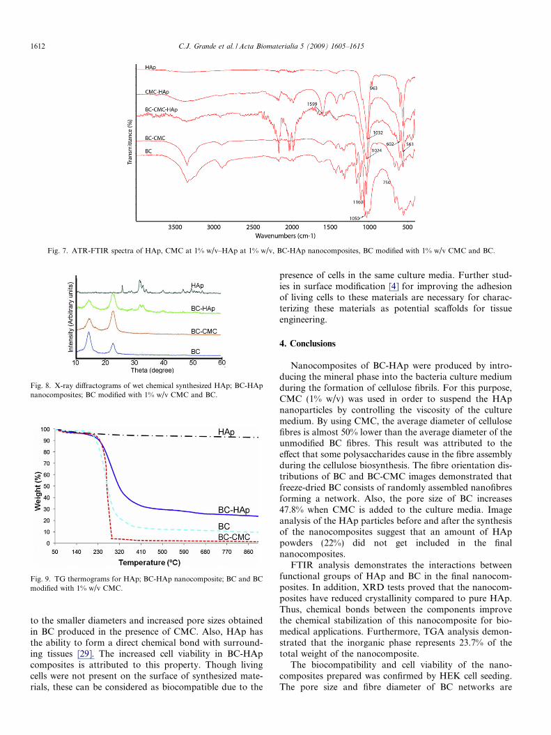
Fig. 7. ATR-FTIR spectra of HAp, CMC at 1% w/v–HAp at 1% w/v, BC-HAp nanocomposites, BC modified with 1% w/v CMC and BC.
Fig. 8. X-ray diffractograms of wet chemical synthesized HAp; BC-HApnanocomposites; BC modified with 1% w/v CMC and BC.
Fig. 9. TG thermograms for HAp; BC-HAp nanocomposite; BC and BCmodified with 1% w/v CMC.
1612 C.J. Grande et al. / Acta Biomaterialia 5 (2009) 1605–1615
to the smaller diameters and increased pore sizes obtainedin BC produced in the presence of CMC. Also, HAp hasthe ability to form a direct chemical bond with surround-ing tissues [29]. The increased cell viability in BC-HApcomposites is attributed to this property. Though livingcells were not present on the surface of synthesized mate-rials, these can be considered as biocompatible due to the
presence of cells in the same culture media. Further stud-ies in surface modification [4] for improving the adhesionof living cells to these materials are necessary for charac-terizing these materials as potential scaffolds for tissueengineering.
4. Conclusions
Nanocomposites of BC-HAp were produced by intro-ducing the mineral phase into the bacteria culture mediumduring the formation of cellulose fibrils. For this purpose,CMC (1% w/v) was used in order to suspend the HApnanoparticles by controlling the viscosity of the culturemedium. By using CMC, the average diameter of cellulosefibres is almost 50% lower than the average diameter of theunmodified BC fibres. This result was attributed to theeffect that some polysaccharides cause in the fibre assemblyduring the cellulose biosynthesis. The fibre orientation dis-tributions of BC and BC-CMC images demonstrated thatfreeze-dried BC consists of randomly assembled nanofibresforming a network. Also, the pore size of BC increases47.8% when CMC is added to the culture media. Imageanalysis of the HAp particles before and after the synthesisof the nanocomposites suggest that an amount of HAppowders (22%) did not get included in the finalnanocomposites.
FTIR analysis demonstrates the interactions betweenfunctional groups of HAp and BC in the final nanocom-posites. In addition, XRD tests proved that the nanocom-posites have reduced crystallinity compared to pure HAp.Thus, chemical bonds between the components improvethe chemical stabilization of this nanocomposite for bio-medical applications. Furthermore, TGA analysis demon-strated that the inorganic phase represents 23.7% of thetotal weight of the nanocomposite.
The biocompatibility and cell viability of the nano-composites prepared was confirmed by HEK cell seeding.The pore size and fibre diameter of BC networks are
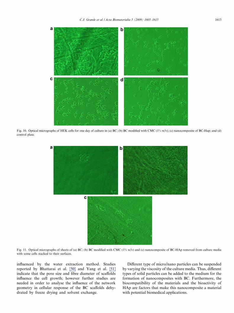
Fig. 10. Optical micrographs of HEK cells for one day of culture in (a) BC; (b) BC modified with CMC (1% w/v); (c) nanocomposite of BC-Hap; and (d)control plate.
Fig. 11. Optical micrographs of sheets of (a) BC; (b) BC modified with CMC (1% w/v) and (c) nanocomposite of BC-HAp removed from culture mediawith some cells stacked to their surfaces.
C.J. Grande et al. / Acta Biomaterialia 5 (2009) 1605–1615 1613
influenced by the water extraction method. Studiesreported by Bhattarai et al. [50] and Yang et al. [51]indicate that the pore size and fibre diameter of scaffoldsinfluence the cell growth; however further studies areneeded in order to analyse the influence of the networkgeometry in cellular response of the BC scaffolds dehy-drated by freeze drying and solvent exchange.
Different type of micro/nano particles can be suspendedby varying the viscosity of the culture media. Thus, differenttypes of solid particles can be added to the medium for theformation of nanocomposites with BC. Furthermore, thebiocompatibility of the materials and the bioactivity ofHAp are factors that make this nanocomposite a materialwith potential biomedical applications.
1614 C.J. Grande et al. / Acta Biomaterialia 5 (2009) 1605–1615
Acknowledgements
CJG would like to thank the International RelationsOffice (Oficina de Relaciones Internacionales) of the Uni-versity of Valencia for the financial support received. Theauthors would also like to thank the Direction of AcademicResearch of the Catholic University of Peru (DAI), TheInternational Foundation for Science (IFS, Sweden)and Generalitat Valenciana, Conselleria de Empresa,Universidad y Ciencia, project number ARVIV/2007/101.O. Troncoso is acknowledged for experimental assistancewith Image Analysis software.
References
[1] Iguchi M, Yamanaka S, Budhiono A. Bacterial cellulose – amasterpiece of nature’s arts. J Mater Sci 2000;35:261–70.
[2] Okiyama A, Motoki M, Yamanaka S. Bacterial cellulose II:processing of the gelatinous cellulose for food materials. FoodHydrocolloids 1992;6:479–87.
[3] Yamanaka S, Watanabe K, Kitamura N, Iguchi M, Mitsuhashi S,Nishi Y, et al. The structure and mechanical properties of sheetsprepared from bacterial cellulose. J Mater Sci 1989;24:3141–5.
[4] Watanabe K, Eto Y, Takano S, Nakamori S, Shibai H, Yamanaka S.A new bacterial cellulose substrate for mammalian cell culture.Cytotechnology 1993;13:107–14.
[5] Helenius G, Backdahl H, Bodin A, Nannmark U, Gatenholm P,Risberg B. In vivo biocompatibility of bacterial cellulose. J BiomedMater Res A 2006;76:431–8.
[6] Svensson A, Nicklasson E, Harrah T, Panilaitis B, Kaplan DL,Brittberg M, et al. Bacterial cellulose as a potential scaffold for tissueengineering of cartilage. Biomaterials 2005;26:419–31.
[7] Klemm D, Schumann D, Udhardt U, Marsch S. Bacterial synthesizedcellulose – aritificial blood vessels for microsurgery. Prog Polym Sci2001;26:1561–603.
[8] Czaja W, Krystynowicz A, Bielecki S, Brown RM. Microbial cellulose– the natural power to heal wounds. Biomaterials 2006;27:145–51.
[9] Czaja W, Young DJ, Kawecki M, Brown RM. The future prospectsof microbial cellulose in biomedical applications. Biomacromolecules2007;8:1–12.
[10] Yasuda K, Ping Gong J, Katsuyama Y, Nakayama A, Tanabe Y,Kondo E, et al. Biomechanical properties of high-toughness doublenetwork hydrogels. Biomaterials 2005;26:4468–75.
[11] Nakayama A, Kakugo A, Ping Gong J, Osada Y, Takai M, Erata T,et al. High mechanical strength double-network hydrogel withbacterial cellulose. Adv Funct Mater 2004;14:1124–8.
[12] Hong L, Wang YL, Jia SR, Huang Y, Gao C, Wan YZ. Hydroxy-apatite/bacterial cellulose nanocomposites synthesized via a biomi-metic route. Mater Lett 2006;60:1710–3.
[13] Wan YZ, Hong L, Jia SR, Huang Y, Zhu Y, Wang YL, et al.Synthesis and characterization of hydroxyapatite-bacterial cellulosenanocomposites. Compos Sci Technol 2006;66:1825–32.
[14] Wan YZ, Huang Y, Yuan CD, Raman S, Zhu Y, Jiang HJ, et al.Biomimetic synthesis of hydroxyapatite/bacterial cellulose nano-composites for biomedical applications. Mater Sci Eng C 2007;27:855–64.
[15] Bodin A, Gustafsson L, Gatenholm P. Surface engineered bacterialcellulose as template for crystallization of calcium phosphate. JBiomater Sci Polym Ed 2006;17:435–47.
[16] Hutchens SA, Benson RS, Evans BR, O’Neill HM, Rawn CJ.Biomimetic synthesis of calcium-deficient hydroxyapatite in a naturalhydrogel. Biomaterials 2006;27:4661–70.
[17] Charpentier PA, Maguire A, Wan WK. Surface modification ofpolyester to produce a bacterial cellulose-based vascular prostheticdevice. Appl Surf Sci 2006;252:6360–7.
[18] Millon LE, Mohammadi H, Wan WK. Anisotropic polyvinyl alcoholhydrogel for cardiovascular applications. J Biomed Mater Res B2006;79:305–11.
[19] Wiegand C, Elsner P, Hipler UC, Klemm D. Protease and ROSactivities influenced by a nanocomposite of bacterial cellulose andcollagen type I in vitro. Cellulose 2006;13:689–96.
[20] Ciechanska D. Multifunctional bacterial cellulose/chitosan nanocom-posite materials for medical applications. Fibres Text East Eur2004;12:69–72.
[21] Chen S, Zou Y, Yan Z, Shen W, Shi S, Zhang X, et al. Carboyme-thylated-bacterial cellulose for copper and lead ion removal. J HazardMater 2008. 10.1016/j.jhazmat.2008.04.098.
[22] Seifert M, Hesse S, Kabrelian V, Klemm D. Controlling the watercontent of never dried and reswollen bacterial cellulose by theaddition of water-soluble polymers to the culture medium. J PolymSci A Polym Chem 2004;42:463–70.
[23] Sakairi N, Suzuki S, Ueno K, Han SM, Nishi N, Tokura S. Biosynthesisof hetero-polysaccharides by Acetobacter xylinum – synthesis andcharacterization of metal-ion adsorptive properties of partially carbo-xymethylated cellulose. Carbohydr Polym 1998;37:409–14.
[24] Haigler CH, White AR, Brown RM, Cooper KM. Alteration ofin vivo cellulose ribbon assembly by carboxymethylcellulose andother cellulose derivatives. J Cell Biol 1982;94:64–9.
[25] Brown EE, Laborie MPG. Bioengineering bacterial cellulose/poly(ethylene oxide) nanocomposites. Biomacromolecules 2007;8:3074–81.
[26] Serafica G, Mormino G, Bungay H. Inclusion of solid particles inbacterial cellulose. Appl Microbiol Biotechnol 2002;58:756–60.
[27] Yano S, Maeda H, Nakajima M, Hagiwara T, Sawaguchi T.Preparation and mechanical properties of bacterial cellulose nano-composites loaded with silica nanoparticles. Cellulose 2008;15:111–20.
[28] Yan Z, Chen S, Wang H, Wang B, Wang C, Jiang J. Cellulosesynthesized by Acetobacter xylinum in the presence of multi-walledcarbon nanotubes. Carbohydr Res 2008;343:73–80.
[29] Murugan R, Ramakrishna S. In situ formation of recombinanthumanlike collagen-hydroxyapatite nanohybrid through bionicapproach. Appl Phys Lett 2006;88:193124.
[30] Thomas V, Dean DR, Jose MV, Mathew B, Chowdhury S, VohraYK. Nanostructured biocomposite scaffolds based on collagencoelectrospun with nanohydroxyapatite. Biomacromolecules 2007;8:631–7.
[31] Krystynowicz A, Czaja W, Wiktorowska-Jezierska A, Goncalves-Miskiewicz M, Turkiewicz M, Bielecki S. Factors affecting the yieldand properties of bacterial cellulose. J Ind Microbiol Biotechnol2002;29:189–95.
[32] Lisdiyanti P, Kawasaki H, Widyastuti Y, Saono S, Seki T, YamadaY, et al. Kozakia baliensis gen. nov., sp. Nov., a novel acetic acidbacterium in the a-proteobacteria. Int J Syst Evol Microbiol 2002;52:813–8.
[33] Yamada Y, Hosono R, Lisdiyanti P, Widyastuti Y, Saono S,Uchimura T, et al. Identification of acetic acid bacteria isolated fromIndonesian sources especially of isolates classified in the generaGluconobacter. J Gen Appl Microbiol 1999;45:23–8.
[34] Grande CJ, Torres FG, Gomez CM, Troncoso OP, Canet-Ferrer J,Martınez-Pastor J. Morphological characterization of bacterial cel-lulose-starch nanocomposites. Polym Polym Compos 2008;16:181–5.
[35] Kuga S, Kim DY, Nishiyama Y, Brown RM. Nanofibrillar carbonfrom native cellulose. Mol Cryst Liq Cryst 2002;387:13–9.
[36] Nge TT, Sugiyama J. Surface functional group dependent apatiteformation on bacterial cellulose microfibrils network in a simulatedbody fluid. J Biomed Mater Res A 2007;81:124–34.
[37] Pretto M, Costa AL, Landi E, Tampieri A, Galassi C. Dispersingbehavior of hydroxyapatite powders produced by wet chemicalsynthesis. J Am Ceram Soc 2003;86:1534–9.
[38] Klemm D, Schumann D, Kramer F, Hebler N, Hornung M,Schmauder H-P, et al. Nanocelluloses as innovative polymers inresearch and application. Adv Polym Sci 2006;205:49–96.
C.J. Grande et al. / Acta Biomaterialia 5 (2009) 1605–1615 1615
[39] Hirai A, Tsuji M, Yamamoto H, Horii F. In situ crystallization ofbacterial cellulose III. Influences of different polymeric additives onthe formation of microfibrils as revealed by transmission electronmicroscopy. Cellulose 1998;5:201–13.
[40] Brown RM. Microbial cellulose modified during synthesis. US Patent4942128; 1990.
[41] Vallet-Regi M, Rodriguez-Lorenzo LM, Salinas AJ. Synthesis andcharacterization of calcium-deficient hydroxyapatite. Solid StateIonics 1997;101–103:1279–85.
[42] Majewicz TG, Erazo-Majewicz PE, Podlas TJ. Cellulose ethers. In:Mark HF, editor. Encyclopedia of polymer science and technology,vol. 5. New York: John Wiley and Sons; 2004. p. 503–31.
[43] Yamamoto H, Horii F, Hirai A. In situcrystallization of bacterial celluloseII. Influences of different polymeric additives on the formation of celluloseIa and Ib at the early stage of incubation. Cellulose 1996;3:229–42.
[44] Koutsopoulos S. Synthesis and characterization of hydroxyapatitecrystals: a review study on the analytical methods. J Biomed MaterRes 2002;62:600–12.
[45] Sukhodub LF, Moseke C, Sukhodub LB, Sulkio-Cleff B, Maleev VY,Semenov MA, et al. Colllagen–hydroxyapatite–water interactions
investigated by XRD, piezogravimetry, infrared and Raman spec-troscopy. J Mol Struct 2004;704:53–8.
[46] Marechal Y, Chanzy H. The hydrogen bond network in Ibcellulose as observed by infrared spectroscopy. J Mol Struct 2000;523:183–96.
[47] Kacurakova M, Smith AC, Gidley MJ, Wilson RH. Molecularinteractions in bacterial cellulose nanocomposites studied by 1DFT-IR and dynamic 2D FT-IR spectroscopy. Carbohydr Res 2002;337:1145–53.
[48] Zhitomirsky I, Gal-Or L. Electrophoretic deposition of hydroxyap-atite. J Mater Sci Mater Med 1997;8:213–9.
[49] Watanabe K, Tabuchi M, Morinaga Y, Yoshinaga F. Structuralfeatures and properties of bacterial cellulose produced in agitatedculture. Cellulose 1998;5:187–200.
[50] Bhattarai SJ, Bhattarai N, Yi HK, Hwang PH, Cha DI, Kim YH.Novel biodegradable electrospun membrane: scaffold for tissueengineering. Biomaterials 2005;25:2595–602.
[51] Yang S, Leong KF, Du Z, Chua CK. The design of scaffolds for usein tissue engineering. Part I. Traditional factors. Tissue Eng 2001;7:679–89.
Related Documents


