Eur. J. Immunol. 2012. 42: 3049–3061 Clinical immunology DOI: 10.1002/eji.201142361 3049 Nano-particle vaccination combined with TLR-7 and -9 ligands triggers memory and effector CD8 + T-cell responses in melanoma patients Simone M. Goldinger 1 , Reinhard Dummer 1 , Petra Baumgaertner 2 , Daniela Mihic-Probst 1 , Katrin Schwarz 3 , Anya Hammann-Haenni 3 , Joerg Willers 3 , Christine Geldhof 2 , John O. Prior 2 , Thomas M. K¨ undig 1 , Olivier Michielin 2 , Martin F. Bachmann 3 and Daniel E. Speiser 2 1 Dermatology and Pathology Departments, University Hospital of Zurich, Zurich, Switzerland 2 Clinical Tumor Biology & Immunotherapy Unit, Ludwig Center of the University of Lausanne, Lausanne, Switzerland 3 Nuclear Medicine Department, University Hospital Center, Lausanne, Switzerland 4 Cytos Biotechnology AG, Schlieren-Zurich, Switzerland Optimal vaccine strategies must be identified for improving T-cell vaccination against infectious and malignant diseases. MelQbG10 is a virus-like nano-particle loaded with A-type CpG-oligonucleotides (CpG-ODN) and coupled to peptide 16–35 derived from Melan- A/MART-1. In this phase IIa clinical study, four groups of stage III-IV melanoma patients were vaccinated with MelQbG10, given (i) with IFA (Montanide) s.c.; (ii) with IFA s.c. and topical Imiquimod; (iii) i.d. with topical Imiquimod; or (iv) as intralymph node injection. In total, 16/21 (76%) patients generated ex vivo detectable Melan-A/MART-1-specific T-cell responses. T-cell frequencies were significantly higher when IFA was used as adjuvant, resulting in detectable T-cell responses in all (11/11) patients, with predominant genera- tion of effector-memory-phenotype cells. In turn, Imiquimod induced higher proportions of central-memory-phenotype cells and increased percentages of CD127 + (IL-7R) T cells. Direct injection of MelQbG10 into lymph nodes resulted in lower T-cell frequencies, asso- ciated with lower proportions of memory and effector-phenotype T cells. Swelling of vac- cine site draining lymph nodes, and increased glucose uptake at PET/CT was observed in 13/15 (87%) of evaluable patients, reflecting vaccine triggered immune reactions in lymph nodes. We conclude that the simultaneous use of both Imiquimod and CpG-ODN induced combined memory and effector CD8 + T-cell responses. Keywords: IFA (Montanide) Imiquimod Melanoma Melan-A/MART-1 antigen Peptide- based vaccination Supporting Information available online Correspondence: Prof. Daniel E. Speiser e-mail: [email protected] C 2012 WILEY-VCH Verlag GmbH & Co. KGaA, Weinheim www.eji-journal.eu

Welcome message from author
This document is posted to help you gain knowledge. Please leave a comment to let me know what you think about it! Share it to your friends and learn new things together.
Transcript

Eur. J. Immunol. 2012. 42: 3049–3061 Clinical immunologyDOI: 10.1002/eji.201142361 3049
Nano-particle vaccination combined with TLR-7 and -9ligands triggers memory and effector CD8+ T-cellresponses in melanoma patients
Simone M. Goldinger1, Reinhard Dummer1, Petra Baumgaertner2,Daniela Mihic-Probst1, Katrin Schwarz3, Anya Hammann-Haenni3,Joerg Willers3, Christine Geldhof2, John O. Prior2, Thomas M. Kundig1,Olivier Michielin2, Martin F. Bachmann3 and Daniel E. Speiser2
1 Dermatology and Pathology Departments, University Hospital of Zurich, Zurich, Switzerland2 Clinical Tumor Biology & Immunotherapy Unit, Ludwig Center of the University of Lausanne,
Lausanne, Switzerland3 Nuclear Medicine Department, University Hospital Center, Lausanne, Switzerland4 Cytos Biotechnology AG, Schlieren-Zurich, Switzerland
Optimal vaccine strategies must be identified for improving T-cell vaccination againstinfectious and malignant diseases. MelQbG10 is a virus-like nano-particle loaded withA-type CpG-oligonucleotides (CpG-ODN) and coupled to peptide16–35 derived from Melan-A/MART-1. In this phase IIa clinical study, four groups of stage III-IV melanoma patientswere vaccinated with MelQbG10, given (i) with IFA (Montanide) s.c.; (ii) with IFA s.c. andtopical Imiquimod; (iii) i.d. with topical Imiquimod; or (iv) as intralymph node injection. Intotal, 16/21 (76%) patients generated ex vivo detectable Melan-A/MART-1-specific T-cellresponses. T-cell frequencies were significantly higher when IFA was used as adjuvant,resulting in detectable T-cell responses in all (11/11) patients, with predominant genera-tion of effector-memory-phenotype cells. In turn, Imiquimod induced higher proportionsof central-memory-phenotype cells and increased percentages of CD127+ (IL-7R) T cells.Direct injection of MelQbG10 into lymph nodes resulted in lower T-cell frequencies, asso-ciated with lower proportions of memory and effector-phenotype T cells. Swelling of vac-cine site draining lymph nodes, and increased glucose uptake at PET/CT was observed in13/15 (87%) of evaluable patients, reflecting vaccine triggered immune reactions in lymphnodes. We conclude that the simultaneous use of both Imiquimod and CpG-ODN inducedcombined memory and effector CD8+ T-cell responses.
Keywords: IFA (Montanide) � Imiquimod � Melanoma � Melan-A/MART-1 antigen � Peptide-based vaccination
Supporting Information available online
Correspondence: Prof. Daniel E. Speisere-mail: [email protected]
C© 2012 WILEY-VCH Verlag GmbH & Co. KGaA, Weinheim www.eji-journal.eu

3050 Simone M. Goldinger et al. Eur. J. Immunol. 2012. 42: 3049–3061
Introduction
Melanoma is associated with frequent spontaneous CD8+ T-cellresponses that may improve the clinical outcome of the disease.Moreover, immunotherapy has demonstrated clinical benefit formelanoma patients, particularly after adoptive T-cell transfer, ortreatment with anti-CTLA-4 antibodies [1–3]. CD8+ cytotoxic Tcells recognize a wide variety of tumor-associated antigens includ-ing melanocytic differentiation antigens, shared tumor-specificantigens, and mutated antigens, as listed in the T cell-definedtumor antigen database at http://cancerimmunity.org/peptide/.These antigens are presented as peptides by human leucocyte anti-gen (HLA) molecules on the surface of tumor cells and antigenpresenting cells (APCs). The most potent inducers of T cells arethe dendritic cells (DCs), which must be activated by innate stimuliand cytokines for optimal immunogenicity.
In contrast to passive immunization, active vaccination is thusfar unable to demonstrate clinical benefit in most studies [4,5].Improvement of cancer vaccines depends on continued invest-ments in research and development, and a better understanding ofinnate and specific immune activation pathways [6,7]. Currently,cancer vaccines are still relatively inefficient in the generationof therapeutic T-cell responses; this is also the case for vaccinesagainst infectious diseases, where efficient and protective memoryand effector T-cell responses are rarely induced.
MelQbG10 is an innovative vaccine that integrates three com-ponents essential for successful immunotherapy. First, MelQbG10consists of an immunogenic virus-like nano-particle (VLP), that isa protein shell with a diameter of 30 nm derived from the bac-teriophage Qbeta that efficiently drains into local lymph nodesfor uptake and processing by DCs and macrophages [8]. Second,the VLPs contain short immunostimulatory oligonucleotides calledG10, an unmodified A-type CpG-ODN triggering toll-like receptor(TLR)-9, protected from DNAse attack by the VLP. And third, thepeptide16–35 of the antigen Melan-A/MART-1 is covalently coupledto the VLP. The peptide is derived from the melanocyte differen-tiation antigen Melan-A/MART-1, is part of the melanosome, andis expressed in early and advanced melanoma lesions [9]. Uponinjection, MelQbG10 is taken up by DCs, which mature and presentMelan-A/MART-1 peptides on their surface and activate cytotoxicT lymphocytes (CTLs) [10], as well as Th cells [11]. T-cell activ-ation is further promoted by the CpG-ODN G10 that trig-gers B cells and plasmocytoid DCs via TLR-9. This resultsin high-level expression of costimulatory molecules (CD80,CD86) and secretion of various cytokines (e.g. IFN-α, TNF-α,IL-12) supporting proliferation and maturation of immunecells.
In a first clinical trial, we showed that vaccination withMelQbG10 was well tolerated and can be used safely in melanomapatients [10]. Proof-of-concept was provided by the induction oftumor-specific CD8+ T cells after MelQbG10 vaccination [10].Nevertheless, it is necessary to further improve immune responsesand clinical responses. The aim of the present study was to fur-ther increase the immunogenicity of MelQbG10. Therefore, weused Montanide ISA-51 (Incomplete Freund’s Adjuvant (IFA)), an
oil-based depot-like adjuvant widely applied to boost immuno-genicity of various antigens in many animal models and in clin-ical trials [12–14], and Imiquimod 5%, a cream that is knownto activate APCs, including plasmacytoid DCs, via TLR-7 inthe skin [15,16] to determine whether the immunogenicity ofMelQbG10 may be further enhanced by additional immune stim-ulatory agents.
Results
Safety and tolerability
Twenty-one patients were enrolled, whereof 17 patients finishedthe clinical trial according to protocol and four patients discon-tinued earlier due to disease progression (one patient per group).Baseline characteristics of the patients are shown in SupportingInformation Table 1. A total of 122 injections were applied accord-ing to the four treatment regimens. The vaccine was generally welltolerated with transient local reactions at the injection sites mostlyrated as of mild intensity. The 21 patients experienced a total of187 adverse events (AEs). Of those, 12 events emerged betweenscreening and first vaccination. Sixty-two AEs (35%) were judgedby the investigator as of causal relationship to the study drug. Ofall AEs, 185 AEs were judged by severity, defined as mild, mod-erate or severe: 154 (83%) of the AEs were mild, 30 (16.5%)moderate and 1 (0.5%) severe. Six events were judged as serious(SAEs according to ICH guidelines) that were hospitalizations dueto tumor progression. No SAE was considered to be related to thestudy treatment. Most recognized AEs were local adverse reactions(induration, pain, erythema, and swelling). Patients of treatmentgroups I and II (with IFA) showed more treatment related local AEs(40 versus 22 AEs) than patients of groups III and IV (without IFA).Injection site reactions were less frequent and milder after intra-nodal injection than after i.d. or s.c. administration (Fig. 1). Painwas predominant in patients receiving IFA, whereas erythema andswelling was predominant in Imiquimod-treated patients. Mostlocal reactions were observed after the third injection in all treat-ment groups. Fatigue and flu-like symptoms were mainly found inIFA-treated patients (Table 1). Vital signs, ECGs, and laboratoryparameters were all unremarkable pre- and posttreatment.
T-cell frequencies
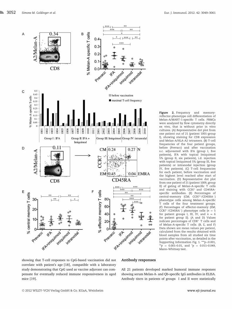
The primary aim of the study was to activate Melan-A-specific CD8+ T cells. PBMCs were analyzed by flow cytometrywith fluorescent peptide/HLA-A2 tetramers. In total, 16of 21 patients (76%) were immune responders to vac-cination with MelQbG10, as defined by at least twofoldincreased percentages of Melan-A tetramer positive cells afterstart of vaccination in comparison to before [17]. T-cellfrequencies were increased in all 11 patients of treatment groupsI and II, that is after vaccination with MelQbG10 together withIFA, with or without local application of Imiquimod (Fig. 2A–C
C© 2012 WILEY-VCH Verlag GmbH & Co. KGaA, Weinheim www.eji-journal.eu

Eur. J. Immunol. 2012. 42: 3049–3061 Clinical immunology 3051
Figure 1. Incidence of injection site reactions per treatment group. The number of patients with pain and/or itching and the number of patientswith a diameter ≥ 1 cm with regard to local erythema, swelling, and/or induration were enumerated in the available diary recordings. The resultsare shown for each injection. (A) Group I: local reactions after 1 mg s.c injection of MelQbG10 with IFA (n = 5) and (B) Group II: with IFA and topicalImiquimod 5% (n = 6); (C) Group III: i.d. injection with topical Imiquimod 5% (n = 5); and (D) Group IV: after ultrasound-guided intralymph nodeinjection (14/42/140 μg MelQbG10, n = 5).
and Supporting Information Table 2). Two patients in treatmentgroup III and three in treatment group IV also reached maximalT-cell frequencies fulfilling the T-cell responder criteria. Never-theless, in patients vaccinated without IFA but treated topicallywith Imiquimod (group III) and by intranodal injection (groupIV) the T-cell frequencies were significantly lower as compared topatients after MelQbG10 vaccinations with IFA (groups I and II,p = 0.003).
Patient age varied slightly from patient group I to IV(means of 58, 49, 61, and 56 years, respectively; Support-ing Information Table 1). We cannot formally exclude thatthese differences biased our results. However, it seems ratherunlikely, as there was no significant correlation; young patientsdid not differ significantly from older patients with regard totheir T-cell frequencies (Supporting Information Table 2). Thisfinding fits with our recent results from another study also
Table 1. All adverse events with an incidence > 3, listed for each group with number of events and number of patients
Group I events Group II events Group III events Group IV eventsPreferred term (patients) (patients) (patients) (patients)
General disorders and administration site conditionsInjection site induration 4(2) 15(5) 1(1)Influenza-like illness 2(2) 2(1) 7(2)Fatigue 4(3) 2(2) 10(4)Edema peripheral 4(2) 1(1)
Skin and subcutaneous tissue disordersSkin neoplasm excision 1(1) 8(3) 3(2)Skin nodule 2(2) 5(3) 1(1) 3(2)
Blood and lymphatic system disordersLymphadenopathya) 1(1) 1(1) 6(4) 4(3)
a)Defined as clinically palpable or radiologically enlarged lymph nodes.
C© 2012 WILEY-VCH Verlag GmbH & Co. KGaA, Weinheim www.eji-journal.eu
3052 Simone M. Goldinger et al. Eur. J. Immunol. 2012. 42: 3049–3061
Figure 2. Frequency and memory-/effector-phenotype cell differentiation ofMelan-A/MART-1-specific T cells. PBMCswere analyzed by flow cytometry directlyex vivo, that is without prior in vitrocultures. (A) Representative dot plot fromone patient out of 21 (patient 1001-groupI), showing staining for CD8 expressionand Melan-A/HLA-A2 tetramers. (B) T-cellfrequencies of the four patient groups,before (Prevacc) and after vaccinations.c. adjuvanted with IFA (group I, fivepatients), IFA with topical Imiquimod5% (group II, six patients), i.d. injectionwith topical Imiquimod 5% (group III, fivepatients) or intranodal injection (groupIV, five patients). (C) T-cell frequenciesfor each patient, before vaccination andthe highest level reached after start ofvaccination. (D) Representative dot plotfrom one patient of 21 (patient 1009, groupII) of gating of Melan-A-specific T cellsand staining with CCR7 and CD45RA-specific antibodies. (E) Percentages ofcentral-memory (CM; CCR7+/CD45RA−)phenotype cells among Melan-A-specificT cells of the four treatment groups.(F) Percentages of effector-memory (EM;CCR7−/CD45RA−) phenotype cells (n = 5for patient groups I, III, IV, and n = 6for patient group II). (A and D) Valuesindicate percentages of CD8+ T cells andof Melan-A-specific T cells. (B, E, and F)Data shown are mean values per patient,calculated from the results obtained withblood samples from all studied six timepoints after vaccination, as detailed in theSupporting Information Fig. 1. ***p<0.001,**p = 0.001–0.01, and *p = 0.011–0.049,Mann–Whitney test.
showing that T-cell responses to CpG-based vaccination did notcorrelate with patient’s age [18], compatible with a laboratorystudy demonstrating that CpG used as vaccine adjuvant can com-pensate for eventually reduced immune responsiveness in agedmice [19].
Antibody responses
All 21 patients developed marked humoral immune responsesshowing serum Melan-A- and Qb-specific IgG antibodies in ELISA.Antibody titers in patients of groups I and II were statistically
C© 2012 WILEY-VCH Verlag GmbH & Co. KGaA, Weinheim www.eji-journal.eu

Eur. J. Immunol. 2012. 42: 3049–3061 Clinical immunology 3053
Figure 3. Expression of surface receptors by Melan-A/MART-1-specific T cells. Melan-A-specific T cellswere gated similarly as in Figure 2, and analyzedfor expression of the costimulatory molecules CD27and CD28, the IL-7 receptor CD127 and the inhibitoryreceptor PD-1. (A) Representative dot plot from onepatient out of n = 5–6 for each treatment group,that is patient 1001 (group I), patient 1009 (groupII), patient 2002 (group III), and patient 2006 (groupIV). As a reference, histograms in the lowest rowshow naıve (CCR7+/CD45RA+/tetramer−) and effector(CCR7−/CD45RA+/tetramer−) phenotype cells, that issubsets of “antigen-nonspecific” CD8+ T cells from therepresentative patient 1009. Values in histograms indi-cate percent positive cells. (B–E) Statistical comparisonsbetween the four treatment groups for expression of(B) CD27, (C) CD28, (D) CD127, and (E) PD-1 by Melan-A-specific T cells. The data shown in panel B–E are meanvalues per patient, calculated from the results obtainedfrom all studied time points after vaccination, similarlyas for Figure 2. *** <0.001, ** 0.001–0.01, and * 0.011–0.049,Mann–Whitney test.
significantly higher than titers in patients of groups III and IV, inaccordance to the cellular responses described above (SupportingInformation Fig. 2).
T-cell differentiation
The proportions of Melan-A-specific T cells at the various dif-ferentiation stages were determined by staining with tetramers,combined with antibodies specific for the short isoform of CD45(CD45RA) and the chemokine receptor CCR7 [20]. Double nega-tive T cells, so-called effector-memory (EM)-phenotype cells, weresignificantly increased after IFA adjuvanted vaccination (Fig. 2F).In contrast, the proportions of central-memory (CM)-phenotype
cells (CCR7+ CD45RA−) were increased in patient groups II andIII who had been treated topically with Imiquimod (Fig. 2E).Multiparameter flow cytometry allowed simultaneous analysis offour additional receptors. The two costimulatory molecules CD27and CD28 are known to be progressively downregulated in CD8+
effector T cells. There was a trend to lower expression of CD27and CD28 after vaccination adjuvanted with only IFA or onlyImiquimod, but the differences to before vaccination were notsignificant (Fig. 3A–C). In accordance with the highest propor-tions of CM-phenotype cells, Imiquimod was also associated withthe highest frequencies of CD127+ (IL-7R) cells, which was sig-nificantly higher in comparison to each of the other three patientgroups (Fig. 3D). Finally, we also determined PD-1 expressionby the Melan-A-specific T cells. Interestingly, the mean values inthe first three patient groups were all approximately 32–34% and
C© 2012 WILEY-VCH Verlag GmbH & Co. KGaA, Weinheim www.eji-journal.eu
3054 Simone M. Goldinger et al. Eur. J. Immunol. 2012. 42: 3049–3061
thus significantly higher than before vaccination and higher thanin patient group IV with a mean value of only 14% (Fig. 3E).Figures 2 and 3 show the patient’s mean values from all timepoints after vaccination. Example data from a single time point(after six vaccinations with MelQbG10) are provided in Support-ing Information Fig. 3, showing similar differences between thefour patient groups. However, in many instances the differencesdid not reach statistical significance, likely due to the low numbersof samples.
Clinical and PET/CT imaging results
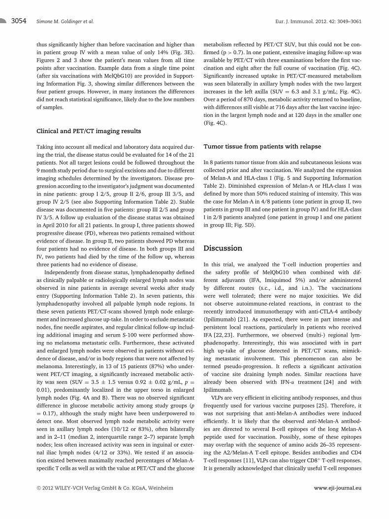
Taking into account all medical and laboratory data acquired dur-ing the trial, the disease status could be evaluated for 14 of the 21patients. Not all target lesions could be followed throughout the9 month study period due to surgical excisions and due to differentimaging schedules determined by the investigators. Disease pro-gression according to the investigator’s judgment was documentedin nine patients: group I 2/5, group II 2/6, group III 3/5, andgroup IV 2/5 (see also Supporting Information Table 2). Stabledisease was documented in five patients: group III 2/5 and groupIV 3/5. A follow up evaluation of the disease status was obtainedin April 2010 for all 21 patients. In group I, three patients showedprogressive disease (PD), whereas two patients remained withoutevidence of disease. In group II, two patients showed PD whereasfour patients had no evidence of disease. In both groups III andIV, two patients had died by the time of the follow up, whereasthree patients had no evidence of disease.
Independently from disease status, lymphadenopathy definedas clinically palpable or radiologically enlarged lymph nodes wasobserved in nine patients in average several weeks after studyentry (Supporting Information Table 2). In seven patients, thislymphadenopathy involved all palpable lymph node regions. Inthese seven patients PET/CT-scans showed lymph node enlarge-ment and increased glucose up-take. In order to exclude metastaticnodes, fine needle aspirates, and regular clinical follow-up includ-ing additional imaging and serum S-100 were performed show-ing no melanoma metastatic cells. Furthermore, these activatedand enlarged lymph nodes were observed in patients without evi-dence of disease, and/or in body regions that were not affected bymelanoma. Interestingly, in 13 of 15 patients (87%) who under-went PET/CT imaging, a significantly increased metabolic activ-ity was seen (SUV = 3.5 ± 1.5 versus 0.92 ± 0.02 g/mL, p =0.01), predominantly localized in the upper torso in enlargedlymph nodes (Fig. 4A and B). There was no observed significantdifference in glucose metabolic activity among study groups (p= 0.17), although the study might have been underpowered todetect one. Most observed lymph node metabolic activity wereseen in axillary lymph nodes (10/12 or 83%), often bilaterallyand in 2–11 (median 2, interquartile range 2–7) separate lymphnodes; less often increased activity was seen in inguinal or exter-nal iliac lymph nodes (4/12 or 33%). We tested if an associa-tion existed between maximally reached percentages of Melan-A-specific T cells as well as with the value at PET/CT and the glucose
metabolism reflected by PET/CT SUV, but this could not be con-firmed (p > 0.7). In one patient, extensive imaging follow-up wasavailable by PET/CT with three examinations before the first vac-cination and eight after the full course of vaccination (Fig. 4C).Significantly increased uptake in PET/CT-measured metabolismwas seen bilaterally in axillary lymph nodes with the two largestincreases in the left axilla (SUV = 6.3 and 3.1 g/mL; Fig. 4C).Over a period of 870 days, metabolic activity returned to baseline,with differences still visible at 716 days after the last vaccine injec-tion in the largest lymph node and at 120 days in the smaller one(Fig. 4C).
Tumor tissue from patients with relapse
In 8 patients tumor tissue from skin and subcutaneous lesions wascollected prior and after vaccination. We analyzed the expressionof Melan-A and HLA-class I (Fig. 5 and Supporting InformationTable 2). Diminished expression of Melan-A or HLA-class I wasdefined by more than 50% reduced staining of intensity. This wasthe case for Melan-A in 4/8 patients (one patient in group II, twopatients in group III and one patient in group IV) and for HLA-classI in 2/8 patients analyzed (one patient in group I and one patientin group III; Fig. 5D).
Discussion
In this trial, we analyzed the T-cell induction properties andthe safety profile of MelQbG10 when combined with dif-ferent adjuvants (IFA, Imiquimod 5%) and/or administeredby different routes (s.c., i.d., and i.n.). The vaccinationswere well tolerated; there were no major toxicities. We didnot observe autoimmune-related reactions, in contrast to therecently introduced immunotherapy with anti-CTLA-4 antibody(Ipilimumab) [21]. As expected, there were in part intense andpersistent local reactions, particularly in patients who receivedIFA [22,23]. Furthermore, we observed (multi-) regional lym-phadenopathy. Interestingly, this was associated with in parthigh up-take of glucose detected in PET/CT scans, mimick-ing metastatic involvement. This phenomenon can also betermed pseudo-progression. It reflects a significant activationof vaccine site draining lymph nodes. Similar reactions havealready been observed with IFN-α treatment [24] and withIpilimumab.
VLPs are very efficient in eliciting antibody responses, and thusfrequently used for various vaccine purposes [25]. Therefore, itwas not surprising that anti-Melan-A antibodies were inducedefficiently. It is likely that the observed anti-Melan-A antibod-ies are directed to several B-cell epitopes of the long Melan-Apeptide used for vaccination. Possibly, some of these epitopesmay overlap with the sequence of amino acids 26–35 represent-ing the A2/Melan-A T-cell epitope. Besides antibodies and CD4T-cell responses [11], VLPs can also trigger CD8+ T-cell responses.It is generally acknowledged that clinically useful T-cell responses
C© 2012 WILEY-VCH Verlag GmbH & Co. KGaA, Weinheim www.eji-journal.eu

Eur. J. Immunol. 2012. 42: 3049–3061 Clinical immunology 3055
Figure 4. PET/CT imaging showing enlarged lymph nodes and increased glucose metabolism after vaccination. Fifteen of the 21 patients ofthis study underwent PET/CT imaging. In parallel to the assessment of disease development (not shown), immune activation was evaluated bymeasuring the glucose metabolism in vaccine-site draining lymph nodes. (A) Patient 2002 (group III) with bilateral axillary lymph nodes with onethat is enlarged (arrow). Images: Maximal intensity projection (left) with PET/CT fusion image (top), PET image (middle), and CT image (bottom).(B) Patient 2004 (group III) with multiple bilateral axillary (arrow, horizontal arrowhead) and subclavicular (vertical arrowhead) lymph nodes withincreased glucose metabolism. (C) Patient 1003 (group I) with long-term longitudinal follow-up before and after the seven vaccinations injectedbetween the third and fourth PET/CT studies. The standardized uptake value (SUV) was measured in two axillary lymph nodes (LN1, LN2) frompatient 1003 (group I) before and after the last vaccination (right). Note also the local inflammation in vaccination sites in the proximal right andleft arms and left thigh (left).
are much more difficult to generate as compared to antibodyresponses. Therefore, T-cell vaccines are much less advanced asopposed to B-cell vaccines. Numerous academic and industrialteams undertake multiple research and development efforts, inorder to elucidate the precise biological parameters that need tobe considered for generating T-cell responses that are capable to
protect from infectious or malignant disease. In this mindset, weare optimizing VLP-based vaccines.
In a previous study in melanoma patients, we have shown thatMelQbG10 can trigger ex vivo detectable and thus relatively strongtumor-specific CD8+ T-cell responses [10]. The present studywas based on the same vaccine (MelQbG10), but extended by
C© 2012 WILEY-VCH Verlag GmbH & Co. KGaA, Weinheim www.eji-journal.eu

3056 Simone M. Goldinger et al. Eur. J. Immunol. 2012. 42: 3049–3061
Figure 5. Reduced HLA-class I and Melan-A/MART-1 expression after vaccination. Immunohistochemistry images (×8 magnification) of differenttumor biopsies before (left) and after (right) vaccination showing reduced Melan-A or HLA-class I expression. (A) Tumor biopsies from plantar left(primary tumor, left) and left leg (in transit cutaneous metastasis, right) with high (left) and low (right) Melan-A intensity staining (Patient 2010,group IV). (B) Biopsies of two dermal abdominal metastases, with high (left) and low (right) Melan-A intensity staining (Patient 2001, group III). (C)Cutaneous metastasis of the right knee (right and left) with low HLA-class I intensity staining (Patient 1003, group I). (D) Melan-A and HLA-class Iexpression before and after vaccination, for each evaluable patient (n = 8) are shown.
additional components. The addition of IFA resulted in signifi-cantly increased T-cell frequencies. These responses were domi-nated by EM-phenotype cells, that is CD45RA−CCR7− cells. Thesecells were also mostly CD127 (IL-7R) negative, demonstratingthat IFA primarily induced effector-phenotype cells, with onlylow proportions of memory-phenotype cells. These results arereminiscent of vaccines composed of synthetic peptide, CpG-ODN, and IFA, which are also dominated by EM-phenotype CD8+
T cells [26]. In contrast, our data show that increased percent-ages of CM-phenotype cells (CD45RA−CCR7+) with enhancedexpression of CD127 were induced by the addition of Imiquimod.Thus, triggering TLR-7 by Imiquimod may promote memory dif-ferentiation, which is promising because memory cells are knownto increase the protective potential of T-cell responses [27–29].Despite its importance, memory cell triggering is often insuffi-cient by synthetic vaccines. The mechanisms by which TLR-7 maypromote memory cell activation are likely linked to DC activa-tion via TLR-7. The generation of memory cells may depend onWnt-TCF-1, mTor, and AMPK signaling pathways [27–29]. Our
data point to possible connections between these mechanisms andTLR-7 induced pathways.
Our previous study showed that even without topicalImiquimod vaccination with MelQbG10 induced increased per-centages of CM-phenotype cells [10]. Here, we show that the com-bination of MelQbG10 plus Imiquimod and IFA (thus triggering ofTLR-7 and -9) leads to enhance promotion of memory-phenotypecells. However, we have never tested whether this can be achievedby triggering of TLR-7 without TLR-9, since MelQbG10 alwayscontained the CpG-ODN G10.
Recent literature reported that the combined stimulationof multiple innate immune receptors may enhance vaccineefficacy [30], reminiscent to the notion that pathogens may trig-ger innate immunity due to the expression of multiple microbe-associated molecular patterns (MAMPs). However, there are stillconsiderable limitations in the availability of clinically gradeddrugs for triggering innate immune receptors. Imiquimod is askin cream and can only be applied topically. It will be inter-esting to test whether systemic and thus more efficient TLR-7
C© 2012 WILEY-VCH Verlag GmbH & Co. KGaA, Weinheim www.eji-journal.eu
Eur. J. Immunol. 2012. 42: 3049–3061 Clinical immunology 3057
triggering can promote even higher numbers of CM-phenotypecells.
The frequencies of Melan-A-specific T cells after vaccinationwith MelQbG10 plus Imiquimod were relatively low. In contrast,the addition of IFA resulted in higher frequencies. Thus, combin-ing CpG-ODN, Imiquimod, and IFA achieved both memory- andeffector-phenotype cells, with relatively high frequencies and highproportions of CM-phenotype cells. However, our data are pre-liminary due to the low numbers of patients, and thus requirefurther confirmation.
Even though the percentages of tetramer positive T cells wererelatively low, they actually revealed large numbers of tumor-specific T cells, because we analyzed the blood samples directlyex vivo. In many cancer vaccine studies, T cells are analyzed afterthey have proliferated in tissue cultures for one or several weeks,providing results with higher T-cell percentages but precludingprecise conclusions on T-cell numbers and qualities in vivo [12].Therefore, recent cancer vaccine studies increasingly include anal-ysis of T cells directly ex vivo [31–38]. Because of considerableinter-patient variability of frequencies of tumor-antigen-specificT cells, at baseline and after immunotherapy, it is important todetermine how many patients are “immune–responders”, basedon the comparison of their values before versus after vaccina-tion. Data from direct ex vivo analysis show that the majority ofvaccination strategies were able to trigger responses of Melan-A-specific CD8+ T cells but usually not in all patients [31–38]. Forexample, a recent study by Ribas et al. showed T-cell responsesin about 50% of melanoma patients vaccinated with DNA fol-lowed by peptides, a novel prime-boost approach [36]. The resultsof our present study with 16/21 immune responder patientsis favorable, particularly when focusing on the 11 patients inwhom vaccination included IFA (groups I and II) who all showedex vivo detectable T-cell responses. However, the T-cell fre-quencies remained lower than in patients vaccinated with pep-tide + CpG 7909 (PF-3512676) emulsified in IFA [18,26], rais-ing the possibility that B-type CpGs (e.g. 7909) may be supe-rior to A-type CpGs (e.g. G10) for CD8+ T-cell vaccination inhumans. Definitive conclusions depend on trials directly com-paring the various vaccination approaches. Importantly, clinicalefficacy needs to be tested in large-scale trials. The currentlyavailable evidence of clinical benefit of CpG-based vaccines [18]justify evaluating their clinical usefulness in phase IIIstudies.
We developed MelQbG10 several years ago, and used the ana-log peptide sequence ELAGIGILTV with L instead of A at position 2as compared with the natural sequence EAAGIGILTV. As for otheranalog peptides, we found that this analog peptide triggered rel-atively high frequencies of T cells. However, more recently wefound that T-cell responses were more robust after vaccinationwith the natural peptide, since the T cells were more stronglyactivated and were of higher functional avidity as compared withT cells after vaccination with the analog peptide [39]. Therefore,it is preferable to vaccinate with peptides corresponding to nativetumor antigens. Unfortunately, such peptides are often insuffi-ciently antigenic, particularly when cross-presentation is required
such as for long peptides as used in this study. Therefore, immu-nization with analog peptides is still justified since reasonablylarge fractions (>50%) of the induced T cells can recognize tumorcells [39]. The question whether the native or the analog Melan-Apeptide should be used for T-cell analysis is much easier to answer.Tetramers constructed with the Melan-A analog peptide are “uni-versal,” as they readily bind virtually all T cells induced by analogor natural peptide, but also endogenously by the tumor itself [39].Thus, A2/Melan-A tetramer binding is highly cross-specific anddoes not discriminate between the very fine differences in speci-ficity and affinity depending on vaccination with analog versusnative antigen.
Of interest, PET/CT could evidence increased lymph nodeactivity in the majority of the patients, as a way of show-ing increased activity in relation to inflammation and immuneresponse, even in the long term (>4 months). This has not beenutilized in vaccine studies so far. It has rather been described asan epiphenomenon in relation to the last vaccination campaignsagainst influenza A/H1N1 pandemic as seen in patients under-going oncologic PET/CT [40–42] or in sporadic immunizationcases [43,44]. Increased glucose metabolism in lymph nodes wasnot observable >14 days after immunization in the study by Burgeret al. [42] or >30–50 days by Thomassen et al. [41], possibly dueto the shorter stimulation time of these vaccines as opposed tothe vaccine formulations used in the present study. Interestingly,Iyengar et al. [45] found an association between PET-measuredlymph node signals and viremia in HIV-infected patients thatwas attributed to CD4 lymphocyte activation in relation to HIVreplication. They observed a predominance for upper torso lymphnodes in recently and chronically HIV-infected patients, but not intheir control group of healthy non-HIV-infected volunteers under-going killed influenza vaccination, where it was observed onlyon the upper torso of the injection site. In future trials, PET/CTcould be an attractive way to noninvasively monitor and investi-gate inflammatory and immune responses to vaccination, althoughthe exact mechanisms and significance of the increased glucosemetabolism remain to be investigated.
In summary, MelQbG10 can be considered safe and well tol-erated when given at a cumulative dose of 6 mg (6 × 1 mg).The use of IFA resulted in more frequent side effects, butinduced the highest Melan-A-specific T-cell frequencies. Bothmemory- and effector-phenotype T-cell responses were observedwhen MelQbG10/IFA vaccines were given in combination withImiquimod. Our data suggest that the induced immune activa-tion was biologically relevant because several relapsing melanomalesions demonstrated diminished expression of Melan-A or of HLA-class I, presumable reflecting mechanisms of immune escape [46].Future vaccines need to target multiple antigens and HLAs simul-taneously.
Vaccine components need to be tested in humans, in orderto determine which of them, and what type of final vaccine for-mulations are most promising for further and large scale clin-ical development. We used the A-type CpG-ODN G10 that wehad integrated in our VLPs, resulting in sizable T-cell responses,which were however less powerful than in rodents [10]. In the
C© 2012 WILEY-VCH Verlag GmbH & Co. KGaA, Weinheim www.eji-journal.eu
3058 Simone M. Goldinger et al. Eur. J. Immunol. 2012. 42: 3049–3061
present paper, we combined this approach with additional adju-vants and/or varied the administration route and found quantita-tively and qualitatively different T-cell responses. The next stepswill be to replace some components, by using, for example a B-typeCpG-ODN, and/or by supplementation with (parenteral) drugsthat trigger additional TLRs and further innate immune receptors.With a step-by-step approach, we will learn which type of vaccineformulation is optimal for triggering CTL and Th1 responses withthe potential of clinical benefit for cancer patients.
Materials and methods
Patients and trial design
Eligible patients were at least 18-years old, with histologically con-firmed stage III or IV malignant melanoma (AJCC), HLA-A*0201positivity, and an expected survival of at least 9 months. The cur-rent general health condition had to allow the patient to undergoall study procedures according to the protocol. Main exclusion cri-teria were known or planned pregnancy or lactation, use of anyinvestigational drug, and previous participation in a clinical trialwith a Qb-based vaccine. The first patient was enrolled in May2008, the last in December 2009. All patients provided writteninformed consent before any study-specific procedure was per-formed.
The exact composition of MelQbG10 was reported earlier [10].The first treatment group (group I) received three initial sub-cutaneous (s.c.) injections of 1 mg MelQbG10 mixed with IFA(Montanide ISA-51, Seppic GmbH, Koeln, Germany) in weeklyintervals, followed by three monthly injections with the vaccine.The second treatment group (group II) was vaccinated identi-cally to the first group but was additionally treated with top-ical Imiquimod 5% cream (AldaraTM, 3M, Switzerland) oncedaily for 10 days (with 2 days of treatment pause after the first5 days). The cream was applied with a thin layer on the injectionsite and covered an area of 5 × 5 cm. The third group (group III)received three intradermal (i.d.) injections of 1 mg MelQbG10 inweekly intervals, followed by three monthly intradermal injectionswith MelQbG10. In addition, the patients were treated with a thinlayer of Imiquimod 5% cream once daily as described for group II.The fourth treatment group (group IV) received three ultrasound-guided intranodal (i.n.) injections of MelQbG10 initially startingwith 14 μg, then 42 μg, and thereafter 140 μg in weekly inter-vals, followed by three monthly injections of 140 μg MelQbG10.All treatment groups received a final boost 4 weeks after the lastvaccination with the Melan-A/MART-1 peptide plus IFA s.c (formore details on trial design see Supporting Information Fig. 1).Before starting treatment group II (Imiquimod + IFA) an inter-nal safety data review was performed with respect to treatmentgroup III (i.d. injection + Imiquimod). Local ethic review com-mittees and responsible health authorities approved the study,which was performed according to GCP guidelines and registeredat http://www.ClinicalTrials.gov: NCT00651703.
Assessment of safety and tolerability and clinicalresults
Adverse events (AEs), concomitant medication, vital signs, physi-cal examinations, ECGs, antinuclear antibodies, and routine clin-ical laboratory blood and urine analyses were monitored care-fully at defined visits. Local reactions at the injection sites wererecorded by patients and investigators. Patients were asked torecord local reactions after every injection of study medication ina paper-based diary for a period of 3 days. Pain and itching weredocumented; swelling, and induration were assessed by record-ing the reactions’ diameter if the diameter was > 1 cm. Localreactions that required medical intervention were documented asAE. Safety and tolerability parameters were primarily evaluatedwithin the single subject and compared with baseline, in addi-tion, trends between the four groups were analyzed. In total 21melanoma patients have been included and represented the safetypopulation, which was identical to the full analysis set. No datahave been extrapolated.
The status of the disease was monitored clinically and/or bycomputer tomography (CT) at screening and at the end of thetrial, if clinically indicated a fluorodeoxyglucose-positron emis-sion tomography (FDG-PET/CT) was performed (GE Healthcare,Milwaukee, WI, USA). To compare PET/CT imaging results,we used the maximal standardized uptake value (SUV ing/mL) corrected for body weight as a way to reflect glucoseconsumption.
Immune monitoring and immunohistochemistry
Primary goal of the clinical trial was to achieve efficient Melan-A-specific T-cell responses by enhancing the immuno-stimulatingpotential of MelQbG10 with various adjuvants and differentadministration modes in patients with malignant melanoma.Simultaneous enumeration and phenotyping of antigen-specificCD8+ T cells was achieved by flow cytometry and were ana-lyzed with FlowJoTM software (TreeStar). Ficoll-Paque centrifugedPBMC (1–2 × 10⁷) were cryopreserved in RPMI 1640, 40%FCS, and 10% DMSO. Phycoerythrin-labeled HLA-A*0201/Melan-A/MART-1 A27L peptide26–35 (ELAGIGILTV) tetramers were pre-pared as described [17]. Anti-CD8, -CD28, -PD-1, and anti-CCR7mAbs were purchased from BD PharMingen (San Diego, CA,USA), anti-CD45RA from Beckman Coulter (Brea, CA, USA), andanti-CD27 and -CD127 from eBioscience (San Diego, CA, USA).All tetramer and antibody batches were titrated to determineoptimal reagent concentrations. PBMC were thawed, and CD8+
T cells were enriched using a MiniMACS device (Miltenyi Biotec,Bergisch Gladbach, Germany) resulting in >90% CD3pos CD8pos
cells. Cells (106) were incubated with tetramers (1 μg/mL,60 min, 4◦C) and then with antibodies (30 min, 4◦C). For deadcell exclusion, cells were stained with Live/Dead Fixable DeadCell violet stain (Molecular Probes/Invitrogen). A total of 5 × 10⁵
CD8+ T cells/sample were acquired with a flow cytometry LSR IITM
machine. The cytometer performance was checked daily using the
C© 2012 WILEY-VCH Verlag GmbH & Co. KGaA, Weinheim www.eji-journal.eu

Eur. J. Immunol. 2012. 42: 3049–3061 Clinical immunology 3059
CST (BDTM Cytometer Setup and Tracking) quality control beadssystem according to the manufacturers’ instructions. To evaluatewhether the CD8 T cells from the 21 patients of this study wererepresentative for larger numbers of patients, we compared theirpre-vaccination values to those of 37 untreated patients, also withstage III–IV metastatic skin melanoma, which we have recentlystudied with an identical flow cytometry approach. For all param-eters studied (Melan-A tetramer, CD27, CD28, CD127, PD-1, andeffector-memory-phenotype and central-memory-phenotype cells)there were no statistical differences (data not shown). Results oftetramer + T cells were calculated and are indicated in percent-ages of circulating CD8+ T cells. Results from T-cell phenotypingwere only used when the numbers of tetramer positive events wereat least 20.
Biopsies of metastases were stained for the melanocytic dif-ferentiation antigen and HLA-class I before and after the treat-ment. For immunohistochemistry the paraffin-embedded tumortissue sections obtained at different time points prior and aftervaccination were stained with anti-Melan-A/MART-1 monoclonalantibody A103 (Novocastra Laboratories Ltd., Newcastle uponTyne, UK) [9] and with anti-HLA-class I antibody 3F10 (1:1000;RDI Research Diagnostics, Inc., Concord, MA, USA) [47]. Thequantification was done by an experienced, board-certified der-matopathologist (RD) on a Zeiss Axiophot HAL100 (Carl ZeissMicroimaging GmbH, Switzerland) with ×40 magnification.Immunoreactivity, evaluated as intensity of the staining in rela-tionship to normal melanocytes in the epidermis (internal control),was scored semiquantitatively in percentages (0–100%). A clearcut difference, interpreted as diminished expression of Melan-A orHLA-class I, was considered starting from a change of 50% or morein the comparison of the sections prior to and after vaccination, asillustrated in Figure 5. Because of expression heterogeneity [9],we analyzed three high power fields of all lesions.
Data and statistical analysis
Evaluation of the disease status was described for each patientindividually. AEs were coded using MedDRA Version 12.0. Sum-mary tables were presented by frequency of AEs and by patient.AEs were listed by subject, including verbatim term, coded Med-DRA term, severity, and relationship to treatment. Standard labo-ratory measurements, vital signs, and ECG parameters were listedby subject and time point of collection. Out of normal range andclinically significant deviating values were summarized by descrip-tive statistics. Anti-Melan-A-specific T cells were expressed as %tetramer positive cells of total CD8+ T cells. Immune responderswere defined as patients whose maximal T-cell frequency at anytime point during the trial was at least double the preimmuneT-cell frequency. T-cell frequencies below 0.01% were consid-ered as not detectable (threshold) [17]. Statistical significancewas assessed by the nonparametric Mann–Whitney and Kruskal–Wallis tests or the parametric Student’s t-test where appropriate.Associations were tested using nonparametric Spearman rank cor-relation. P values less than 0.05 were considered significant and
reported in the figures. The scatter dot plots in Figure 2 and 3show individual values, mean values, and standard deviations.
Acknowledgments: The authors thank the patients for their ded-icated collaboration and Cytos Biotechnology AG for their coop-eration during the study. We gratefully acknowledge L.J. Old, J.O’Donnell-Tormey, E. Devevre, and H. Bouchaab for their contri-butions, H.R. MacDonald, P. Romero, J. Skipper, and H.F. Oettgenfor support, P. Guillaume and I. Luescher for tetramers, and M.Braun, C. Jandus, J.-P. Rivals, L. Derre, D. Rimoldi, H. Bouzourene,F. Bosman, H.-A. Lehr, L.Cagnon, M. Iancu, L. Derre, T. Lovgren,A. Baur, J.-Y. Meuwly, L. Pan, and R. Venhaus for collabora-tion and advice. This work was supported by grants from CytosBiotechnology AG (Switzerland), by the Swiss Cancer League(grant nr 02279–08-2008), the Swiss National Science Founda-tion (grant nr 310030–135553), the Ludwig Institute for CancerResearch, the Cancer Research Institute (USA), the Cancer VaccineCollaborative (USA), the Gottfried and Julia Bangeter Founda-tion (Switzerland), and the Schnyder Foundation (Switzerland).Partly presented at the Melanoma Centers meeting, ClearwaterBeach, Florida, 2008; at the 7th World Congress on Melanoma/5thCongress of the EADO, Vienna, 2009; at the 6th Congress of theEADO, Athens, 2010 and at the 7th Congress of the EADO, Nantes,2011.
Conflict of interest: K.S., A.H., J.W., and M.B. are employeesof Cytos Biotechnology AG. The remaining authors declare nofinancial or commercial conflict of interest.
References
1 Dudley, M. E., Yang, J. C., Sherry, R., Hughes, M. S., Royal, R., Kam-
mula, U., Robbins, P. F. et al., Adoptive cell therapy for patients with
metastatic melanoma: evaluation of intensive myeloablative chemora-
diation preparative regimens. J. Clin. Oncol. 2008. 26: 5233–5239.
2 Hodi, F. S., O’Day, S. J., McDermott, D. F., Weber, R. W., Sosman, J. A.,
Haanen, J. B., Gonzalez, R. et al., Improved survival with ipilimumab in
patients with metastatic melanoma. N. Engl. J. Med. 2011. 363: 711–723.
3 Robert, C., Thomas, L., Bondarenko, I., O’Day, S., M, D. J., Garbe, C.,
Lebbe, C., Baurain, J. F. et al., Ipilimumab plus dacarbazine for previously
untreated metastatic melanoma. N. Engl. J. Med. 2011. 364: 2517–26.
4 Rosenberg, S. A., Yang, J. C. and Restifo, N. P., Cancer immunotherapy:
moving beyond current vaccines. Nat. Med. 2004. 10: 909–915.
5 Eggermont, A. M., Immunotherapy: vaccine trials in melanoma – time
for reflection. Nat. Rev. Clin. Oncol. 2009. 6: 256–258.
6 Fox, B. A., Schendel, D. J., Butterfield, L. H., Aamdal, S., Allison, J. P.,
Ascierto, P. A., Atkins, M. B. et al., Defining the critical hurdles in cancer
immunotherapy. J. Transl. Med. 2011. 9: 214.
7 Baitsch, L., Fuertes-Marraco, S. A., Legat, A., Meyer, C. and Speiser, D. E.,
The three main stumbling blocks for anticancer T cells. Trends Immunol.
2012. 33: 364–372.
C© 2012 WILEY-VCH Verlag GmbH & Co. KGaA, Weinheim www.eji-journal.eu
3060 Simone M. Goldinger et al. Eur. J. Immunol. 2012. 42: 3049–3061
8 Manolova, V., Flace, A., Bauer, M., Schwarz, K., Saudan, P. and Bach-
mann, M. F., Nanoparticles target distinct dendritic cell populations
according to their size. Eur. J. Immunol. 2008. 38: 1404–1413.
9 Hofbauer, G. F., Kamarashev, J., Geertsen, R., Boni, R. and Dum-
mer, R., Melan A/MART-1 immunoreactivity in formalin-fixed paraffin-
embedded primary and metastatic melanoma: frequency and distribu-
tion. Melanoma Res. 1998. 8: 337–343.
10 Speiser, D. E., Schwarz, K., Baumgaertner, P., Manolova, V., Devevre, E.,
Sterry, W., Walden, P. et al., Memory and effector CD8 T-cell responses
after nanoparticle vaccination of melanoma patients. J. Immunother. 2010.
33: 848–858.
11 Braun, M., Jandus, C., Maurer, P., Hammann-Haenni, A., Schwarz, K.,
Bachmann, M. F., Speiser, D. E. et al., Virus-like particles induce robust
human T-helper cell responses. Eur. J. Immunol. 2012. 42: 330–340.
12 Speiser, D. E., Pittet, M. J., Rimoldi, D., Guillaume, P., Luescher, I. F.,
Lienard, D., Lejeune, F. et al., Evaluation of melanoma vaccines with
molecularly defined antigens by ex vivo monitoring of tumor-specific T
cells. Semin. Cancer Biol. 2003. 13: 461–472.
13 Appay, V., Speiser, D. E., Rufer, N., Reynard, S., Barbey, C., Cerottini, J. C.,
Leyvraz, et al., Decreased specific CD8+ T-cell cross-reactivity of antigen
recognition following vaccination with Melan-A peptide. Eur. J. Immunol.
2006. 36: 1805–1814.
14 Fourcade, J., Kudela, P., Andrade Filho, P. A., Janjic, B., Land, S. R., Sander,
C., Krieg, A. et al., Immunization with analog peptide in combination
with CpG and montanide expands tumor antigen-specific CD8+ T cells
in melanoma patients. J. Immunother. 2008. 31: 781–791.
15 Adams, S., O’Neill, D. W., Nonaka, D., Hardin, E., Chiriboga, L., Siu, K.,
Cruz, C. M. et al., Immunization of malignant melanoma patients with
full-length NY-ESO-1 protein using TLR7 agonist imiquimod as vaccine
adjuvant. J. Immunol. 2008. 181: 776–784.
16 Urosevic, M., Dummer, R., Conrad, C., Beyeler, M., Laine, E., Burg, G.
and Gilliet, M., Disease-independent skin recruitment and activation of
plasmacytoid predendritic cells following imiquimod treatment. J. Natl.
Cancer Inst. 2005. 97: 1143–1153.
17 Speiser, D. E., Pittet, M. J., Guillaume, P., Lubenow, N., Hoffman, E., Cerot-
tini, J. C. and Romero, P., Ex vivo analysis of human antigen-specific CD8+T-cell responses: quality assessment of fluorescent HLA-A2 multimer
and interferon-gamma ELISPOT assays for patient immune monitoring.
J. Immunother. 2004. 27: 298–308.
18 Baumgaertner, P., Jandus, C., Rivals, J. P., Derre, L., Lovgren, T., Baitsch,
L., Guillaume, P. et al., Vaccination-induced functional competence of
circulating human tumor-specific CD8 T-cells. Int. J. Cancer 2012. 130:
2607–2617.
19 Posnett, D. N., Engelhorn, M. E., Lin, Y., Merghoub, T., Duan, F., Wolchok,
J. D. and Houghton, A. N., Development of effective vaccines for old mice
in a tumor model. Vaccine 2009. 27: 1093–1100.
20 Sallusto, F., Lenig, D., Forster, R., Lipp, M. and Lanzavecchia, A., Two
subsets of memory T lymphocytes with distinct homing potentials and
effector functions. Nature 1999. 401: 708–712.
21 Bouwhuis, M. G., Ten Hagen, T. L., Suciu, S. and Eggermont, A. M.,
Autoimmunity and treatment outcome in melanoma. Curr. Opin. Oncol.
2011. 23: 170–176.
22 Chianese-Bullock, K. A., Pressley, J., Garbee, C., Hibbitts, S., Murphy, C.,
Yamshchikov, G., Petroni, G. R. et al., MAGE-A1-, MAGE-A10-, and gp100-
derived peptides are immunogenic when combined with granulocyte-
macrophage colony-stimulating factor and montanide ISA-51 adjuvant
and administered as part of a multipeptide vaccine for melanoma. J.
Immunol. 2005. 174: 3080–3086.
23 Lienard, D., Rimoldi, D., Marchand, M., Dietrich, P. Y., van Baren,
N., Geldhof, C., Batard, P. et al., Ex vivo detectable activation of
Melan-A-specific T cells correlating with inflammatory skin reactions
in melanoma patients vaccinated with peptides in IFA. Cancer Immun.
2004. 4: 4.
24 Heinzerling, L. M., Anliker, M. D., Muller, J., Schlaeppi, M. and von Moos,
R., Sarcoidosis induced by interferon-α in melanoma patients: incidence,
clinical manifestations, and management strategies. J. Immunother. 33:
834–839.
25 Bachmann, M. F. and Jennings, G. T., Vaccine delivery: a matter of size,
geometry, kinetics and molecular patterns. Nat. Rev. Immunol. 2010. 10:
787–796.
26 Speiser, D. E., Lienard, D., Rufer, N., Rubio-Godoy, V., Rimoldi, D., Leje-
une, F., Krieg, A. M. et al., Rapid and strong human CD8+ T-cell responses
to vaccination with peptide, IFA, and CpG oligodeoxynucleotide 7909. J.
Clin. Invest. 2005. 115: 739–746.
27 Gattinoni, L., Zhong, X. S., Palmer, D. C., Ji, Y., Hinrichs, C. S., Yu, Z.,
Wrzesinski, C. et al., Wnt signaling arrests effector T-cell differentia-
tion and generates CD8+ memory stem cells. Nat. Med. 2009. 15: 808–
813.
28 Araki, K., Youngblood, B. and Ahmed, R., The role of mTOR in memory
CD8 T-cell differentiation. Immunol. Rev. 2010. 235: 234–243.
29 Finlay, D. and Cantrell, D. A., Metabolism, migration and memory in
cytotoxic T cells. Nat. Rev. Immunol. 2011. 11: 109–117.
30 Kasturi, S. P., Skountzou, I., Albrecht, R. A., Koutsonanos, D., Hua, T.,
Nakaya, H. I., Ravindran, R. et al., Programming the magnitude and per-
sistence of antibody responses with innate immunity. Nature 2011. 470:
543–547.
31 Bins, A., Mallo, H., Sein, J., van den Bogaard, C., Nooijen, W., Vyth-
Dreese, F., Nuijen, B. et al., Phase I clinical study with multiple peptide
vaccines in combination with tetanus toxoid and GM-CSF in advanced-
stage HLA-A*0201-positive melanoma patients. J. Immunother. 2007. 30:
234–239.
32 Butterfield, L. H., Comin-Anduix, B., Vujanovic, L., Lee, Y., Dissette, V. B.,
Yang, J. Q., Vu, H. T. et al., Adenovirus MART-1-engineered autologous
dendritic cell vaccine for metastatic melanoma. J. Immunother. 2008. 31:
294–309.
33 Chakraborty, N. G., Chattopadhyay, S., Mehrotra, S., Chhabra, A. and
Mukherji, B., Regulatory T-cell response and tumor vaccine-induced
cytotoxic T lymphocytes in human melanoma. Hum. Immunol. 2004. 65:
794–802.
34 Di Pucchio, T., Pilla, L., Capone, I., Ferrantini, M., Montefiore, E., Urbani,
F., Patuzzo, R. et al., Immunization of stage IV melanoma patients with
Melan-A/MART-1 and gp100 peptides plus IFN-alpha results in the acti-
vation of specific CD8(+) T cells and monocyte/dendritic cell precursors.
Cancer Res. 2006. 66: 4943–4951.
35 Lesimple, T., Neidhard, E. M., Vignard, V., Lefeuvre, C., Adamski, H.,
Labarriere, N., Carsin, A. et al., Immunologic and clinical effects of inject-
ing mature peptide-loaded dendritic cells by intralymphatic and intran-
odal routes in metastatic melanoma patients. Clin. Cancer Res. 2006. 12:
7380–7388.
36 Ribas, A., Weber, J. S., Chmielowski, B., Comin-Anduix, B., Lu, D.,
Douek, M., Ragavendra, N. et al., Intra-lymph node prime-boost vac-
cination against Melan A and tyrosinase for the treatment of metastatic
melanoma: results of a phase 1 clinical trial. Clin. Cancer Res. 2011. 17:
2987–2996.
37 Tuettenberg, A., Becker, C., Huter, E., Knop, J., Enk, A. H. and Jonuleit, H.,
Induction of strong and persistent Melan A/MART-1-specific immune
responses by adjuvant dendritic cell-based vaccination of stage II
melanoma patients. Int. J. Cancer 2006. 118: 2617–2627.
38 Weber, J., Boswell, W., Smith, J., Hersh, E., Snively, J., Diaz, M., Miles,
S. et al., Phase 1 trial of intranodal injection of a Melan-A/MART-1 DNA
C© 2012 WILEY-VCH Verlag GmbH & Co. KGaA, Weinheim www.eji-journal.eu

Eur. J. Immunol. 2012. 42: 3049–3061 Clinical immunology 3061
plasmid vaccine in patients with stage IV melanoma. J. Immunother. 2008.
31: 215–223.
39 Speiser, D. E., Baumgaertner, P., Voelter, V., Devevre, E., Barbey, C., Rufer,
N. and Romero, P., Unmodified self antigen triggers human CD8 T cells
with stronger tumor reactivity than altered antigen. Proc. Natl. Acad. Sci.
USA 2008. 105: 3849–3854.
40 Panagiotidis, E., Exarhos, D., Housianakou, I., Bournazos, A. and Dat-
seris, I., FDG uptake in axillary lymph nodes after vaccination against
pandemic (H1N1). Eur. Radiol. 2010. 20: 1251–1253.
41 Thomassen, A., Lerberg Nielsen, A., Gerke, O., Johansen, A. and Petersen,
H., Duration of 18F-FDG avidity in lymph nodes after pandemic H1N1v
and seasonal influenza vaccination. Eur. J. Nucl. Med. Mol. Imaging 2011.
38: 894–898.
42 Burger, I. A., Husmann, L., Hany, T. F., Schmid, D. T. and Schaefer, N. G.,
Incidence and intensity of F-18 FDG uptake after vaccination with H1N1
vaccine. Clin. Nucl. Med. 2011. 36: 848–853.
43 Williams, G., Joyce, R. M. and Parker, J. A., False-+ axillary lymph node
on FDG-PET/CT scan resulting from immunization. Clin. Nucl. Med. 2006.
31: 731–732.
44 Sheehy, N. and Drubach, L., (18)F-FDG uptake at vaccination site. Pediatr.
Radiol. 2008. 38: 246.
45 Iyengar, S., Chin, B., Margolick, J. B., Sabundayo, B. P. and Schwartz,
D. H., Anatomical loci of HIV-associated immune activation and associ-
ation with viraemia. Lancet 2003. 362: 945–950.
46 Geertsen, R., Hofbauer, G., Kamarashev, J., Yue, F. Y. and Dummer, R.,
Immune escape mechanisms in malignant melanoma. Int. J. Mol. Med.
1999. 3: 49–57.
47 Walter, A., Barysch, M. J., Behnke, S., Dziunycz, P., Schmid, B., Ritter,
E., Gnjatic, S., Kristiansen, G., Moch, H., Knuth, A., Dummer, R. and
van den Broek, M., Cancer-testis antigens and immunosurveillance in
human cutaneous squamous cell and basal cell carcinomas. Clin. Cancer
Res. 2010. 16: 3562–3570.
Abbreviations: CT: computer tomography · FDG-PET: fluorodeox
yglucose-positron emission tomography · IFA: Incomplete Freund’s
Adjuvant · ODN: oligonucleotide · (S)AE: (serious) adverse event · VLP:
virus-like nano-particle
Full correspondence: Prof. Daniel E. Speiser, Clinical Tumor Biology &Immunotherapy Unit, Ludwig Center of the University of Lausanne,CHUV HO 05/1552, Av. Pierre-Decker 4, CH-1011 Lausanne, SwitzerlandFax: +41-21-314-74-77e-mail: [email protected]
Received: 30/12/2011Revised: 15/6/2012Accepted: 9/7/2012Accepted article online: 18/7/2012
C© 2012 WILEY-VCH Verlag GmbH & Co. KGaA, Weinheim www.eji-journal.eu
Related Documents